Article

Polyurethane Membranes Modified with Isopropyl Myristate as a Potential Candidate for Encapsulating Electronic Implants: A Study of Biocompatibility and Water Permeability

1
School of Engineering and Materials Science, Interdisciplinary Research Centre in Biomedical Materials, Queen Mary University of London, Mile End Road, London, E1 4NS, UK
2
Advanced Prosthodontics, Herman Ostrow School of Dentistry, University of Southern California, Los Angeles, CA, USA
3
Centre for Academic Surgery, Institute of Cell and Molecular Science, Barts and The London School of Medicine and Dentistry, 4 Newark Street, London, E1 2AT, UK
*
Author to whom correspondence should be addressed.
Polymers 2010, 2(3), 102-119; https://doi.org/10.3390/polym2030102
Submission received: 31 May 2010 / Revised: 8 July 2010 / Accepted: 8 July 2010 / Published: 12 July 2010

Abstract

:
Medical polyurethanes have shown good bio-stability and mechanical properties and have been used as coating for implantable medical devices. However, despite their excellent properties, they are relatively permeable to liquid water and water vapour which is a drawback for electronic implant encapsulation. In this study polyether polyurethanes with different soft segment molecular weights were modified by incorporating isopropyl myristate (IPM), as a hydrophobic modifying agent, and the effect of IPM on water resistant and biocompatibility of membranes were investigated. IPM changed the surface properties of the polyurethane film and reduced its surface energy. Polyurethane films were found to be stable with IPM concentrations of 1–5 wt% based upon their chemistry; however it leached out in BSA at higher concentrations. Though, low concentrations of IPM reduced both liquid water and water vapour permeability; at higher IPM content liquid permeability did not improved significantly. In general, the polyurethane materials showed much lower water permeability compared with currently used silicone packaging material for electronic implants. In addition, cytotoxicity assessment of IPM containing polyurethanes showed no evidence of cytotoxcity up to 5 wt% IPM.

1. Introduction

In recent years considerable development effort has put into implantable medical devices and micro electronics, were focused on designing and fabrication of sophisticated electronic devices with the aim of achieving certain functionality. However, there are encapsulation problems that need to be overcome in order to improve the functionality and survival of the implantable device over an extended period. In particular electronic devices the ingress of water as vapour phase or liquid water if associated with electrolyte leads to rapid corrosion. In addition, the device needs to be packaged, sterilized, and compatible with the host biological matrix. The study by Kotzar et al. [1], indicated concerns about biocompatibility and toxicity of micro devices components in implantable applications without proper packaging. There is accumulated experience of using polymers such as silicone rubber, PVC, PTFE and Paralyne as coating for implantable devices [2,3], however these materials remain far from perfect with respect to water permeability.
Polyurethanes and in particular polyether-urethanes are currently used to fabricate medical devices due to their biocompatibility and desirable mechanical properties such as strength, abrasion resistance and flexibility [4]. Medical grade polyurethanes have also shown good bio-stability and mechanical properties when used to protect implantable medical devices within the body e.g., mitigating the inflammatory response. However, in spite of these excellent properties relating to their morphology and the controlled inclusion of soft segments, they are relatively permeable to gases, water and water vapour [5,6]. As we have previously reported, the surface properties and polymer packing are playing major roles in water permeability of polymeric membranes [7]. Thus surface modification combine with increased polymer packing will have significant effect on film permeability. An alternative more traditional approach to this problem is to modify the chemistry of the copolymer, mainly of soft segment in polyurethanes which is also proved effective [7,8]. Despite the fact that synthetic polymer chemistry is a powerful and convenient materials modification tool, this leads to major regulatory challenges when any new chemistry is introduced [9].
Another effective approach to reduce the permeability of an elastomer is to disperse an additive in the polymer phase to reduce its permeability in particular to water [10,11]. In recent years, polymer blending and use of additives has received increasing attention from both scientific and industrial communities since it is widely accepted as an efficient method to offer an attractive, low cost substitute for establishing new properties [12,13]. Such additives are normally incorporated into a monomer or polymer solution or variously during synthesis, compounding, melt processing or moulding, notably for the production of plastics articles. Their function may be to improve manufacturing operations or to meet specification for end-use targets. For biomedical applications, the biological response of such additives should receive no less attention than that of the polymer matrix [14].
Surface active agents (surfactants), oleic acids and natural oils such as lipids have been widely used for surface and bulk modification of biomedical elastomers through grafting [15], or blending as additives [16,17]. Phospholipids have also been used to modify polyurethanes for cardiovascular applications [16,18]. It has also been possible to modify surface properties of a polymer film by adding bulk modifying agent [19].
In this study, isopropyl myristate, a synthetic lipid, was used to alter both surface and bulk properties of polyether-polyurethane. Isopropyl myristate (IPM) is a long chain hydrophobic ester (Figure 1) which is used as an oil phase for formulation of hydrophobic drugs typically applied in vivo [20,21]. We have previously used IPM as plasticizer in PVC to both control solid permeability of biosensors and to reduce surface fouling [22,23,24].
Figure 1. Chemical structure of Isopropyl myristate (IPM).
Polymers 02 00102 g001
Both polyurethane and IPM have the advantage that they are already approved for clinical use and physical blending eliminates the safety issues associated with chemical grafting or synthesis. Such a modified material should thus experience a shorter approval pathway.
It is proposed that IPM will not only plasticize the polyurethane to improve its conformability but could possibly reduce water permeability by raising the hydrophobicity of the film over all. The outcome of which should be to reduce water uptake and diffusion. Furthermore, presence of IPM in the polymer film is likely to increase packing ability of the polyurethane chains [25]. Finally, if IPM leaches out from polymer matrix it will not have adverse effect on surrounded tissue due to its biocompatibility. We have already reported on a significant improvement in the mechanical properties and water uptake for IPM containing polyurethane [26]. The previous study indicate that the optimum is derived for over 1–2 wt% IPM. Below that the effect is less marked and above this range, IPM aggregates in the polymer and more easily lost. Therefore, it is envisaged that properties can be enhanced to a significant degree for a little extra expense, especially when compared with block or graft copolymers.
In this study, two series of polyether-urethanes with different soft segment length and hard segment contents were synthesised and loaded with different concentrations of IPM as previously reported [26]. The aim of this work was to evaluate the effect of IPM incorporation on in vitro cell compatibility and water transport properties of the blend. We also report on concentration dependant stability of film, aging during wet storage.

2. Materials and Methods

2.1. Materials

Polytetramethylene oxide (PTMO, Mw~ 1000 and 2000 g/mol, Sigma-Aldrich, UK) was dehydrated at 80 °C in a vacuum oven for 24 hours before use. The hydroxyl value of the polyols was corrected using the method described by Stetzler et al. [22]. 1,4-Butanediol ( BD, Sigma-Aldrich ,UK), 4,4’-methylene diphenyl diisocyanate (MDI, Sigma-Aldrich, UK) and dibutyltin dilaurate catalyst (DBTBL, Sigma-Aldrich, UK) were used as received. Tetrahydrofuran (THF, Sigma-Aldrich, UK) and dimethylformamide(DMF, Sigma-Aldrich ,UK) were dried over molecular sieves (4 Å, BDH,UK).

2.1.1. Synthesis of polyurethane

Polyurethane was synthesised via a two-step, solution polymerization of PTMO, MDI and chain extended with BD. This was carried out in a three-neck flask equipped with a stirrer, a nitrogen inlet and a condenser guarded by a calcium chloride drying tube. The reaction was carried out at molar ratios of (PTMO: MDI: BD) 1:2:1. The temperature of the reaction in the first step was 50 °C whilst all soft segments were dissolved in the DMF. MDI dissolved in DMF was then added drop-wise to the reactor. The temperature was increased to 80 °C and kept for 1 hour to form the prepolymer. The prepolymer was then chain extended using BD at 120 °C for four hours. After cooling to room temperature the copolymer was precipitated in propanol:water (1:1) solution, then washed with methanol and water several times, filtered and dried in a vacuum oven at 80 °C for 24 hour. The hard and soft segment mass ratios in the resulting polyurethanes were respectively 35:65 (PTMO1000) and 25:75 (PTMO 2000).

2.1.2. Preparation of membranes

Synthetic polyurethane and isopropyl myristate (Fluka, UK) were dissolved in THF and stirred for at least 2 hours for the polymer to dissolve. Films were obtained by casting about 25 mL of IPM-polyurethane solution (4 wt%) in glass Petri dishes. Dishes were loosely covered with glass lids, and films dried under forced convection at room temperature for 72 hours. The dried films were then peeled off from the Petri dish and further dried in a vacuum oven at 0.1 torr and 80 °C for 24 hours for immediate testing or stored in a dessicator for delayed testing. The compositions of polyurethane membranes are listed in table 1.
As a control group in some tests, medical grade silicone rubber (MED-4211, Nusil Technology Ltd, USA) part A was dissolved in heptane (VWR Int., UK) at a concentration of 20% w/v and stirred until solution homogeneity was obtained (2–3 hours). Next, silicone part B was added in the ratio 1:10 (A:B) to part A and stirred. After 1hour, 4 mL of the solution was cast on to a polystyrene Petri dish followed by 65 °C incubation for 4hour. After that, silicone films were gently removed from the Perti dish and placed on a glass plate for further heat treatment at 100 °C for 2 hour and then at 150 °C for 45 min.
Table 1. IPM loaded polyether-urethanes (combination as used in [26]).
Sample IDHard segment in PEU (%)PTMO (Mw)IPM loading (wt%)
PEU-1351,000-
0.5IPM-PEU-1351,0000.5
1IPM-PEU-1351,0001
2IPM-PEU-1351,0002
5IPM-PEU-1351,0005
10IPM-PEU-1351,00010
PEU-2252,000-
0.5IPM-PEU-2252,0000.5
1IPM-PEU-2252,0001
2IPM-PEU-2252,0002
5IPM-PEU-2252,0005
10IPM-PEU-2252,00010

2.2. Characterization

2.2.1. ATR-FTIR

ATR-FTIR spectra were obtained using a Nicolet 8700 FTIR spectrometer (Thermo Electron Corporation, UK) in conjunction with Attenuated Total Reflectance (ATR) accessory at room temperature. Data were collected on KRS-5 using a variable-angle ATR unit at a nominal incident angle of 45°. Spectra were recorded in the mid-infrared region (4,000–400 cm−1) at 4 cm−1 resolution and 128 scans. Samples were cut rando mLy from polymer films cut to ATR crystal size and mounted on trapezoid crystal.

2.2.2. Contact angle and surface energy

Water and α-Bromonaphthalene (Fluka, UK) were used as probe liquids for determination of the surface free energy of polyurethane films. The static contact angle of these liquids on the surface of polymer films were measured with a KSV CAM200 contact angle setup (KSV Instruments Ltd, Finland) using the sessile drop method at 25 °C. The average of five reading was reported. The polar term ( γ S P ) and dispersive term ( γ S D ) of surface energy ( γ S ) for membranes were calculated using Fowkes method combined with Young’s equation as detailed by Kowk et al. [27].

2.2.3. Stability of IPM in PEU

To examine the stability of the IPM additives in PEU films, polymer films were cut in to 60 mm × 10 mm × 0.3–0.4 mm and immersed in 40 mL Bovine Serum Albumin (BSA) in Phosphate Buffer Solution (PBS) and PBS alone at 37 °C for 10 days. The composition of PBS was: disodium hydrogen phosphate (Na2HPO4, BDH, UK), 52.8 mM, 7.50 g/L; sodium dihydrogen phosphate (NaH2PO4, BDH, UK), 15.6 mM, 1.87 g/L and sodium chloride (NaCl, Sigma, UK), 5.1 mM, 2.98 g/L. All the buffer salts were dissolved in 1 litre of distilled water with pH adjusted to 7.4 by drop-wise addition of concentrated sodium hydroxide solution (5 M NaOH, BDH, UK).To prepare BSA 20 g of bovine serum albumin (Sigma, UK) was dissolved in 500 mL buffer solution to obtain 40 g/L solution. 0.005 g of Sodium Azide (Sigma, UK) was added to prevent bacterial growth in BSA.
After aging, the test specimens were carefully washed in ethanol and then in deionised water. Finally, samples were dried in vacuum oven at 80 °C for 12 hours and then weighed again (Wdry,final). The extraction of the additives (weight loss) from the films was determined as follows [28]:
Weight loss (%) = W d r y W d r y , f i n a l W d r y × 100

2.2.4. Scanning Electron Microscopy (SEM)

Scanning Electron Microscopy (FEI Quanta, USA) with 10 KV and 10 mm WD was used to study the surface of membranes after aging in BSA. The specimens were gold coated (agar Auto Sputter Coater 103 A) before observation under SEM.

2.2.5. Liquid water transport

Liquid water permeability of the membranes (modified and un-modified, thickness ~0.25 mm) was determined at 37 °C. Dry films were mounted and sealed to open mouths of test bottles (r = 22.5 mm) containing hygroscopic agent (Copper sulphate II, Sigma-Aldrich, UK) and immersed in water for 24 hours and one week. Weighing the assembly allow for the determination of water transmission rate, from which the permeability was calculated. A reference aluminium foil film was used as control to monitor the experimental error. The amount observed with aluminium as control represents the small experimental error due to initial CuSO4 exposure during chamber assembly. Data were compared using one-way analysis of variance (ANOVA) and p values less than or equal to 0.05 were considered statistically significant. Data are presented as mean ± standard deviation.

2.2.6. Water vapour transport

Water vapour transmission was determined by fixing a polyurethane membrane over the open mouth of a 25 mL conical flask containing a known amount of water (~10 mL). Measurements of water vapour transport, based on gravimetric determination—vaporised water [29], were taken after one week exposure to ambient air.
Membranes for both water vapour and liquid water transport measurements were vacuum dried at 80 °C for 4 h to remove any residual water prior to studies.

2.2.7. In vitro cytotoxicity evaluation

The direct cell contact assay carried out in conformity with ISO10993-5:1999 using 3T3 mouse fibroblasts to evaluate the biocompatibility of the materials. Since the end-point of the standard test is a relatively subjective observation of cell morphology, a quantitative evaluation of cell proliferation (rezazurin-based AlamarBlue® dye reduction assay) was used.
Samples of 10 × 10 mm were cut for testing. Both positive and negative toxicity polymer samples were included in the test. The negative toxicity controls were 13 mm ThermanoxTM coverslips (Nalgene Nunc, USA, type 174950). ISO 10993-5 standard organo-tin plasticised PVC (Portex UK, type 499/400/000) was used for positive toxicity controls. Cell growth in the presence of test and control materials was compared to growth in standard multi-well tissue culture plates.

2.2.7.1. Cell culture

The Swiss albino murine fibroblast cell line 3T3 (ECACC Ref No: 93061524) was obtained from mycoplasma-free stocks held within the Institute of Cell and Molecular Biology. Cells were cultured in Eagle minimal essential medium (EMEM, D5546, Sigma, UK) supplemented with 1% penicillin/sterptomycin solution (10,000 U/mL of each antibiotic, Gibco Invitrogen, type 15140−122) and 10% heat-inactivated calf serum (Biosera, South America origin, cat. No. S 1810/500).

2.2.7.2. Toxicity test

The test materials were immersed overnight in 100% ethanol (Analytical reagent grade, Fisher Scientific) in a petri dish and kept overnight within a class II laminar flow cabinet. The materials were then washed three times in phosphate buffered saline and subsequently washed with EMEM containing 3% penicillin/streptomycin.
The test and control materials were placed into 24 well plates; each containing three replicates of three of the test materials, and negative and positive controls. Each test material was used in two replicate cell culture plates, so that a total of 6 replicate determinations were carried out for each material. Once the test and control materials had been positioned in the plate, 1 mL of cell culture medium containing 3T3 cells at a density of 1 × 10⁴ cells mL−1 was added to the wells. The tests for this group of materials were carried out in two parts, with duplicate cell culture plates used in each part. 1 mL of a 10% solution of AlamarBlue® (Biosource, type DAL1100) in cell culture medium was added to each well. The plates were placed back into the incubator for 4 hours at 37 °C to allow reduction of the dye. A sample of the medium was then removed from each well and its optical density measured using a fluorescence plate reader (Bio-TEK Synergy HT) with excitation filters at 530–560 nm and emission at 590 nm. The resulting fluorescence signal is proportional to the amount the AlamarBlue® dye that had been converted to the reduced form by the metabolic action of the cells and so is a measure of the number of metabolically active cells.
At the end of each dye incubation period, the dye containing medium was removed, the cells washed once, and then 1 mL of fresh cell culture medium was added. The cells were then returned to the incubator until the next measurement point.An indicative statistical analysis was performed for the 96 hour incubations using one-way ANOVA coupled with Bonferroni's Multiple Comparison Test to evaluate differences between the materials (GraphPad Prism Version 4.02). Significance was taken at p ≤ 0.05.

3. Results and Discussion

3.1. Surface Properties

It is well documented that the surface chemistry and morphology of polymers play an important role in their biocompatibility as well as in transport control of penetrants [30]. The effect of IPM modification on surface chemistry and wettability of polyurethane films was therefore investigated.

3.1.1. ATR-FTIR

In this study, the air-facing surface of the polymer was considered as the contacting surface with the biological environment and therefore, all surface properties were related to air-facing surface. Figure 2 shows the ATR-FTIR spectra of 2IPM-PEU-2 and PEU-2 at 3,100–2,700 cm−1 and 1,800–700 cm−1. The IPM containing polymer shows an absorption band at 2,950 cm−1 due to asymmetric stretching of CH3 in lipid which is superimposed on the asymmetric stretch of CH2 (2,930 and 2,860 cm−1) in the polyurethane. Additional FTIR bands at 1,465 and 720 cm−1 (bending vibration of CH2 in lipid) and 1,250 cm−1 assigned to ester band of lipid are showing the existence of IPM on polymer surface [26]. We have shown that, IPM is well dispersed in PEU-2 with concentrations ≤2 wt% and that there is sufficient IPM at or near surface to influence surface energy [26]. It could be explained by considering the desire of molecules to orient in a way that minimizes the interfacial energy. Thus due to hydrophobic nature of IPM, it may arrange in a way to reduce the surface energy of the polyurethane at air interface.
Figure 2. ATR-FTIR spectra of IPM containing polyurethane (2 wt%) compared with unmodified polyurethane, (a) in the range 3,100–2,700 cm−1, (b) 1,800–700 cm−1.
Polymers 02 00102 g002

3.1.2. Contact angle measurement

Contact angle analysis provides information about the hydrophilicity and could indicate the level of molecular mobility at the surface. Hydrophobic surfaces such as silicone or fluorocarbon polymers, have contact angles of >70° and the contact angle at hydrophilic surfaces such as glass is <30°. Studies have shown that wettablity of the surface can affect protein absorption on surface and biocompatibility of the materials [31,32].
Incorporation of IPM in PEU increased the hyrophobicity of the membrane surface, which is reflected in the increased water contact angle and decreased surface energy values (Table 2.). PEU-2 which has the higher soft segment is clearly the more hydrophobic one. In general, the water contact angles increased with increasing the concentration of IPM until a plateau value was reached. This is attributed to the presence of IPM at the surface of the membranes and more directly to the hydrophobic nature of the IPM. In other words, the IPM migrated to the surface of the polymer mixture, yielding a new hydrophobic surface. This phenomenon shows that IPM is acting as a surface modifying agent as well as plasticizing.
Pervious researches have shown that a very small amount, not more than 1–2 wt%, of SMAs (surface modifying agents), is required to cover the membrane surface completely. Factors such as the SMA formulation, its concentration, the base polymer, membrane thickness, solvent evaporation temperature and time, chemistry and type of the polymer, may affect the migration of the SMA to the surface [14,33].
Table 2. Contact angle and surface energy values of IPM modified polyurethanes.
Sampleθ° Water*θ° Bromonaphtalene γ S d (mNm−1) γ S P (mNm−1) γ S (mNm−1)
PEU-178.43 ± 1.4437.96 ± 0.644.9135.6640.57
0.5IPM-PEU-180.95 ± 1.6238.02 ± 1.023.9935.6339.62
1IPM-PEU-186.94 ± 1.6740.01 ± 0.562.3034.7737.07
2IPM-PEU-185.32 ± 0.8139.65 ± 0.922.7334.9237.65
5IPM-PEU-184.77 ± 1.1739.94 ± 1.422.9234.7937.71
10IPM-PEU-185.43 ± 0.5740.42 ± 0.432.7634.5837.34
PEU-289.86 ± 1.0441.95 ± 1.091.7133.9035.61
0.5IPM-PEU-292.56 ± 0.9643.71 ± 1.281.2433.1034.34
1IPM-PEU-294.89 ± 1.0044.54 ± 0.720.8632.7133.57
2IPM-PEU-297.36 ± 0.8047.37 ± 1.360.6231.3631.98
5IPM-PEU-296.56 ± 1.1746.85 ± 1.080.7131.6132.32
10IPM-PEU-295.65 ± 0.5746.05 ± 0.840.8131.2032.81
* Water contact angle values were reported in Ref. [26].

3.2. Aging and Stability

The addition of IPM to polyurethane matrix changed both surface and bulk properties of the polyurethanes. Retaining of the IPM in the polyurethane phase is of importance as the composition is used as part of the implant. The integrity of the material was examined by measuring weight changes of the film after 10 days immersion in PBS and BSA (Figure 3).
Figure 3. IPM leaching from polyurethane after 10 days immersion in (a) PBS; (b) BSA at 37 °C.
Polymers 02 00102 g003
The amount of IPM leaching in BSA solution was more than PBS in both series of PEU after 10 days. In fact IPM appears to be stable in polymer when exposed to PBS as a result of hydrophobic nature of the IPM molecule.
However the additional loss of mass from the material with IPM confirms that at least some of the lipid has already passed to the bulk media and a concentration dependence of this process may be due to either high relative level near the surface or to longer amounts of localized collections of IPM that are not effectively distributed within the polyurethane.
Figure 4 shows the surface morphology of the 1 wt% IPM containing polyurethane before and after immersion in BSA. The 10 day aging results in no evident of destruction of the film and no major acquisition of albumin in BSA exposed sample.
Figure 4. Scanning electron micrograph (SEM) showing surface of IPM modified polyurethanes after 10 days immersion in BSA at 37 °C (a) 1IPM-PEU-1 before immersion (b) 1IPM-PEU-1 after immersion. Magnification = 20 000 ×
Polymers 02 00102 g004
Aging IPM containing polyurethanes in PBS and BSA showed that, IPM leaches out from the polyurethane matrix if IPM content is more than certain amount. The rate of IPM leaching in BSA is more than PBS, since BSA contains proteins and has different solubility compared with PBS which increases the possibility of IPM solubility in BSA solution. In addition, BSA has affected unmodified polymer as well as IPM containing polymers (Figure 3), which could be because of dissolving low molecular weight polymers in synthetic polyurethane.
Though, the key observation is that the BSA is playing a facilitating role in removing both leachables in polyurethane and the IPM itself. This is likely to better reflect the in vivo situation (due to protein content of BSA) for not only this construct but for other biomaterials.

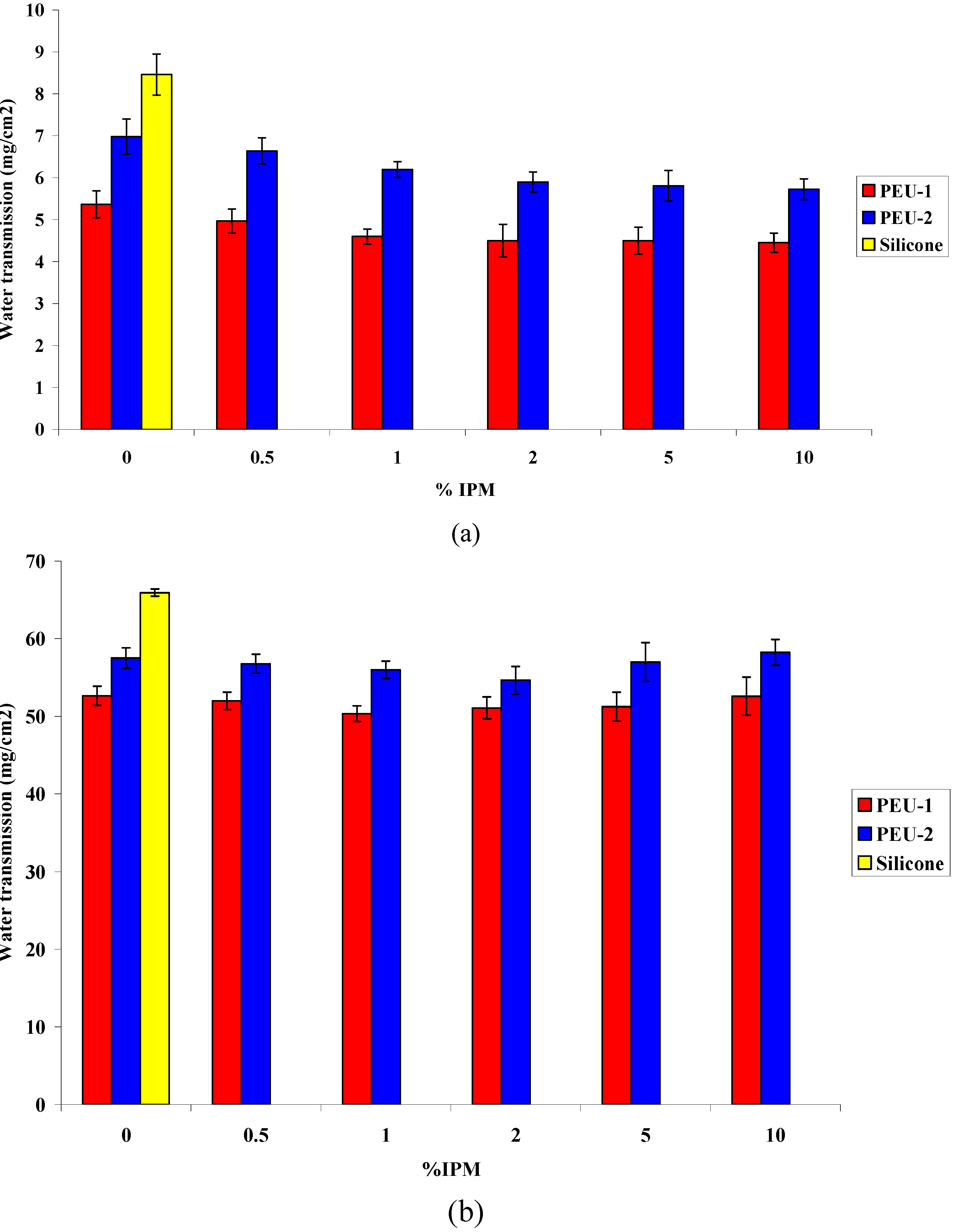
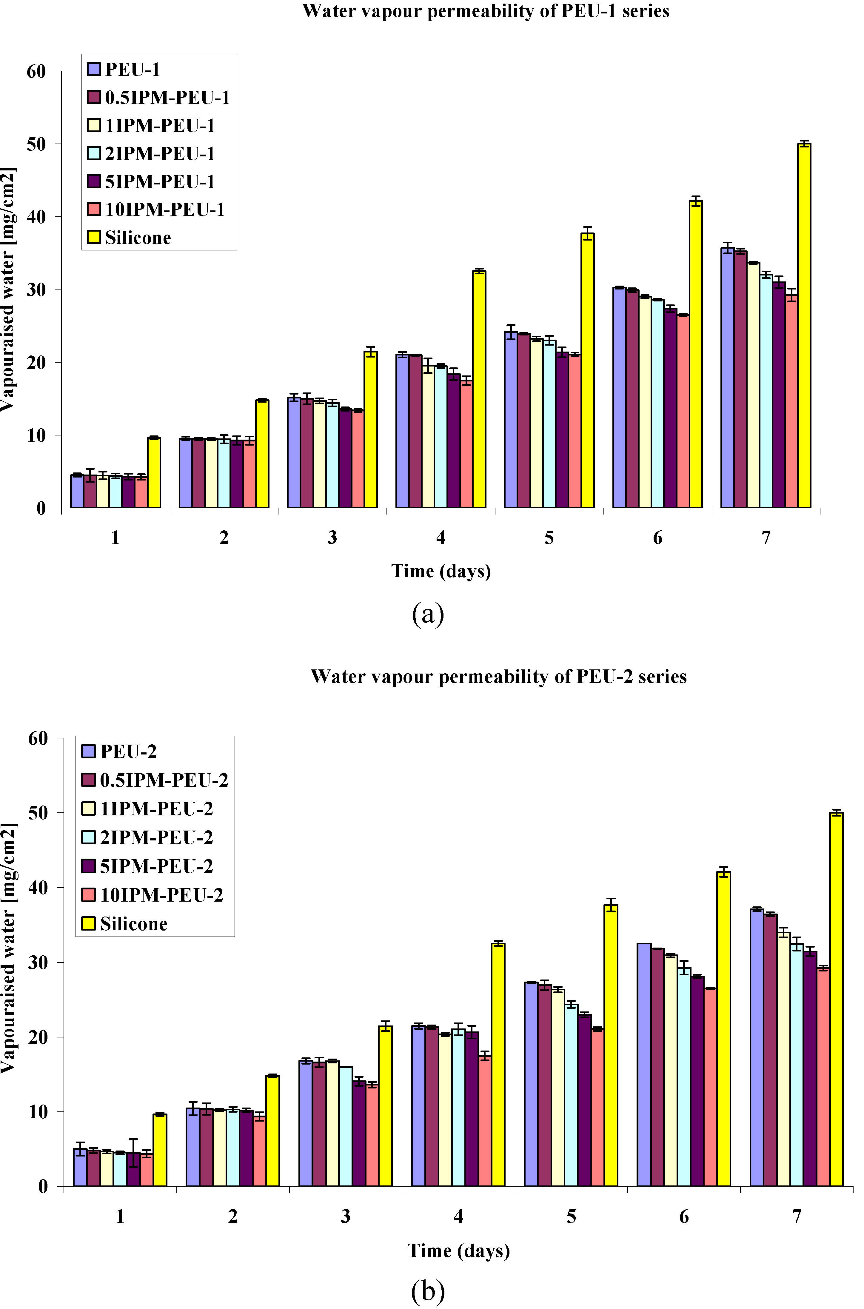
3.3. Water Permeability

The water permeability of both modified and unmodified polyurethane membranes in liquid water are presented in Figure 5. Interestingly, polyurethane series are all less permeable to water compared with the silicone which could be due to higher chain packing of polyurethane and the crystallinity of its hard segment. In polyurethanes, the structure of soft segment plays an important role in permeability of other substances. Indeed it is notable that PEU-1 series with short soft segment chains and higher hard segment content are showing less permeability to water compared to PEU-2 series, however, PEU-2 series are absorbing less water due to their less polarity [26].
Figure 5. Water transmission through IPM modified membranes in liquid water at 37 °C, (a) after 24 hours; (b) after 7 days immersion in deionised water.
Polymers 02 00102 g005
The water permeability of polyurethanes in liquid water was decreased by incorporating IPM in polymer structure especially in first 24 hours (Figure 5a.). This might be because of the increased surface hydrophobicity which reduces the rate of water absorption at the surface of the membrane. It is clear from the results that even adding small amount of IPM (ca. 1 wt%) in polyurethane decreased its water permeability. The water transition through all IPM-PEU formulations were lower than that of PEU alone (p value <0.05) and interestingly they were independent of IPM content in first 24 hours. The difference between rates of water permeation through IPM modified and unmodified group were reduced by longer immersion and marginally improved in low concentration of IPM (Figure 5.b).
The water vapour permeability of polymer films was evaluated and results are presented in Figure 6. There is a clear positive influence on the moisture vapour permeability of the films by incorporating the IPM.
Figure 6. Vaporised water transmission through IPM modified membranes at 25 °C (a) PEU-1 series (b) PEU-2 series.
Polymers 02 00102 g006
Figure 6 shows unlike the liquid water case, permeability to water vapour reduced by the IPM, it is also constant in terms of rate of transport in period of 3–7 days as the daily rate (24 hours) water vapour transmitted through the polyurethanes is constant. Also the differential effect of the IPM over this time period may be due to segregation and distribution of IPM which has taken time to reach stable state. At the end of 7 days approximately 35–50% more water has transported a cross the membrane from liquid reservoir versus vapour reservoir. This is not expected finding, and strongly suggest that mechanisms for transport will be different.
Liquid water resistant of polyurethane has been increased when IPM was added up to certain amount (1 wt% in PEU-1 series and 2–5 wt% in PEU-2 series) and then decreased. It could be concluded that IPM increases the packing of the polymer when it is added to an optimum concentration [26], and this increased packing will reduce the water penetration trough the membrane. In addition, the non-polar nature of IPM will reduce the water solubility in the polymer. The increase of water transmission of polyurethanes (in liquid water) by adding more than 5% IPM might be because of non-stability of the IPM in the system and leaching of the IPM which can form free spaces for water diffusion. This phenomenon did not happen when samples were exposed to water vapour. It could be concluded that some IPM leached out from the polyurethane to water when its concentration was more than 5 wt%.
In PEU films, the water vapour permeation occurs primarily through the soft segment rather than the hard (crystalline) segment. As a result, the water vapour permeability is inversely proportional to hard segment content and packing of the polymer chains. Therefore PEU-1 series with more hard segments and shorter soft segments are less permeable than PEU-2 series. On the other hand, vapour permeability of the films in both series are reduced by increasing the IPM content. As noted earlier, this is a consequence of higher packing of the polymer chains by addition of IPM and because membranes are not exposed to liquid environment, IPM will stabilize in the membrane. IPM also increases hydrophobicity of the polymer film and consequently reduces the possibility of water molecule diffusion trough the film.

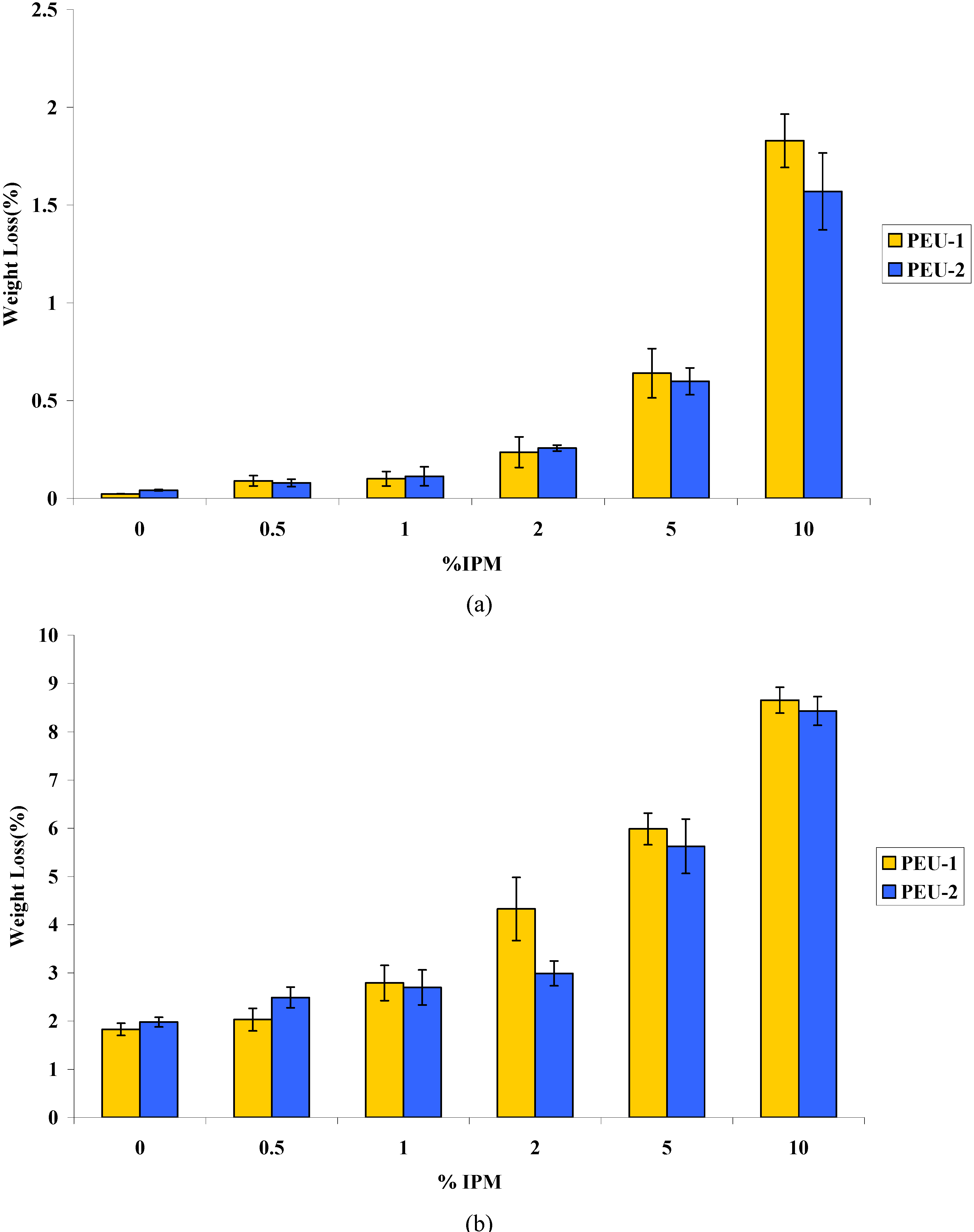
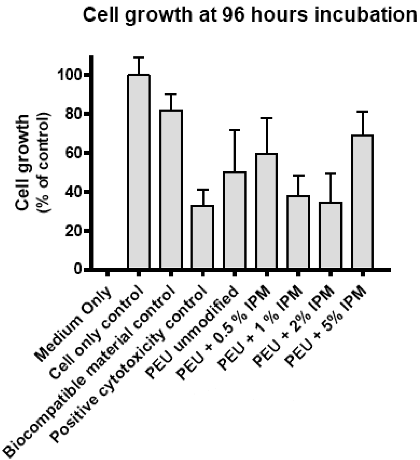
3.4. Cytotoxicity evaluation

Cytotoxicity testing is a part of the complex biological evaluation required for materials used in medicine according to standards [34]. The high sensitivity of this test is the result of cell culture isolation, and the lack of defence mechanisms presents in cells in vivo. Negative results of cytotoxic test mean that either the material is free of harmful substances or it contains too little of them to be detected. Positive results can be an early warning that the material contains more toxic substances than allowed at the clinical level.
A quantitative assessment of cell growth in each well as a whole is provided by the alamarBlue® dye reduction test. The test produces a fluorescence signal of random units that is proportional to the number of viable cells in each well.
All the test materials and the biocompatible material control displayed less cell growth at 48 hours incubation than the cell-only control. The comparative effects of these materials on cell growth are best assessed if growth is normalised to that in the negative toxicity (no material present) control. Therefore, the mean of the background fluorescence seen in wells without cells (coefficient of variation <5%) was subtracted from all other measurements and cell growth is expressed as a proportion of the matching cell-only control in the subsequent analysis.
Figure 7 shows the relative growth for each of the test materials grouped after 96 hours incubation. The comparative cell growth at 96 hours can be considered the main end point of the alamarBlue® cytotoxicity test. These results effectively summarise the findings of the experiment.
Figure 7. Relative cell growth on PEU-1 series at 96 hours. (n = 12 for controls and 6 for test materials).
Polymers 02 00102 g007
An indicative statistical analysis was performed for the 96 hour incubations using one-way ANOVA coupled with Bonferroni's Multiple Comparison Test to evaluate differences between the materials (GraphPad Prism Version 4.02). Significance was taken at p ≤ 0.05. The biocompatible control material was not significantly different from the cell-only control and both supported significantly more cell growth than the positive cytotoxicity control. The unmodified PEU supported less growth than the cell only and biocompatible material controls and was significantly different from the positive cytotoxicity control. The samples of PEU with increasing concentrations of IPM showed differing responses. The 0.5 and 5 wt% IPM containing PEU supported more cell growth than the unmodified polymer, similar to that of the biocompatible material control, whilst the 1 and 2 wt% IPM samples supported less cell growth.
The addition of IPM at concentrations of 5 wt% and below to PEU was seen to both increase and decrease cell growth, with no clear pattern with increasing concentrations of IPM. There is no evidence that the addition of IPM up to 5 wt% has any adverse effect on the biocompatibility of PEU. Unmodified PEU was seen to support less cell growth than Thermanox®, the biocompatible material control. Similar results have been seen with a number of polymers presented as thin films. It is possible that this is a result of residues of the synthesis process within the samples together with effect of low surface energy of modified membranes which reduces cell attachment. But it may also result from the fact that these materials proved difficult to anchor within the test wells, some floating freely during the test.

4. Conclusion

In this study IPM containing polyurethanes were investigated in respect to solution stability, water permeability and biocompatibility as a potentially useful candidate material for encapsulating implantable medical devices. The incorporation of Isopropyl myristate (IPM) to polyether-urethanes with different soft segment molecular weights and effect of IPM content on properties of polyurethane were investigated. The compatibility between IPM and polyurethane was investigated in detail in previous article [26]. IPM was blended in polyurethane successfully, and a possible hydrogen bonding between polyurethane hard segment and ester group in IPM assist the IPM dispersion in polyurethane. High content of IPM in polyurethane (depend on chemical structure of PEU) creates a separated phase and have a negative effect on liquid water permeability of polyurethanes. The apparent difference in water permeability due to a liquid versus water vapour source indicates quite different transport mechanisms and for practical characterisation the need to use both liquid and vapour phase analysis recommended. Although, IPM leaches out from polyurethane when immersed in BSA solution and the rate of leaching raised by increasing the IPM content, 1–2 wt% IPM in polyurethane appear to be stable for at least 10 days. Notably, study shows that it is necessary to undertake leaching studies in biological or at least protein containing samples as a marked effect was seen when BSA was in contact with the membranes. Cytotoxicity assessment of IPM containing polyurethanes did not show any evidence that the addition of IPM up to 5% has any adverse effect on the biocompatibility of PEU.
Although, findings of this study encourage the application of IPM modified polyurethane for coating implantable devices, future investigation on long term biocompatibility and bacterial adhesion of these materials will help to expand the application range of IPM modified polyurethanes for medical devices.

Acknowledgment

This work was supported by European Union through Healthy Aims consortium (Grant no.IST-2002−1-001837).

References

  1. Kotzar, G.; Freas, M.; Abel, P.; Fleischman, A.; Roy, S.; Zorman, C.; Moran, J.M.; Melzak, J. Evaluation of MEMS materials of construction for implantable medical devices. Biomaterials 2002, 23, 2737–2750. [Google Scholar] [CrossRef] [PubMed]
  2. Receveur, R.A.M.; Lindemans, F.W.; de Rooij, N.F. Microsystem technologies for implantable applications. J. Micromech. Microeng. 2007, 17, 50–80. [Google Scholar] [CrossRef]
  3. Rodger, D.C.; Tai, Y.C. Microelectronic packaging for retinal prostheses. IEEE Eng. Med. Biol. Mag. 2005, 24, 52–57. [Google Scholar] [CrossRef] [PubMed]
  4. Bergeron, M.; Levesque, S.; Guidion, R.G. Biomedical applications of polyurethanes. In Biomedical Applications of Polyurethanes; Vermette, P., Griesser, H.J., Laroche, G., Guidoin, R., Eds.; Landes Bioscience: Georgetown, TX, USA, 2001; pp. 220–251. [Google Scholar]
  5. Donaldson, P. Encapsulating microelectronic implants in one-part silicone rubbers. Med. Biol. Eng. Comput. 1989, 27, 93–94. [Google Scholar] [CrossRef] [PubMed]
  6. Galland, G.; Lam, T.M. Permeability and diffusion of gases in segmented polyurethanes—structure properties relations. J. Appl. Polym. Sci. 1993, 50, 1041–1058. [Google Scholar] [CrossRef]
  7. Roohpour, N.; Wasikiewicz, J.M.; Paul, D.; Vadgama, P.; Rehman, I.U. Synthesis and characterisation of enhanced barrier polyurethane for encapsulation of implantable medical devices. J. Mater. Sci. Mater. Med. 2009, 20, 1803–1814. [Google Scholar] [CrossRef] [PubMed]
  8. Weisberg, D.M.; Gordon, B.; Rosenberg, G.; Snyder, A.J.; Benesi, A.; Runt, J. Synthesis and characterization of amphiphilic poly(urethaneurea)-comb-polyisobutylene copolymers. Macromolecules 2000, 33, 4380–4389. [Google Scholar] [CrossRef]
  9. Ratner, B.D.; Hoffman, A.; Schoen, F.; Lemons, J. Biomaterials science: An introduction to materials in medicine; Elsevier Academic Press: San Diego, CA, USA, 2004. [Google Scholar]
  10. Xu, R.J.; Manias, E.; Snyder, A.J.; Runt, J. Low penneability biomedical polyurethane nanocomposites. J. Biomed. Mater. Res. A 2003, 64A, 114–119. [Google Scholar] [CrossRef]
  11. Wang, X.D.; Luo, X.; Wang, X.F. Study on blends of thermoplastic polyurethane and aliphatic polyester: Morphology, rheology, and properties as moisture vapor permeable films. Polym. Test. 2005, 24, 18–24. [Google Scholar] [CrossRef]
  12. Hsieh, K.H.; Liao, D.C.; Chen, C.Y.; Chiu, W,Y. Interpenetrating polymer networks of polyurethane and maleimide-terminated polyurethane for biomedical applications. Polym. Adv. Technol. 1996, 7, 265–272. [Google Scholar] [CrossRef]
  13. Wang, D.A.; Ji, J.; Gao, C.Y.; Yu, G.H.; Feng, L.X. Surface coating of stearyl poly(ethylene oxide) coupling-polymer on polyurethane guiding catheters with poly(ether urethane) film-building additive for biomedical applications. Biomaterials 2001, 22, 1549–1562. [Google Scholar] [CrossRef] [PubMed]
  14. Dube, N.; Al-Malaika, S.; Laroche, G.; Vermette, P. Additives in biomedical polyurethanes In Biomedical Applications of Polyurethanes; Vermette, P., Griesser, H.J., Laroche, G., Guidoin, R., Eds.; Landes Bioscience: Georgetown, TX, USA, 2001; pp. 55–76. [Google Scholar]
  15. Krishna, O.D.; Kim, K.; Byun, Y. Covalently grafted phospholipid monolayer on silicone catheter surface for reduction in platelet adhesion. Biomaterials 2005, 26, 7115–7123. [Google Scholar] [CrossRef] [PubMed]
  16. Yoo, H.J.; Kim, H.D. Characteristics of crosslinked blends of Pellethene((R)) and multiblock polyurethanes containing phospholipid. Biomaterials 2005, 26, 2877–2886. [Google Scholar] [CrossRef] [PubMed]
  17. Sharma, V.; Kundu, P.P. Addition polymers from natural oils—A review. Prog. Polym. Sci. 2006, 31, 983–1008. [Google Scholar] [CrossRef]
  18. Morimoto, N.; Watanabe, A.; Iwasaki, Y.; Akiyoshi, K.; Ishihara, K. Nano-scale surface modification of a segmented polyurethane with a phospholipid polymer. Biomaterials 2004, 25, 5353–5361. [Google Scholar] [CrossRef] [PubMed]
  19. Jeong, H.Y.; Lee, M.H.; Kim, B.K. Surface modification of waterborne polyurethane. Colloids Surf. A Physicochem. Eng. Asp. 2006, 290, 178–185. [Google Scholar] [CrossRef]
  20. Abraham, M.H.; Acree, W.E. Characterisation of the water-isopropyl myristate system. Int. J. Pharm. 2005, 294, 121–128. [Google Scholar] [CrossRef] [PubMed]
  21. Gupta, S.; Moulik, S.P. Biocompatible microemulsions and their prospective uses in drug delivery. J. Pharm. Sci. 2008, 97, 22–45. [Google Scholar] [CrossRef] [PubMed]
  22. Pauliukaite, R.; Schoenleber, M.; Vadgama, P.; Brett, C.M.A. Development of electrochemical biosensors based on sol-gel enzyme encapsulation and protective polymer membranes. Anal. Bioanal. Chem. 2008, 390, 1121–1131. [Google Scholar] [CrossRef] [PubMed]
  23. Christie, I.M.; Treloar, P.H.; Vadgama, P. Plasticized poly(vinyl chloride) as a permselective barrier membrane for high-selectivity amperometric sensors and biosensors. Anal. Chim. Acta 1992, 269, 65–73. [Google Scholar] [CrossRef]
  24. Reddy, S.M.; Vadgama, P.M. Surfactant-modified poly(vinyl chloride) membranes as biocompatible interfaces for amperometric enzyme electrodes. Anal. Chim. Acta 1997, 350, 77–89. [Google Scholar] [CrossRef]
  25. Bremner, T.; Hill, D.J.T.; Killeen, M.I.; Odonnell, J.H.; Pomery, P.J.; StJohn, D.; Whittaker, A.K. Development of wear-resistant thermoplastic polyurethanes by blending with poly(dimethyl siloxane). II. A packing model. J. Appl. Polym. Sci. 1997, 65, 939–950. [Google Scholar] [CrossRef]
  26. Roohpour, N.; Wasikiewicz, J.; Moshaverinia, A.; Paul, D.; Rehman, I.; Vadgama, P. Isopropyl myristate-modified polyether-urethane coatings as protective barriers for implantable medical devices. Materials 2009, 2, 719–733. [Google Scholar] [CrossRef]
  27. Kwok, D.Y.; Neumann, A.W. Contact angle measurement and contact angle interpretation. Adv. Colloid Interface Sci. 1999, 81, 167–249. [Google Scholar] [CrossRef]
  28. Lee, J.H.; Ju, Y,M,; Kim, D.M. Platelet adhesion onto segmented polyurethane film surfaces modified by addition and crosslinking of PEO-containing block copolymers. Biomaterials 2000, 21, 683–691. [Google Scholar] [CrossRef] [PubMed]
  29. Standard Test Methods for Water Vapor Transmission of Materials (ASTM E 96); American Society for Testing and Materials: Washington, DC, USA, 1980.
  30. Castner, D.G.; Ratner, B.D. Biomedical surface science: Foundations to frontiers. Surf. Sci. 2002, 500, 28–60. [Google Scholar] [CrossRef]
  31. Lin, D.T.; Young, T.H.; Fang, Y. Studies on the effect of surface properties on the biocompatibility of polyurethane membranes. Biomaterials 2001, 22, 1521–1529. [Google Scholar] [CrossRef] [PubMed]
  32. Freij-Larsson, C.; Jannasch, P.; Wesslen, B. Polyurethane surfaces modified by amphiphilic polymers: effects on protein adsorption. Biomaterials 2000, 21, 307–315. [Google Scholar] [CrossRef]
  33. Khayet, M.; Suk, D.E.; Narbaitz, R.M.; Santerre, J.P.; Matsuura, T. Study on surface modification by surface-modifying macromolecules and its applications in membrane-separation processes. J. Appl. Polym. Sci. 2003, 89, 2902–2916. [Google Scholar] [CrossRef]
  34. Szelest-Lewandowska, A.; Masiulanis, B.; Szymonowicz, M.; Pielka, S.; Paluch, D. Modified polycarbonate urethane: Synthesis, properties and biological investigation in vitro. J. Biomed. Mater. Res. A 2007, 82A, 509–520. [Google Scholar] [CrossRef]

