1. Introduction
Rifampicin or rifampin (Rif) is an antibiotic that has been used in the treatment of tuberculosis for the past four decades. Tuberculosis (TB) is still a common infectious disease with high mortality and morbidity rates in developing countries, and in the developed world as the result of immune systems being compromised by substance addictions and immunosuppressive drugs. There is also a growing problem of tuberculosis exhibiting resistance to TB drugs, including Rif.
Mycobacterium tuberculosis, the bacteria responsible for TB, invades and replicates in alveolar macrophages. In order to improve tuberculosis treatment using available drugs, Rif has been investigated with new drug delivery methods to increase targeted delivery to alveolar macrophages. The rationale for targeted Rif delivery is to achieve enhanced uptake of Rif into target cells to reduce drug dosage and possibly the duration of treatment. This approach also has the potential to reduce antibiotic resistance and Rif adverse effects, such as hepatotoxicity. New drug delivery methods that have been evaluated with Rif include; liposomes [
1], PLGA microparticles [
2,
3], nanoparticles [
4], and dendrimers [
5]. These systems have been shown effective for enhancement of drug delivery to macrophages
in vitro, but an improved method for effective
in vivo delivery of Rif has yet to be found.
Here we report preliminary results on the use of glucan particles (GPs) to deliver Rif. GPs are 2–4 μm spherical, hollow, porous shells extracted from
Saccharomyces cerevisiae (Baker’s yeast). The hollow cavity of the particles allows for adsorption and encapsulation of payload molecules, and the surface composition of the GPs (>90% 1,3-β-glucan) facilitates receptor-mediated phagocytic cell uptake by cells bearing glucan receptors (e.g., dectin-1 and complement receptor 3, CR3), such as macrophages and dendritic cells [
6]. These properties make GPs an ideal drug delivery vehicle to target phagocytic cells in the immune system. Since
M. tuberculosis is primarily an intracellular pathogen of macrophages the ability of glucan particles to traffic to organs in the body containing these types of cells provides the rationale for testing glucan particles as an alternative method to effectively deliver lethal doses of Rif.
The use of GPs for delivery of macromolecules, such as DNA, siRNA and protein has been successfully demonstrated both
in vitro and
in vivo [
7,
8]
. These types of macromolecules are efficiently trapped within GPs by formation of complexes (polyplexes) held together by electrostatic interactions. For example encapsulation of anionic plasmid DNA or siRNA is achieved by polyplex formation or layer-by-layer synthesis with cationic trapping polymers (
i.e., polyethylenimine, PEI). Unlike polyelectrolyte materials, the trapping of small neutral drug molecules, such as Rif within glucan particles cannot be achieved by polyplex formation or layer-by-layer synthetic methods. An alternative strategy described in this paper is physical entrapment of the drug within the glucan particles by embedding the payload in a hydrogel that partially seals the glucan particle pores to slow drug release. Hydrogels are high-water content materials prepared from crosslinked polymers that have applications in drug delivery due to their ability to encapsulate and slowly release the target drug [
9]. Ideally, hydrogels are prepared with polymers that are non-toxic, biocompatible, and biodegradable. Natural marine derived polysaccharides, such as chitosan or alginate, and synthetic derivatives of these polymers have been extensively studied for applications in tissue engineering [
10], and controlled, localized drug delivery of both small drug molecules and macromolecules [
11,
12,
13,
14,
15,
16].
2. Results and Discussion
Loading of soluble drugs inside glucan particles (GPs) is accomplished by incubating GPs with a soluble drug solution in a volume just sufficient to swell the GPs (hydrodynamic volume), and then encapsulating the drug molecule inside the GPs by physical or chemical trapping. Electrostatic binding between charged polymers has been effectively used for the encapsulation of various payload macromolecule classes (
i.e., DNA, siRNA, proteins) [
7,
8]. In the case of neutral, small drug molecules, such as Rif it is difficult to achieve high drug loading into electrostatically bound complexes. An alternative encapsulation strategy described in this report uses a hydrogel matrix inside the GPs to physically entrap the payload drug and provide for slow drug release. It was hypothesized that hydrogel matrices composed of calcium alginate or chitosan would seal the GP pores blocking or slowing down drug release after resuspension of the particles in an aqueous buffer. As shown in
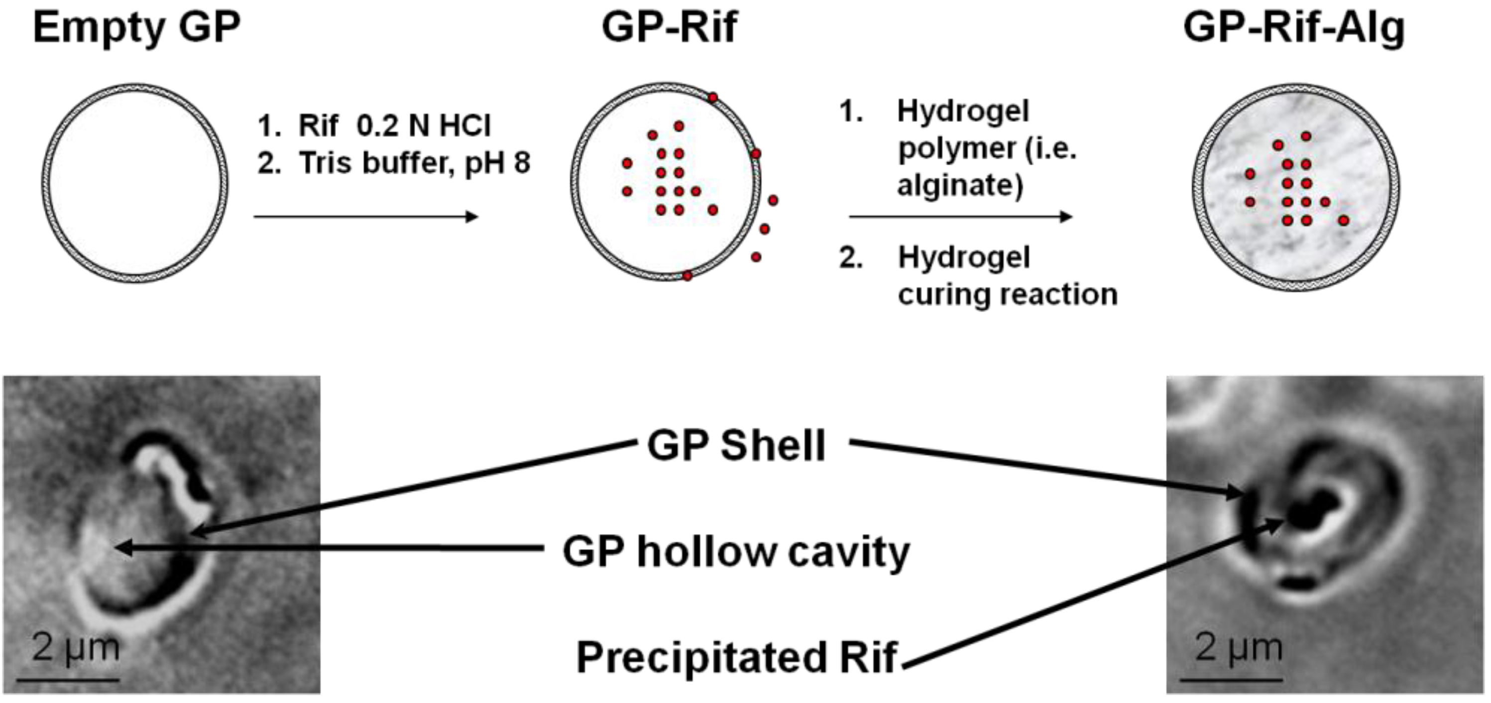
Figure 1, Rif was loaded in a hydrodynamic volume into glucan particles at low pH. The Rif was then precipitated inside the particles following a rapid pH change to pH 8. The lyophilized GP-Rif pellet was subsequently incubated with alginate (pH 7) or chitosan (pH 5) to form the hydrogel matrix and seal the GP pores. In the case of alginate sealed formulations, GP-Rif was first incubated in a hydrodynamic volume of sodium alginate and then treated with calcium chloride to form a calcium alginate hydrogel seal around the precipitated Rif within the GP hollow cavity. Chitosan sealed formulations were synthesized by incubating GP-Rif in a hydrodynamic volume of soluble chitosan (pH 5), and then precipitating the chitosan by raising the pH with tris buffer to pH 8 to form a chitosan hydrogel seal around the precipitated Rif within the GP hollow cavity. Formulations with single and multiple sealing steps were synthesized to determine the effect of hydrogel density on Rif retention inside the particles. The resulting GP-Rif formulations had a strong red color and microscopic evaluation (
Figure 1) showed the presence of a core of precipitated Rif inside GPs. These sealed GP-Rif samples were stored dry at −20 °C until ready to use to prevent premature drug release.
Figure 1.
Schematic representation of Rif (red circles) loading into GPs and sealing with a hydrogel. Microscope brightfield images of empty GPs and GP-Rif samples.
Figure 1.
Schematic representation of Rif (red circles) loading into GPs and sealing with a hydrogel. Microscope brightfield images of empty GPs and GP-Rif samples.
The concentration of Rif loaded into GPs was determined by spectrophotometric measurement of Rif recovered from GP-Rif formulations after enzymatic digestion of the glucan particles with the endoglucanase, zymolyase. A range of GP-Rif formulations were prepared from 0–33% w/w Rif/GP. As shown in
Table 1, Rif loading efficiency was >80% across the concentration range tested. At a 10% w/w Rif/GP load each GP contained ~0.2 pg Rif, while at a 33% w/w Rif/GP load each GP contained ~0.66 pg Rif.
Table 1.
Correlation between loaded amount of Rif/GP and actual Rif recovered as determined from spectrophotometric measurement following enzymatic digestion of GP-Rif samples. (% recovered Rif indicates the average value of three measurements from two different samples for each formulation).
Table 1.
Correlation between loaded amount of Rif/GP and actual Rif recovered as determined from spectrophotometric measurement following enzymatic digestion of GP-Rif samples. (% recovered Rif indicates the average value of three measurements from two different samples for each formulation).
| Loaded Rif/GP (% w/w) | Recovered Rif (% w/w) |
|---|
| 0 | 0 |
| 0.5 | 0.4 ± 0.1 |
| 1.0 | 1.0 ± 0.3 |
| 2.0 | 1.9 ± 0.1 |
| 5.0 | 4.8 ± 0.2 |
| 10.0 | 9.5 ± 0.5 |
| 33.3 | 31.6 ± 0.4 |
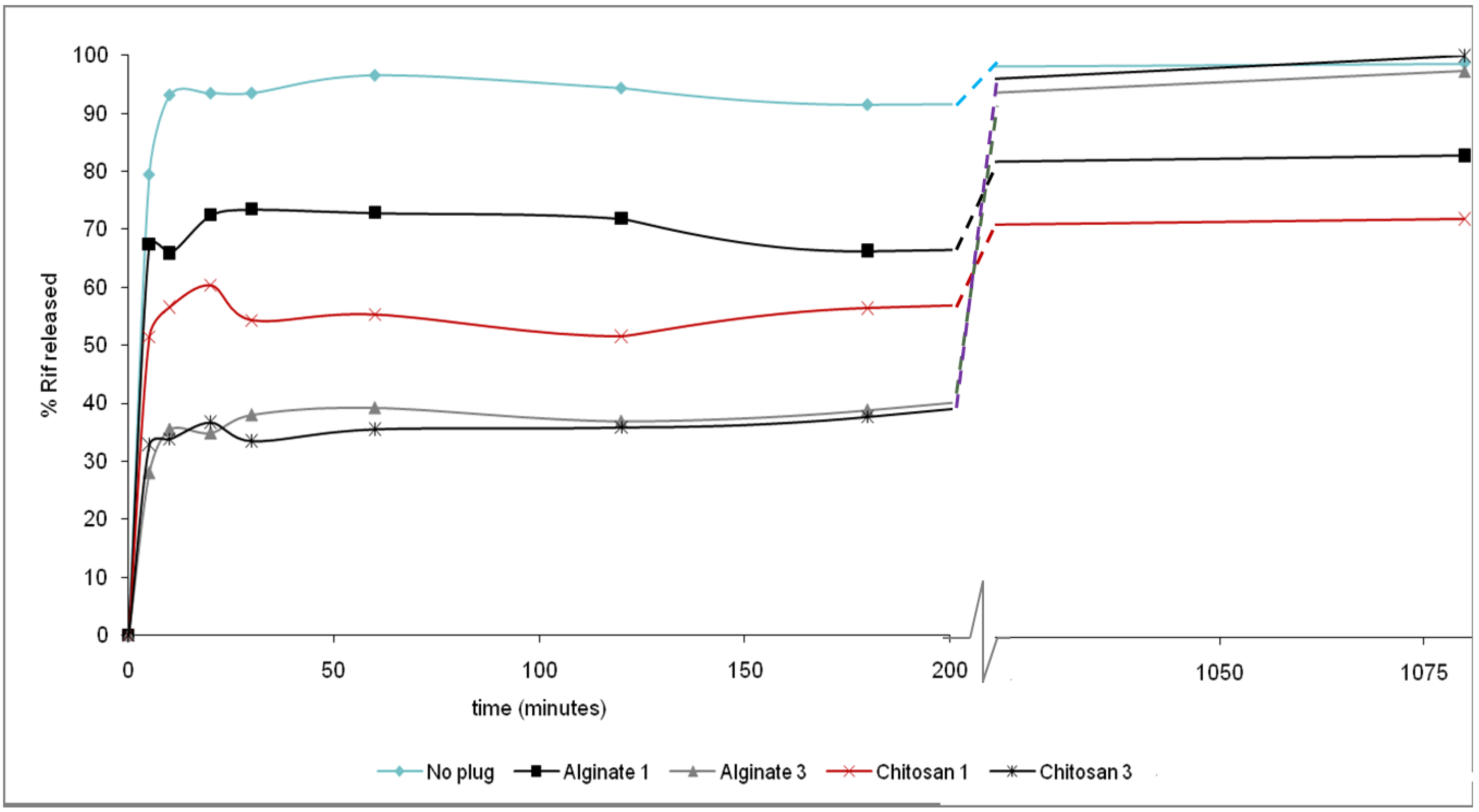
Release of Rif from unsealed and sealed formulations was measured at pH 7. As shown in
Figure 2, there was an immediate burst of drug release upon sample resuspension in PBS, likely due to some Rif not being effectively trapped inside the particles. GP samples without an alginate or chitosan seal were ineffective at retaining Rif and more than 90% of the drug was released in less than 30 minutes. Both chitosan or alginate sealed formulations showed a slower Rif release rate, with formulations that were repetitively sealed with either polymer being more effective at slowing Rif release. This release behavior indicates stabilization of the encapsulated drug after the burst release. The main mechanism controlling Rif release from the formulations is diffusion through the hydrogel matrix, as there are no electrostatic interactions between the anionic (alginate) or cationic (chitosan) matrices and uncharged Rif. The concentration of Rif released during the following 3 h remained constant suggesting that the alginate or chitosan seals effectively prevented premature drug release. Over 36–48 h incubation in PBS the GP-Rif formulations released 70–95% of the loaded Rif. A faster release was observed at pH 5, especially from the chitosan sealed samples, likely due to chitosan dissolution at low pH. However, GP-Rif samples generated though three coatings of either alginate or chitosan were effective at reducing premature Rif release over a period of 24 h, and these samples were chosen to assess
in vitro receptor-targeted GP-Rif delivery to
M. tuberculosis-infected macrophages. All GP-Rif formulations were also evaluated for particle uptake and cytotoxicity with cells (NIH3T3-D1 and RAW264) that express glucan receptors. No difference in particle uptake was observed for the GP-Rif samples coated with either alginate or chitosan compared to the glucan particle control.
GP-Rif formulations were tested for their effect on preventing growth of the live attenuated
Mycobacterium tuberculosis strain, mc
26020 [
17,
18,
19,
20].
Figure 2.
% of Rif released from unsealed and sealed GP formulations at pH 7. Results correspond to samples containing 10% w/w Rif/GP and were sealed with 0, 1 or 3 layers of the indicated polymers.
Figure 2.
% of Rif released from unsealed and sealed GP formulations at pH 7. Results correspond to samples containing 10% w/w Rif/GP and were sealed with 0, 1 or 3 layers of the indicated polymers.
GP-Rif formulations containing 5% w/w Rif/GP (~0.1 pg Rif/GP) or lower concentrations were not effective at reducing
M. tuberculosis growth in bone-marrow derived macrophages (data not shown). Macrophages incubated with alginate or chitosan sealed GP-Rif containing 10% or 33.3% w/w RIF showed increased killing of
M. tuberculosis compared to empty GP and GP-free Rif controls.
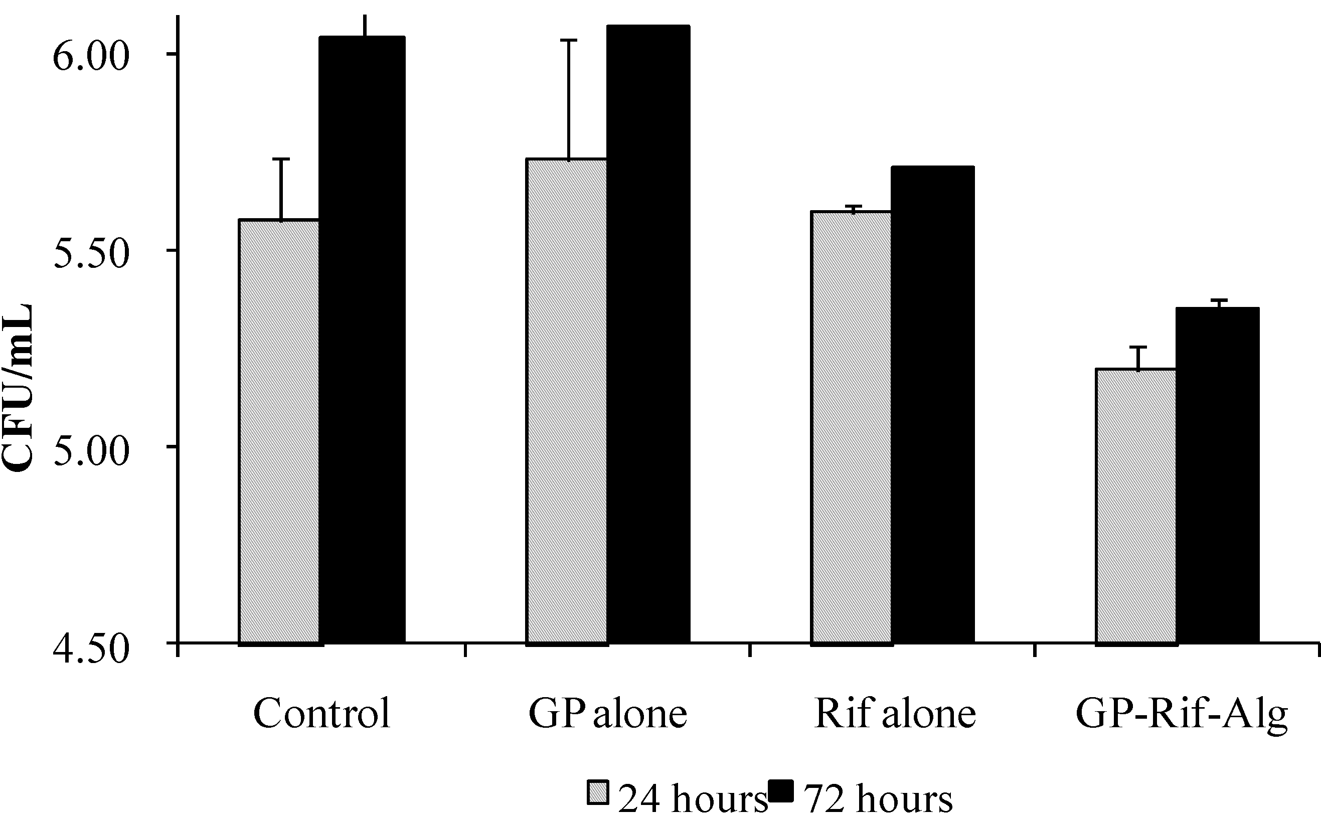
M. tuberculosis growth was measured by enumerating colony forming units/mL lysate (CFU/mL). A representative experiment testing a 10% w/w GP-Rif-Alg sealed formulation is shown in
Figure 3. The alginate sealed GP-Rif formulation was compared against free Rif at the same concentration of Rif that was determined spectrophotometrically to be trapped in the particles. The 10% w/w GP-Rif-Alg formulation contained ~0.2 pg Rif/glucan particle, which represents 80% of the minimum inhibitory concentration (MIC) of Rif (0.25 μg/mL). Empty GPs were also used as a control in these experiments. Intracellular CFU results at 24 h show that the untreated and empty GP controls had no effect on bacterial viability and at 72 h showed an increase in intracellular bacterial load due to intracellular
M. tuberculosis growth. Free Rif alone at a sub-MIC concentration had a small effect (5–10% inhibition of CFU relative to controls). In contrast, the GP-Rif-Alg formulations (10% w/w Rif/GP) showed an 80–90% inhibition of CFU recovered from macrophage lysates after 24 or 72 h of incubation. The increased intracellular CFU inhibitory effect of GP-Rif-Alg compared to free Rif treatment at a sub-MIC concentration of Rif demonstrates the value of GP targeted delivery of Rif to macrophages. Other drug delivery systems for Rif,
i.e., PLGA microparticles [
2,
3], have shown a similar effect
in vitro to GP-Rif. PLGA microparticles containing 0.16–0.5 pg Rif/microparticle have shown CFU reduction of ~1.3 log CFU units. The size of PLGA microparticles is optimal for macrophage uptake, but unlike GPs the macrophage uptake does not occur by receptor mediated process.
Figure 3.
Effect of GP-Rif-Alg (10% w/w Rif/GP) on intracellular M. tuberculosis viability in macrophage lysates.
Figure 3.
Effect of GP-Rif-Alg (10% w/w Rif/GP) on intracellular M. tuberculosis viability in macrophage lysates.
Future work will focus on developing GP-Rif formulations with drug loading levels above the Rif MIC concentration and provide for pH-controlled release.
3. Experimental Section
Materials: Glucan particles derived from
S. cerevisiae were prepared according to a previously published procedure [
7]. Rif and high molecular weight chitosan (85% degree of deacetylation) were purchased from Sigma-Aldrich (Allentown, PA) and used as received. Food grade high molecular weight sodium alginate F-200 extracted from brown seaweed (molecular weight of less than 600,000) was obtained from Multi-Kem Corporation (Ridgefield, NJ). Zymolyase 20T enzyme (21,200 U/g) from
Arthrobacter luteus was purchased from Seikagaku Corporation (Tokyo, Japan). Solvents and buffer solutions were purchased from Sigma Aldrich or Fischer Scientific and used without further purification.
Preparation of GP-Rif samples: Dry glucan particles were mixed with 5 μL Rif/mg GP solution to obtain a uniform paste and incubated at room temperature for 2 h. The amount of Rif was varied from 0 to 0.5 mg Rif/mg GP by mixing a stock Rif solution (100 mg Rif/mL in 0.2 N HCl) with 0.2 N HCl. Tris buffer (1 M, pH 8) was added to precipitate the Rif inside the glucan particles. The samples were then centrifuged, unprecipitated Rif and buffer removed, and the GP-Rif pellets lyophilized.
Preparation of Alginate (Alg) sealed samples (GP-Rif-Alg): The lyophilized GP-Rif samples were mixed with 0.25% sodium alginate (5 μL/mg GP, pH 8). The samples were allowed to swell, incubated at room temperature for 1 h and lyophilized. This sealing process was repeated twice for one set of samples, resulting in GP-Rif-Alg samples sealed once or three times. After the last Alg sealing step calcium chloride (2%, 5 μL/mg GP) was added, mixed to obtain a uniform paste and allowed to gel overnight at room temperature. The GP Rif-Alg-Ca samples were lyophilized and stored dry at −20 °C.
Preparation of Chitosan (CN) sealed samples (GP-Rif-CN): The lyophilized GP-Rif samples were mixed with 1% chitosan solution in sodium acetate buffer (5 μL/mg GP, pH 5). The samples were allowed to swell and incubated at room temperature for 1 h. This sealing process was repeated twice for one set of samples resulting in GP-Rif-CN samples sealed once or three times. After the last chitosan sealing, tris saline buffer (0.5 M tris, 0.15M NaCl, pH 8, 5 μL/mg GP) was added, mixed to obtain a uniform paste and allowed to gel overnight at room temperature. The GP-Rif-CN samples were lyophilized and stored dry at −20 °C.
Spectrophotometric analysis of GP-Rif formulations: Rif loading was carried out at varying ratios (0–33 % w/w Rif/GP). Samples (1 mg) of GP-Rif were resuspended in 1 mL of a phosphate buffer (pH 6.5) solution containing 2 mg of zymolyase. The samples were incubated at 45 °C for 1 h to completely digest the glucan particles. The samples were centrifuged to remove any insoluble material and the absorbance of the digested GP-Rif samples was measured at 470 nm to quantitate the total amount of Rif loaded into the GPs.
Spectrophotometric absorbance of supernatant solutions was used to measure the release of Rif from the GP-Rif formulations at pH 5 and 7 over time. Samples of GP-Rif (2 mg) were resuspended in 1.5 mL of PBS (pH 7) or sodium acetate buffer (pH 5), incubated at room temperature, aliquots (150 μL) collected at different times (0–72 h), centrifuged and the absorbance of the supernatants measured to calculate the amount of released Rif.
Effect of GP-Rif on M. tuberculosis-infected macrophages: Bone marrow cells from C57BL/6 mice were differentiated into macrophages in 10% L929 conditioned media for 7 d. The macrophages were plated in 48-well plates at a density of 2 × 10
5 cells/well and infected with
M. tuberculosis mc
26020 [
20] at a MOI of 10. Media were supplemented with lysine and pantothenate to sustain this
ΔlysAΔpanCD double auxotrophic strain. After 3 h cells were washed with media to remove unbound
M. tuberculosis bacilli. GP-Rif samples were resuspended in 0.9% saline, counted in a hematocytometer, and diluted to 1 × 10
8 particles/mL in 0.9% saline. GP-Rif samples were added to the infected cells at 10:1 particle/cell ratio at 3 h post-infection. After 24 h, cells were lysed with PBS containing 0.25% saponin/0.05% Tween 80 for 20 min at room temperature, and the lysates were transferred to 1.5 mL centrifuge tubes and centrifuged. The supernatants that contained released Rif were removed and the pellets resuspended in PBS containing 0.05% Tween80 to count bacterial number. CFU were counted by 10-fold serial dilution and plating on 7H11 agar. The number of colonies on the plates was counted at 13 and 21 days.


