Review

Antimicrobial Polymers in Solution and on Surfaces: Overview and Functional Principles

by
Felix Siedenbiedel
and
Joerg C. Tiller
*
BCI, TU Dortmund, Emil-Figge-Str. 66, 44227 Dortmund, Germany
*
Author to whom correspondence should be addressed.
Polymers 2012, 4(1), 46-71; https://doi.org/10.3390/polym4010046
Submission received: 28 November 2011 / Revised: 23 December 2011 / Accepted: 4 January 2012 / Published: 9 January 2012
(This article belongs to the Special Issue Bioinspired Polymers)

Abstract

:
The control of microbial infections is a very important issue in modern society. In general there are two ways to stop microbes from infecting humans or deteriorating materials—disinfection and antimicrobial surfaces. The first is usually realized by disinfectants, which are a considerable environmental pollution problem and also support the development of resistant microbial strains. Antimicrobial surfaces are usually designed by impregnation of materials with biocides that are released into the surroundings whereupon microbes are killed. Antimicrobial polymers are the up and coming new class of disinfectants, which can be used even as an alternative to antibiotics in some cases. Interestingly, antimicrobial polymers can be tethered to surfaces without losing their biological activity, which enables the design of surfaces that kill microbes without releasing biocides. The present review considers the working mechanisms of antimicrobial polymers and of contact-active antimicrobial surfaces based on examples of recent research as well as on multifunctional antimicrobial materials.

Graphical Abstract

1. Introduction

The control of the growth of microbes, such as bacteria, fungi, yeast and algae, in nature is one of the fundamental concepts for the survival of higher species. Plants, animals, even microbes themselves have developed a great variety of mechanisms that keep microbes at bay [1]. In human society these control mechanisms often do not work efficiently, which makes microbial infections the number one killer in the world. The treatment of microbial infections becomes more and more difficult, because the number of resistant microbial strains as well as that of antibiotic-immune patients grows a lot faster than the number of useable antibiotics [2,3].
Antimicrobial polymers represent a class of biocides that has become increasingly important as an alternative to existing biocides and in some cases even to antibiotics. The working mechanism of the large number of structurally different polymers is often not fully understood. However, some of them are known for their low potential of building up resistant microbial strains [4].
Man-made materials completely lack defence against microbial growth. Thus, microbial cells attached to any artificial surface in a moist environment can survive and proliferate. While the cell number increases on the surface the microbial cells usually start to build up a biofilm, which consists of a polysaccharide matrix with embedded cells (see Figure 1). Such biofilms allow microbial cells to survive under harsh conditions and the embedded cells are up to 1,000 times less susceptible to most antibiotics and other biocides [5]. Further, many toxins excreted from biofilms make the latter pathogenic and resilient infections are spread [6]. Further, antibiotic genes can be exchanged between bacteria within biofilms, which additionally enhances the formation of multi-resistant bacterial strains [7]. A recent example is the enterohemorrhagic Escherichia coli (EHEC) epidemic in Europe [8]. Additionally, almost every man-made material can be degraded by microbial biofilms [9]. Therefore, the control of microbial growth on surfaces is one of the key issues in material science as well as medicine.
Figure 1. Typical biofilms:(a)mold in households, (b) algae growth on a ship’s hull, (c) bacterial biofilm on a catheter.
Polymers 04 00046 g001
One way to prevent surface contamination is to keep the environment sterile, for instance by using disinfectants, such as hypochlorite, hydrogen peroxide or other reactive oxygen species (ROS). Alternatively silver salts, quarternary ammonium compounds or alcohols are in use. Another widely used disinfectant is triclosan. Unfortunately, the sterile state does not last for long and the frequent use of such disinfectants poses a great environmental problem, particularly dramatic in the case of triclosan [10]. Further, disinfectants have been shown to support the formation of resistant antimicrobial strains [11,12], e.g., methicillin resistant Staphylococcus aureus (MRSA), which causes the majority of nosocomial infections and notably causes more deaths in the USA than HIV [13].
Antimicrobial surfaces that prevent the growth of biofilms are an alternative way to inhibt the spread of microbial infections. Such surfaces either repel microbes, so they cannot attach to the surface, or they kill microbes in the vicinity. The latter is achieved by surfaces loaded with biocides, such as silver, antimicrobial ammonium compounds, antibiotics, active chlorine, or triclosan [14]. Although such surfaces work quite efficiently, they eventually exhaust and the released biocides cause the above described problems. Alternatively, surfaces that catalytically produce biocides using externally applied chemical, electrical, or optical energy are an intriguing alternative (recently reviewed in [15]). Another possibility is the design of surfaces that kill microbes on contact, i.e., they do not release biocides and do not exhaust, at least in principle. Such contact-active antimicrobial surfaces can be realized by tethering antimicrobial polymers [16]. Studies suggest that the polymers on these surfaces work differently to those in solution, whilst their true functional principle is still under speculation [17].
The present study gives an overview on recent developments in the field of antimicrobial polymers with particular focus on their function both in solution and on surfaces.

2. Antimicrobial Polymers

Antimicrobial polymers have been known since 1965, when Cornell and Dunraruma described polymers and copolymers prepared from 2-methacryloxytroponones that kill bacteria [18]. In the 1970s several groups synthesized various polymeric structures that showed antimicrobial action, e.g., Vogl et al., who polymerized salicylic acid [19], or Panarin et al. who synthesized polymers with quarternary ammonium groups [20]. A large number of such macromolecules are known to date. Their function is not always understood. Nevertheless, the number of FDA-approved disinfecting polymers has significantly increased in the past decade, which indicates the need for alternatives to antibiotics and environmentally critical disinfectants. Recently, several reviews have summarized the state of the art [15,21,22,23,24]. The most recent review of Timofeeva et al. thoroughly discusses the influence of relevant parameters, such as molecular weight, type and degree of alkylation, and distribution of charge on the bactericidal action of antimicrobial polymers [23].
Figure 2. General working principles of antimicrobial polymers: (a) polymeric biocides; (b) biocidal polymers; (c) biocide-releasing polymers.
Polymers 04 00046 g002
Careful study of the literature-known antimicrobial macromolecules reveals three general types of antimicrobial polymers: polymeric biocides, biocidal polymers, and biocide-releasing polymers. As shown in Figure 2, the first class is based on the concept that biocidal groups attached to a polymer act in the same way as analogous low molecular weight compounds, i.e., the repeating unit is a biocide. Taking into account the sterical hindrance exerted by the polymer backbone, polymeric biocides should be less active than the respective low molecular weight compounds. For biocidal polymers on the other hand the active principle is embodied by the whole macromolecule, not necessarily requiring antimicrobial repeating units. Biocide releasing polymers do not act through the actual polymeric part, instead the latter functions as carrier for biocides that are somehow transferred to the attacked microbial cells. Such polymers are usually the most active systems, because they can release their biocides close to the cell in high local concentrations.

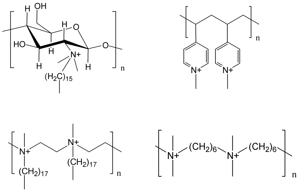
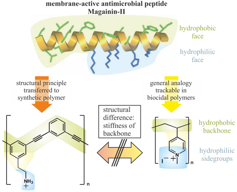
2.1. Polymeric Biocides

Polymeric biocides are polymers that consist of bioactive repeating units, i.e., the polymers are just multiple interconnected biocides, which act similarly to the monomers. Often, the polymerization of biocidal monomers does not lead to active antimicrobial polymers, either, because the polymers are water-insoluble or the biocidal functions do not reach their target. For example, the polymerization of the antimicrobial 4-vinyl-N-benzylpyridinium chloride and subsequent crosslinking resulted in a non-biocidal water-insoluble polymer that only captures microbes but does not kill them [25].
The polymerization of antibiotics, which is usually performed to obtain prolonged activity and less toxicity, is very sensitive to the tethering method and the nature of the antibiotic. For instance, Nathan et al. showed that the antibiotics Penicillin V and Cephradine are not active when conjugated with PEG-Lysine via a hydrolytically stable bond, indicating that the polymer itself is not active [26]. However, the conjugates exhibited full antimicrobial activity if the biocides were tethered via a hydrolytically labile bond, which proved that the antibiotics are reactivated upon release. On the other hand recent examples indicate the possibility to polymerize antibiotics retaining their activity at the polymer backbone, e.g., by copolymerization of methacrylate modified Norfloxacin and PEG-methacrylates [27]. Direct modification of Vancomycin with PEG-methacrylate and subsequent polymerization resulted in active polymers that are at least 6-fold less active when comparing them to the unmodified antibiotic [28]. The activity decreased with greater PEG-length.
These examples show the negative effect of PEG on most biocides, which was also found for quarternary ammonium groups attached to the polyether [29]. Interestingly, it was reported that penicillin attached to polyacrylate nanoparticles showed higher activity against Methicillin-resistant Staphylococcus aureus (MRSA) than the free antibiotic in solution [30]. Polymers with antimicrobial side groups, based on hydrophobic quaternary ammonium functions can be considered as polymeric biocides as well [31]. However, some of those polymers are more active than the respective monomers, indicating that the mode of action is not that of the biocidal group alone.

2.2. Biocidal Polymers

As pointed out by Timofeeva and others, microbial cells generally carry a negative net charge at the surface due to their membrane proteins, teichoic acids of Gram-positive bacteria, and negatively charged phospholipids at the outer membrane of Gram-negative bacteria. This way, polycations are attracted and if they have a proportionate amphiphilic character, they are able to disrupt the outer as well as the cytoplasmic membrane and afford lysis of the cell resulting in cell death. So far this has been the only established way of action of polycationic antimicrobial polymers confirmed by numerous studies nicely reviewed by Timofeeva et al. [23]. Many examples of such antimicrobial polycations with quarternary ammonium [32] and phosphonium [33,34], tertiary sulfonium [35] and guanidinium functions are known. The first polymer that became a commercial disinfectant was poly(hexamethylene biguanidinium hydrochloride), which was thoroughly investigated by Ikeda et al. [36]. The same group presented one of the most potent biocidal polymers against S. aureus, a poly(methacrylate) that contains chlorhexidine-like side groups [37].
Closer observation of the known antimicrobial polymeric structures reveals that they are not always polymerized cationic biocides. Numerous biocidal macromolecules that do not contain biocidal repeating units are known to date. The antimicrobial activity of such polymers obviously originates from the whole molecule. Figure 3 shows some examples of polymers that carry a quarternary ammonium group in the main chain or the side chain. In both cases some molecules require biocidal repeating units (left side) in the polymer chain, while others just need a quaternary ammonium group (right side).
Figure 3. Typical structures of biocidal polymers with quarternary ammonium groups.
Polymers 04 00046 g003
It is obvious that biocidal repeating units are not required if the polymer backbone has a hydrophobic character. On the other hand, polymers with a hydrophilic polymer backbone require a hydrophobic region parallel to the backbone, which is provided by the hydrophobic side groups. DeGrado and particularly Tew have intensively and very successfully worked on polymeric mimics for the antimicrobial peptides, such as magainin and defensin [21,38,39,40,41]. Such antimicrobial peptides (AMP) are membrane active antimicrobial compounds and their mode of action is based on two basic elements: Firstly, both peptides have a highly rigid backbone and secondly the side groups are organized in a way such that these stiff molecules have one hydrophobic side and one side with a cationic net charge. Such molecules are highly efficient in disrupting a microbial cell membrane by being inserted with the whole backbone. This intrusion is devastating to the membrane and rips it apart resulting in rapid cell death [42,43]. Poly(phenylene ethynylene)-based conjugated polymers with amino side groups and also other polymers with stiff backbones and cationic side groups are perfect mimics for those peptides and show high antimicrobial activity and low toxicity. Interestingly, also random peptide sequences prepared by ring-opening polymerization of beta-lactams can be tuned to be mimics of antimicrobial peptides [44]. However, taking a look at the known antimicrobial polycations reveals that basically all these polymers could be mimics for magainin (see Figure 4). The only missing element is most often the stiff backbone. Thus, the polycations cannot sufficiently separate the hydrophobic side (backbone or side groups) from cationic groups, which explains the generally lower activity of most biocidal polymers compared to the antimicrobial peptides.
Figure 4. Biocidal polymers as antimicrobial peptide-mimics.
Polymers 04 00046 g004
Besides the established quarternary ammonium groups, effective antimicrobial polymers with protonated tertiary and primary amino groups have been described recently. Examples are the above mentioned Poly(phenylene ethynylene)-based polymers. Another example is a random copolymer class of dimethylaminomethyl styrene and octylstyrene, which is antimicrobially active upon protonation of the tertiary amino groups, while the respective quarternized derivatives are less active [45]. Palermo and Kurada synthesized similar copolymers by copolymerizing dimethylaminoethylacrylamide and aminoethylacrylamide, respectively, with n-butylacrylamide and showed that these polymers are antimicrobially active and less toxic to blood cells compared to the respective quarternary ammonium derivatives [46,47]. Another recent development in the group of Timoveeva are poly(diallylammonium salts) that contain either secondary or tertiary amino groups [48]. These polymers show excellent activity against S. aureus and Candida albicans. Besides linear polymers, dendritic and hyperbranched polymers have also been described to exhibit strong antimicrobial properties, e.g., quarternized, hyperbranched PEI [49].
A seemingly low molecular weight biocide developed by Das et al. might actually work as an antimicrobial polymer, because the molecule is a hydrogelator that forms helical polymeric structures in aqueous solution [50,51]. This might be the reason why this non-toxic molecule shows antimicrobial activity against Gram-positive and Gram-negative bacteria comparable and even superior to antibiotics.
Although polymeric biocides work most effectively when cationic groups are positioned along the polymer backbone, there is another class of antimicrobial polymers that have only one biocidal end group. These polymers are mostly realized by cationic ring-opening polymerization of 2-alkyl-1,3-oxazolines and terminating the macromolecule with a cationic surfactant. One advantage of this polymerization technique is the controlled introduction of a specific group at one end and another group at the distal end of the polymers by choosing a suitable initiator and termination agent. It could be shown that the molecular weight of poly(2-methyl-1,3-oxazolines) (PMOx) and the respective ethyl-derivative (PEtOx) does not influence the activity of the biocidal end group, dimethyldodecylammonium bromide (DDA), in a range of 2,000–10,000 g/mol [29]. Further, a similar derivative containing polyethylene glycol (PEG) showed a 5–10 times lower antimicrobial activity compared to the polyoxazolines. Interestingly, the group distal to the biocide, the so-called satellite group, controls the activity of the whole molecule in a range of 3 orders of magnitude [52]. We propose that this effect is due to the amphipatic behaviour of the macromolecules, i.e., they form unimolecular micelles in solutions and refold at the surface of microbial cell membranes. The satellite group controls micelle formation and insertion of the biocide into the membrane (see Figure 5).
Figure 5. Working concepts of telechelic poly(2-alkyl-1,3-oxazolines) with antimicrobial end group and satellite group. Redrawn according to [52,53].
Polymers 04 00046 g005
The proposed mechanism was supported by experiments on liposome/polymer interactions, which showed that the highly active PMOx derivative with an antimicrobial DDA group and a non-bioactive decyl satellite group quickly perforates an E. coli-like liposome membrane [53]. When changing the satellite to a hexadecyl group, the whole molecule completely looses its antimicrobial activity, which was again supported by the liposome experiment. It was also demonstrated that the latter inactive macromolecules bind more strongly to the liposomes than the active ones, but without penetrating the membrane. This example illustrates the strong dependence of bioactivity of polymers on structural detail, i.e., the change of 4 methylene groups in a macromolecule with a mass of 10,000 g/mol can dramatically influence the supramolecular structure and its bioactivity. Recently, it was found that the bioactivity of antimicrobial peptide mimics is also strongly influenced by the nature of the end groups [54].

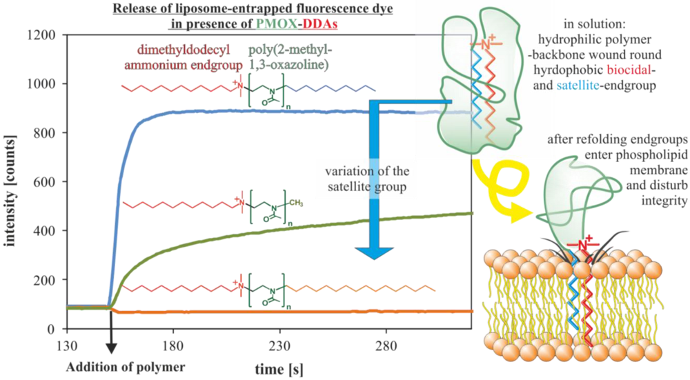
2.3. Biocide-Releasing Polymers

The polymerization of biocide-releasing macromolecules, first achieved by Vogl and Tirell, who polymerized salicylic acid, did not result in polymers with antimicrobial properties [19]. However, they demonstrated that the degradation of the polyester liberates salicylic acid in a controlled fashion. Similar effects were found for acrylate based polymers with salicylic acid side groups [55] and salicylic acid based poly(anhydride esters) [56].
For a long time, tributyltin esters of polyacrylates were believed to be contact-active polymers, because the released TBT kills microbial cells in the surroundings below the detection limit [57].
Worley et al. have developed a class of antimicrobial polymers by using N-halamine groups attached to various polymeric backbones [58,59]. The design of these N-halamines allows the long-term storage of active chlorine. This makes these polymers potent oxidants, which in some cases release chlorine or hypochlorite and in the optimized form, as far as the experimental setups allow such insights, they are able to transfer the active chlorine directly to microbial cells killing them by oxidizing the phospholipids of the cell membrane [60].
Recently, another class of biocidal polymers that release NO (nitric oxide) has been introduced by Schoenfisch [61,62,63]. The polymers contain diazeniumdiolate groups, which can be formed by addition of NO to an amino group containing polymer under great pressure. The optimization of these groups resulted in polymers that release the NO within several hours. However, more work is needed to get to truly long term active antimicrobial polymers.
Polyphenols from natural sources such as teas have been found to show antimicrobial properties [64]. Mimics of these natural polymers have been synthesized by Kenawy et al. [65]. Investigations on the activity pattern revealed that the polymers have to release phenols in order to kill microbial cells.
A recent intriguing antimicrobial polymer class was introduced by Schanze, Whitten and coworkers, who designed cationic conjugated polyelectrolytes based on poly(phenylene ethynylene)-type conjugated polymers that are functionalized with cationic quaternary ammonium solubilizing groups [66]. These polymers were found to be highly effective in the presence of light by forming and releasing efficiently sensitize singlet oxygen (1O2), but they were also effective in the dark, because of their AMP mimicking structure [67]. The singlet oxygen formation also gave the polymer antiviral properties [68].

3. Antimicrobial Surfaces via Attachment of Antimicrobial Polymers

Biofilms on materials are extremely hard to remove and show great resistance to all kinds of biocides. Thus, the prevention of biofilm formation by antimicrobial surfaces is the best way to avoid spreading of diseases and material deterioration. In order to do this, the material must avoid the primary adhesion of living planktonic microbial cells from the surroundings. In general, this can be achieved by either repelling or killing the approaching cells (see Figure 6). Repelling microbes was realized with hydrogel coatings mostly based on PEG or similar hydrogel forming polymers, by highly negatively charged polymers or ultrahydrophobic modifications. The killing of microbes can be achieved by either releasing a biocide from a matrix, which is either previously embedded or actively formed for instance by formation of ROS by photocatalytic TiO2. Alternatively, surfaces can be rendered contact-active antimicrobial upon tethering antimicrobial polymers. In the following, only surfaces with attached antimicrobial polymers and multiple working mechanisms will be discussed. Further biocide release systems in general and microbe repelling surfaces are extensively discussed in recent reviews [14,15,21,22,23,24].
Figure 6. General principles of antimicrobial surfaces.
Polymers 04 00046 g006

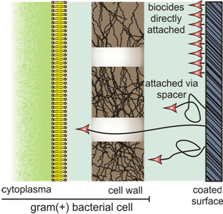
3.1. Techniques towards Surface Attached Polymers

Numerous techniques are known to attach polymers to surfaces, including chemical grafting techniques [69,70], layer-by-layer deposition [71,72] and plasma polymerization [73]. At first, antimicrobial polymers were covalently attached via elaborate techniques. For example, poly-4-vinyl-N-hexylpyridinium was surface-grafted by subsequent modification of a glass slide with an aminosilane and acryloyl chloride. Then the acrylated slide was copolymerized with 4-vinylpyridine, and the product was finally alkylated with hexylbromide [16]. This elaborate procedure required many chemical steps and the use of organic solvents. The side-on attachment of hexylated P4VP to aminoglass was effected by simultaneously reacting the polymer with 1,4-dibromobutane and hexylbromide in the presence of aminoglass [16]. Polyethylenimine (PEI) was also surface-attached to aminoglass via 1,4-dibromobutane, subsequent methylation and alkylation with long chain alkyl halides [74]. Alternatively, the grafting-from method could be used for attaching cationic polyacrylates to various surfaces, including cellulose filters, via atom transfer radical polymerization (ATRP) of N,N-dimethyl-2-aminoethylacrylamide followed by quarternization [75].
All these elaborate techniques are useful for preparations in the laboratory but not in the industry, because chemically achieved finishing is often too expensive. More recently, surface-attached non-releasing polymers could be realized by incorporating polymeric additives for polyurethane [76,77] as well as acrylate [78] coatings that migrate to the surface of the coating during the preparation process. This way, antimicrobial, contact-active materials can be obtained without a finishing procedure. While the first approach uses 1,3-propylene oxide soft blocks with alkylammonium groups as crosslinked soft segments, the second approach applies macromonomers based on polyoxazolines with biocidal quarternary ammonium end groups [29,52,53], which are copolymerized with the acrylate monomers and crosslinkers (see Figure 7).
Figure 7. Surface-grafting of biocidal polymers via self-induced migration to the surface. Created according to [78].
Polymers 04 00046 g007
Besides chemical grafting it is often convenient to coat surfaces with antimicrobial polymers, e.g., by Layer-by-Layer (LbL)-techniques [79]. Also coatings with water-insoluble block copolymers or comb-like block copolymers containing an antimicrobial block, or with water-insoluble quarternized PEIs all from solutions in organic solvents have resulted in long-lasting antimicrobial coatings [80]. In order to avoid organic solvents, block copolymers with a hydrophobic and a hydrophilic, antimicrobial block were used as emulsifiers for the emulsion copolymerization of styrene and acrylates. The resulting paint-like coating can be derived from aqueous suspensions [81].
Another way of coating is the surface attachment of antimicrobial hydrogels. Several examples of gelling antimicrobial peptides, a Pyrene-derivative of Vancomycin and biocidal cationic surfactants have been reported [51,82]. These compounds form hydrogels from their aqueous solutions. The structure of the gels suggests that the compounds form polymers first and those build up networks incorporating the water. The concept of surface-induced hydrogelation [83,84] allows such compounds to gel on surfaces from their diluted aqueous solution. Even compounds that do not form hydrogels have been reported to gel on an affine surface [85]. Another contact-active surface is a hydrogel formed by a cationic hair-pin like antimicrobial peptide that is also a hydrogelator [86].
The techniques for obtaining contact-active antimicrobial surfaces and representative examples are summarized in Table 1.
Table 1. Examples of surface attached biocidal polymers.
MethodPolymerRef.
Grafting fromImmobilized initiatorQPAM[75]
PEtOx[87]
Grafting toImmobilized comonomerQP4VP [16]
End-onAMP[88,89]
Side-onQPEI[90]
NB[91]
Parallel grafting to and modificationQP4VP[17]
In situ end-onPMOx[78]
In situ side-onQPU[76]
CoatingLayer-by-layerPolylysine[79]
PAA[92]
PHGH[93]
Chitosan[94]
Particles with grafted polymerMagnetide with QPEI[95]
PA-particles with QP4VP[81]
Hyperbranched PolymersQPEI[80,96]
Plasma PolymerizationPDAA[97]
Polyterpenol[98]
Surface induced HydrogelationVancomycin[82]
AMP[86]
Abbreviations: QP4VP: Quarternized poly(4-vinylpridine); QPAM: Quarternized poly(N,N-dimethylaminoethylacrylamide); PAA: poly(allylammonium chloride); QPEI: Quarternized polyethyleneimine; PS: poly(styrene); PEtOx: poly(2-ethyloxazoline); PMOx: poly(2-methyloxazoline); QPU: Quarternized polyurethanes; PN: Norbonene-based polymers; AMP: antimicrobial peptides; PA: poly(acrylate); PHGH: poly(hexamethylene guanidinium hydrochloride).

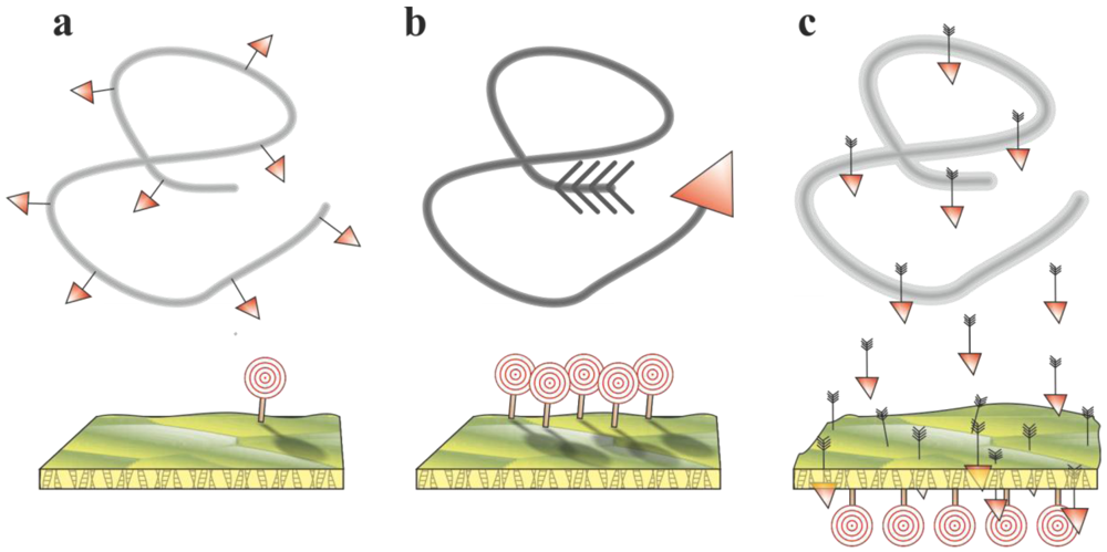
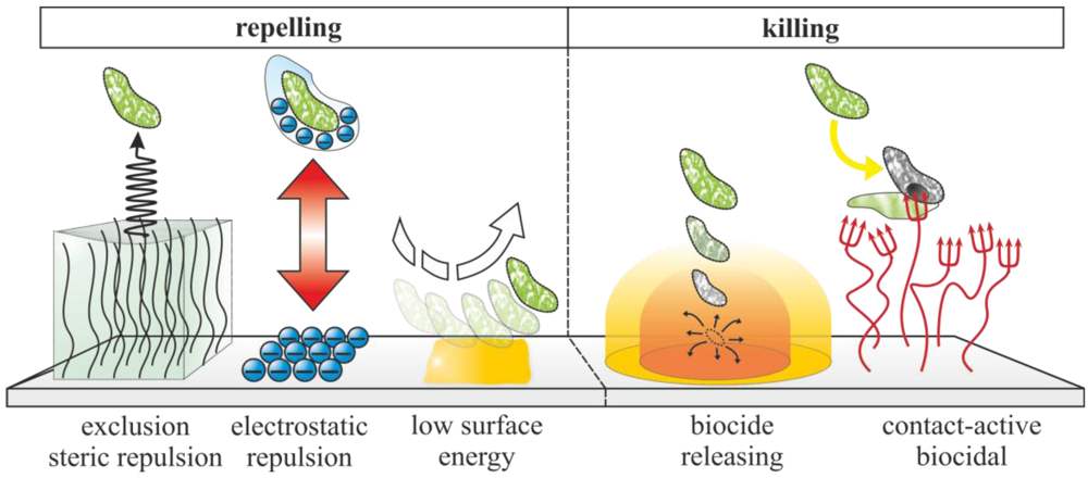
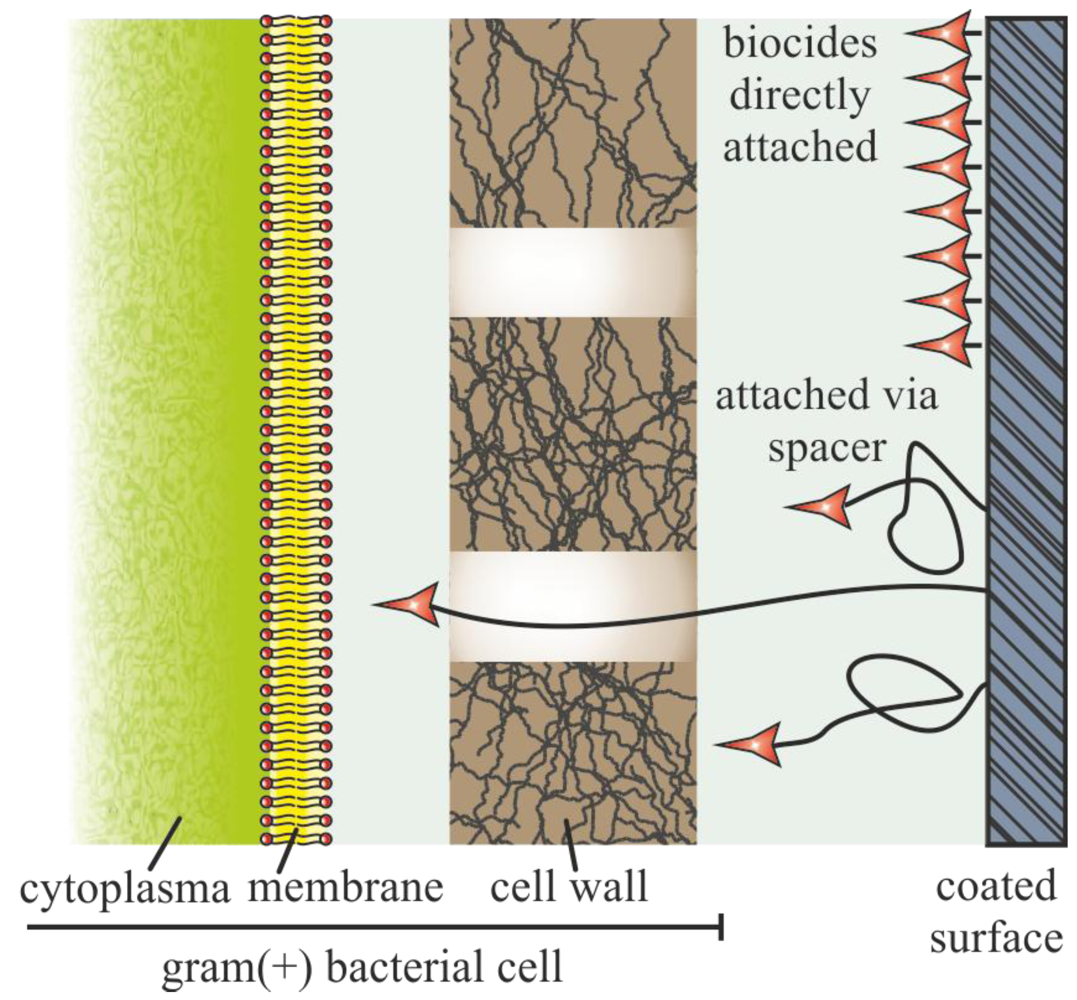
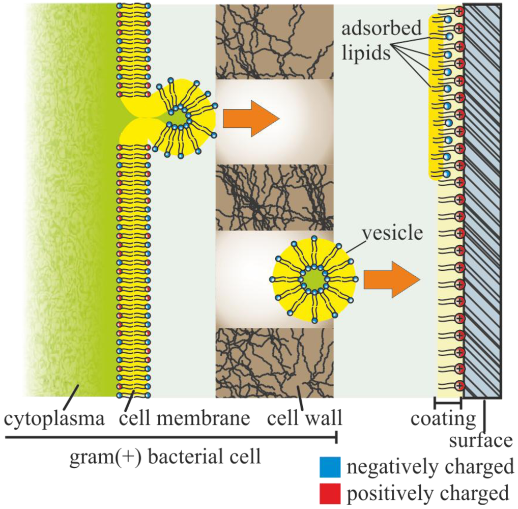
3.2. Functional Principles of Contact-Active Antimicrobial Polymers

As discussed above, the attachment of polymeric biocides or biocidal non-releasing polymers can result in surfaces that kill microbial cells on contact without releasing a biocide. Although, the claim has been made by many, it is rather difficult to prove. Common tests for antimicrobial activity cannot distinguish between contact-activity and release systems. The reason for this is that a biocide on or in a material will diffuse out of the material if it is not covalently attached. However, in order to be lethal for microbes, the concentration has to be above the minimal inhibitory concentration (MIC) or even the minimal biocidal concentration (MBC) only in a region of one µm above the surface. If the biocide concentration is below MIC outside the small kill zone, all existing tests conclude contact-activity. So far there is no test that can distinguish between those cases. The only way to get a better picture is to perform long-term washing tests and constantly monitor and concentrate the washing solutions as well as testing the antimicrobial performance.
One of the first models of how surface attached antimicrobial polymers might act as a contact-active surface was proposed in 2001 (see Figure 8) [16].
Figure 8. Concept of contact-killing via the polymeric spacer effect. Created according to [16].
Polymers 04 00046 g008
The concept of the polymeric spacer effect presumes that a surface grafted biocidal polymer might be capable of penetrating the bacterial cell wall of an adhered bacterial cell. If it reaches the cytoplasmic membrane the cell can be killed by disruption of the phospholipid bilayer. Further, surface grafted polymers are presented in such a high concentration to an adhering cell that they might even kill microbes that are not very susceptible to those polymers in solution. This killing efficiency of surface grafted N-hexylated P4VP could be shown for Gram-positive Staphylococci as well as Gram-negative Pseudomonas aeruginosa. Remarkably, the latter microbial cells cannot be killed by the polymer in solution, which thus supports the high surface concentration hypothesis. It further indicates that the antimicrobial surface-bound polymer works differently to the polymer in solution. Interestingly, there is no significant difference between non-resistant S. aureus cells and MRSA that contain a particularly high numbers of proton pumps, which obviously cannot work at a surface with a grafted antimicrobial polymer [99]. Quarternized surface grafted polyethyleneimine, which effectively kills microbes [74] and even deactivates certain influenza viruses [90] shows a dependence on the molecular weight of the polymer. As calculations indicate, the polymer must be sufficiently large to work efficiently. This strongly supports the polymeric spacer effect. Subsequent work showed that surface grafted quarternized PEI did not build up resistant S. aureus or E. coli strains [4]. Coatings based on single-walled carbon nanotubes were claimed to be antimicrobially active, because the nanotubes would poke through the cell walls of approaching microbial cells, i.e., they also work according to the polymeric spacer effect [100]. As shown by Dathe et al. who immobilized various cationic AMPs on Penta Gels [88] and others who grafted the well-known membrane active AMP magainin I via a polymeric spacer using ATRP [101], the concept of the polymeric spacer seems to be valid for peptides as well. On the other hand, the immobilization of magainin I without a polymeric spacer as SAM on gold showed reduced bacterial adherence and killed adhered bacteria as well although biofilm formation could only be delayed [102].
Another recently published work has explored comb-like polymer coatings, where an antimicrobial DDA-group is grafted to cellulose via a PEtOx of spacer varying in length [87]. Surprisingly, all coatings, even those without a polymeric spacer, were active against S. aureus cells. When treating these coatings with negatively charged phospholipids in order to simulate contact with a microbial cell membrane, all coatings adsorbed similar amounts of the phospholipid but only the coatings with polymeric spacers of less than some 90 repeating unit of the PEtOx were no longer antimicrobially active after this treatment. This indicates that there might be more than one functional principle of such antimicrobial surfaces.
In order to gain insights into a second possible mechanism, a series of coatings of cellulose derivatives with different quarternary ammonium groups and additional hydrophobic groups has been investigated [103]. The result of the antimicrobial assessment showed that a surface can kill S. aureus cells mainly controlled by the cationic/hydrophobic balance and not so much by the charge density. Treatment with negatively charged phospholipids did quench the antimicrobial activity of all investigated surfaces as well as that of the well known antimicrobial surface derived by coating surfaces with the silane 3-(trimethoxysilyl)-propyldimethyloctadecylammonium chloride referred to as DOW 5700 [104]. From these results, it was proposed that the antimicrobial action of coatings that are not modified with biocidal functions on long polymeric spacers originates from their ability to adsorb negatively charged phospholipids (see Figure 9) [103]. Removal of such molecules from cell membranes might indeed lead to their disruption. The weak point of the proposed phospholipid sponge effect is that it cannot explain how the water-insoluble phospholipids cross the cell wall and reach the antimicrobial surface. One possible hypothesis is that the phospholipids travel as liposomes through larger holes in the microbial cell wall.
The increasing number of contact-active antimicrobial surfaces with short chain antimicrobial polymers or just surface attached biocidal groups has resulted in some other models for the functional principle of antimicrobial surfaces [91,105,106]. Kugler et al. have grafted P-4VP to glass surfaces and investigated the influence of the degree of quarternization on the antimicrobial action [107]. The very sharp transition between the active and inactive state at a minimal degree of alkylation was interpreted in terms of a mechanism that somehow involves the exchange of divalent ions of the cell wall. Other authors have followed this unlikely scenario [108,109]. Another proposed mechanism suggests that the hydrophobic surface and the negative surface net charge of microbes leads to an attraction of the cells to cationic surfaces, which has been shown in many scenarios, and somehow destroys their cell envelop [110].
Figure 9. Concept of contact-killing via the phospholipid sponge effect. Created according to [103].
Polymers 04 00046 g009
An alternative way to create contact-active surfaces is to immobilize antimicrobial proteins. The first enzyme tested for the attachment to surfaces was the mureinase lysozyme, which was first described as an antimicrobial protein by Fleming in 1922 [111]. Although most approaches to lysozyme-immobilization have resulted in complete loss of its cell wall-lytic properties, some alternative ways to immobilize lysozyme, including polymeric spacers, have led to surfaces capable of killing Micrococcus lysodeikticus and Bacillus subtilis, and growth-inhibiting or even killing S. aureus [112,113,114,115,116].
Recently, more effective antimicrobial enzymes, such as the endopeptidase lysostaphin [117], enzymes with mureinase and endopeptidease activity [118], lysins [119] and the fast killing, pneumococcal bacteriophage lytic enzyme (Pal) [120] have been found.
Dordick et al. published a coating based on lysostaphin, immobilized on carbon nanotubes [121]. The coating is long-term active and efficiently kills MRSA on contact. It is conceivable that the carbon nanotubes stick out of the surface and in this way reach deep into the microbial cell wall.

4. Multifunctional Antimicrobial Surfaces

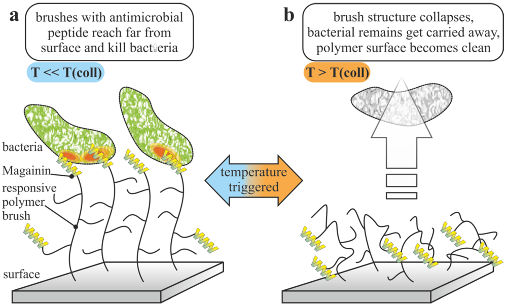
4.1. Repelling and Releasing

Coatings based on biocide-releasing polymers often change their surface properties after liberating the biocide. If the resulting polymers are water-soluble or a least hydrophilic, the surface can repel the attached microbe corpses and hereby refresh the material. The most prominent examples are antifouling paints. They combine the release of a biocide with frequent renewing of the surface (self-polishing), which washes away all attached microbes and other marine organisms dead or alive. Such coatings work most effectively on ship’s hulls, because the shear of the moving ship strongly supports the self-polishing. Modern antifouling paints are coatings of copper ion-releasing, strongly hydrophobic organo copper esters of poly(methacrylic acid) copolymers. The most important aspect is that the hydrophobic coating does not swell in water. Thus, only organo copper ester groups in the upper 10-100 nm are hydrolyzed. The resulting hydrophilic poly(methacrylate) is removed by shear and a fresh contamination free surface is revealed. Unfortunately, the relatively harmless copper is not sufficient to kill all attached marine organisms and thus the coatings are filled with so-called booster biocides, which are usually pesticides [122]. Although these antifouling paints release large amounts of biocides they are only 80 to 90% as effective as the Tributyltin-based coatings which were banned in 2003 [123]. Alternative benign but still effective antifouling coatings, for instance paints with natural biocides instead of the synthetic pesticides [124] are not yet on the market.
Besides antifouling paints, several approaches can be considered as release and repel systems. For instance typical hydrogel coatings for implants usually based on polymers such as polyvinylalcohol, PEG-copolymers or poly acrylic acid derivatives, tend to reduce the microbial adhesion by nearly two orders of magnitude compared to the uncoated control. Loading these hydrogels with antibiotics or other biocides, makes these coatings more active and in principle capable of simultaneously repelling and releasing systems. A more complex design described is a coating based on a hydrophilic polymer network of poly(2-hydroxyethylacrylate) with PEI crosslinking points (see Figure 10). The latter are capable of selectively taking up silver ions from aqueous solution and act as a template for the formation of silver nanoparticles [125]. PEGylation of these conetworks results in materials that efficiently kill S. aureus cells and still repel them after exhaustion of the silver.
Figure 10. Concept of repel and release of a designed network. Created according to [125].
Polymers 04 00046 g010

4.2. Contact-Killing and Repelling

Contact-active surfaces are often considered to be self-deactivating, because killed microbes may adhere to the active layer and new approaching microbes can adhere and proliferate on these corpses. An improvement to such surfaces would be a material that kills microbes and repels the dead cells simultaneously. Chen et al. have created an antimicrobial contact-active surface against E. coli by attaching poly(N,N-dimethyl-N-(ethoxycarbonylmethyl)-N-[2′-(methacryloyloxy)ethyl]-ammonium bromide) via ATRP [126]. The constant contact with the aqueous environment results in hydrolytic cleaving of the ethyl ester next to the quarternary ammonium head group. The resulting zwitterionic head group is no longer biocidal, but effectively repels all attached microbial cells, dead or alive. This interesting effect is not yet reversible, but impressively shows the opportunities of such surfaces. More recently Laloyaux et al. have presented a surface that consists of surface attached magainin grafted with oligo(ethylene glycol) methacrylates [127]. The polymer chains are stretched at room temperature and effectively kill microbial cells. When heated over 35 °C the polymers collapse representing the PEG at the surface, which repels the attached and non-attached Gram-positive cells efficiently. By lowering the temperature, the killing properties are reactivated. In principle, this allows to kill or repel microbial cells by frequent temperature sweeps (see Figure 11).
Figure 11. Concept thermoresponsive surfaces with magainin grafted via thermoresponsive oligo(ethylene glycol) methacrylates that (a) kill bacterial cells below and (b)repel them above the transition temperature. Created according to [127].
Polymers 04 00046 g011

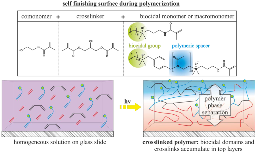
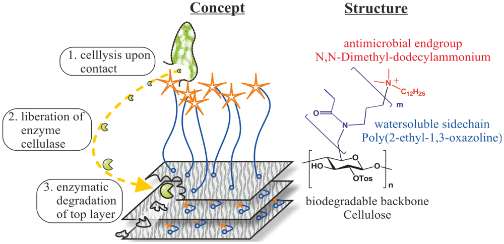
4.3. Releasing and Contact-Killing

Another way to prolong the activity of an antimicrobial release system is to combine this concept with a contact-active killing approach. Worley and coworkers have pioneered the release of oxidative chlorine compounds as slow release and even as release on demand systems [128]. By combining the above described halamine-containing groups with biocidal quarternary ammonium groups in a polyurethane coating, the antimicrobial effect caused by the release of hypochlorite was significantly prolonged by the additional non-releasing antimicrobial groups, which continue to kill the microbes even after the release system is exhausted [129]. A similar behaviour could be afforded by the incorporation of silver nanoparticles into a LbL coating and finishing it with quaternary ammonium groups [130]. The release of silver ions is accompanied by the contact-killing of the layer that contains quarternary ammonium groups. Another benefit of this coating is the successfully demonstrated reloading with silver nanoparticles using silver salts. The same is hold true for the halamine-coatings as demonstrated by Liang et al. [129].
As demonstrated by Shamby et al., poly(4-vinylpyridine) alkylated with hexylbromide reacts with silver nitrate in aqueous solution to form water-insoluble conjugates of the antimicrobial polymer and silver bromide nanoparticles. Coatings of these composites simultaneously kill microbes by releasing silver ions and upon contact with the quarternary ammonium groups [131]. Gyomard et al. claim that the natural antimicrobial peptide gramicidin A immobilized into a LbL matrix kills E. faecalis cells in the surroundings upon release and in the surface-immobilized form [79].
More recently, we have developed a coating based on cellulose with an antimicrobial DDA group grafted via PEtOx, which kills approaching microbial cells on contact [87]. Once a microbial cell is disrupted on the surface, the cell liberates the ubiquitous enzyme cellulase. We could show that this enzyme is capable of degrading the coating and reactivating it in a way similar to the above described release on demand systems (see Figure 12). In contrast to common self-polishing antifouling paints, the cellulose-based coating acts as a contact-active system, is biologically compatible, degradable, and additionally might release biocides in case of a biological contamination only.
Figure 12. Concept of a cellulose-based coating with an attached biocidal polymer that can be degraded by cellulase. Created according to [87].
Polymers 04 00046 g012

5. Conclusions

The present manuscript considers the major functional principles of antimicrobial polymers in solution and on surfaces. Most cationic antimicrobial polymers seem to work in a way similar to membrane-active antimicrobial peptides, such as magainin. Alternative polymers get their biological activity from either presenting biocidal side groups that act the same as in their free form or the polymers have to release biocidal groups. In general, antimicrobial polymers can be classified as polymeric biocides, biocidal polymers, and biocide-releasing polymers. The attachment of the second class results in contact-active antimicrobial surfaces. However, the functional principle of such surfaces is controversially discussed in the literature. So far, it seems that there are at least two mechanisms, the polymeric spacer effect and the phospholipids-sponge effect. However, other possible mechanisms cannot be excluded and a significant amount of work has to be done in order to clarify the proposed mechanisms. For future developments of antimicrobial surfaces, it seems desirable to combine different working mechanisms and to optimize release on demand systems.

References

  1. Nicolas, P.; Mor, A. Peptides as Weapons against Microorganisms in the Chemical Defense System of Vertebrates. Annu. Rev. Microbiol. 1995, 49, 277–304. [Google Scholar] [CrossRef]
  2. Lode, H.M. Clinical Impact of Antibiotic-Resistant Gram-Positive Pathogens. Clin. Microbiol. Infect. 2009, 15, 212–217. [Google Scholar] [CrossRef]
  3. Gonzales, F.P.; Maisch, T. XF Drugs: A New Family of Antibacterials. Drug News Perspect. 2010, 23, 167–174. [Google Scholar] [CrossRef]
  4. Milovic, N.M.; Wang, J.; Lewis, K.; Klibanov, A.M. Immobilized N-Alkylated Polyethylenimine Avidly Kills Bacteria by Rupturing Cell Membranes with No Resistance Developed. Biotechnol. Bioeng. 2005, 90, 715–722. [Google Scholar] [CrossRef]
  5. Mah, T.-F.; Pitts, B.; Pellock, B.; Walker, G.C.; Stewart, P.S.; O’Toole, G.A. A genetic Basis for Pseudomonas Aeruginosa Biofilm Antibiotic Resistance. Nature 2003, 426, 306–310. [Google Scholar] [CrossRef]
  6. Gabriel, G.J.; Som, A.; Madkour, A.E.; Eren, T.; Tew, G.N. Infectious Disease: Connecting Innate Immunity to Biocidal Polymers. Mater. Sci. Eng. R 2007, R57, 28–64. [Google Scholar]
  7. Klein, E.; Smith, D.L.; Laxminarayan, R. Hospitalizations and Deaths Caused by Methicillin-Resistant Staphylococcus aureus, United States, 1999–2005. Emerg. Infect. Dis 2007, 13, 1840–1846. [Google Scholar] [CrossRef]
  8. Viazis, S.; Diez-Gonzalez, F. Enterohemorrhagic Escherichia coli: The Twentieth Century’s Emerging Foodborne Pathogen: A Review. In Advances in Agronomy; Sparks, D.L., Ed.; Elsevier Academic Press Inc.: San Diego, CA, USA, 2011; Volume 111, pp. 1–50. [Google Scholar]
  9. Macedo, M.F.; Miller, A.Z.; Dionisio, A.; Saiz-Jimenez, C. Biodiversity of Cyanobacteria and Green Algae on Monuments in the Mediterranean Basin: An Overview. Microbiology 2009, 155, 3476–3490. [Google Scholar]
  10. Andresen, J.A.; Muir, D.; Ueno, D.; Darling, C.; Theobald, N.; Bester, K. Emerging Pollutants in the North Sea in Comparison to Lake Ontario, Canada, Data. Environ. Toxicol. Chem. 2007, 26, 1081–1089. [Google Scholar]
  11. Romao, C.; Miranda, C.A.; Silva, J.; Clementino, M.M.; de Filippis, I.; Asensi, M. Presence of qacE Delta 1 Gene and Susceptibility to a Hospital Biocide in Clinical Isolates of Pseudomonas aeruginosa Resistant to Antibiotics. Curr. Microbiol. 2011, 63, 16–21. [Google Scholar]
  12. Buffet-Bataillon, S.; Branger, B.; Cormier, M.; Bonnaure-Mallet, M.; Jolivet-Gougeon, A. Effect of Higher Minimum Inhibitory Concentrations of Quaternary Ammonium Compounds in Clinical E. coli Isolates on Antibiotic Susceptibilities and Clinical Outcomes. J. Hosp. Infect. 2011, 79, 141–146. [Google Scholar] [CrossRef]
  13. Klevens, R.M.; Morrison, M.A.; Nadle, J.; Petit, S.; Gershman, K.; Ray, S.; Harrison, L.H.; Lynfield, R.; Dumyati, G.; Townes, J.M.; et al. Invasive Methicillin-Resistant Staphylococcus aureus Infections in the United States. J. Am. Med. Assoc. 2007, 298, 1763–1771. [Google Scholar]
  14. Tiller, J.C. Coatings for Prevention or Deactivation of Biological Contamination. In Developments in Surface Contamination and Cleaning; Kohli, R., Mittal, K.L., Eds.; William Andrew: Norwich, NY, USA, 2008; pp. 1013–1065. [Google Scholar]
  15. Tiller, J.C. Antimicrobial Surfaces. In Bioactive Surfaces; Borner, H.G., Lutz, J.F., Eds.; Springer-Verlag Berlin: Berlin, Germany, 2011; Volume 240, pp. 193–217. [Google Scholar]
  16. Tiller, J.C.; Liao, C.-J.; Lewis, K.; Klibanov, A.M. Designing Surfaces That Kill Bacteria on Contact. Proc. Natl. Acad. Sci. USA 2001, 98, 5981–5985. [Google Scholar]
  17. Tiller, J.C.; Lee, S.B.; Lewis, K.; Klibanov, A.M. Polymer Surfaces Derivatized with Poly(vinyl-N-hexylpyridinium) Kill Airborne and Waterborne Bacteria. Biotechnol. Bioeng. 2002, 79, 465–471. [Google Scholar] [CrossRef]
  18. Cornell, R.J.; Donaruma, L.G. 2-Methacryloxytroponones. Intermediates for Synthesis of Biologically Active Polymers. J. Med. Chem. 1965, 8, 388–390. [Google Scholar] [CrossRef]
  19. Vogl, O.; Tirrell, D. Functional Polymers with Biologically-Active Groups. J. Macromol. Sci.-Chem. 1979, A13, 415–439. [Google Scholar]
  20. Panarin, E.F.; Solovski, M.; Ekzemply, O. Synthesis and Antimicrobial Properties of Polymers Containing Quaternary Ammonium Groups. Khim.-Farm. Zh. 1971, 5, 24–28. [Google Scholar]
  21. Tew, G.N.; Scott, R.W.; Klein, M.L.; De Grado, W.F. De Novo Design of Antimicrobial Polymers, Foldamers, and Small Molecules: From Discovery to Practical Applications. Account. Chem. Res. 2010, 43, 30–39. [Google Scholar] [CrossRef]
  22. Kenawy, E.-R.; Worley, S.D.; Broughton, R. The Chemistry and Applications of Antimicrobial Polymers: A State-of-the-Art Review. Biomacromolecules 2007, 8, 1359–1384. [Google Scholar] [CrossRef]
  23. Timofeeva, L.; Kleshcheva, N. Antimicrobial Polymers: Mechanism of Action, Factors of Activity, and Applications. Appl. Microbiol. Biotechnol. 2011, 89, 475–492. [Google Scholar] [CrossRef]
  24. Banerjee, I.; Pangule, R.C.; Kane, R.S. Antifouling Coatings: Recent Developments in the Design of Surfaces That Prevent Fouling by Proteins, Bacteria, and Marine Organisms. Adv. Mater. 2011, 23, 690–718. [Google Scholar] [CrossRef]
  25. Kawabata, N. Capture of Microorganisms and Viruses by Pyridinium-Type Polymers and Application to Biotechnology and Water-Purification. Prog. Polym. Sci. 1992, 17, 1–34. [Google Scholar] [CrossRef]
  26. Nathan, A.; Zalipsky, S.; Ertel, S.I.; Agathos, S.N.; Yarmush, M.L.; Kohn, J. Copolymers of Lysine and Polyethylene Glycol: A New Family of Functionalized Drug Carriers. Bioconjug. Chem. 1993, 4, 54–62. [Google Scholar] [CrossRef]
  27. Dizman, B.; Elasri, M.O.; Mathias, L.J. Synthesis, Characterization, and Antibacterial Activities of Novel Methacrylate Polymers Containing Norfloxacin. Biomacromolecules 2005, 6, 514–520. [Google Scholar] [CrossRef]
  28. Lawson, M.C.; Shoemaker, R.; Hoth, K.B.; Bowman, C.N.; Anseth, K.S. Polymerizable Vancomycin Derivatives for Bactericidal Biomaterial Surface Modification: Structure-Function Evaluation. Biomacromolecules 2009, 10, 2221–2234. [Google Scholar] [CrossRef]
  29. Waschinski, C.J.; Tiller, J.C. Poly(oxazoline)s with Telechelic Antimicrobial Functions. Biomacromolecules 2005, 6, 235–243. [Google Scholar] [CrossRef]
  30. Turos, E.; Shim, J.Y.; Wang, Y.; Greenhalgh, K.; Reddy, G.S.K.; Dickey, S.; Lim, D.V. Antibiotic-Conjugated Polyacrylate Nanoparticles: New Opportunities for Development of Anti-MRSA Agents. Bioorg. Med. Chem. Lett. 2007, 17, 53–56. [Google Scholar]
  31. Tashiro, T. Antibacterial and Bacterium Adsorbing Macromolecules. Macromol. Mater. Eng. 2001, 286, 63–87. [Google Scholar] [CrossRef]
  32. Kawabata, N.; Nishiguchi, M. Antibacterial Activity of Soluble Pyridinium-Type Polymers. Appl. Environ. Microbiol. 1988, 54, 2532–2535. [Google Scholar]
  33. Kanazawa, A.; Ikeda, T.; Endo, T. Polymeric Phosphonium Salts as a Novel Class of Cationic Biocides. IV. Synthesis and Antibacterial Activity of Polymers with Phosphonium Salts in the Main Chain. J. Polym. Sci. Part A: Polym. Chem. 1993, 31, 3031–3038. [Google Scholar] [CrossRef]
  34. Kanazawa, A.; Ikeda, T.; Endo, T. Novel Polycationic Biocides: Synthesis and Antibacterial Activity of Polymeric Phosphonium Salts. J. Polym. Sci. Part A: Polym. Chem. 1993, 31, 335–343. [Google Scholar]
  35. Kanazawa, A.; Ikeda, T.; Endo, T. Antibacterial Activity of Polymeric Sulfonium Salts. J. Polym. Sci. Part A: Polym. Chem. 1993, 31, 2873–2876. [Google Scholar] [CrossRef]
  36. Ikeda, T.; Ledwith, A.; Bamford, C.H.; Hann, R.A. Interaction of a Polymeric Biguanide Biocide with Phospholipid Membranes. Biochim. Biophys. Acta Biomembr. 1984, 769, 57–66. [Google Scholar] [CrossRef]
  37. Ikeda, T.; Hirayama, H.; Yamaguchi, H.; Tazuke, S.; Watanabe, M. Polycationic Biocides with Pendant Active Groups: Molecular Weight Dependence of Antibacterial Activity. Antimicrob. Agents Chemother. 1986, 30, 132–136. [Google Scholar]
  38. Tew, G.N.; Liu, D.; Chen, B.; Doerksen, R.J.; Kaplan, J.; Carroll, P.J.; Klein, M.L.; DeGrado, W.F. De Novo Design of Biomimetic Antimicrobial Polymers. Proc. Natl. Acad. Sci. USA 2002, 99, 5110–5114. [Google Scholar]
  39. Ilker, M.F.; Nuesslein, K.; Tew, G.N.; Coughlin, E.B. Tuning the Hemolytic and Antibacterial Activities of Amphiphilic Polynorbornene Derivatives. J. Am. Chem. Soc. 2004, 126, 15870–15875. [Google Scholar]
  40. Gabriel, G.J.; Madkour, A.E.; Dabkowski, J.M.; Nelson, C.F.; Nusslein, K.; Tew, G.N. Synthetic Mimic of Antimicrobial Peptide with Nonmembrane-Disrupting Antibacterial Properties. Biomacromolecules 2008, 9, 2980–2983. [Google Scholar]
  41. Lienkamp, K.; Madkour, A.E.; Musante, A.; Nelson, C.F.; Nusslein, K.; Tew, G.N. Antimicrobial Polymers Prepared by ROMP with Unprecedented Selectivity: A Molecular Construction Kit Approach. J. Am. Chem. Soc. 2008, 130, 9836–9843. [Google Scholar]
  42. Zasloff, M. Antimicrobial Peptides of Multicellular Organisms. Nature 2002, 415, 389–395. [Google Scholar] [CrossRef]
  43. Shai, Y. Mechanism of the Binding, Insertion and Destabilization of Phospholipid Bilayer Membranes by Alpha-Helical Antimicrobial and Cell Non-Selective membrane-Lytic Peptides. Biochim. Biophys. Acta-Biomembr. 1999, 1462, 55–70. [Google Scholar] [CrossRef]
  44. Mowery, B.P.; Lee, S.E.; Kissounko, D.A.; Epand, R.F.; Epand, R.M.; Weisblum, B.; Stahl, S.S.; Gellman, S.H. Mimicry of Antimicrobial Host-Defense Peptides by Random Copolymers. J. Am. Chem. Soc. 2007, 129, 15474–15476. [Google Scholar]
  45. Gelman, M.A.; Weisblum, B.; Lynn, D.M.; Gellman, S.H. Biocidal Activity of Polystyrenes That Are Cationic by Virtue of Protonation. Org. Lett. 2004, 6, 557–560. [Google Scholar]
  46. Palermo, E.F.; Kuroda, K. Chemical Structure of Cationic Groups in Amphiphilic Polymethacrylates Modulates the Antimicrobial and Hemolytic Activities. Biomacromolecules 2009, 10, 1416–1428. [Google Scholar] [CrossRef]
  47. Palermo, E.F.; Sovadinova, I.; Kuroda, K. Structural Determinants of Antimicrobial Activity and Biocompatibility in Membrane-Disrupting Methacrylamide Random Copolymers. Biomacromolecules 2009, 10, 3098–3107. [Google Scholar] [CrossRef]
  48. Timofeeva, L.M.; Kleshcheva, N.A.; Moroz, A.F.; Didenko, L.V. Secondary and Tertiary Polydiallylammonium Salts: Novel Polymers with High Antimicrobial Activity. Biomacromolecules 2009, 10, 2976–2986. [Google Scholar] [CrossRef]
  49. Pasquier, N.; Keul, H.; Heine, E.; Moeller, M.; Angelov, B.; Linser, S.; Willumeit, R. Amphiphilic Branched Polymers as Antimicrobial Agents. Macromol. Biosci. 2008, 8, 903–915. [Google Scholar] [CrossRef]
  50. Debnath, S.; Shome, A.; Das, D.; Das, P.K. Hydrogelation through Self-Assembly of Fmoc-Peptide Functionalized Cationic Amphiphiles: Potent Antibacterial Agent. J. Phys. Chem. B 2010, 114, 4407–4415. [Google Scholar]
  51. Roy, S.; Das, P.K. Antibacterial Hydrogels of Amino Acid-Based Cationic Amphiphiles. Biotechnol. Bioeng. 2008, 100, 756–764. [Google Scholar] [CrossRef]
  52. Waschinski, C.J.; Herdes, V.; Schueler, F.; Tiller, J.C. Influence of Satellite Groups on Telechelic Antimicrobial Functions of Polyoxazolines. Macromol. Biosci. 2005, 5, 149–156. [Google Scholar] [CrossRef]
  53. Waschinski, C.J.; Barnert, S.; Theobald, A.; Schubert, R.; Kleinschmidt, F.; Hoffmann, A.; Saalwaechter, K.; Tiller, J.C. Insights in the Antibacterial Action of Poly(methyloxazoline)s with a Biocidal End Group and Varying Satellite Groups. Biomacromolecules 2008, 9, 1764–1771. [Google Scholar]
  54. Mowery, B.P.; Lindner, A.H.; Weisblum, B.; Stahl, S.S.; Gellman, S.H. Structure-activity Relationships among Random Nylon-3 Copolymers That Mimic Antibacterial Host-Defense Peptides. J. Am. Chem. Soc. 2009, 131, 9735–9745. [Google Scholar]
  55. Bortnowska-Barela, B.; Polowinska, A.; Polowiski, S.; Vaskova, V.; Barton, J. Bioactive Emulsions of o-Carboxyphenyl Methacrylate Copolymers. Acta Polym. 1987, 38, 652–654. [Google Scholar] [CrossRef]
  56. Rosenberg, L.E.; Carbone, A.L.; Roemling, U.; Uhrich, K.E.; Chikindas, M.L. Salicylic Acid-Based Poly(anhydride esters) for Control of Biofilm Formation in Salmonella Enterica Serovar Typhimurium. Lett. Appl. Microbiol. 2008, 46, 593–599. [Google Scholar] [CrossRef]
  57. Huttinger, K.J.; Muller, H.; Bomar, M.T. Synthesis and Effect of Carrier-Bound Disinfectants. J. Colloid Interface Sci. 1982, 88, 274–285. [Google Scholar] [CrossRef]
  58. Sun, G.; Wheatley, W.B.; Worley, S.D. A New Cyclic N-Halamine Biocidal Polymer. Ind. Eng. Chem. Res. 1994, 33, 168–170. [Google Scholar] [CrossRef]
  59. Eknoian, M.W.; Worley, S.D.; Harris, J.M. New Biocidal N-Halamine-PEG Polymers. J. Bioact. Compat. Polym. 1998, 13, 136–145. [Google Scholar]
  60. Liang, J.; Barnes, K.; Akdag, A.; Worley, S.D.; Lee, J.; Broughton, R.M.; Huang, T.-S. Improved Antimicrobial Siloxane. Ind. Eng. Chem. Res. 2007, 46, 1861–1866. [Google Scholar]
  61. Coneski, P.N.; Rao, K.S.; Schoenfisch, M.H. Degradable Nitric Oxide-Releasing Biomaterials via Post-Polymerization Functionalization of Cross-Linked Polyesters. Biomacromolecules 2010, 11, 3208–3215. [Google Scholar] [CrossRef]
  62. Stasko, N.A.; Schoenfisch, M.H. Dendrimers as a Scaffold for Nitric Oxide Release. J. Am. Chem. Soc. 2006, 128, 8265–8271. [Google Scholar]
  63. Charville, G.W.; Hetrick, E.M.; Geer, C.B.; Schoenfisch, M.H. Reduced Bacterial Adhesion to Fibrinogen-Coated Substrates via Nitric Oxide Release. Biomaterials 2008, 29, 4039–4044. [Google Scholar]
  64. Ferrazzano, G.F.; Roberto, L.; Amato, I.; Cantile, T.; Sangianantoni, G.; Ingenito, A. Antimicrobial Properties of Green Tea Extract against Cariogenic Microflora: An in vivo Study. J. Med. Food 2011, 14, 907–911. [Google Scholar]
  65. Kenawy, E.-R.; El-Shanshoury, A.E.-R.R.; Omar Shaker, N.; El-Sadek, B.M.; Khattab, A.H.B.; Badr, B.I. Biocidal Polymers: Synthesis, Antimicrobial Activity, and Possible Toxicity of Poly (hydroxystyrene-co-methylmethacrylate) Derivatives. J. Appl. Polym. Sci. 2011, 120, 2734–2742. [Google Scholar]
  66. Chemburu, S.; Corbitt, T.S.; Ista, L.K.; Ji, E.; Fulghum, J.; Lopez, G.P.; Ogawa, K.; Schanze, K.S.; Whitten, D.G. Light-Induced Biocidal Action of Conjugated Polyelectrolytes Supported on Colloids. Langmuir 2008, 24, 11053–11062. [Google Scholar]
  67. Ji, E.-K.; Corbitt, T.S.; Parthasarathy, A.; Schanze, K.S.; Whitten, D.G. Light and Dark-Activated Biocidal Activity of Conjugated Polyelectrolytes. ACS Appl. Mater. Interfaces 2011, 3, 2820–2829. [Google Scholar]
  68. Wang, Y.; Canady, T.D.; Zhou, Z.J.; Tang, Y.L.; Price, D.N.; Bear, D.G.; Chi, E.Y.; Schanze, K.S.; Whitten, D.G. Cationic Phenylene Ethynylene Polymers and Oligomers Exhibit Efficient Antiviral Activity. ACS Appl. Mater. Interfaces 2011, 3, 2209–2214. [Google Scholar]
  69. Advincula, R. Polymer Brushes by Anionic and Cationic Surface-Initiated Polymerization (SIP). Adv. Polym. Sci. 2006, 197, 107–136. [Google Scholar] [CrossRef]
  70. Edmondson, S.; Osborne, V.L.; Huck, W.T.S. Polymer Brushes via Surface-Initiated Polymerizations. Chem. Soc. Rev. 2004, 33, 14–22. [Google Scholar]
  71. Hammond, P.T. Engineering Materials Layer-by-Layer: Challenges and Opportunities in Multilayer Assembly. AIChE J. 2011, 57, 2928–2940. [Google Scholar] [CrossRef]
  72. Lyklema, J.; Deschenes, L. The First Step in Layer-by-Layer Deposition: Electrostatics and/or Non-Electrostatics? Adv. Colloid Interface Sci. 2011, 168, 135–148. [Google Scholar] [CrossRef]
  73. Friedrich, J. Mechanisms of Plasma Polymerization—Reviewed from a Chemical Point of View. Plasma Processes Polym. 2011, 8, 783–802. [Google Scholar] [CrossRef]
  74. Lin, J.; Qiu, S.Y.; Lewis, K.; Klibanov, A.M. Bactericidal Properties of Flat Surfaces and Nanoparticles Derivatized with Alkylated Polyethylenimines. Biotechnol. Prog. 2002, 18, 1082–1086. [Google Scholar] [CrossRef]
  75. Lee, S.B.; Koepsel, R.R.; Morley, S.W.; Matyjaszewski, K.; Sun, Y.J.; Russell, A.J. Permanent, Nonleaching Antibacterial Surfaces. 1. Synthesis by Atom Transfer Radical Polymerization. Biomacromolecules 2004, 5, 877–882. [Google Scholar] [CrossRef]
  76. Kurt, P.; Wood, L.; Ohman, D.E.; Wynne, K.J. Highly Effective Contact Antimicrobial Surfaces via Polymer Surface Modifiers. Langmuir 2007, 23, 4719–4723. [Google Scholar]
  77. Wynne, J.H.; Fulmer, P.A.; McCluskey, D.M.; Mackey, N.M.; Buchanan, J.P. Synthesis and Development of a Multifunctional Self-Decontaminating Polyurethane Coating. ACS Appl. Mater. Interfaces 2011, 3, 2005–2011. [Google Scholar] [CrossRef]
  78. Waschinski, C.J.; Zimmermann, J.; Salz, U.; Hutzler, R.; Sadowski, G.; Tiller, J.C. Design of Contact-Active Antimicrobial Acrylate-Based Materials Using Biocidal Macromers. Adv. Mater. 2008, 20, 104–108. [Google Scholar]
  79. Guyomard, A.; De, E.; Jouenne, T.; Malandain, J.J.; Muller, G.; Glinel, K. Incorporation of a Hydrophobic Antibacterial Peptide into Amphiphilic Polyelectrolyte Multilayers: A Bioinspired Approach to Prepare Biocidal Thin Coatings. Advanced Functional Materials 2008, 18, 758–765. [Google Scholar] [CrossRef]
  80. Park, D.; Wang, J.; Klibanov, A.M. One-Step, Painting-Like Coating Procedures to Make Surfaces Highly and Permanently Bactericidal. Biotechnol. Prog. 2006, 22, 584–589. [Google Scholar] [CrossRef]
  81. Fuchs, A.D.; Tiller, J.C. Contact-Active Antimicrobial Coatings Derived from Aqueous Suspensions. Angew. Chem. Int. Ed. 2006, 45, 6759–6762. [Google Scholar]
  82. Xing, B.G.; Yu, C.W.; Chow, K.H.; Ho, P.L.; Fu, D.G.; Xu, B. Hydrophobic Interaction and Hydrogen Bonding Cooperatively Confer a Vancomycin Hydrogel: A Potential Candidate for Biomaterials. J. Am. Chem. Soc. 2002, 124, 14846–14847. [Google Scholar]
  83. Tiller, J.C. Increasing the Local Concentration of Drugs by Hydrogel Formation. Angew. Chem. Int. Ed. 2003, 42, 3072–3075. [Google Scholar] [CrossRef]
  84. Bieser, A.M.; Tiller, J.C. Surface-Induced Hydrogelation. Chem. Commun. 2005. [Google Scholar] [CrossRef]
  85. Bieser, A.M.; Tiller, J.C. Supramolecular Self-organization of Potential Hydrogelators on Attracting Surfaces. Supramol. Chem. 2008, 20, 363–367. [Google Scholar]
  86. Salick, D.A.; Kretsinger, J.K.; Pochan, D.J.; Schneider, J.P. Inherent Antibacterial Activity of a Peptide-Based Beta-Hairpin Hydrogel. J. Am. Chem. Soc. 2007, 129, 14793–14799. [Google Scholar]
  87. Bieser, A.M.; Thomann, Y.; Tiller, J.C. Contact-Active Antimicrobial and Potentially Self-Polishing Coatings Based on Cellulose. Macromol. Biosci. 2011, 11, 111–121. [Google Scholar] [CrossRef]
  88. Bagheri, M.; Beyermann, M.; Dathe, M. Immobilization Reduces the Activity of Surface-Bound Cationic Antimicrobial Peptides with No Influence upon the Activity Spectrum. Antimicrob. Agents Chemother. 2009, 53, 1132–1141. [Google Scholar] [CrossRef]
  89. Costa, F.; Carvalho, I.F.; Montelaro, R.C.; Gomes, P.; Martins, M.C.L. Covalent Immobilization of Antimicrobial Peptides (AMPs) onto Biomaterial Surfaces. Acta Biomater. 2011, 7, 1431–1440. [Google Scholar] [CrossRef]
  90. Haldar, J.; An, D.Q.; de Cienfuegos, L.A.; Chen, J.Z.; Klibanov, A.M. Polymeric Coatings That Inactivate Both Influenza Virus and Pathogenic Bacteria. Proc. Nal. Acad. Sci. USA 2006, 103, 17667–17671. [Google Scholar]
  91. Madkour, A.E.; Dabkowski, J.M.; Nusslein, K.; Tew, G.N. Fast Disinfecting Antimicrobial Surfaces. Langmuir 2009, 25, 1060–1067. [Google Scholar]
  92. Lichter, J.A.; Rubner, M.F. Polyelectrolyte Multilayers with Intrinsic Antimicrobial Functionality: The Importance of Mobile Polycations. Langmuir 2009, 25, 7686–7694. [Google Scholar] [CrossRef]
  93. Pan, Y.; Xiao, H. Rendering Rayon Fibres Antimicrobial and Thermal-Responsive via Layer-by-Layer Self-Assembly of Functional Polymers. Adv. Mater. Res. 2011, 236-238, 1103–1106. [Google Scholar]
  94. Cecius, M.; Jerome, C. A fully Aqueous Sustainable Process for Strongly Adhering Antimicrobial Coatings on Stainless Steel. Prog. Org. Coat. 2011, 70, 220–223. [Google Scholar]
  95. Lin, J.; Qiu, S.; Lewis, K.; Klibanov, A.M. Bactericidal Properties of Flat Surfaces and Nanoparticles Derivatized with Alkylated Polyethylenimines. Biotechnol. Prog. 2002, 18, 1082–1086. [Google Scholar] [CrossRef]
  96. Pasquier, N.; Keul, H.; Heine, E.; Moeller, M. From Multifunctionalized Poly(ethylene imine)s toward Antimicrobial Coatings. Biomacromolecules 2007, 8, 2874–2882. [Google Scholar] [CrossRef]
  97. Thome, J.; Hollander, A.; Jaeger, W.; Trick, I.; Oehr, C. Ultrathin Antibacterial Polyammonium Coatings on Polymer Surfaces. Surf. Coat. Technol. 2003, 174-175, 584–587. [Google Scholar] [CrossRef]
  98. Bazaka, K.; Jacob, M.V.; Truong, V.K.; Crawford, R.J.; Ivanova, E.P. The Effect of Polyterpenol Thin Film Surfaces on Bacterial Viability and Adhesion. Polymers 2011, 3, 388–404. [Google Scholar] [CrossRef]
  99. Lin, J.; Tiller, J.C.; Lee, S.B.; Lewis, K.; Klibanov, A.M. Insights into Bactericidal Action of Surface-Attached Poly(vinyl-N-hexylpyridinium) Chains. Biotechnol. Lett. 2002, 24, 801–805. [Google Scholar] [CrossRef]
  100. Kang, S.; Pinault, M.; Pfefferle, L.D.; Elimelech, M. Single-Walled Carbon Nanotubes Exhibit Strong Antimicrobial Activity. Langmuir 2007, 23, 8670–8673. [Google Scholar]
  101. Glinel, K.; Jonas, A.M.; Jouenne, T.; Leprince, J.; Galas, L.; Huck, W.T.S. Antibacterial and Antifouling Polymer Brushes Incorporating Antimicrobial Peptide. Bioconjug. Chem. 2009, 20, 71–77. [Google Scholar] [CrossRef]
  102. Humblot, V.; Yala, J.F.; Thebault, P.; Boukerma, K.; Hequet, A.; Berjeaud, J.M.; Pradier, C.M. The Antibacterial Activity of Magainin I Immobilized onto Mixed Thiols Self-Assembled Monolayers. Biomaterials 2009, 30, 3503–3512. [Google Scholar] [CrossRef]
  103. Bieser, A.M.; Tiller, J.C. Mechanistic Considerations on Contact-Active Antimicrobial Surfaces with Controlled Functional Group Densities. Macromol. Biosci. 2011, 11, 526–534. [Google Scholar] [CrossRef]
  104. Isquith, A.J.; Abbott, E.A.; Walters, P.A. Surface-Bonded Antimicrobial Activity of an Organosilicon Quaternary Ammonium Chloride. Appl. Microbiol. 1972, 24, 859–863. [Google Scholar]
  105. Gottenbos, B.; van der Mei, H.C.; Klatter, F.; Nieuwenhuis, P.; Busscher, H.J. In vitro and in vivo Antimicrobial Activity of Covalently Coupled Quaternary Ammonium Silane Coatings on Silicone Rubber. Biomaterials 2002, 23, 1417–1423. [Google Scholar] [CrossRef]
  106. Bouloussa, O.; Rondelez, F.; Semetey, V. A New, Simple Approach to Confer Permanent Antimicrobial Properties to Hydroxylated Surfaces by Surface Functionalization. Chem. Commun. 2008. [Google Scholar] [CrossRef]
  107. Kugler, R.; Bouloussa, O.; Rondelez, F. Evidence of a Charge-Density Threshold for Optimum Efficiency of Biocidal Cationic Surfaces. Microbiology-(UK) 2005, 151, 1341–1348. [Google Scholar] [CrossRef]
  108. Murata, H.; Koepsel, R.R.; Matyjaszewski, K.; Russell, A.J. Permanent, Non-Leaching Antibacterial Surfaces—2: How High Density Cationic Surfaces Kill Bacterial Cells. Biomaterials 2007, 28, 4870–4879. [Google Scholar] [CrossRef]
  109. Huang, J.Y.; Koepsel, R.R.; Murata, H.; Wu, W.; Lee, S.B.; Kowalewski, T.; Russell, A.J.; Matyjaszewski, K. Nonleaching Antibacterial Glass Surfaces via “Grafting Onto”: The Effect of the Number of Quaternary Ammonium Groups on Biocidal Activity. Langmuir 2008, 24, 6785–6795. [Google Scholar]
  110. Page, K.; Wilson, M.; Parkin, I.P. Antimicrobial Surfaces and Their Potential in Reducing the Role of the Inanimate Environment in the Incidence of Hospital-Acquired Infections. J. Mater. Chem. 2009, 19, 3819–3831. [Google Scholar] [CrossRef]
  111. Fleming, A. Containing Papers of a Biological Character. Proc. Roy. Soc. London Ser. B 1922, 93, 306–317. [Google Scholar] [CrossRef]
  112. Edwards, J.V.; Sethumadhavan, K.; Ullah, A.H.J. Conjugation and Modeled Structure/Function Analysis of Lysozyme on Glycine Esterified Cotton Cellulose-Fibers. Bioconjug. Chem. 2000, 11, 469–473. [Google Scholar] [CrossRef]
  113. Luckarift, H.R.; Dickerson, M.B.; Sandhage, K.H.; Spain, J.C. Rapid, Room-Temperature Synthesis of Antibacterial Bionanocomposites of Lysozyme with Amorphous Silica or Titania. Small 2006, 2, 640–643. [Google Scholar] [CrossRef]
  114. Wang, Q.; Fan, X.R.; Hu, Y.J.; Yuan, J.G.; Cui, L.; Wang, P. Antibacterial Functionalization of Wool Fabric via Immobilizing Lysozymes. Bioprocess. Biosyst. Eng. 2009, 32, 633–639. [Google Scholar] [CrossRef]
  115. Watanabe, S.; Kato, H.; Shimizu, Y.; Teramatsu, T.; Hino, T. Antibacterial Biomaterials by Immobilization of Hen Egg-White Lysozyme onto Collagen—Synthetic Polymer Composites—Histological Findings of Immobilized Lysozyme in the Tissue of a Different Species. Artif. Organs 1981, 5, 309–309. [Google Scholar]
  116. Caro, A.; Humblot, V.; Methivier, C.; Minier, M.; Salmain, M.; Pradier, C.M. Grafting of Lysozyme and/or Poly(ethylene glycol) to Prevent Biofilm Growth on Stainless Steel Surfaces. J. Phys. Chem. B 2009, 113, 2101–2109. [Google Scholar]
  117. Schindler, C.A.; Schuhardt, V.T. Lysostaphin—New Bacteriolytic Agent for Staphylococcus. Proc. Natl. Acad. Sci. USA 1964, 51, 414–421. [Google Scholar] [CrossRef]
  118. Navarre, W.W.; Ton-That, H.; Faull, K.F.; Schneewind, O. Multiple Enzymatic Activities of the Murein Hydrolase from Staphylococcal Phage phi 11—Identification of a D-alanyl-glycine Endopeptidase Activity. J. Biol. Chem. 1999, 274, 15847–15856. [Google Scholar]
  119. Schuch, R.; Nelson, D.; Fischetti, V.A. A Bacteriolytic Agent that Detects and Kills Bacillus anthracis. Nature 2002, 418, 884–889. [Google Scholar] [CrossRef]
  120. Loeffler, J.M.; Nelson, D.; Fischetti, V.A. Rapid Killing of Streptococcus Pneumoniae with a Bacteriophage Cell Wall Hydrolase. Science 2001, 294, 2170–2172. [Google Scholar] [CrossRef]
  121. Pangule, R.C.; Brooks, S.J.; Dinu, C.Z.; Bale, S.S.; Salmon, S.L.; Zhu, G.; Metzger, D.W.; Kane, R.S.; Dordick, J.S. Antistaphylococcal Nanocomposite Films Based on Enzyme-Nanotube Conjugates. ACS Nano 2010, 4, 3993–4000. [Google Scholar]
  122. Kuo, P.L.; Chuang, T.F.; Wang, H.L. Surface-Fragmenting, Self-Polishing, Tin-Free Antifouling Coatings. J. Coat. Technol. 1999, 71, 77–83. [Google Scholar] [CrossRef]
  123. Ibbitson, D.; Johnson, A.F.; Morley, N.J.; Penman, A.K. Structure-Property Relationships in Tin-Based Antifouling Paints. Acs Symp. Ser. 1986, 322, 326–340. [Google Scholar]
  124. Qian, P.Y.; Xu, Y.; Fusetani, N. Natural Products as Antifouling Compounds: Recent Progress and Future Perspectives. Biofouling 2010, 26, 223–234. [Google Scholar] [CrossRef]
  125. Ho, C.H.; Tobis, J.; Sprich, C.; Thomann, R.; Tiller, J.C. Nanoseparated Polymeric Networks with Multiple Antimicrobial Properties. Adv. Mater. 2004, 16, 957–961. [Google Scholar] [CrossRef]
  126. Cheng, G.; Xue, H.; Zhang, Z.; Chen, S.; Jiang, S. A Switchable Biocompatible Polymer Surface with Self-Sterilizing and Nonfouling Capabilities. Angew. Chem. Int. Ed. 2008, 47, 8831–8834. [Google Scholar]
  127. Laloyaux, X.; Fautre, E.; Blin, T.; Purohit, V.; Leprince, J.; Jouenne, T.; Jonas, A.M.; Glinel, K. Temperature-Responsive Polymer Brushes Switching from Bactericidal to Cell-Repellent. Adv. Mater. 2010, 22, 5024–5028. [Google Scholar]
  128. Kocer, H.B.; Cerkez, I.; Worley, S.D.; Broughton, R.M.; Huang, T.S. Polymeric Antimicrobial N-Halamine Epoxides. ACS Appl. Mater. Interfaces 2011, 3, 2845–2850. [Google Scholar] [CrossRef]
  129. Liang, J.; Chen, Y.; Barnes, K.; Wu, R.; Worley, S.D.; Huang, T.S. N-Halamine/Quat Siloxane Copolymers for Use in Biocidal Coatings. Biomaterials 2006, 27, 2495–2501. [Google Scholar] [CrossRef]
  130. Li, Z.; Lee, D.; Sheng, X.X.; Cohen, R.E.; Rubner, M.F. Two-Level Antibacterial Coating with both Release-Killing and Contact-Killing Capabilities. Langmuir 2006, 22, 9820–9823. [Google Scholar] [CrossRef]
  131. Sambhy, V.; MacBride, M.M.; Peterson, B.R.; Sen, A. Silver Bromide Nanoparticle/Polymer Composites: Dual Action Tunable Antimicrobial Materials. J. Am. Chem. Soc. 2006, 128, 9798–9808. [Google Scholar]

