Article

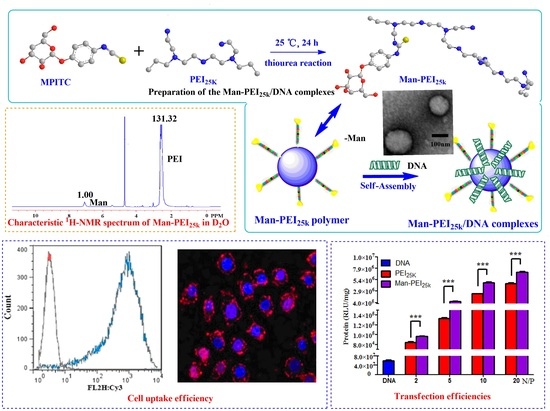
Synthesis of Mannosylated Polyethylenimine and Its Potential Application as Cell-Targeting Non-Viral Vector for Gene Therapy

1
Zhejiang Pharmaceutical College, Ningbo 315100, Zhejiang, China
2
Department of Medicine, Wenzhou Medical University, Wenzhou 325000, Zhejiang, China
3
Department of Medicine, Zhejiang Academy of Traditional Chinese Medicine, Hangzhou 310006, Zhejiang, China
4
College of Pharmaceutical Sciences, Zhejiang University, Hangzhou 310023, Zhejiang, China
*
Authors to whom correspondence should be addressed.
These authors contributed equally to this work.
Polymers 2014, 6(10), 2573-2587; https://doi.org/10.3390/polym6102573
Submission received: 11 August 2014 / Revised: 25 September 2014 / Accepted: 2 October 2014 / Published: 17 October 2014
(This article belongs to the Special Issue Polymers and Polymeric Nanoparticles for Therapy and Imaging)

Abstract

:
Mannose polyethylenimine with a molecular weight of 25 k (Man-PEI25k) was synthesized via a phenylisothiocyanate bridge using mannopyranosylphenyl isothiocyanate as a coupling reagent, and characterized by 1H NMR (nuclear magnetic resonance) and FT-IR (Fourier transform infrared spectroscopy) analysis. Spherical nanoparticles were formed with diameters of 80–250 nm when the copolymer was mixed with DNA at various charge ratios of copolymer/DNA (N/P). Gel electrophoresis demonstrated that the DNA had been condensed and retained by the PEI derivates at low N/P ratios. The Man-PEI25k/DNA complexes were less cytotoxic than the PEI complexes with a molecular weight of 25 k (PEI25k) at the same N/P ratio. Laser scan confocal microscopy and flow cytometry confirmed that the Man-PEI25k/DNA complexes gave higher cell uptake efficiency in (Dendritic cells) DC2.4 cells than HeLa cells. The transfection efficiency of Man-PEI25k was higher than that of PEI25k towards DC2.4 cells. These results indicated that Man-PEI25k could be used as a potential DC-targeting non-viral vector for gene therapy.

Graphical Abstract

1. Introduction

Polyethylenimine (PEI) is one of the most successful and efficient non-viral gene delivery systems to have been reported to date. It has been well documented that PEI can condense oligonucleotides, as well as plasmid DNA and siRNA to form complexes and promote endosomal escape via the “proton sponge” effect [1]. Branched PEI25k has been used as a standard reference during the development of new polymer systems because of its excellent performance as a vehicle for gene delivery [2]. The use of PEI as a gene carrier, however, has been limited because of its high cytotoxicity and non-specific interactions in vivo [3]. Various modifications have been applied to PEI to improve its biocompatibility, reduce its toxicity and increase its target specificity [4]. One efficient strategy for overcoming these limitations could be to add targeting ligands to polyplexes that would enhance cell-specific gene delivery through receptor-mediated cellular uptake [5,6]. Mannose is often used as a ligand in this context because it binds to the mannose receptors on the surfaces of cells to induce receptor-mediated endocytosis, which leads to an increase in the delivery efficacy of these systems [7]. Mannose receptor is expressed on the surfaces of antigen presenting cells (APCs; i.e., dendritic cells and macrophages) in the immune system [8,9]. A number of studies have shown that mannosylated nanoparticles enter Raw264.7 cells via receptor-mediated endocytosis [10]. Sun et al. reported that mannosylated biodegradable polyethyleneimine (man-PEI-TEG) exhibited a low level of cytotoxicity and performed well in both cellular uptake and transfection assays in vitro. They also demonstrated that man-PEI-TEG can be used as a DC-targeting gene-delivery system [11].
We designed and synthesized a perfectly appropriate gene delivery system that incorporates PEI and mannose at the specially appointed ratio based on preliminary experiments. For the more free amine groups were used to conjugate mannose, the less surface positive charges of the PEI-Man copolymers remained, in other words, with the increasing of the ratio of mannose group displays lower cytotoxicity but limited delivering efficiency, and based on the previously report, the amount of mannose was sufficient at 5% for receptor mediated gene delivery to the cells [12]. In this study, Man-PEI25k copolymers were synthesized and characterized. It was envisaged that these copolymers would exhibit a low cytotoxicity and enhance transfection efficiency through receptor-mediated endocytosis. The cytotoxicities and transfection efficiencies of the Man-PEI25k/DNA complexes were also evaluated in vitro to determine the suitability of these copolymers as gene vectors.

2. Experimental Section

2.1. Materials and Reagents

Branched PEI25k, α-d-mannopyranosylphenyl isothiocyanate (MPITC), 3-(4,5-dimethyl-thiazole-2-yl)-2,5-diphenyl tetrazolium bromide (MTT), agarose, and hydroxyethyl-piperazineethane-sulfonic acid buffer (HEPES) were purchased from Sigma-Aldrich (St. Louis, MO, USA). RPMI-1640 cell culture medium and fetal bovine serum were obtained from Life Technologies (Grand Island, NY, USA). Murine myeloid dendritic cell line DC2.4 and human cervical carcinoma cell line HeLa were purchased from the Chinese Academy of Sciences Kunming cell library (Kunming, Yunnan, China). The plasmid pGL3 encoding luciferase was obtained from Clonetech Co. (Mountain View, CA, USA) and was isolated and purified from DH5α Escherichia coli using the Qiagen End-free Plasmid Purification Kit (Germantown, MD, USA) prior to use. The Cy3-labeled DNA was synthesized by Sangon Biotech Co. Ltd. (Shanghai, China). The luciferase assay system was purchased from Promega (Madison, WI, USA). The bicinchoninic acid (BCA) protein assay kit was purchased from the Beyotime Institute of Biotechnology (Haimen, Jiangsu, China). All other chemicals and solvents used in the current study were obtained commercially and used without further purification.

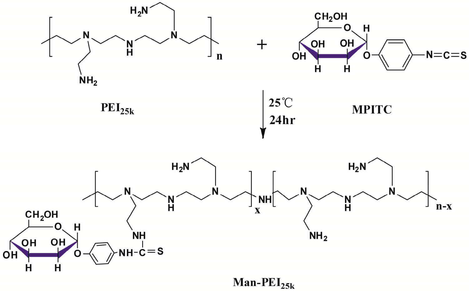
2.2. Synthesis of Man-PEI25k

The Man-PEI25k copolymer contained mannose, which was linked to PEI via a phenylisothiocyanate bridge using mannopyranosylphenyl isothiocyanate as a coupling reagent, and was synthesized via a previously published thiourea reaction [13]. Briefly, a α-d-mannopyranosylphenyl isothiocyanate (MPITC) to PEI feed ratio of 4.5:1 (mol/mol) was used to obtain the mannosylated derivates. Thus, 84 mg (or 270 μmol) of MPITC was dissolved in methanol (10 mL), and the resulting mixture was added to a solution of PEI25k (108 mg or 60 μmol) in methanol (0.5 mL) in a drop-wise manner. The reaction was then agitated at room temperature until no MPITC could be detected by thin layer chromatography (TLC) analysis. The reaction mixture was then evaporated under vacuum to allow for the removal of methanol.

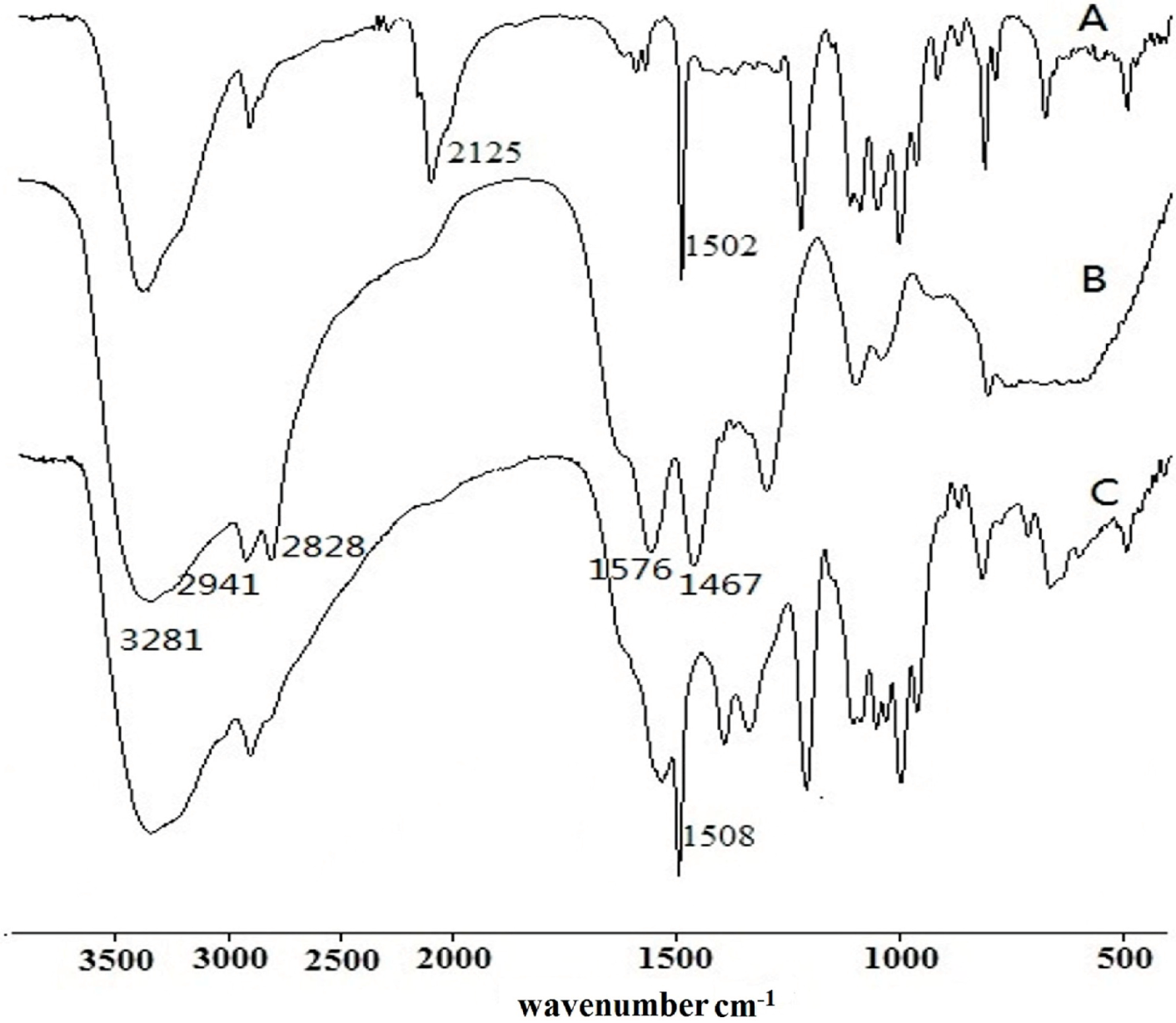
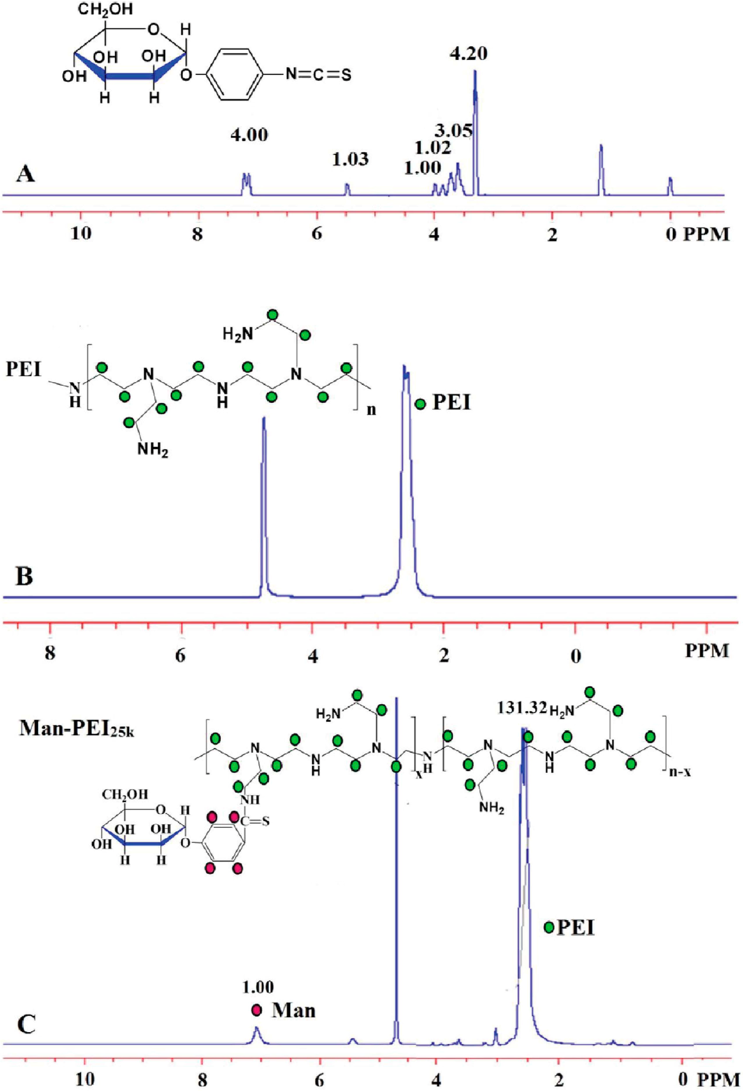
2.3. Characterization of Copolymers

NMR (nuclear magnetic resonance) spectra were recorded on a Bruker Avance 500 spectrometer (Bruker, Billerica, Germany) using D2O (0.5 mL) with tetramethylsilane as internal reference. IR spectra of the PEI25k and PEI25k derivatives were recorded on a Tenson 27 Fourier-transform infrared (FTIR) spectrometer (Bruker) as KBr disks. FTIR spectra were acquired over 16 scans between 4000 and 400 cm−1. The molar ratios of the mannose residues in the Man-PEI25k copolymers were calculated based on the size of the integrals corresponding to the proton resonance signals of interest in the 1H NMR spectra.

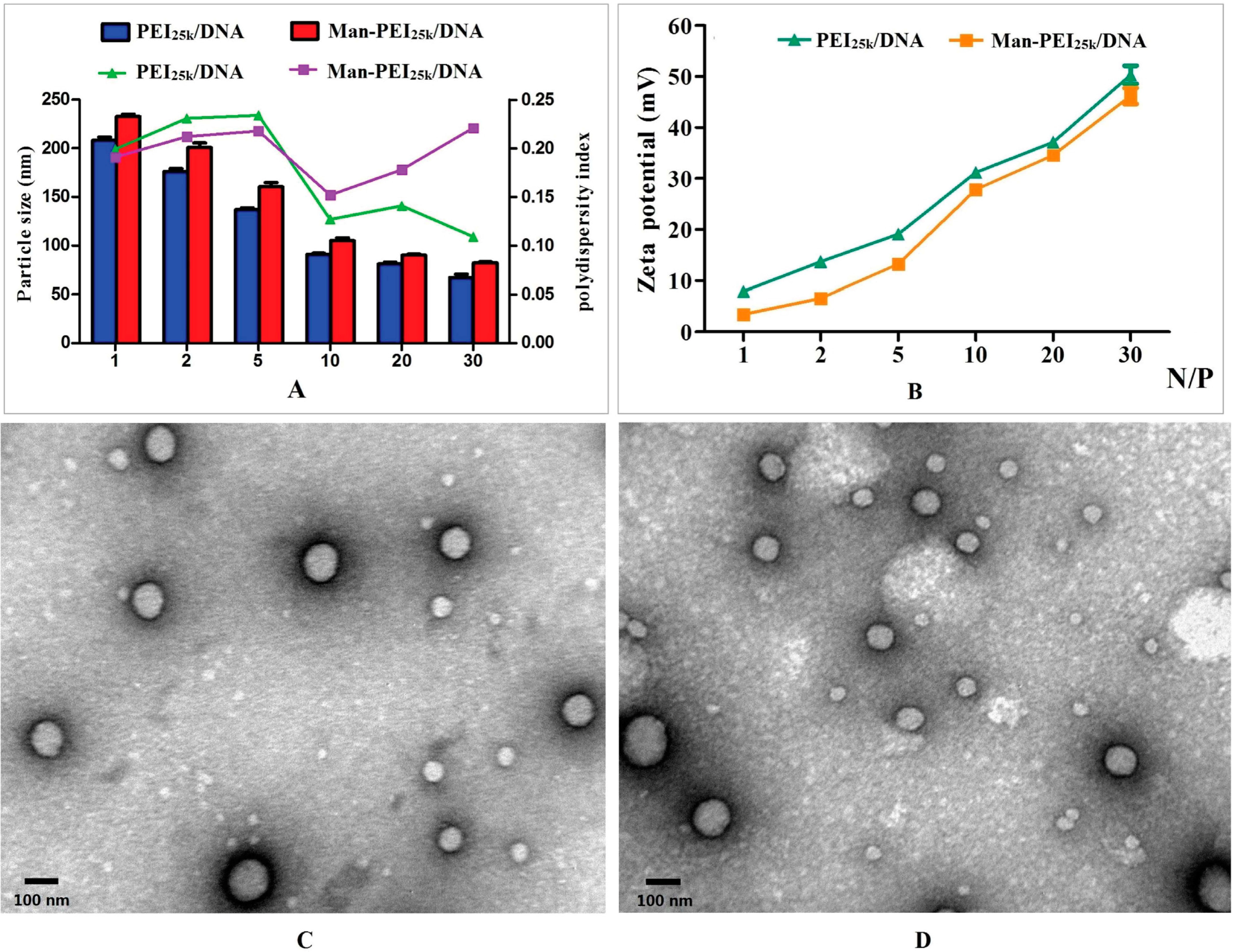
2.4. Characterization of the PEI/DNA Complexes

The mean particle size and zeta potential values of the PEI/DNA complexes were measured by dynamic laser scattering (DLS) using a Malvern Zetasizer Nano ZS90 (Malvern, UK) with a scattering angle of 90° at room temperature. Prior to analysis, the PEI/DNA complexes with N/P ratios in the range of 1:1 to 30:1 were prepared according to the conditions described previously [14]. All of these analyses were conducted in triplicate. The morphological characteristics of the PEI/DNA complexes (with an N/P ratio of 10) were observed by transmission electron microscope (TEM) using a JEM-1200EX TEM system (NEC, Tokyo, Japan), which was operated at 50 kV. A drop of an aqueous solution of the complex was deposited onto a 200-mesh carbon-coated copper grid, and any excess aqueous solution was removed by blotting with filter paper after 2 min. Each sample of the complex contained 10 μg of DNA, which was diluted with PBS buffer to a final volume of 1 mL.

2.5. Gel Retardation Assay

Electrophoresis was performed to estimate the ability of copolymers to condense DNA. A series of PEI/DNA complexes were prepared with N/P ratios in the range of 1 to 10 prior to the addition of the loading buffer. These samples were loaded onto 1% agarose gels in a Tris–acetate–ethylenediamine tetraacetic acid (TAE) buffer (40 mM Tris/HCl, 1 mM EDTA, pH 7.4) containing 0.5 μg·mL−1 of ethidium bromide and ran with TAE buffer at 90 V for 60 min. DNA retardation was visualized and photographed using a ChemiDoc™ XRS molecular imaging system (Bio-Rad, Hercules, CA, USA).

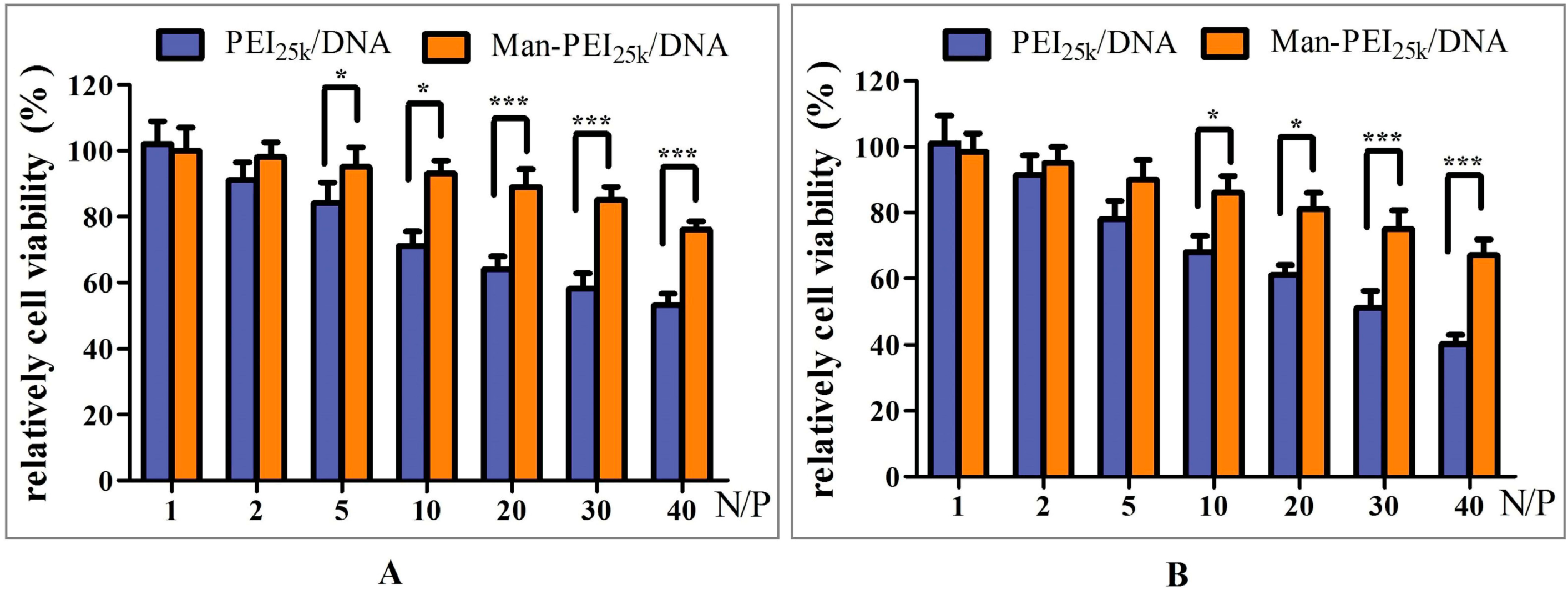
2.6. Cytotoxicity Assay

The cytotoxicity of the modified Man-PEI25k/DNA complexes and unmodified PEI/DNA complexes were evaluated using a 3-(4,5-dimethylthiazol-2-yl)-2,5-diphenyltetrazolium bromide (MTT) assay, as previously described. DC2.4 and HeLa cells were seeded into 96-well plates (5 × 10³ cells/well) and incubated for 24 h at 37 °C with 5% CO2, as previously described [14]. By keeping the amount of DNA as constant (1 μg/well), the PEI/DNA complexes of the different PEI derivates with N/P ratios in the range of 1 to 40 were then added to the cells, and the resulting mixtures were incubated for 24 h at 37 °C with 5% CO2. Twenty microliters of MTT solution (0.5 mg·mL−1 final concentration) in serum-free RPMI-1640 medium was then added to each well, and the resulting mixtures were incubated in the dark for 4 h under normal growing conditions. The medium in each well was then replaced with 150 μL of dimethylsulfoxide (DMSO), and the resulting mixtures were incubated for 15 min. The absorbance values of each well were then measured at 570 nm on a microplate reader (550; Bio-Rad, Philadelphia, PA, USA). Untreated cells were used as a control (Acontrol). The relative cell viability (%) compared with the control cells were calculated by Asample/Acontrol × 100%. All of the cytotoxicity assay experiments were performed in quintuplicate.

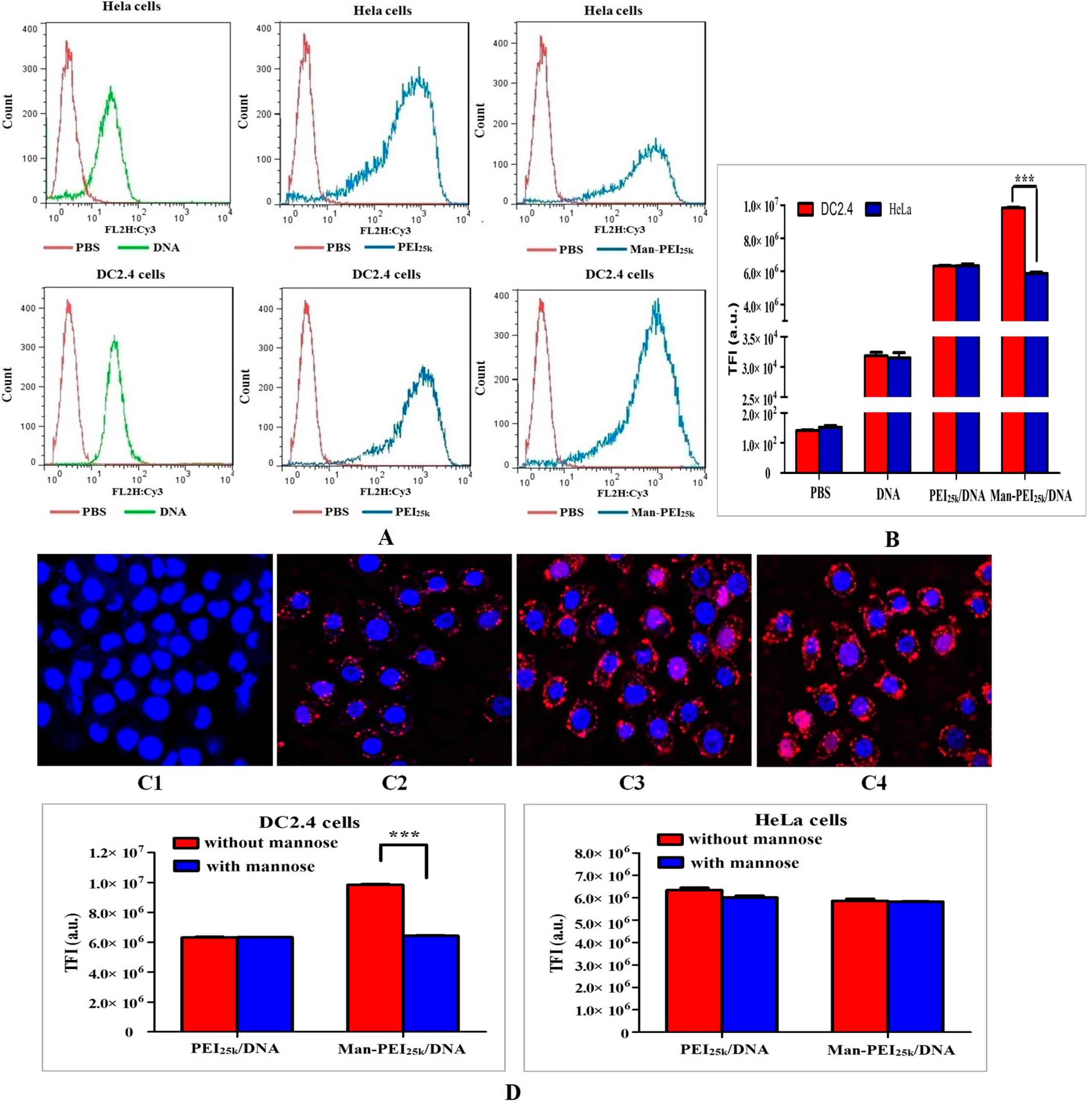
2.7. Cell Uptake Study

Cellular uptake in the DC2.4 and HeLa cells was examined by confocal laser scanning microscopy and flow cytometry, respectively. The cells were seeded in 6-well culture plates (5 × 10⁵ cells/well) and incubated for 12 h at 37 °C with 5% CO2 to obtain approximately 80% cell confluency. 0.2 μg·μL−1 of Cy3 labeled DNA solution were used to assemble the PEI/Cy3-DNA complexes with various N/P ratios, and then added (containing 10 μL of DNA solution/well), and the resulting mixtures were incubated for 4 h. The cells were then carefully rinsed three times with PBS (1 mL) to remove any residual complexes, before being trypsinized, re-suspended in PBS (500 μL) and analyzed by flow cytometry (BD Bioscience, San Jose, CA, USA). Untreated cells were used as negative controls. All of these experiments were performed in triplicate.
For the intracellular distribution studies, DC2.4 cells were seeded with a density of 5 × 10⁴ cells/dish in the confocal imaging dishes (Glass Bottom microwells, MatTek Corp., Ashland, MA, USA) and incubated for 12 h at 37 °C with 5% CO2 prior to the experiment. PEI/DNA complexes containing 2 μg of Cy3-DNA at an N/P ratio of 10 were prepared and co-cultured with the cells for 4 h. The cells were then washed five times with PBS (1 mL), fixed with 4% (w/v) paraformaldehyde (200 μL) for 30 min and rinsed three times with PBS (1 mL). The nuclei were then counterstained with 4',6-diamidino-2-phenylindole (DAPI) for 20 min and washed three times with PBS. The intracellular distributions of the complexes were analyzed by confocal laser scanning microscopy (CLSM; Olympus, FV-1000; Edwardstown, Australia).
A competition assay was also conducted to investigate the cell uptake efficiencies of the Man-PEI25k/DNA complexes (at an N/P ratio of 10) that were mediated by the mannose receptor via the addition of mannose to the cell culture medium. Both DC2.4 and HeLa cells were preincubated for 15 min with fixed amounts of mannose (20 mM) in 1 mL culture medium and the cell uptake assays were conducted as mentioned above. All of the cell uptake experiments were performed in triplicate.

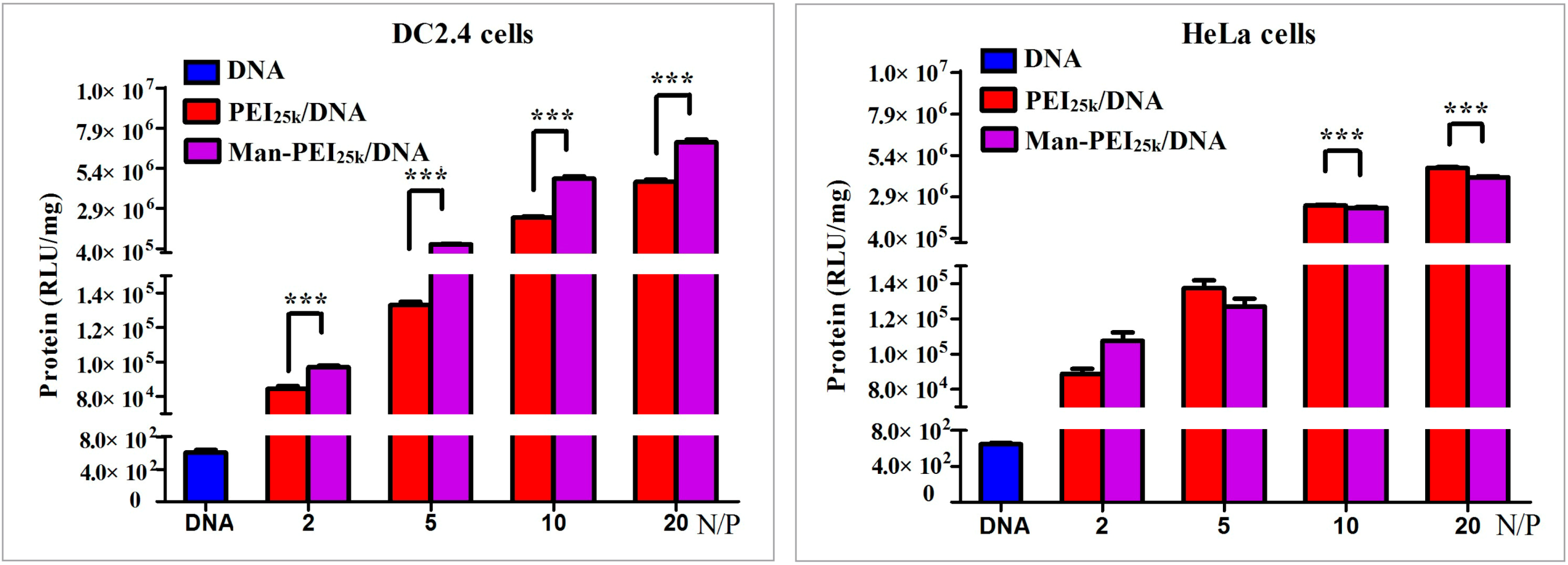
2.8. In Vitro Transfection

Plasmid pGL3 encoding luciferase was used as the reporter gene in the transfection efficiency assay. DC2.4 and HeLa cells were seeded into 6-well culture plates (5 × 10⁵ cells/well) and incubated for 12 h prior to the transfection experiments. When the cells were grown to 70%–80% confluence, the culture media were replaced with serum-free media containing PEI/pGL3 complexes at various N/P ratios and incubated for another 4 h. The serum-free media were replaced with fresh complete media containing serum, and the cells were incubated for a further 48 h. The luciferase activity was measured in terms of relative light units (RLU) using a luciferase assay system following the manufacturer’s instructions. The total protein concentration of transfected cell lysate was determined using a BCA protein assay kit (Beyotime, Haimen, Jiangsu, China). The final transfection efficiency was calculated as the relative lights units (RLU)/mg of total protein.

2.9. Statistical Analysis

The data presented in the current experiment have been reported as mean ± SD. Statistically significant differences were determined using the two-sample Student’s t-tests and ANOVA. Statistical significance is represented by * p < 0.05 and *** p < 0.01.

3. Results and Discussion

3.1. Synthesis and Characterization of Man-PEI25k

Man-PEI25k was prepared through a phenylisothiocyanate bridge using mannopyranosylphenyl isothiocyanate as a coupling reagent, which was synthesized via a thiourea reaction (Scheme 1). The reaction can happen quickly, for TLC analysis showed the completion of the reaction shortly after the adding of MPITC. Therefore the duration of reaction was within five minutes after dropwise add. The methanol in the reaction mixture was removed by rotary evaporation to dryness easily. MPITC (A), PEI25k (B), and Man-PEI25k (C) were characterized by FTIR spectroscopy, as shown in Figure 1. Figure 1A shows the FTIR spectrum of MPITC, where the characteristic peak of the –N=C=S stretching vibration can be clearly seen at 2125 cm−1. The band at 1502 cm−1 was attributed to the C=C stretching vibration of the phenyl group. The FTIR spectrum of PEI25k in Figure 1B contained bands at 3281 and 1576 cm−1, which were attributed to the stretching and bending vibrations of the N–H and –NH2 groups, respectively. The bands at 2941 and 2828 cm−1, and 1467 cm−1 were attributed to C–H stretching and scissor vibrations, respectively. Compared with the spectra of MPITC (A) and PEI25k (B), the spectrum of Man-PEI25k (C), which is shown in Figure 1C revealed several significant changes. Most notably, the band at 2125 cm−1 corresponding to the –N=C=S group had disappeared, which indicated that the –N=C=S sites on the PEI backbone must have undergone some reaction. Furthermore, a new peak appeared in the spectrum of Man-PEI25k (C) at 1508 cm−1, which was attributed to the phenyl group and confirmed that Man-PEI25k had been successfully synthesized.
Scheme 1. Reaction scheme of Man-PEI25k.
Polymers 06 02573 g008
Figure 1. FTIR (Fourier transform infrared spectroscopy) spectra of MPITC (α-d-mannopyranosylphenyl isothiocyanate) (A) PEI25; (B) Man-PEI25k; (C) (KBr pellets).
Polymers 06 02573 g001
The 1H NMR spectra of MPITC (A), PEI25k (B), and Man-PEI25k (C) are shown in Figure 2. The 1H NMR spectrum of MPITC (Figure 2A) contained a peak at 7.2 ppm, which was attributed to the hydrogen atoms of the phenyl groups. The 1H NMR spectrum of PEI25k (Figure 2B) contained a peak from 2.4 to 2.8 ppm, which was attributed to the –NCH2CH2– groups. The 1H NMR spectrum of Man-PEI25k (Figure 2C) contained similar signals to those of MIPTC and PEI25k, with peaks being observed at 2.4–2.8 and 7.1 ppm. This data effectively verified the structure of Man-PEI25k. Consideration of the integrals for these peaks allowed for a comparison of the intensity of peak at 7.1 ppm (assigned to phenyl group with four hydrogen atoms) with that of the peak at 2.4–2.8 ppm (assigned to PEI25k with about 600 –NCH2CH2– units). This result indicated that the mannose had been successfully conjugated to the PEI25k backbone via the reaction of the isothiocyanate groups in MPITC with the amine groups in PEI. The molar ratio (MR) of the mannose residues in the Man-PEI copolymer can be calculated using the following equation (Equation (1)):
MR = [2400 × (MMPITC7.1)/4 × (MPEI2.4–2.8)] × 100%
Using this equation, the molar ratio of the mannose residues in Man-PEI25k was calculated to be 457% from Figure 2C, which was consistent with the MPITC:PEI25k feed ratio of 4.5:1 (mol:mol).
Figure 2. 1H NMR spectra of MPITC (A) and PEI25k (B) in CD3OD. 1H NMR spectrum of Man-PEI25k (feed ratio of MPITC:PEI25k = 4.5:1 mol/mol) in D2O (C).
Polymers 06 02573 g002

3.2. Characterization of Complexes

The particle sizes of the PEI/DNA complexes were analyzed by DLS to investigate the degree of compaction within the DNA, which represents an important property for a gene delivery system [15]. The process of endocytosis in a variety of different mammalian cells requires the particles to be no more than 150 nm in diameter [16]. From Figure 3A, it can be seen that the sizes of the complexes generally decreased as the N/P ratio of the complexes increased, which indicated that stronger electrostatic interactions were being formed between the copolymer and the DNA at higher N/P ratios. The mean particle size across all of the PEI/DNA complexes was found to be in the range of 80 to 250 nm, with a narrow size distribution (polydispersity index (PDI) < 0.25). The Man-PEI25k/DNA complexes used in the current study showed slightly larger particle sizes than the PEI25k/DNA complexes at an N/P ratio of 10 (p > 0.05). The net positive charge of the copolymer/DNA complex plays a major role in determining the extent of its interaction with the negatively charged phospholipid surface of the cell membrane, which can have a significant impact on the gene transfection efficiency [17]. Strong cationic charges on the complexes, however, can result in cytotoxic activity [18]. Figure 3B indicated that the Man-PEI25k/DNA complexes had a low cationic charge density on the surface (when they had an N/P ratio in the range of 1–30), which was compared with that of the unmodified PEI25k/DNA complexes. Furthermore, all of the PEI/DNA complexes were positively charged, with the size of the positive charge increasing as the N/P ratio increased from one to 30. Figure 3C,D shows representative TEM images of the PEI/DNA complexes (at N/P ratio of 10). These images reveal the particles to be largely spherical, with mean diameters ranging from 150 to 200 nm. The particle sizes measured by TEM were in good agreement with the DLS results. The complexes were spherical and similar in size to the results mentioned above. These findings indicated that DNA had condensed successfully with the PEI derivates.

3.3. Gel Retardation Assays

Gel retardation assays were performed to investigate the ability of the PEI derivates to condense DNA via the formation of electrostatic interactions between the PEI derivates and the DNA at a variety of different N/P ratios (Figure 4). Figure 4A shows that the DNA was effectively condensed by Man-PEI25k when the N/P ratio was greater than three, indicating that all of the polymers could electrostatically neutralize the plasmid DNA and limit any electrophoretic mobility. This result demonstrated that the polymers were binding to the DNA and neutralizing its charge. If the N/P ratio of the polymer and DNA exceeded the neutralization composition, the resulting complexes would be positively charged, and they would stop migrating towards the anode. This experiment showed that all of the Man-PEI25k copolymers could condense the DNA effectively and neutralize its charge at an N/P ratio of three. Furthermore, the DNA could be retarded by the unconjugated PEI25k at an N/P ratio of two. This result indicated that more Man-PEI25k was needed than PEI25k to condense DNA completely. The difference between these results was attributed to more of the primary PEI25k amines being substituted by mannose, which would result in there being fewer positively charged primary amines to interact with the negatively charged DNA phosphates.
Figure 3. Characterization of the PEI/DNA complexes. (A) Nanoparticle size and polydispersity index of the two complexes formulations; (B) Zeta potentials of the two complex formulations at various N/P ratios; (C) TEM images of Man-PEI25k/DNA complexes at an N/P charge ratio of 10; and (D) TEM images of the PEI25k/DNA complexes at an N/P charge ratio of 10. Notes: Each data point represents the mean ± standard deviation (n = 3).
Polymers 06 02573 g003
Figure 4. Agarose gel electrophoresis of different complexes at various N/P (polymer particle/DNA) ratios: (A) Man-PEI25k and (B) PEI25k using 1% agarose in Tris-acetate running buffer.
Polymers 06 02573 g004

3.4. Cytotoxicity Assay

The cytotoxicities of the unmodified and modified PEI/DNA complexes were evaluated in DC2.4 and Hela cells using the MTT assay. As shown in Figure 5, the Man-PEI25k/DNA complexes exhibited much lower levels of cytotoxicity towards the DC2.4 and HeLa cells than the unmodified PEI25k/DNA complexes at all of the concentrations tested. These results also showed that the cytotoxicity increased in all of the PEI/DNA complexes as the N/P ratio increased. The viabilities of the two cell lines treated with Man-PEI25k/DNA were significantly higher than those treated with commercial PEI25k/DNA. The viabilities of the cells treated with Man-PEI25k complexes were greater than 80%, even up to an N/P ratio of 40, whereas the relative cell viabilities of PEI25k began to decrease quickly (Figure 5). Treatment of the cells with the PEI25k/DNA complexes resulted in high cytotoxicity at a high concentration, whereas treatment with the Man-PEI25k/DNA complexes resulted in reduced cytotoxicity. The difference in these results could be related to the incorporation of the mannose residues, which could effectively reduce the toxicity of carriers [11].
Figure 5. Cytotoxicities of the complexes at various N/P ratios in (A) DC2.4 and (B) HeLa cells by MTT assay. Each data point represents the mean value ± standard deviation (n = 6). *** p < 0.01, * p < 0.05.
Polymers 06 02573 g005

3.5. Cell Uptake and Intracellular Distribution

The cellular uptake properties of carriers are important to the delivery efficiency of a drug-targeted delivery system. Carriers with high cellular uptake properties generally make efficient drug delivery systems. The influence of mannose on cellular uptake via receptor-mediated endocytosis was investigated in the current paper. DNA was labeled with Cy3 red fluorophores to allow the cellular uptake and distribution of the mannosylated PEI/DNA complexes (N/P ratio of 10) to be tracked. The mean fluorescence intensity and uptake positive incidence were analyzed by flow cytometry (Figure 6A) following 4 h of incubation. The Man-PEI25k/DNA complexes exhibited higher cellular uptake efficiency in the DC2.4 cells than the PEI25k/DNA complexes. The cellular uptake efficiencies of the Man-PEI25k/DNA complexes were compared in the DC2.4 and HeLa cells to demonstrate the effect of mannose on receptor-mediated endocytosis. The DC2.4 cells also showed higher uptake efficiency than HeLa cells when they were treated with Man-PEI25k/DNA complexes with the same N/P ratio (Figure 6B), because HeLa cells have no mannose receptors on their surface [10]. DC2.4 cells were selected for the in vitro experiments in the current study because they express mannose receptors and are generally difficult to transfect. Furthermore, these cells are dendritic cells that behave as APCs in the immune system, and therefore represent a good model for the potential target of this delivery system [11]. The results of the current study suggested that mannose-modified PEI/DNA complexes could be taken-up selectively by DC2.4 cells by enhancing the level of transfer across the cell membrane.
The intracellular gene distribution was investigated by CLSM, and the results are shown in Figure 6C. The nuclei were stained with DAPI (blue) and the DNA was stained with Cy3 (red). For the PEI/DNA complexes, the DNA was mainly distributed in the cytoplasm and only small amounts of DNA were found in the nuclei following an incubation period of 4 h. The results indicated that the red fluorescence intensity levels inside the cells improved significantly following the treatment of the DC2.4 cells with Man-PEI25k/DNA complexes. Only minimal fluorescent intensities were observed around the cell membrane for the Cy3-labeled naked DNA molecules (Figure 6C2). The results indicated that more Man-PEI25k/DNA complexes had entered into the cells, which was similar to the results obtained for the PEI25k/DNA complexes (Figure 6C3,C4). The effective entry of the complexes into the cells is a prerequisite for efficient gene transcription.
To confirm the receptor-mediated endocytosis of the Man-PEI25k/DNA complexes, the uptake efficiencies of the Man-PEI25k/DNA and PEI25k/DNA complexes prepared with an N/P ratio of 10 were investigated in the presence of mannose as a competitor for the complexes (Figure 6D). The uptake efficiency of the Man-PEI25k/DNA complexes was significantly inhibited in the presence of mannose, whereas the uptake efficiency of the PEI25k/DNA complexes remained unchanged. The availability of mannose receptors was therefore essential for the entry of the Man-PEI25k/DNA complexes into the DC2.4 cells via the receptor-mediated delivery system.

3.6. Cell Transfection Assay

DC2.4 cells with abundant mannose receptors and HeLa cells without mannose receptors were evaluated using a luciferase assay system to determine the transfection efficiencies of the PEI/DNA complexes [19]. The results of these assays are shown in Figure 7, and indicated that the level of luciferase expression in the cells transfected with the positive control PEI25k/DNA complexes was about 150 times greater than the level of expression in the naked plasmid DNA-treated group (p < 0.01). The Man-PEI25k/DNA complexes (with an N/P ratio of 10) exhibited a transfection efficiency that was 3-fold greater than that of the complexes made from PEI25k (p < 0.01) at the same N/P ratio in DC2.4 cells. Compared with the PEI25k complexes, the luciferase activities were significantly higher when the DC2.4 cells were transfected with Man-PEI25k/pGL3 complexes at any N/P ratio, whereas the luciferase activities were significantly higher when HeLa cells were transfected with PEI25k/DNA complexes at a higher N/P ratio. These results indicated that the Man-PEI25k/DNA complexes had been internalized into cells by mannose receptor-mediated endocytosis.
Figure 6. Cell uptake efficiency: (A) Representative series of flow cytometry histograms; (B) Total fluorescence intensity of each group in the two different cell lines; (C) Confocal micrographs showing the cellular uptake of the complexes in DC2.4 cells; The DNA was labeled with Cy3 (red). The nuclei were stained with DAPI (blue). C1, DC2.4 cells without treatment; C2, naked DNA; C3, PEI25k/DNA; C4, Man-PEI25k/DNA; and (D) Competition assay with mannose in the two different cell lines. Each data point represents the mean ± standard deviation (n = 6). *** p < 0.01.
Polymers 06 02573 g006
Figure 7. Transfection efficiencies of the PEI/DNA complexes with various N/P ratios in the two different cell lines. Each data point represents the mean ± standard deviation (n = 6). *** p < 0.01.
Polymers 06 02573 g007

4. Conclusions

Man-PEI25k has been synthesized as a novel nonviral gene delivery vector by crosslinking PEI25k with mannose to form a copolymer for Dendritic cells (DC)-targeting and decreasing cellular toxicity. We have confirmed that Man-PEI25k complexes were able to form complexes with DNA using a variety of physicochemical methods and that the complexes themselves possessed physicochemical properties suitable for gene delivery. This new gene delivery vector showed much lower cytotoxicity and higher gene transfection efficiency properties than PEI25k towards DC2.4 cells. Taken together, these results demonstrate that Man-PEI25k has the potential to be used as a DC-targeting non-virus vector for gene therapy.

Acknowledgments

This study was financially supported by the Natural Science Foundation of China (Grant No. 30973648), and the Natural Science Foundation of Zhejiang Province, China (Grant No. LY12H30002, Z14H300001).

Author Contributions

Ying Hu and Jian-Qing Gao were mainly engaged in the research design; the main part of the research experiments were done by Bei-Hua Xu and Jiao-jiao Xu; Dan Shou was a writer and validator of the paper.

Conflicts of Interest

The authors declare no conflict of interest.

References

  1. Höbel, S.; Aigner, A. Polyethylenimine (PEI)/siRNA-mediated gene knockdown in vitro and in vivo. Methods Mol. Biol. 2010, 623, 283–297. [Google Scholar] [PubMed]
  2. Swami, A.; Kurupati, R.K.; Pathak, A.; Singh, Y.; Kumar, P.; Gupta, K.C. A unique and highly efficient non-viral DNA/siRNA delivery system based on PEI-bisepoxide nanoparticles. Biochem. Biophys. Res. Commun. 2007, 362, 835–841. [Google Scholar] [CrossRef] [PubMed]
  3. Kunath, K.; Merdan, T.; Hegener, O.; Häberlein, H.; Kissel, T. Integrin targeting using RGD-PEI conjugates for in vitro gene transfer. J. Gene Med. 2003, 5, 588–599. [Google Scholar] [CrossRef] [PubMed]
  4. Kawakami, S.; Hashida, M. Targeted delivery systems of small interfering RNA by systemic administration. Drug Metab. Pharmacokinet. 2007, 22, 142–151. [Google Scholar] [CrossRef] [PubMed]
  5. Cheng, H.; Zhu, J.L.; Zeng, X.; Jing, Y.; Zhang, X.Z.; Zhuo, R.X. Targeted gene delivery mediated by folate-polyethylenimine-block-poly(ethylene glycol) with receptor selectivity. Bioconjug. Chem. 2009, 20, 481–487. [Google Scholar] [CrossRef] [PubMed]
  6. Liang, B.; He, M.L.; Chan, C.Y.; Chen, Y.C.; Li, X.P.; Li, Y.; Zheng, D.; Lin, M.C.; Kung, H.F.; Shuai, X.T.; et al. The use of folate-PEG-grafted-hybranched-PEI nonviral vector for the inhibition of glioma growth in the rat. Biomaterials 2009, 30, 4014–4020. [Google Scholar] [CrossRef] [PubMed]
  7. Diebold, S.S.; Kursa, M.; Wagner, E.; Cotton, M.; Zenke, M. Mannose polyethylenimine conjugates for targeted DNA delivery into dendritic cells. J. Bio. Chem. 1999, 274, 19087–19094. [Google Scholar] [CrossRef]
  8. Chen, Y.Z.; Ruan, G.X.; Yao, X.L.; Li, L.M.; Hu, Y.; Tabata, Y.; Gao, J.Q. Co-transfection gene delivery of dendritic cells induced effective lymph node targeting and anti-tumor vaccination. Pharm. Res. 2013, 30, 1502–1512. [Google Scholar] [CrossRef] [PubMed]
  9. Chen, Y.Z.; Yao, X.L.; Ruan, G.X.; Zhao, Q.Q.; Tang, G.P.; Tabata, Y.; Gao, J.Q. Gene-carried chitosan-linked polyethylenimine induced high gene transfection efficiency on dendritic cells. Biotechnol. Appl. Biochem. 2012, 59, 346–352. [Google Scholar] [CrossRef] [PubMed]
  10. Jiang, H.L.; Kim, Y.K.; Arote, R.; Jere, D.; Quan, J.S.; Yu, J.H.; Choi, Y.J.; Nah, J.W.; Cho, M.H.; Cho, C.S. Mannosylated chitosan-graft-polyethylenimine as a gene carrier for Raw 264.7 cell targeting. Int. J. Pharm. 2009, 375, 133–139. [Google Scholar] [CrossRef] [PubMed]
  11. Sun, X.; Chen, S.; Han, J.; Zhang, Z. Mannosylated biodegradable polyethyleneimine for targeted DNA delivery to dendritic cells. Int. J. Nanomed. 2012, 7, 2929–2942. [Google Scholar] [CrossRef]
  12. Zanta, M.A.; Boussif, O.; Adib, A.; Behr, J.P. In vitro gene delivery to hepatocytes with galactosylated polyethylenimine. Bioconjug. Chem. 1997, 8, 839–844. [Google Scholar] [CrossRef] [PubMed]
  13. Diebold, S.S.; Plank, C.; Cotton, M.; Wagner, E.; Zenke, M. Mannose receptor-mediated gene delivery into antigen presenting dendritic cells. Somat. Cell Mol. Genet. 2002, 27, 65–74. [Google Scholar] [CrossRef]
  14. Hu, Y.; Xu, B.; Ji, Q.; Shou, D.; Sun, X.; Xu, J.; Gao, J.; Liang, W. A mannosylated cell-penetrating peptide-graft-polyethylenimine as a gene delivery vector. Biomaterials 2014, 35, 4236–4246. [Google Scholar] [CrossRef] [PubMed]
  15. Nimesh, S.; Thibault, M.M.; Lavertu, M.; Buschmann, M.D. Enhanced gene delivery mediated by low molecular weight chitosan/DNA complexes: Effect of pH and serum. Mol. Biotechnol. 2010, 46, 182–196. [Google Scholar] [CrossRef] [PubMed]
  16. Ahn, H.H.; Lee, M.S.; Cho, M.H.; Shin, Y.N.; Lee, J.H.; Kim, K.S.; Kim, M.S.; Khang, G.; Hwang, K.C.; Lee, I.W.; et al. DNA/PEI nano-particles for gene delivery of rat bone marrow stem cells. Colloids Surf. Physicochem. Eng. Aspect 2008, 313–314, 116–120. [Google Scholar] [CrossRef]
  17. Ong, Z.Y.; Fukushima, K.; Coady, D.J.; Yang, Y.Y.; Ee, P.L.; Hedrick, J.L. Rational design of biodegradable cationic polycarbonates for gene delivery. J. Control. Release 2011, 152, 120–126. [Google Scholar] [PubMed]
  18. Liang, W.; Gong, H.; Yin, D.; Lu, S.; Fu, Q. High-molecular-weight polyethyleneimine conjuncted pluronic for gene transfer agents. Chem. Pharm. Bull. 2011, 59, 1094–1101. [Google Scholar] [CrossRef] [PubMed]
  19. Mendoza, L.; Olaso, E.; Anasagasti, M.J.; Fuentes, A.M.; Vidal-Vanaclocha, F. Mannose receptor-mediated endothelial cell activation contributes to B16 melanoma cell adhesion and metastasis in liver. J. Cell. Physiol. 1998, 174, 322–330. [Google Scholar] [CrossRef] [PubMed]

