Article

Optimization of Biopolymer Based Transdermal Films of Metoclopramide as an Alternative Delivery Approach

1
Department of Pharmaceutical Technology, Faculty of Pharmacy, Istanbul University, Istanbul 34116, Turkey
2
Department of Analytical Chemistry, Faculty of Pharmacy, Istanbul University, Istanbul 34116, Turkey
*
Author to whom correspondence should be addressed.
Polymers 2014, 6(5), 1350-1365; https://doi.org/10.3390/polym6051350
Submission received: 27 January 2014 / Revised: 29 April 2014 / Accepted: 30 April 2014 / Published: 9 May 2014
(This article belongs to the Special Issue Natural Polymers 2014)

Abstract

:
The objectives of this study were to develop and to characterize sodium alginate based matrix-type transdermal films of metoclopramide hydrochloride (MTC) in order to improve patient compliance to treatment. The suitability of sodium alginate was shown to be a natural film former in terms of the physicochemical, mechanical, and bioadhesive features of the MTC loaded transdermal films. Terpinolene provided the highest drug release among the different terpenes (nerolidol, eucalyptol, dl-limonene, or terpinolene) assessed as enhancer. Attenuated Total Reflectance Infrared (ATR-FTIR) spectroscopy analysis performed to evaluate the effect of the transdermal films on skin barrier confirmed enhancer induced lipid bilayer disruption in stratum corneum, indicating its permeation enhancement effect.

1. Introduction

Biopolymers obtained from natural sources are usually biodegradable and their biocompatibility is excellent [1]. Natural biopolymers in protein and polysaccharide structures are widely used as backings, adhesives or matrix carriers in the compositions of dermal and transdermal delivery systems [2,3,4,5]. Natural polysaccharides represent a group of polymers playing a fundamental role in controlling the mechanism and rate of drug release from a dosage form [2]. Alginic acid is a natural linear polysaccharide isolated from marine brown algae and from bacterial fermentation [6]. It is a block copolymer of two different monosaccharides, β-d-(1,4)-mannuronic acid and α-l-(1,4)-glucuronic acid. Its salt, sodium alginate (Figure 1), is an anionic polymer, which plays an important role in the design of controlled drug delivery formulations. It is widely used in topical pharmaceutical products as a thickening and suspending, as well as stabilizing agent and as a wound dressing due to its excellent swelling properties and biocompatibility [6,7]. The choice of polymers used in transdermal delivery systems has strong impact on drug release, permeability, elasticity, and wearing properties of the formulations. The bioadhesion strength of polymers with ionisable carboxyl groups, such as sodium alginate was found to be stronger than that of those with neutral or non-ionisable groups [8]. The gels prepared with sodium alginate to fabricate transdermal films have been found appropriate in terms of mechanical strength, flexibility, and bioadhesiveness of the films [5].
Figure 1. Structural formula of sodium alginate [9].
Polymers 06 01350 g001
Transdermal delivery of drugs have numerous benefits including avoidance of hepatic first pass effect, maintenance of constant drug plasma level, and improved patient compliance to the treatment [10,11]. However, transdermal transport of drugs is limited due to low permeability of the uppermost layer of the skin, stratum corneum. In order to overcome that barrier and to enhance the permeability of the drugs across the skin, permeation enhancers and co-solvents, which reversibly perturb the permeability of stratum corneum or increase partition of drug to stratum corneum are included in the transdermal formulation [12,13,14].
Terpenes are considered as effective class of chemical permeation enhancers and, they have been approved by the Food and Drug Administration (FDA) as generally regarded as safe (GRAS) [15,16,17]. Terpenes are derived from plant essential oils, have been shown to increase the percutaneous absorption of both hydrophilic and lipophilic drugs [12,15,18,19]. It was reported that terpenes enhance the permeation of drug across the skin mainly by disrupting the highly ordered intercellular chain packing of stratum corneum lipids and thereby increasing drug diffusivity [20,21]. It has been shown that the activity of terpenes as permeation enhancer is primarily dependent on the lipophilicity and structure of both drugs and terpenes [22,23,24,25]. In our study, four different types of terpenes, nerolidol (amphiphilic sesquiterpene, logP: 5.32), eucalyptol (oxygen containing monoterpene, logP: 2.82), terpinolene (hydrocarbon lipophilic monoterpene, logP: 4.52), and limonene (hydrocarbon lipophilic monoterpene, logP: 4.58) were selected.
Metoclopramide (MTC) (Figure 2) is an antiemetic drug, effective in the treatment of nausea and vomiting associated with cancer therapy, migraine, pregnancy, etc. [26]. The oral bioavailability of MTC is variable due to the hepatic first pass metabolism and oral dosage forms of MTC often get vomited out before systemic absorption occurs. Parenteral and rectal administration routes both result in low patient compliance [27]. Therefore, the transdermal delivery of MTC seems to be an attractive alternative route.
Figure 2. Chemical structure and properties of MTC [28,29].
Polymers 06 01350 g002
The aim of the present study was to develop transdermal films of MTC with sodium alginate as matrix agent and propylene glycol as plasticizer. In this perspective, the physicochemical, mechanical and bioadhesive properties of the developed formulations were assessed and the impact of different types of terpenes as permeation enhancers on in vitro release of MTC was examined. ATR-FTIR spectroscopic studies on excised pig skin were also performed in order to evaluate the effect of terpenes on skin barrier properties.

2. Experimental Section
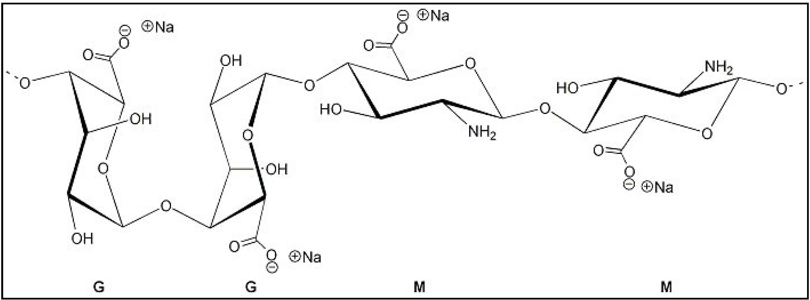
2.1. Materials

MTC was kindly provided from Recordati Drug Company (Istanbul, Turkey). Acetonitrile, dl-limonene and Tween® 80 were supplied from Merck (Darmstadt, Germany). Nerolidol, terpinolene and eucalyptol were acquired from Sigma-Aldrich (Dorset, UK). The matrix forming polymer, sodium alginate, composed of approximately 61% mannuronic acid (M) and 39% guluronic acid (G) with a M/G ratio of 1.56, a polydispersity index of 2.32 and a average molecular weight (Mw) which ranges between 80,000 and 120,000 Da was purchased from Sigma-Aldrich. All other reagents were used without additional purification.

2.2. Methods

2.2.1. Solubility of MTC in Terpenes and Co-Solvents

A mixture of terpenes (nerolidol, eucalpytol, terpinolene or dl-limonene), propylene glycol and Tween® 80 at the concentrations of 1%, 7.5%, and 1%, respectively, was prepared. The volume of each mixture was adjusted to 10 mL with water. Excess amount of MTC was added to 0.5 mL of these mixtures and shaken at 250 rpm at room temperature for 24 h. After 5 min centrifugation at 3000 rpm the supernatant was withdrawn with a membrane filter (0.45 µM, Millex LH, Merck Millipore, Darmstadt, Germany) and suitable dilution was made. The amount of MTC was analyzed by the high performance liquid chromatography (HPLC) (Shimadzu HPLC System, Kyoto, Japan) method given below. Each experiment was performed at least three times.

2.2.2. Formulation of Gels

The codes and composition of MTC gel formulations are shown in Table 1. The concentrations of the natural matrix polymer (sodium alginate), plasticizer (propylene glycol) and terpenes (nerolidol, eucalyptol, terpinolene, and limonene) are determined in pre-formulation studies.
Briefly, undialyzed sodium alginate was dispersed in water for 24 h. A mixture of Tween® 80 and terpene was homogenized in water at 8000 rpm during 5 min. MTC was dissolved in water and combined with the prepared emulsion system. This MTC containing emulsion was added to the sodium alginate gel base and finally propylene glycol was added as a plasticizer agent and stirred continuously until a homogeneous gel was obtained.
Table 1. The codes and composition of gel formulations (G) and transdermal films (TF) of metoclopramide hydrochloride (MTC) prepared from the gel formulations (w/w, % on wet basis).
CompositionsGel and Transdermal Formulations
G-NRTF-NRG-EUTF-EUG-TPTF-TPG-LMTF-LM
Sodium alginate4.54.54.54.5
Propylene glycol7.57.57.57.5
Nerolidol1.0
Eucalptol1.0
Terpinolene1.0
dl-Limonene1.0
Tween 801.01.01.01.0
Distilled water86868686
Metoclopramide HCl
% w/w a1.01.01.01.0
mg/cm2 b3.33.33.33.3
Note: a On wet basis, amount of gel prepared = 25 g per batch; b As a base finished product.

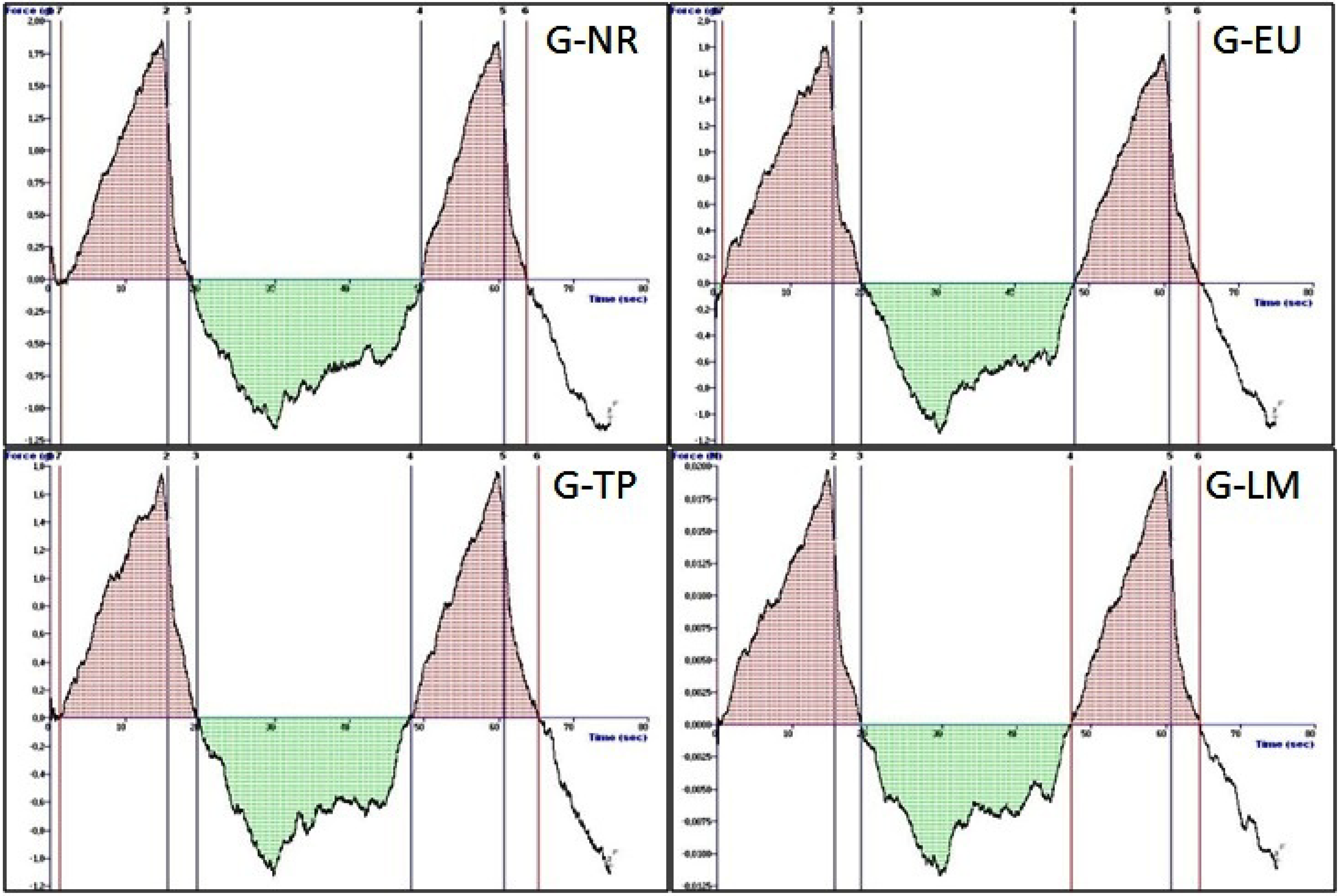
2.2.3. Mechanical Properties of the Sodium Alginate Gels-Texture Profile Analysis

Evaluation of the mechanical properties (hardness, compressibility, adhesiveness, elasticity, and cohesiveness) of sodium alginate gel formulations coded with G-NR, G-EU, G-TP, G-LM consist of different type of terpene penetration enhancer were performed using texture profile analyzer (TPA) (TA-XT Plus, Stable Micro Systems, Surrey, UK). 20 mL volume glass beakers filled with gel formulations were placed in the ultrasonic water bath for 30 min prior to experiments to remove any air bubbles. After the temperature of the formulations maintained at 37 ± 0.5 °C, the hemispherical analytical probe was twice inserted into each sample to a depth of 15 mm, at a defined rate of 2.0 mm·s−1. The delay period between the end of the first and beginning of the second compression was 15 s. All analyses were performed six times and data collection and calculation were performed using the Texture Exponent 6.0.7.0 software (Stable Micro Systems, Surrey, UK) package of the instrument. The resultant force-time plots were used for determination of the mechanical properties. Data evaluation was performed using the Texture Exponent software package of the instrument [5,30,31].

2.2.4. Formulation of MTC Transdermal Films

The codes and composition of transdermal films developed (TF-NR, TF-EU, TF-TP and TF-LM) are given in Table 1. The transdermal films of MTC were obtained by casting the gel dispersions mentioned in Table on petri dishes (θ: 9.8 cm) followed by drying at 40 ± 2 °C for 15 h. All transdermal formulations were prepared 24 h before the in vitro release studies. Dried films were stored in a desiccator wrapped in aluminium foil.

2.2.5. Physicochemical Characteristics of MTC Transdermal Films

Organoleptic Examination

Transdermal films of MTC were evaluated by visual inspection in terms of color, transparency, smoothness, flexibility, and homogeneity.

Thickness

Film thickness of MTC transdermal films was measured with a digital micrometer (QLR Digit IP4, Qinghai, China). Mean values and standard deviations were calculated.

Uniformity of Weight

The uniformity of weight for each formulation was calculated in six pieces of 1.77 cm2 film by calculating their average weight, and the deviation from average weight was determined.

Uniformity of Drug Content

A known weight of film was dissolved and subsequently diluted with 0.9% (w/w) saline solution. Samples were filtered through membrane filters (0.45 µM, Millex LH, Merck Millipore, Darmstadt, Germany) prior to HPLC analysis. Each film formulation was tested in triplicate and the results were expressed as the mean and standard deviation.

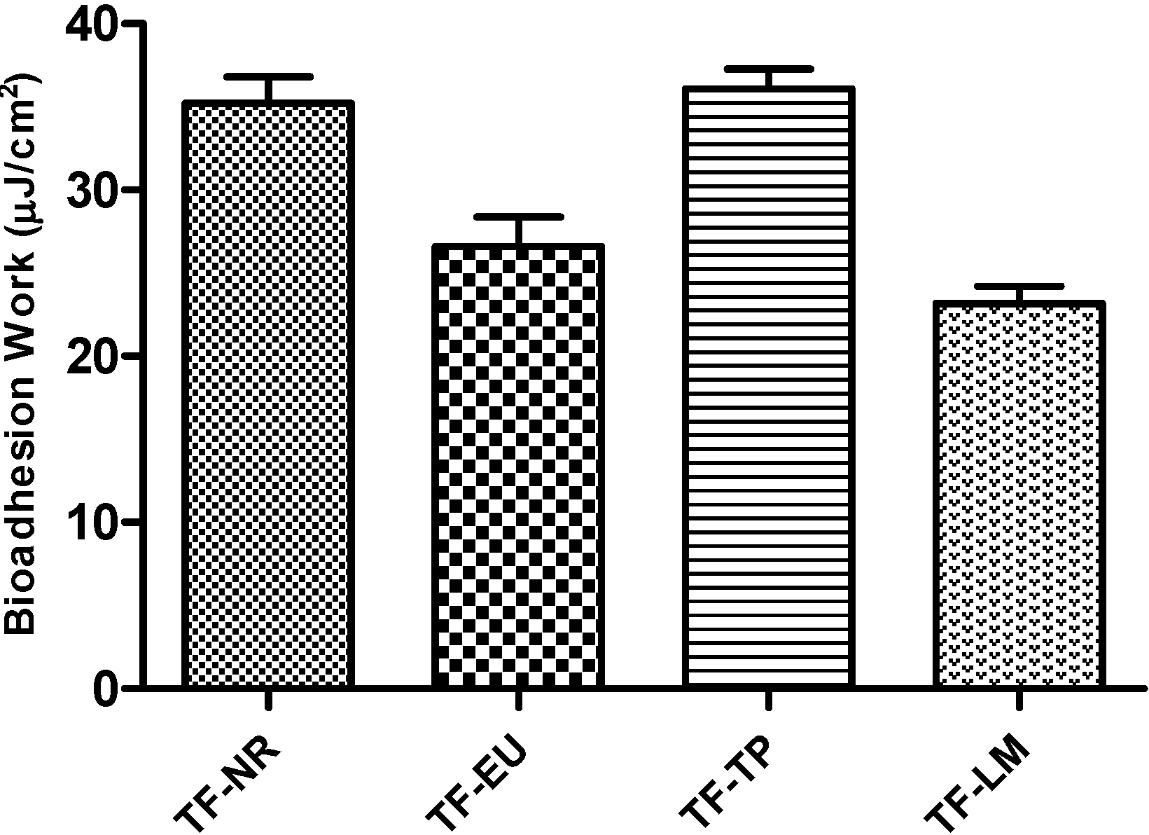
2.2.6. In Vitro Adhesion Test

The adhesive properties of the sodium alginate based transdermal films of MTC (TF-NR, TF-EU, TF-TP and TF-LM) and control formulation (TF-CONT) prepared in this study were assessed on asynthetic cellulose membrane (Visking Tubing 27/32, London, UK) using the TPA. The membrane was allowed to hydrate with phosphate buffer pH 7.4 at 37 °C prior to the experiments. The membrane was held on the lower platform of the instrument and the transdermal film was applied on it. The upper probe was immersed on the film surface, kept in contact for 120 s, and then it moved at a constant speed of 1 mm·s−1. The force required to detach the film from the membrane was determined as the peak value in resultant force-distance plot. The in vitro adhesion work in 1 cm2 was calculated with the following equation and the adhesion values were expressed as the mean of three replicates:
Bioadhesion work (mJ/cm2) = AUC1-2r2
πr2 = Area of the transdermal formulation in contact with the skin
AUC1-2 = Area under the force-distance plot

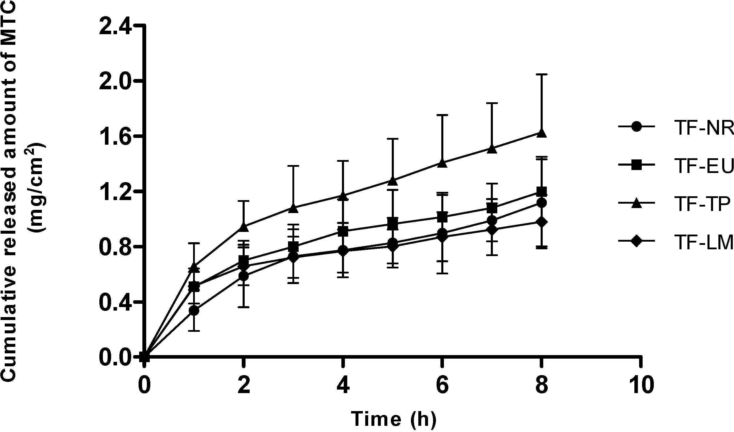
2.2.7. In Vitro Release Studies of MTC from Transdermal Films

Modified Franz-type diffusion cells of an area of 3.14 cm2 and receptor volume of 33.2 mL were used for assessing the release behavior of MTC from the sodium alginate based transdermal films. A mixture of ethanol:water (1:50) was used as a receptor fluid in order to maintain sink conditions in the receiver compartment. The receptor medium was continuously stirred with a teflon coated magnetic stirrer at 600 rpm and thermostated at 37 ± 0.5 °C throughout the experiments. Cellulose membrane (Visking Tubing 27/32, London, UK) was allowed to hydrate with distilled water 24 h before being mounted on the cells. A circular section (r = 1 cm) of transdermal film was fixed on the membrane and all cell donors were occluded with a Parafilm®. 1 mL sample of the receptor was taken at predetermined time intervals over 8 h and replenished immediately with equal volume of the receptor fluid. MTC amount was analyzed by the HPLC method after diluting at appropriate ratios. The results were expressed as the mean ± SD of three experiments. The cumulative amount of diffused drug was plotted against time. Drug release mechanism from each transdermal formulation (TF-NR, TF-EU, TF-TP, and TF-LM) was evaluated kinetically with zero-order, first-order and Higuchi release models [32]. The model with the highest coefficient correlation (r2) was judged to be a more appropriate model for the release data.

2.2.8. HPLC Analysis

The quantitative determination of MTC in this study was performed using a Shimadzu HPLC system (Shimadzu, Kyoto, Japan) equipped with an SPD-M20A UV/Vis detector, a LC 20A pump and a reversed phase C18 column (250 mm × 4.6 mm; 5 µm, Waters Symmetry). The mobile phase was a mixture of acetonitrile: sodium acetate (pH 4.7) (35:65 v/v) filtered through membrane filters (0.45 µM, Millex LH, Merck Millipore, Darmstadt, Germany) and eluted at a flow rate of 1 mL/min. Analyses were performed using a detection wavelength of 273 nm. The method was validated for selectivity, linearity, accuracy, and precision. It was found to be linear over the concentration range 0.2–10 µg/mL with a high correlation coefficient (r2 > 0.999) and precise (intra and inter day variation < 2%) and accurate (recovery > 98%). There were no interfering peaks with MTC confirming the selectivity of the method. Stability studies showed that MTC was stable during 48 h in the mobile phase.

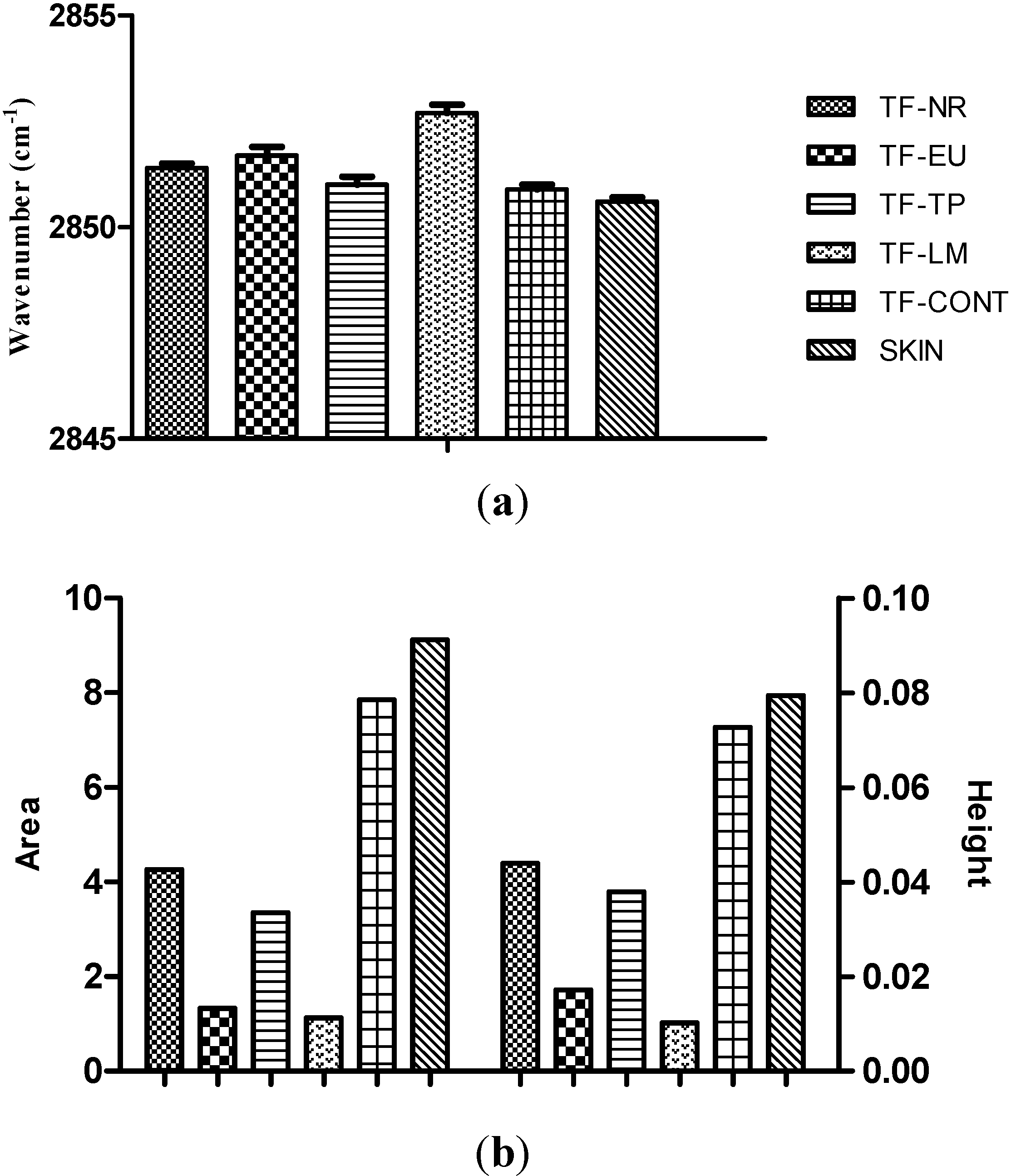
2.2.9. Attenuated Total Reflectance Infrared (ATR-FTIR) Spectroscopy Studies

The interaction between the sodium alginate based transdermal films of MTC and the skin were investigated using ATR-FTIR spectroscopy. ATR-FTIR spectroscopy has been widely employed to report on stratum corneum intercellular lipid ordering through the C–H stretching absorbances from the methylene groups of the lipid acyl chains [33,34]. The spectrographs of skin obtained from ATR-FTIR studies provide information about the influence of chemical penetration enhancers on reduced order of skin lipids and proteins [20]. For this purpose in our study the subcutaneous fat tissue of excised pig skin was removed with a scalpel and the hairs were clipped with hair clippers. The skin was stored frozen at −25 °C (wrapped in Parafilm® and packed in ZipLock bags) for not longer than 2 months prior to use. Before collection of ATR-FTIR spectra, samples were defrosted 8 h at 4 °C. After defrosting, skin surface was wiped two times with wet cotton swab and excess water was gently dried with paper tissue. Transdermal films of MTC were applied immediately on the surface of the skin and kept for 3 h at 25 °C under non-occlusive conditions. After the treatment period, excess of formulation was gently blotted away from the surface, then spectra were collected with a spectral resolution of 4 cm−1 in the 4000–650 cm−1 range using a Perkin Elmer Spectrum 100 FT-IR spectrometer (Cambridge, UK) equipped with a diamond ATR crystal. Treated skin, cut to dimensions 2 cm × 2 cm, was placed stratum corneum side down onto the ATR crystal. To ensure reproducible contact between the sample and the crystal, always the same pressure on top of samples was applied (force gauge 80 N). Attention was focused on detecting the shifts of the symmetric (SSV) C–H stretching bands near 2850 cm−1. For each treatment, peak height and area was measured and a non-treated skin served as control. In order to evaluate the effects of penetration enhancers, a control film was also prepared following the same method and components without adding penetration enhancer (TF-CONT). Spectrum Version 6.0.2 software (Perkin Elmer, Cambridge, UK) was used for calculations with ±0.1 cm−1 accuracy.

2.2.10. Statistical Analysis

The significance of the differences between values was assessed using GraphPad Prism 5 software (La Jolla, CA, USA). One-way ANOVA, followed by the Newman-Keuls multiple comparison tests were performed.

3. Results and Discussion

The solubility of MTC in the terpene, propylene glycol, and Tween 80 mixture are determined. The solubility data indicated that while nerolidol had significantly highest MTC solubility capacity (977.58 ± 2.09 mg/mL) than those of other terpenes examined (p < 0.05), terpinolene had the lowest solubility capacity (826.38 ± 1.65 mg/mL). The solubility of MTC in eucalyptol and dl-limonene was 927.42 ± 0.53 mg/mL and 872.12 ± 0.71 mg/mL, respectively.
In this study, firstly the difference in the mechanical characteristics of the sodium alginate based MTC gels was investigated firstly using TPA. Our results are given in Table 2 and Figure 3.
Table 2. The mechanical properties of gel formulations (G-NR, G-EU, G-TP, G-LM) used to formulate transdermal films (n=6).
CodeHardness (n)Elasticity (mJ)CohesivenessAdhesiveness (µj)Compressibility (n·mm)
G-NR0.02 ± 0.010.71 ± 0.040.91 ± 0.04104.33 ± 9.020.13 ± 0.03
G-EU0.02 ± 0.010.87 ± 0.050.88 ± 0.0357.33 ± 10.700.17 ± 0.02
G-TP0.02 ± 0.010.80 ± 0.060.85 ± 0.1285.33 ± 4.040.17 ± 0.05
G-LM0.01 ± 0.020.79 ± 0.080.91 ± 0.0484.67 ± 3.790.14 ± 0.02
Figure 3. The force-time plots of sodium alginate based gel formulations G-NR, G-EU, G-TP, and G-LM.
Polymers 06 01350 g003
The hardness parameter represents the strength of the gel structure under compression and introduces the required force to provide the deformation of a gel formulation [30]. In the present study it is found that the hardness values of MTC gels were between 0.02 ± 0.01 and 0.01 ± 0.02. The hardness value of the gel formulation G-LM was the lowest at body temperature. A low hardness value has been reported as an advantage for administration to the skin or mucosa easily [35]. The hardness influences the pourability and spreadability of the gel into film molds [30]. The gel formulations G-NR, G-EU, G-TP, and G-LM in our study flowed properly and spread homogenously when poured into the film molds indicating that the obtained hardness values were appropriate.
Elasticity is defined as the direction of reconstruction of the gel after its deformation by compression in a defined period of time. It significantly affects the elasticity and homogeneity of the films to be prepared by using these gels. The increase in the quantitative value of elasticity showed the decrease in the elasticity of the gel formulation [30,36]. We observed that eucalyptol containing gel formulation G-EU showed the highest elasticity value (0.87 ± 0.05). Bektaş et al., showed that sodium alginate as polymer at concentration of 4.5% and propylene glycol at a concentration of 7.5% as plasticizer were suitable for fabrication of transdermal films of nifedipine [5]. Our results are in accordance with this finding.
Cohesiveness parameter is a measure of the degree of difficulty in breaking down the gels internal structure and significantly affects the strength and the elasticity of the film formulations. We observed that there was not a significant difference between the gel formulations G-NR, G-EU, G-TP, and G-LM in the mean of cohesiveness (p > 0.05). The cohesiveness of the sodium alginate based gels in our study ranged between 0.85 ± 0.12 and 0.91 ± 0.04 which are in accordance with literature [5].
The adhesiveness is calculated from the negative force area obtained during the first immersion period and represents the work required to overcome the attractive forces between the surface of the gel and the surface of the probe [36,37]. In our study the adhesiveness value of gel formulation G-NR (104.33 ± 9.02) was significantly higher when compared with the adhesiveness of G-EU (57.33 ± 10.70). Thus, it can be suggested that the bioadhesive property of the gel formulation coded with G-NR seems to be the highest one among the gel formulations studied, whereas G-EU is expected to show the weakest bioadhesive characteristics [37].
The compressibility defines the work required for the compaction of the gel along a definite distance [36,38]. According to our data, it can be assumed that the spreadability of G-LM and G-NR can be better than that of G-EU and G-TP because of their low compressibility value.
It was evident from the TPA data that all of the sodium alginate based gel formulations showed suitable mechanical characteristics and they could be used to prepare the transdermal films of MTC.
All transdermal films (TF-NR, TF-EU, TF-TP, and TF-LM) of MTC prepared using sodium alginate as matrix agent in this study were thin, flexible and smooth. The thickness, weight and drug content of the film formulations are represented in Table 3.
Table 3. Thickness, weight and drug content uniformity of the MTC transdermal films (mean±SD, n = 3–6).
Transdermal FilmThickness (mm)Weight (mg·cm−2)MTC Content (%)
TF-NR0.55 ± 0.0560.12 ± 1.46102.48 ± 0.73
TF-EU0.67 ± 0.0458.60 ± 1.51101.56 ± 1.43
TF-TP0.51 ± 0.0263.59 ± 1.7798.29 ± 1.76
TF-LM0.47 ± 0.0351.18 ± 1.1692.70 ± 0.74
The thickness of MTC transdermal films was found between 0.47 ± 0.03 and 0.67 ± 0.04 mm and the standard deviation of weight was less than 2%, indicating homogenously spreadability of gels while preparing transdermal films. The MTC content of the transdermal films (TF-NR, TF-EU, TF-TP and TF-LM) varied between 92.70% ± 0.74% and 102.48% ± 0.73%, confirming the reproducibility of the manufacturing process [11]. The content uniformity of all formulations satisfied pharmacopeia requirements for transdermal drug delivery systems evidenced by the low standard deviation values [39]. This implies also that the mechanical properties of the sodium alginate gel formulations were convenient to provide homogeneous drug distribution throughout the film casting and drying process.
The adhesion of transdermal drug delivery systems is one of the most critical parameters for product safety, efficiency and quality [11,40]. In matrix type transdermal systems, where the drug is dispersed or solubilized in a polymer matrix, the quality of contact between the film and the membrane (skin) determines the consistency of drug delivery [41].
The in vitro adhesion results of sodium alginate based transdermal films of MTC showed that the highest adhesion force values were obtained with the film formulations TF-TP (36.10 ± 1.20) and TF-NR (35.23 ± 1.59), containing terpinolene and nerolidol as penetration enhancer, respectively (Figure 4). These values are significantly higher than those of obtained with the other two film formulations TF-EU (26.57 ± 1.80) and TF-LM (23.17 ± 1.06) (p < 0.05).
Figure 4. In vitro adhesion force of transdermal films.
Polymers 06 01350 g004
A plasticizer is added to a polymer formulation to enhance its flexibility and to help its processing [42]. The addition of a plasticizer to a polymer results an increase in molecular motion so that drug molecules can diffuse through the plasticized polymer matrix at a higher rate [7,42]. In pre-formulation studies we observed that the unplasticized sodium alginate films were non-flexible (data not given). Correspondingly, the use of propylene glycol (7.5%) as plasticizer improved the mechanical properties of the developed films in terms of flexibility and elasticity. During the in vitro adhesion test, the films were stripped cleanly from the plate and left no visually residue.
The release profiles of MTC from transdermal film formulations (TF-NR, TF-EU, TF-TP and TF-LM) through the cellulose membrane are shown in Figure 5. There was no significant difference in transdermal films containing nerolidol (TF-NR, 1.12 ± 0.32 mg/cm2), eucalyptol (TF-EU, 1.20 ± 0.25 mg/cm2) and dl-limonene (TF-LM, 0.98 ± 0.19 mg/cm2) in terms of the released amount of drug (p > 0.05) whereas transdermal film containing terpinolene (TF-TP, 1.63 ± 0.42 mg/cm2) had significantly higher drug release rate than those of other formulations (p < 0.05). This significant increase in the extent of drug release was observed with transdermal film composed of terpinolene can be attributable to the lower solubility capacity of terpinolene than those of other terpenes and this could be explained by the fact that MTC showed reduced affinity for the vehicle.
The release mechanism of the drug from transdermal films was examined by fitting the obtained release data into zero order, first order and Higuchi matrix model [32,43]. Our results indicate that MTC release from transdermal films followed Higuchi model (r2 = 0.98–0.99). This observation implies that the drug released from all transdermal films prepared follows matrix diffusion mechanism, in which the rate controlling step is the process of diffusion through matrix polymer base [44,45,46]. The regular release of MTC from all formulations led to prove the homogeneous physical structure of the films.
ATR-FTIR spectroscopy measurements were performed to investigate the effects of sodium alginate based film formulations containing different terpenes on in vitro conformational order of stratum corneum intercellular lipids. Excised pig skin was used as model membrane which is accepted possessing similar properties to human skin. The absorbance frequency shift, area and height of C–H symmetric absorbance peaks were analysed and the results are given in Figure 6a, b.
Figure 5. In vitro release of MTC from transdermal film formulations.
Polymers 06 01350 g005
Figure 6. (a) Peak positions; (b) peak height and areas of skin lipids C–H symmetric absorbances after application of MTC transdermal films.
Polymers 06 01350 g006
Chemical enhancers are commonly incorporated in transdermal drug delivery systems in order to improve the diffusion kinetics of the drugs administered [47]. Terpenes act to alter the lipid or protein structures in the stratum corneum by modifying the intercellular packing, disrupting the order of lipids, and/or increasing drug diffusivity into the tissue [20,21,48]. It has been reported that the C–H symmetric stretching band is more susceptible for the introduction of conformational disorder and a blue shift of C–H symmetric band to higher frequencies is characteristic of a high content of gauche conformers indicating higher lipids disorder [33,49,50]. For untreated skin the C–H symmetric frequency was observed at 2850.6 ± 0.2 cm−1. ATR-FTIR results showed a more fluidized stratum corneum lipid state in the presence of terpenes as enhancer (p > 0.05).
In general, a decrease in peak height and area of C–H stretching is attributed to reduced lipid content in skin [48,50,51]. As shown in Figure 6b, transdermal film formulations TF-NR, TF-EU, TF-TP, and TF-LM produced a greater decrease in peak heights and areas for C–H symmetric stretching absorbances in comparison with untreated skin and the skin treated with TF-CONT. It was reported that the presence of various co-solvents can affect the interaction of terpenes with stratum corneum and the combination of limonene in propylene glycol has been found to produce a synergistic action and thereby resulting in higher stratum corneum lipid extraction [51,52,53]. Our results are in accordance with this finding, the transdermal film formulation TF-LM containing 1% dl-limonene as penetration enhancer and 7.5% propylene glycol led to a prominent decrease in symmetric stretching peak height and area in the ATR-FTIR spectra of the skin, explained by improved partitioning of the terpene in the stratum corneum [20].

4. Conclusions

Based on the results obtained in the present work, it can be concluded that sodium alginate based matrix type of transdermal films can be considered as suitable alternative delivery systems of MTC. Although ATR-FTIR analysis showed that dl-limonene was the most effective terpene in terms of extracting the stratum corneum lipids, in vitro release data indicated that the released amount of MTC from transdermal films composed of terpinolene as permeation enhancer was significantly higher than those of transdermal films containing nerolidol, eucalyptol or dl-limonene. Thus the enhancing effect of terpenes through synthetic membrane was not discriminated. To confirm the validity of these in vitro results and to assess in vivo performance of the transdermal films of MTC, future studies will be focusing on in vitro permeation studies across excised pig skin and in vivo studies in rats, respectively.

Author Contributions

Study conception and design was conducted by Yıldız Özsoy and Sevgi Güngör. Acquisition of data was implemented by Betül Aktar, Olcay Sagirli and Meryem Sedef Erdal. Drafting of manuscript was performed by Meryem Sedef Erdal, Sevgi Güngör and Yıldız Özsoy.

Conflicts of Interest

The authors declare no conflict of interest.

References

  1. Pillai, O.; Panchagnula, R. Polymers in drug delivery. Curr. Opin. Chem. Biol. 2001, 5, 447–451. [Google Scholar] [CrossRef]
  2. Erdal, M.S.; Güngör, S.; Özsoy, Y. Biopolymers: Dermal and Transdermal Drug Delivery Systems. In Encyclopaedia of Biomedical Polymers and Polymeric Biomaterials; Mishra, M.K., Ed.; Taylor & Francis: New York, NY, USA, 2014; in press. [Google Scholar]
  3. Güngör, S.; Bektaş, A.; Alp, F.I.; Uydeş-Doğan, B.S.; Ozdemir, O.; Araman, A.; Özsoy, Y. Matrix-type transdermal patches of verapamil hydrochloride: In vitro permeation studies through excised rat skin and pharmacodynamic evaluation in rats. Pharm. Dev. Technol. 2008, 13, 283–289. [Google Scholar] [CrossRef]
  4. Can, S.; Erdal, M.S.; Güngör, S.; Özsoy, Y. Optimization and characterization of chitosan films for transdermal delivery of ondansetron. Molecules 2013, 18, 5455–5471. [Google Scholar] [CrossRef]
  5. Bektaş, A.; Cevher, E.; Güngör, S.; Özsoy, Y. Design and evaluation of polysaccharide-based transdermal films for the controlled delivery of nifedipine. Chem. Pharm. Bull. 2014, 6, 1–9. [Google Scholar]
  6. Gruber, J.W. Polysaccharide based polymers in cosmetics. In Principles of Polymer Science and Technology in Cosmetics and Personal Care; Goddard, E.D., Gruber, J.V., Eds.; Marcel Dekker Inc.: New York, NY, USA, 1999; pp. 339–402. [Google Scholar]
  7. Sinko, P.J.; Singh, Y. Martin’s Physical Pharmacy and Pharmaceutical Sciences, 6th ed.; Wolters Kluwer: Baltimore, MD, USA; pp. 492–515.
  8. Roy, S.K.; Prabhakar, B. Bioadhesive polymeric platforms for transmucosal drug delivery systems—A review. Trop. J. Pharm. Res. 2010, 9, 91–104. [Google Scholar]
  9. Andersen, T.; Strand, B.L.; Formo, K.; Alsberg, E.; Christensen, B.E. Alginates as biomaterials in tissue engineering. Carbohydr. Chem. 2012, 37, 227–258. [Google Scholar]
  10. Kitagawa, S.; Li, H.; Sato, S. Skin permeation of parabens in excised guinea pig dorsal skin, its modification by penetration enhancers and their relationship with n-octanol/water partition coefficients. Chem. Pharm. Bull. 1997, 45, 1354–1357. [Google Scholar] [CrossRef]
  11. Li, T.; Ren, C.; Wang, M.; Zhao, L.; Wang, X.; Fang, L. Optimized preparation and evaluation of indomethacin transdermal patch. Asian J. Pharm. Sci. 2007, 2, 249–259. [Google Scholar]
  12. Krisnaiah, Y.S.R.; Raju, V.; Kumar, M.S.; Rama, B.; Raghumurthy, V.; Ramana Murthy, K.V. Studies on optimizing in vitro transdermal permeation of ondansetron hydrochloride using nerodilol, carvone, and limonene as penetration enhancers. Pharm. Dev. Technol. 2008, 13, 177–185. [Google Scholar] [CrossRef]
  13. Escobar-Chavez, J.J.; Quintanar-Guerrero, D.; Ganem-Quintanar, A. In vivo skin permeation of sodium naproxen formulated in pluronic F-127 gels: Effect of Azone and Transcutol. Drug Dev. Ind. Pharm. 2005, 31, 447–454. [Google Scholar] [CrossRef]
  14. Benson, H.A.E. Transdermaldrug delivery: Penetration enhancement techniques. Curr. Drug Deliv. 2005, 2, 23–33. [Google Scholar] [CrossRef]
  15. Levison, K.K.; Takayama, K.; Isowa, K.; Okabe, K.; Nagai, T. Formulation optimization of indomethacin gels containing a combination of three kinds of cyclic monoterpenes as percutaneous penetration enhancers. J. Pharm. Sci. 1994, 83, 1367–1372. [Google Scholar] [CrossRef]
  16. Aqil, M.; Ahad, A.; Sultana, Y.; Ali, A. Status of terpenes as skin penetration enhancers. Drug Discov. Today 2007, 12, 1061–1067. [Google Scholar] [CrossRef]
  17. Kang, L.; Yap, C.W.; Lim, P.F.C.; Chen, Y.Z.; Ho, P.C.; Chan, Y.W., Wong; Chan, S.Y. Formulation development of transdermal dosage forms: Quantitative structure-activity relationship model for predicting activities of terpenes that enhance drug penetration through human skin. J. Control. Release 2007, 120, 211–219. [Google Scholar] [CrossRef]
  18. Rizwan, M.; Aqil, M.; Ahad, A.; Sultana, Y.; Ali, M.M. Transdermal delivery of valsartan: I. Effect of various terpenes. Drug Dev. Ind. Pharm. 2008, 34, 618–626. [Google Scholar] [CrossRef]
  19. Williams, A.C.; Edwards, H.G.M.; Lawson, E.E.; Barry, B.W. Molecular interactions between the penetration enhancer 1,8-cineole and human skin. J. Raman Spectrosc. 2006, 37, 361–366. [Google Scholar] [CrossRef]
  20. Babita, K.; Kumar, V.; Rana, V.; Jain, S.; Tiwory, A.K. Thermotropic and spectroscopic behavior of skin: Relationship with percutaneous permeation enhancement. Curr. Drug Deliv. 2006, 3, 95–113. [Google Scholar] [CrossRef]
  21. Chang, J.S.; Tsai, Y.H.; Wu, P.C.; Huang, Y.B. The effect of mixed-solvent and terpenes on percutaneous absorption of meloxicam gel. Drug Dev. Ind. Pharm. 2007, 33, 984–989. [Google Scholar] [CrossRef]
  22. Williams, A.C.; Barry, B.W. Terpenes and the lipid-protein partition theory of skin penetration enhancement. Pharm. Res. 1991, 8, 17–24. [Google Scholar] [CrossRef]
  23. Williams, A.C.; Barry, B.W. The enhancement index concept applied to terpene penetration enhancers for human skin and model lipophilic (oestradiol) and hydrophilic (5-fluorouracil) drugs. Int. J. Pharm. 1991, 74, 157–168. [Google Scholar] [CrossRef]
  24. Moghimi, H.R.; Williams, A.C.; Barry, B.W. Enhancement by terpenes of 5-fluororuracil permeation through the stratum corneum: model solvent approach. J. Pharm. Pharmacol. 1998, 50, 955–964. [Google Scholar] [CrossRef]
  25. Hori, M.; Satoh, S.; Maibach, H.I.; Guy, R.H. Enhancement of propranolol hydrochloride and diazepam skin absorption in vitro: Effect of enhancer lipophilicity. J. Pharm. Sci. 1991, 80, 32–35. [Google Scholar] [CrossRef]
  26. Zaki, N.M.; Awaad, G.A.S.; Mortada, N.M.; Abd El Hady, S.S. Rapid-onset intranasal delivery of metoclopramide hydrochloride. Part I. Influence of formulation variables on drug absorption in anesthetized rats. Int. J. Pharm. 2006, 327, 89–96. [Google Scholar] [CrossRef]
  27. Zaki, N.M.; Mortada, N.M.; Awaad, G.A.S.; Abd El Hady, S.S. Rapid-onset intranasal delivery of metoclopramide hydrochloride Part II: Safety of various absorption enhancers and pharmacokinetic evaluation. Int. J. Pharm. 2006, 327, 97–103. [Google Scholar] [CrossRef]
  28. Pitrè, D.; Stradi, R. Metoclopramide hydrochloride. In Analytical Profiles of Drug Substances; Florey, K., Ed.; Academic Press: San Diego, CA, USA, 1987; Volume 16, pp. 327–360. [Google Scholar]
  29. Stosik, A.G.; Junginger, H.E.; Kopp, S.; Midha, K.K.; Shah, V.P.; Stavchansky, S.; Dressman, J.B.; Barends, D.M. Biowaiver monographs for immediate release solid oral dosage forms: Metoclopramide hydrochloride. J. Pharm. Sci. 2008, 97, 3700–3708. [Google Scholar] [CrossRef]
  30. Cevher, E.; Sensoy, D.; Taha, M.A.M. Effect of thiolated polymers to textural and mucoadhesive properties of vaginal gel formulations prepared with polycarbophil and chitosan. AAPS PharmSciTech 2008, 9, 953–965. [Google Scholar] [CrossRef]
  31. Jones, D.S.; Irwin, C.R.; Woolfson, A.D.; Djokic, J.; Adams, V. Physicochemical characterization and preliminary in vivo efficiacy of bioadhesive, semisolid formulations containing flurbiprofen for the treatment of gingivitis. J. Pharm. Sci. 1999, 88, 592–598. [Google Scholar] [CrossRef]
  32. Higuchi, T. Mechanism of sustained-action medication. Theoretical analysis of rate of release of solid drugs dispersed in solid matrices. J. Pharm. Sci. 1963, 52, 1145–1149. [Google Scholar] [CrossRef]
  33. Bommannan, D.; Potts, R.O.; Guy, R.H. Examination of stratum corneum barrier function in vivo by infrared spectroscopy. J. Investig. Dermatol. 1990, 95, 403–408. [Google Scholar]
  34. Gorcea, M.; Hadgraft, J.; Moore, D.J.; Lane, M.E. In vivo barrier challenge and initial recovery in human facial skin. Skin Res. Technol. 2013, 19, e375–e382. [Google Scholar] [CrossRef]
  35. Baloğlu, E.; Karavana, S.Y.; Hysein, I.Y.; Köse, T. Design and formulation of mebeverine HCl semisolid formulations for intraorally administration. AAPS PharmSciTech 2010, 11, 181–188. [Google Scholar] [CrossRef]
  36. Jones, D.S.; Woolfson, A.D.; Djokic, J.; Coulter, W.A. Development and mechanical characterization of bioadhesive semi-solid, polymeric systems containing tetracycline for the treatment of periodontal diseases. Pharm. Res. 1996, 13, 1734–1738. [Google Scholar] [CrossRef]
  37. Cevher, E.; Taha, M.A.M.; Orlu, M.; Araman, A. Evaluation of mechanical and mucoadhesive properties of clomiphene citrate gel formulations containing carbomers and their thiolated derivatives. Drug Deliv. 2008, 15, 57–67. [Google Scholar] [CrossRef]
  38. Jones, D.S.; Woolfson, A.D.; Brown, A.F.; O’Neill, M.J. Mucoadhesive, syringeable drug delivery systems for controlled application of metronidazole to the periodontal pocket: In vitro release kinetics, syringeability, mechanical and mucoadhesive properties. J. Control. Release 1997, 49, 71–79. [Google Scholar] [CrossRef]
  39. The United States Pharmacopeia 31, 26th ed.; U.S. Pharmacopeial Convention: Twinbrook Parkway, Rockwille, MD, USA, 2008; p. 683.
  40. Gutschke, E.; Bracht, S.; Nagel, S.; Weitschies, W. Adhesion testing of transdermal matrix patches with a probe tack test—In vitro and in vivo evaluation. Eur. J. Pharm. Biopharm. 2010, 75, 399–404. [Google Scholar] [CrossRef]
  41. Wokovich, A.M.; Prodduturi, S.; Doub, W.H.; Hussain, A.S.; Buhse, L.F. Transdermal drug delivery system (TDDS) adhesion as a critical safety, efficacy and quality attribute. Eur. J. Pharm. Biopharm. 2006, 64, 1–8. [Google Scholar] [CrossRef]
  42. Güngör, S.; Erdal, M.S.; Özsoy, Y. Plasticizers in Transdermal Drug Delivery Systems. In Recent Advances in Plasticizers; Luqman, M., Ed.; InTech: Rijeka, Croatia, 2012; pp. 91–112. [Google Scholar]
  43. Costa, P.; Lobo, S.J.M. Modeling and comparison of dissolution profiles. Eur. J. Pharm. Sci. 2001, 13, 123–133. [Google Scholar] [CrossRef]
  44. Bhatt, D.C.; Dhake, A.S.; Khar, R.K.; Mishra, D.N. Development and in vitro evaluation of transdermal matrix films of metoprolol tartrate. Yakugaku Zasshi. 2008, 128, 1325–1331. [Google Scholar] [CrossRef]
  45. Mamatha, T.; Venkateswara Rao, J.; Mukkanti, K.; Ramesh, G. Development of matrix type transdermal patches of lercanidipine hydrochloride: physicochemical and in vitro characterization. Daru. J. Pharm. Sci. 2010, 18, 9–16. [Google Scholar]
  46. Tirunagari, M.; Jangala, V.R.; Khagga, M.; Gannu, R. Transdermal therapeutic system of isradipine: Effect of hydrophilic and hydrophobic matrix on in vitro and ex vivo characteristics. Arch. Pharm. Res. 2010, 33, 1025–1033. [Google Scholar] [CrossRef]
  47. Melero, A.; Garrigues, T.M.; Almudever, P.; Villodre, A.M.; Lehr, C.M.; Schaefer, U. Nortriptyline hydrochloride skin absorption: Development of a transdermal patch. Eur. J. Pharm. Biopharm. 2008, 69, 588–596. [Google Scholar] [CrossRef]
  48. Zhao, K.; Singh, J. Mechanism(s) of in vitro percutaneous absorption enhancement of Tamoxifen by Enhancers. J. Pharm. Sci. 2000, 89, 771–780. [Google Scholar] [CrossRef]
  49. Levi, K.; Kwan, A.; Rhines, A.S.; Gorcea, M.; Moore, D.J.; Dauskardt, R.H. Emollient molecule effects on the drying stresses in human stratum corneum. Br. J. Dermatol. 2010, 163, 695–703. [Google Scholar] [CrossRef]
  50. Erdal, M.S.; Peköz Yıldız, A.; Aksu, B.; Araman, A. Impacts of chemical enhancers on skin permeation and deposition of terbinafine. Pharm. Dev. Technol. 2014, 19, 565–570. [Google Scholar] [CrossRef]
  51. Zhao, K.; Singh, J. Mechanisms of percutaneous absorption of tamoxifen by terpenes: Eugenol, d-limonene and menthone. J. Control. Rel. 1998, 55, 253–260. [Google Scholar] [CrossRef]
  52. Takahashi, K.; Sakano, H.; Yoshida, M.; Numata, N.; Mizuno, N. Characterization of the influence of polyol fatty acid esters on the permeation of diclofenac through rat skin. J. Control. Rel. 2001, 73, 351–358. [Google Scholar] [CrossRef]
  53. Fang, J.Y.; Hwang, T.L.; Leu, Y.L. Effect of enhancers and retarders on percutaneous absorption of flurbiprofen from hydrogels. Int. J. Pharm. 2003, 250, 313–325. [Google Scholar] [CrossRef]

