Review

Polyelectrolyte Multilayers in Microfluidic Systems for Biological Applications

by
Saugandhika Minnikanti
1,3,
Aveek Gangopadhyay
2,3 and
Darwin R. Reyes
3,*
1
Dakota Consulting Inc., 1110 Bonifant Street, Suite 310, Silver Spring, MD 20910, USA
2
Electrical & Computer Engineering Department, George Mason University, 4400 University Drive, Fairfax, VA 22030, USA
3
Physical Measurement Laboratory, National Institute of Standards and Technology, 100 Bureau Drive, Gaithersburg, MD 20899, USA
*
Author to whom correspondence should be addressed.
Polymers 2014, 6(8), 2100-2115; https://doi.org/10.3390/polym6082100
Submission received: 22 May 2014 / Revised: 15 July 2014 / Accepted: 22 July 2014 / Published: 31 July 2014
(This article belongs to the Special Issue Polyelectrolytes 2014)

Abstract

:
The formation of polyelectrolyte multilayers (PEMs) for the first time, two decades ago, demonstrating the assembly on charged substrates in a very simple and efficient way, has proven to be a reliable method to obtain structures tunable at the nanometer scale. Much effort has been put into the assembly of these structures for their use in biological applications. A number of these efforts have been in combination with microfluidic systems, which add to the nanoassembly that is already possible with polyelectrolytes, a new dimension in the construction of valuable structures, some of them not possible with conventional systems. This review focuses on the advancements demonstrated by the combination of PEMs and microfluidic systems, and their use in biological applications.

Graphical Abstract

1. Introduction

The advent of new and innovative self-assembly and template-assisted assembly methods over the last twenty years have ushered in a new era for the fabrication of nanoarchitectures for a wide variety of applications. After the proof-of-concept of layer-by-layer (LbL) deposition in 1966 by Iler, using micron size particles, Decher and colleagues brought this approach to the nanoscale arena when they generated nanoarchitectures made out of polyelectrolytes. They used a template-assisted assembly process that relied on electrostatic interactions between polyions of opposite charges, creating the first nanoscale polyelectrolyte multilayer (PEM) [1,2,3].
Self-assembly of PEMs occurs spontaneously on substrates by sequentially immersing the substrate in the polyelectrolyte solutions. With this approach, electrostatic interactions hold the layers in place as the multilayers are built-up. The first polyelectrolyte layer is chosen to have the opposite charge of the substrate, thus that there will be a strong electrostatic interaction. Polyelectrolytes of opposite charges are subsequently deposited in an alternate fashion with water or buffer solution rinses between each layer. Although at first the LbL deposited assemblies relied on electrostatic forces, other chemical interactions were subsequently employed to build PEMs (e.g., hydrogen bonding, covalent bonds, etc.). The physico-chemical characterization of these nanoarchitectures has played an important role in the efforts to understand and extend the applications of this nanoassembly technology [4,5,6,7,8,9]. The use of materials expanded from synthetic polyelectrolytes to a variety of other building blocks such as DNA, proteins, enzymes, cells, and many others. This came as a result of an increase in applications, most notably in biotechnology. Along with the expansion in biological applications, the use of nanoarchitectures in microfluidic devices further increased the applications and added benefits from the combined use of both PEMs and microfluidics. Physical properties of the combined technologies render unique benefits, such as nanometer-scale control of the buildup in the plane of the substrate, easy combination of multiple polyelectrolytes in PEMs, fine control over flow, short diffusion times in microchannels, low Reynolds numbers allowing for laminar flow, use of very small volumes, and larger (orders of magnitude) surface to volume ratio (microchannels) versus conventional systems (Petri dish), all in the same system [10,11]. This review article focuses on the use of PEMs in microfluidic devices for surface modification, electrical characterization of electronic-based biosensors, cell adhesion and patterning, and sensing.

2. Biosensors

The use of microfluidic devices for biological assays can be devised with advanced system integration while maintaining reliability and low manufacturing cost. A closed microenvironment with increased surface area-to-volume ratio can be leveraged to achieve greater sensitivity while keeping low sample consumption. In miniaturized systems (e.g., microfluidic devices) diffusion plays an important role in many applications. The distance that molecules and particles travel in microfluidic systems is so small that the time for diffusion processes are reduced by two to three orders of magnitude relative to conventional systems, depending upon the size of the channels. Therefore, gradients with complex profiles are formed without much effort, while mixing is particularly diffusive in nature due to the laminar flow observed in these systems [12,13]. A very common required feature of devices for biological applications is the compatibility (and in some cases biocompatibility) at the interface level between the substrate material and the coating materials that functionalize it for the desired purpose. If used as a sensor, an efficient process for surface functionalization with necessary biomolecules for sensitive detection is crucial. Depositing PEMs in microfluidic channels is one of a number of ways to bio-functionalize surfaces. PEMs offer advantages such as ease of deposition, tailored composition and biomimetic architecture wherein the attached/inserted biomolecules can maintain their native conformation.

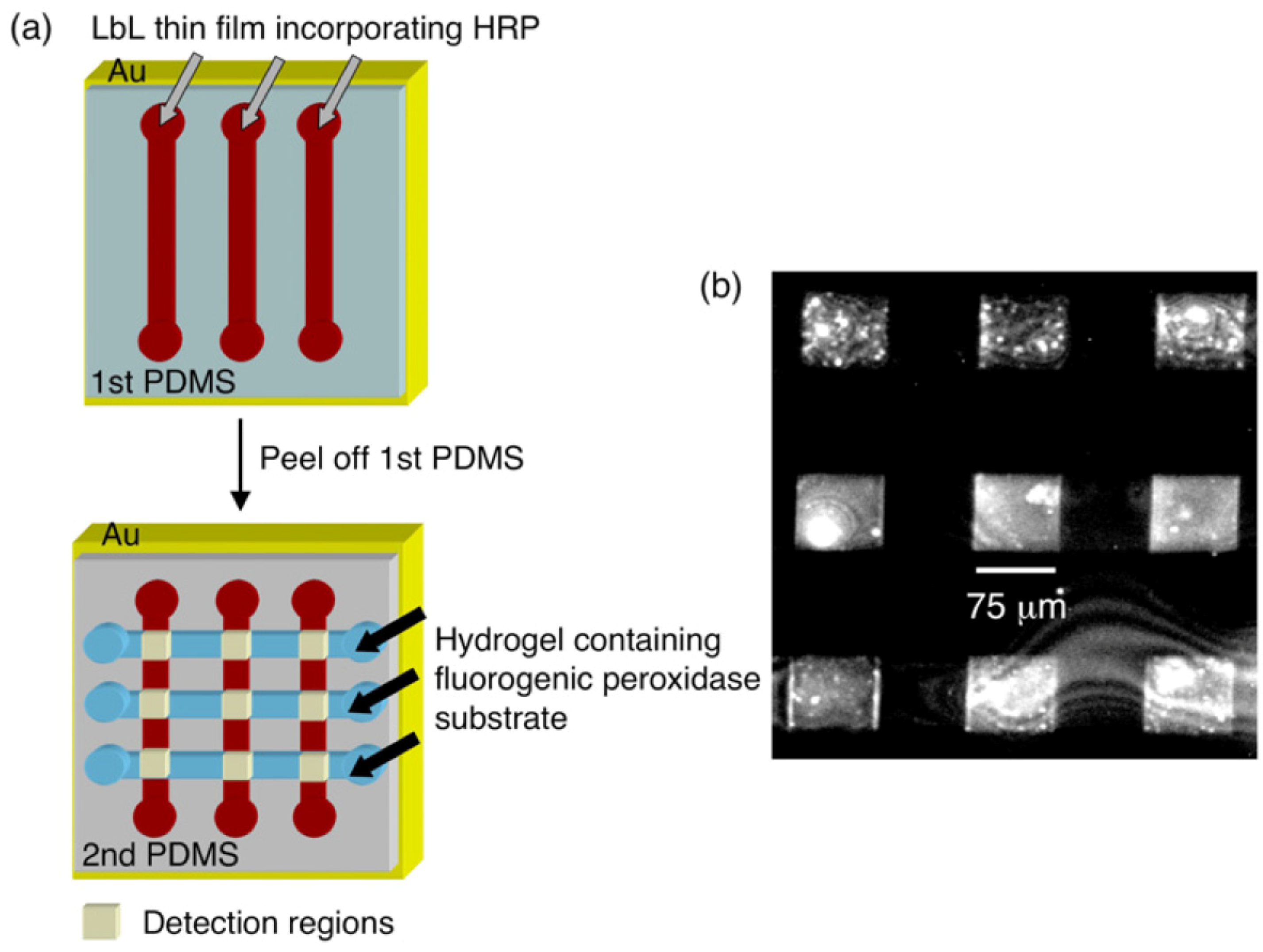
2.1. Enzymatic Biochips

Protein digestion biochips present a large surface-area-to-volume ratio that allows for the entrapment of a large number of enzymes. This high surface-area-to-volume ratio translates into rapid diffusion of molecules and therefore a larger number of interactions (in a fixed time) between substrate molecules and enzymes on the surface. Thus, the use of PEMs provides compatible surfaces for reactions to occur while at the same time allowing the functionalization of the surfaces for the required purposes. Work that takes advantage of these combined benefits from microfluidics and PEMs was presented by Liu et al., 2006, where they were able to identify casein extracted from bovine milk and attenuated hepatitis A virus vaccine using mass spectrometry in a poly(ethylene terephthalate) (PET) microfluidic chip coated with PEMs [14]. An increased catalytic efficiency over bulk form, enhanced enzyme loading and stable bioactivity was reported. Another example of an enzyme reactor was demonstrated with hydrolyzed PET channels coated with multilayers of cationic chitosan (CS) and anionic hyaluronic acid (HA) and terminated with a trypsin only layer (Figure 1). The PEMs rendered the flat and smooth PET surface into a protruding network of nanoporous structures, enabling enzyme adsorption while not compromising access to proteins and peptide movement. An important observation of this work is that the trypsin activity increases sharply from layer one to three and reaches a plateau by the fourth layer. The same group reported a PET microchip with alternate layers of poly(diallyldimethyl ammonium chloride) (PDDA) and gold nanoparticles with a final adsorption step of trypsin. This enzyme digests complex protein extracts into peptides that are ultimately detected using MALDI-TOF-MS/MS (Matrix-Assisted Laser Desorption Ionization-Time-of-Flight-Mass Spectrometer/Mass Spectrometer) or LC-ESI-MS/MS (Liquid Chromatography-Electrospray Ionization-Mass Spectrometer/Mass Spectrometer) [15].
The large surface-area-to-volume ratio in microfluidics systems could instead be detrimental in the analytical performance of assays if non-specific binding occurs to a large extent. This problem is particularly challenging in instances when the analyte of interest is at a low concentration and is excessively lost due to non-specific binding in areas other than where the sensor is located. For polydimethylsiloxane (PDMS) microfluidic channels, rendering the PDMS surface hydrophilic reduces hydrophobic interactions of non-specific binding [16]. Typically, a blocking agent, such as bovine serum albumin (BSA) protein, is used to decrease non-specific binding in enzyme-linked immunosorbent assay (ELISA). Sung et al., 2008, modified the PDMS surface with poly(ethyleneimine) (PEI) and poly(acrylic acid) (PAA) and later cross-linking of the PEMs with amide bonds. The use of this device for detecting low levels of proteins was established by detecting transforming growth factor beta (TGF-β), a cytokine, using ELISA [17].
Using the same concept, Weng et al., 2011, designed a PEM-coated microfluidic platform for identifying dengue virus (DV) using ELISA [18]. The focus of this work was on the design of a device that allows for sample transport and mixing capabilities integrated into a single unit to reduce the time commonly required in conventional ELISA. However, the stability and efficiency are improved by the use of PEMs on the channel surfaces. Weng et al., 2011, reported that the PEM/PDMS microfluidic system reduces the time required for surface modification from 24 h [17] to 2 h [18]. The PAA and PEI layers (6 nm total thicknesses) were cross-linked with amide bonds by exposing them to glutaraldehyde. A top protein layer was deposited with antibodies for the binding of DV. Four different types of viruses were tested: enterovirus 71/4643, influenza A (H3/93S243H3), hepatitis B virus 99N2485, and DV, and the device were selective in detecting DV only.
Figure 1. (a) Steps for on-chip protein digestion and identification; (b) Enlarged area of a microchannel showing the multilayer-assembled enzymes; (c) Enzyme immobilization process. Reprinted with permission from [14]. © 2006 Wiley-VCH Verlag GmbH&Co. KGaA.
Polymers 06 02100 g001

2.2. Fluorescently-Based Biosensors

Quantum dots (QD) are well-accepted fluorescent labels due to their high quantum yields and long-term photostability. PEMs offer a simple surface for QD attachment and functionalization. Biofunctional multicolored QD-encoded fluorescent microspheres comprised of CdTe nanocrystals conjugated with mercaptopropyl acid were electrostatically bound to three layers of polyelectrolytes: poly(allylamine hydrochloride)/poly(styrene sulfonate)/poly(allylamine hydrochloride) (PAH/PSS/PAH) [19]. These QD-encoded fluorescent microspheres were used to detect antigens efficiently. In another study, CdSe/ZnS QDs were embedded in PEM (PAH/PSS) coatings in PDMS microfluidic channels, along with a fluorophore at a set distance from the QDs, to create thin films that allow for fluorescence resonance energy transfer (FRET) detection [20]. The quantum dots act as the fluorescent donors, while the molecular fluorophores act as acceptors in the PEMs. They reported FRET signal reduction during cleavage of the peptide neurotensin by the enzyme trypsin. Overall, PEM-based microfluidic biological assays provide a system to analyze low volume samples.
A DNA sequencing mechanism in a microfluidic-integrated system was proposed by Kartalov and Quake [21]. The authors demonstrated sequencing of four consecutive base pair sequences (bps) using a fluorescence detection system. PEMs were used to anchor the DNA to the PDMS channel in order to protect against non-specific binding and any loss of DNA during “feeds and flushes”. The authors report that the non-specific binding decreased with PEMs due to higher surface charge density. It was found that a minimum of four layers opposed most of the non-specific attachment, but the highest SNR (Signal-to-Noise Ratio) was achieved for 12 layers.

2.3. Electronically-Based Biosensors

In addition to the use of PEMs as a matrix to insert sensing elements, their buildup can be used to characterize biosensors in terms of their performance, sensitivity, limit of detection, and viability. The buildup of PEMs was used to measure the sensitivity of a nanoscale optofluidic biosensor comprised of evanescently-coupled 1-D photonic crystal resonator arrays [22]. The sensor exhibited changes in refractive index as the mass of PEMs and proteins increased in an optical cavity. Multilayers of PEI and polyacrylic acid (PAA) were deposited on top of the resonator as a way to determine the sensitivity of the device. A shift in wavelength of 0.35 nm was observed for every nanometer of PEM growth. The device was ultimately tested for the detection of streptavidin/anti-streptavidin, and the binding of other antibodies. A limit of detection of 63 ag (attogram) was estimated for the device.
An interesting property of PEMs is that the electrostatic forces of alternating polyelectrolyte layers influences their dielectric properties during layer formation [23]. The viability of a CMOS-based capacitive sensor developed for detecting liquids flowing through a microchannel using this dielectric PEM property was tested [24]. The sensor consisted of an array of capacitance voltage converters incorporated into a microfluidic channel, with sensing electrodes and an analog-to-digital converter, fabricated using 0.18 μm CMOS and direct-write processes. To characterize the device, positively and negatively charged polyelectrolytes were introduced into the channel to form a five-layer stack consisting of PEI/(alginate/chitosan)2 (Figure 2). The ultimate goal was to create a generic capacitive detector for lab-on-a-chip applications. Using the same principle as the variation in surface charge due to incremental layers, Hou et al., 2006, developed a microfluidic device for label-free polymerase chain reaction (PCR) quantification [25]. The silicon based field-effect sensor detected unprocessed PCR products by means of the measurement of intrinsic charges. The channel was coated with a thin layer of poly-l-lysine (PLL) and a PCR product was injected through the channel to measure a particular concentration. The surface was reset by depositing a new layer of PLL on the sensor surface, a process that did not decrease the sensitivity. The sensor ultimately could detect concentration differences of DNA in the PCR in the range of 1 ng·μL−1 to 80 ng·μL−1.
An electrochemical enzymatic biosensor made by electro-deposition of chitosan on gold electrodes on the sidewalls of a PDMS microchannel and sequential LbL deposition of alginate/glucose oxidase and chitosan was demonstrated by Wang et al., 2011 [26]. Electrochemical testing was conducted by applying 0.6 V (vs. Ag/AgCl) to one PEM coated gold electrode, while the opposing electrode served as a counter electrode. Baseline anodic current was monitored using a test solution; and when a glucose containing solution (6 mM) was introduced into the channel, the anodic current increased, demonstrating the device’s sensing capabilities (Figure 3).
Figure 2. (a) Deposition sequence of PEI/(alginate/chitosan)2 on the CMOS chip; (b) Detection of the deposition of polyelectrolytes on the chip; The y axis (qn) denotes the response from the device when the polyelectrolytes are deposited on it in terms of the bit stream that results from converting the signal from analog to digital. Reprinted with permission from [24]. © 2008, Elsevier.
Polymers 06 02100 g002
Figure 3. Experimental set up and output upon exposure to glucose solution. Reprinted with permission from [26]. © 2011 Wiley-VCH Verlag GmbH&Co. KGaA.
Polymers 06 02100 g003
Another type of enzymatic biosensor, which employs changes in enzymatic activity when heavy metals interact with whole bacteria to induce a decrease in viability, was developed by Gammoudi et al. [27]. This system incorporates a Love wave sensor in a microfluidic device to, first, measure the deposition of polyelectrolytes (PAH and PSS) on the substrate use to immobilize E. coli, and to later measure the difference in oscillator frequencies produced by changes in viscoelastic properties when Cd(II) and Hg(II) is taken in by E. coli. This device is an improvement over previous work from this group [28] when they had the same detection approach but with a stagnant system instead of a microfluidic system. The microfluidic version [27] significantly reduced the detection time with an effective detection within 60 s (versus approximately 3 min in the stagnant system) and detection limits below 10−12 M.
Cell behavior such as proliferation, differentiation and death can be interpreted by impedance measurements between a cell and an underlying electrode [29,30,31]. PEMs provide a stable, biocompatible and adhesive environment for cells to grow. These properties were used in the fabrication of an impedance-based cell proliferation monitoring microfluidic device [32]. In this study, optically-transparent gold electrodes were initially coated with PEI as an anchoring layer followed by PSS/PAH. NIH-3T3 fibroblast cells were allowed to grow on the microfluidic device, while the increased magnitude of impedance at 1 kHz was measured from cell seeding, to proliferation (96 h). Upon exposure to trypsin/EDTA solution, the impedance magnitude decreased steeply due to cell detachment. In an example of the use PEMs for electrophoretic separation, Wang et al., 2006, used PEM-coated PDMS microchips to improve the electrophoretic separation efficiency of analytes [33]. The PEM structure consisted of chitosan-Au-nanoparticles/albumin and lysozyme/albumin. The device was used to separate neurotransmitters (dopamine and epinephrine) and environmental pollutants (p-phenylenediamine, hydroquinone) and was coupled with amperometric detection.

3. Patterning with PEMs

Patterning of cells or proteins onto a surface opens up the opportunity to investigate cell-cell and cell-substrate interactions and, in general, these interactions are important in biochip applications. Patterned PEMs have demonstrated successful immobilization of cells. In addition, their properties can be engineered to generate gradients with varying properties such as stiffness, pH or biomolecular functionalization. In this section we will be discussing PEMs patterned in microfluidics for cell immobilization and biosensing.

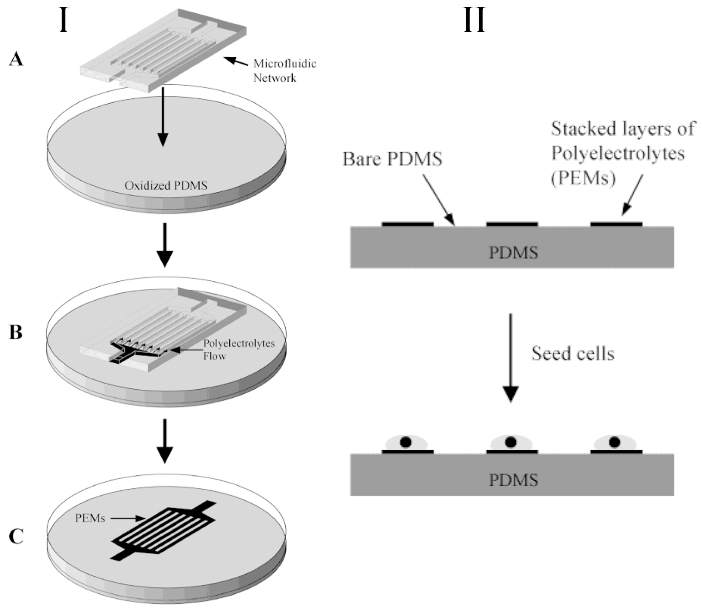
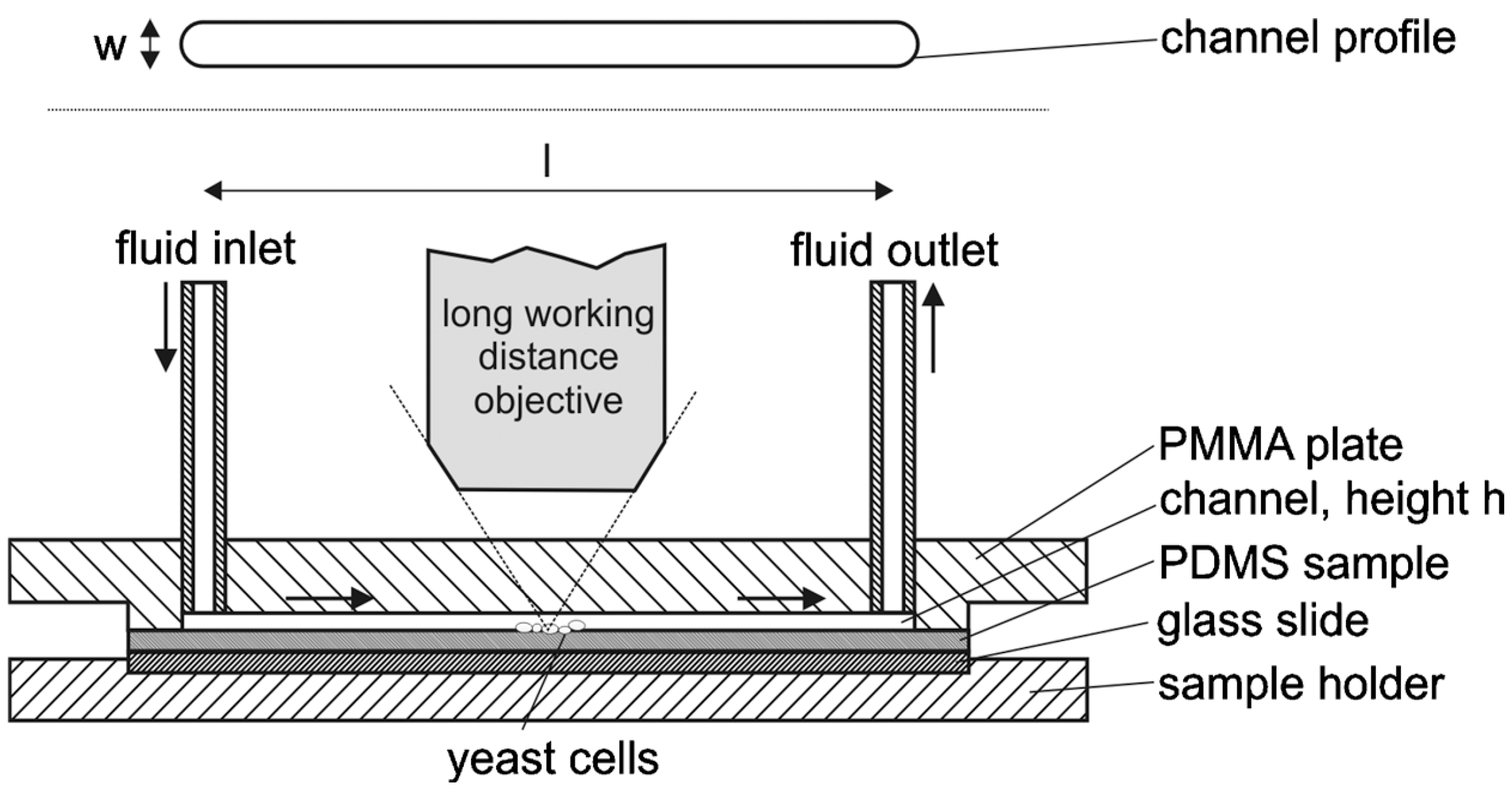
3.1. Cellular Interfaces and Patterning

Rat retinal cells (R28) were seeded on patterned PEI/PSS and PAH/PSS multilayer-coated PDMS microfluidic devices and the cells preferentially attached and grew on the PEM areas [34]. Areas of uncharged or neutral-state PDMS repelled the cells, while the cells adhered well to the oxidized PDMS coated with PEMs (Figure 4). Cell viability tests demonstrated the nontoxic nature of the PEMs, irrespective of number of layers.
In a related work, induced P19 cells were seeded on dishes with PEMs patterned via microfluidic networks [35]. P19 cells attached to these PAH/PSS multilayers were healthy, with good adherence, and showed directed neuronal extension. This work demonstrated that a supporting fibroblast monolayer was not needed for neurite extension as long as a PEM treated surface was present. A different approach for patterning cells, demonstrated by the same group, used PEMs as anchorage material to immobilize cells following dielectrophoretic trapping. Coating the area on top of the electrodes with PEMs generated a charged surface that enabled rapid cell attachment and kept the cells anchored after the dielectrophoresis (DEP) electrodes were de-energized. This set up allows for the quick return to cell culture conditions without the cells being loosed from the PEM surface [36]. Further development of this approach to make it completely biocompatible under DEP conditions (e.g., sucrose and electric fields) required the engineering of what was called a hybrid cell adhesive material, or hCAM [37]. This hCAM was comprised of PEMs, fibronectin and an outermost layer of PAH, and was deposited on top of indium tin oxide (ITO) electrodes; P19 cells were DEP trapped using the ITO electrodes, and electrostatically anchored to the hCAM. Differentiation of P19 into neuron-like cells was possible on the hCAM after DEP trapping, demonstrating that cell functionality was not affected. The same group also demonstrated the use of PEMs to hold cells during and after DEP on devices where the substrate was polyethylene terephthalate (PET) [38]. Gold electrodes were patterned on porous PET membranes and five layers of polyelectrolytes were deposited on top of the gold/PET to continue to hold cells electrostatically after DEP forces ceased.
Figure 4. (I) Steps to pattern PEMs on PDMS. A thin layer of PDMS cured on the dish was plasma oxidized (A) and a PDMS microfluidic network was placed on top of it. Polyelectrolytes pass through the microfluidic network to deposit PEMs within the microfluidic channels limits (B); The PDMS microfluidic network was lifted off exposing the PEMs on the PDMS substrate (C). (II) Diagram showing the cells attaching to areas where the PEMs were deposited, versus the areas of hydrophobic, oxidized PDMS. Reprinted with permission from [34]. © 2004 American Chemical Society.
Polymers 06 02100 g004
PEM layers can be terminated with various functional groups and this capability has been used to understand the interaction of cells with substrates. Such an approach was used by Johann et al., 2007, to grow human embryonic kidney (HEK 293) cells on patterned PAH/PSS multilayers on glass slides [39]. It was observed that HEK 293 cells grew preferentially according to the following order: PAH/NH2 > ox-PDMS (–OH) >> PSS (–SO3-) > PDMS (–CH3).
Microgrooves of varying dimensions (5, 10, 30, 100) μm coated with PEMs (PLL/HA) were used to study the differentiation of muscle cells into myotubes [40]. It was found that myoblasts pre-aligned to the microgrooves post differentiation, and their formation was generally independent of the microgroove width; however, in the case of the 5 μm microgrooves, cell nuclei deformation and impaired maturation was observed.
Micromolding in capillaries, or MIMIC, was used to generate cytophilic and cytophobic regions on a substrate by depositing (PAH/PSS)xPAH and PEG-PLA (polyethylene glycol-polylactic acid), respectively [41]. Cells seeded on the orthogonally functionalized surfaces attached preferentially to the (PAH/PSS)xPAH regions and not to the PEG-PLA. Cell proliferation occurred on the PEM areas for up to 48 h, but cells did not grow onto the PEG-PLA surfaces. It was also reported that the cell adhesive material necessary for cell growth decreased as chain length of PEG and PLA increased. Similarly, Palama et al. patterned a PEM coating on polyurethane microchannel surfaces using the MIMIC technique [42]. The patterned PEMs were comprised of two different polyion pairs: PAH/PSS, and pARG/DXS (poly-l-arginine sulfate salt/dextran sulfate sodium salt), the latter being a biodegradable layer. Murine skeletal myoblast (C2C12) cells were cultured in these microchannels. This arrangement allowed studies of myogenic differentiation under geometrical confinement conditions.
A process to form a 3D cell adhesive material was developed by alternating the extent of the width of laminar flows creating two distinct areas: one with collagen and the other with collagen/hydroxylethylmethacrylate-methacrylate-methylacrylic acid. This formed a cell-conformal cell-matrix layer whose physico-chemical properties, such as polyelectrolyte composition, could be tuned as needed [43].
A Braille-display-actuated microfluidic device was set up and aligned with a fabricated PDMS microfluidic channel to create multiple types of PEM deposition in different channels. Sequential deposition of poly(diallyldimethyl ammonium chloride) (PDADMAC) and clay was used to build a stack with desired number of bilayers, followed by a final bilayer of collagen and fibronectin. The number of bilayers deposited was varied, as well as the outer most PEM layer to determine the best combination for bone marrow cell culture. Bone marrow cells adhered best to the negatively charged clay-topped surface, followed by protein deposition [44].
A number of PEM approaches use polyelectrolytes with functional groups to induce cell adhesion, and other functional groups to repel cells from the surfaces. Schmolke and collaborators used PEM and PEM-PEG to assess the strength of the interactions between cells and these surfaces in a flow chamber, and compared them to the cell repellant capabilities of PEG-silane engineered PDMS surfaces [45] (Figure 5). Here, the PEMs consist of PDADMAC/PAA and PAH/PAA as well as a copolymer comprised of PEG with (PAA-g-PEG) as side chains, as the outermost layer. S. cerevisiae cells were introduced in the channel and allowed to settle on the treated surface before washing with fresh media to remove non-adhered cells. Both types of PEMs, PEM-PEG and PEG silane, were able to reduce the adhesion strength.

3.2. PEMS with Surface Gradients
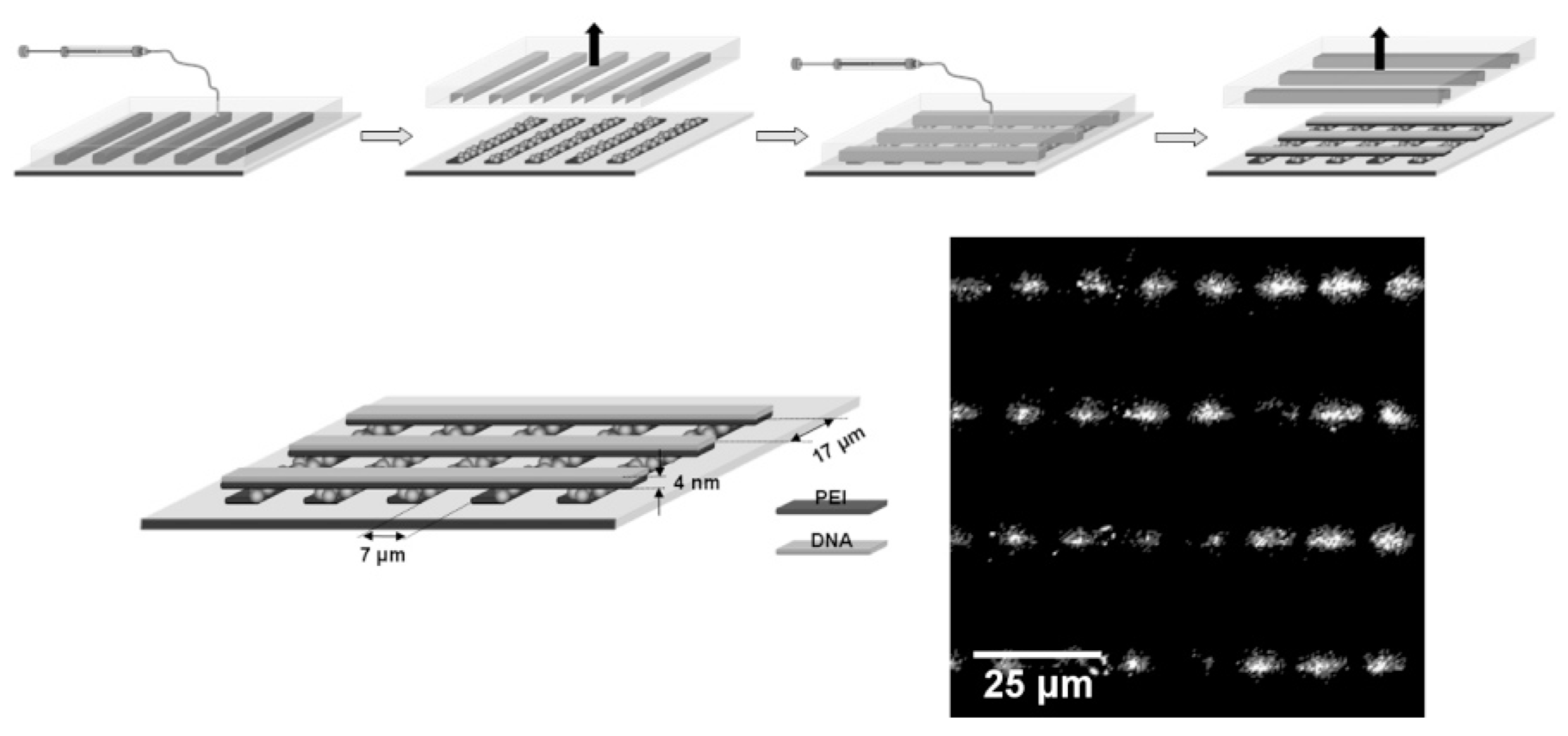
PEM structures with surface gradients spanning centimeters were demonstrated using a PLL/hyaluronan HA model [46]. The surface gradients were generated on the PEM coated substrate within a PDMS microfluidic device. The gradients explored in this study involved: topology using fluorescent microbeads, biochemical cues via fluorescent PLL, poly(glutamic acid) conjugated to arginine-glycine-aspartic-acid (RGD) for cell adhesion, and physical cues involving carbodiimide cross-linking to provide a stiffness gradient from 200 to 600 kPa. Pre-osteoblast cells preferred regions of maximum cross-linking and adhesion reduced along the decreasing cross-linking gradient. A similar trend was observed for C2C12 cells in response to the RGD biochemical gradient. The same group engineered (poly-l-lysine)/HA multilayers with a gradient of matrix-bound bone morphogenetic proteins (BMP-2 and BMP-7) [47]. They showed C2C12 myoblasts cell differentiated into osteogenic lineage linearly on BMP-7, but non-linearly on BMP-2 embedded PLL (poly-l-lysine)/HA, while myogenic marker expression increased as the concentration of BMP decreased in the gradient layer.
Figure 5. Scheme of the parallel flow channel used to study the strength of yeast cell attachment to different surfaces (bottom: sectional view; top: channel profile). Reprinted with permission from [45]. © 2010 AIP Publishing LLC.
Polymers 06 02100 g005
For weak polyelectrolytes, the charge and conformation are dependent on the pH, which then affects the adsorption kinetics. Thus, by modulating pH, one can tailor the properties of PEMs such as viscoelasticity, thickness, charge, and wettability. Kirchhof et al. combined the LbL deposition of PEMs with microfluidics to generate PEMs with a pH gradient [48]. By introducing the same polyelectrolyte at pH 5 and 9 at opposite sides of a microfluidic chamber, multilayers with a pH gradient from 5 to 9 were form. The authors demonstrated that a higher density of adhered MG-63 cells was observed on the more basic pH layers, and cells migrated from the pH 5 to the pH 9 regions, thus following the pH gradient patterned on the PEM layer. In addition, the mechanical properties of the PEMs varied with pH; at higher pH the water content within the PEM is lower making it stiffer, while at lower pH, higher water content makes the PEM softer.

3.3. DNA-Based PEMs

DNA, proteins, and glycosaminoglycans are biologically relevant polyions that can be used to build up PEMs. Not only can they be tailored with functional molecules for novel sensing films, but they also can create biomimetic environments for cell growth or patterning. Dootz et al. analyzed the interactions of PEI and DNA polyelectrolyte layers patterned via microfluidics using Raman and surface enhanced Raman spectroscopy (SERS) [49]. The micropatterning in conjunction with SERS allows for characterization of the PEM structure (Figure 6). This pattern is useful for detecting molecules across a surface using SERS.
Lee, Wark, and Corn demonstrated the formation of DNA/PLL multilayers on gold surfaces [50]. The biopolymer-based PEMs were functionalized with gold nanorods, fluorogenic peroxidase and electroactive molecules. Horizontally-aligned microchannels were used to create horseradish peroxidase (HRP) functionalized double-stranded-DNA/PLL multilayers, and then perpendicularly oriented microchannels were used to pattern a hydrogel interface (Figure 7). The PEM/hydrogel interface was characterized using fluorescence imaging, which showed fluorogenic peroxidase reaction with the HRP at the interface.
Figure 6. Scheme of the set up used for SERS to characterize the PEM structure formed at the regions where the two perpendicular stripes of polyelectrolytes, PEI and DNA, overlap and form a PEM. The micrograph shows a lateral Raman scan at a fixed wavenumber (757 cm−1), where the PEM regions can be detected with SERS. Reprinted with permission from [49] © 2006 American Chemical Society.
Polymers 06 02100 g006
Figure 7. (a) Illustration of the steps to form the PEM/HRP nanostructures, and the fluorogenic-peroxidase-containing hydrogel generated perpendicular to it; (b) Micrographs showing fluorescence at the intersection of the structures (PEM/HRP and hydrogel) due to the enzymatic reaction. Reprinted with permission from [50] © IOP Publishing.
Polymers 06 02100 g007

4. Conclusions and Outlook

New polyelectrolyte-based structures that can only be produced using the physical phenomena that occur in microfluidic systems (such as laminar flow and rapid diffusion of molecules) will allow for the creation of new micro- and nanofeatures not possible otherwise. This unique microfluidic/PEM toolbox will be particularly useful for the development of in vitro assays, as well as for fundamental studies in the areas of cell biology, tissue engineering, stem cell research, and organ-on-a-chip. This type of system can provide fine microenvironmental cell-culture controls where conditions such as flow, extracellular matrix and closeness of different cell lines in co-culture can be adjusted down to the nanoscale, thus, providing the means to generate an environment very similar to in vivo conditions.
Synthetic polyelectrolytes use as biomimetic matrices, in combination with microfluidics, could allow for the defined and precise presentation of specific biochemical moieties in studies of, for example, interactions between cells and matrices. In addition, the mechanical properties of PEMs can be tuned based on the deposition conditions. This physical parameter has been found, in recent years, to be a key regulator of cellular behavior and fate along with soluble cues, thus presenting a unique opportunity for the study of mechanical and soluble biochemical cues under well-controlled conditions.
Microfluidics will greatly benefit from the integration of features (of nanometer size in the z-direction so that changes in flow are negligible) inside microchannel networks where parameters such as pressure, flow and temperature could be measured locally. These could not only improve areas in microfluidics research outside biology, but could further the fine control over those parameters that are frequently monitored not locally, but externally, during biological experiments. Changes in these parameters during the duration of a biological experiment could have detrimental effects over certain biomolecules and cells.
The integration of smart PEMs to, for example, release certain biomolecules in microfluidic devices as part of an assay for Point of Care (POC) devices or disposable microfluidic devices for diagnostics in the developing world, could indeed open the door for more applications of such importance to be developed. Emerging opportunities in areas, such as nanomanufacturing, as well as biomanufacturing, could benefit immensely from the integration of these two areas. In nanomanufacturing, areas such as the formation of nano-liposomes using microfluidics has already show promising results for drug delivery opportunities. In this case, the use of microfluidics has provided the means to finely tweak the size of liposomes in a way that certain diameters can be produced with small diameter spreading. Similarly, the formation of nanoparticles or other type of nanostructures could be adjusted so that they are formulated using the unique features provided by these techniques. Certainly, the continuation of research in these merging fields will be needed to produce the new discoveries and advancements that will provide the scientific and industrial communities with the required tools to answer the unforeseen challenges of the future.

Acknowledgments

This project was funded internally by the National Institute of Standards and Technology (NIST).

Author Contributions

All the authors contributed in the writing of this manuscript.

Conflicts of Interest

The authors declare no conflict of interest.

References

  1. Iler, R.K. Multilayers of colloidal particles. J. Colloid Interf. Sci. 1966, 21, 569–594. [Google Scholar] [CrossRef]
  2. Decher, G.; Schmitt, J. Fine-tuning of the film thickness of ultrathin multilayer films composed of consecutively alternating layers of anionic and cationic polyelectrolytes. In Trends in Colloid and Interface Science VI; Helm, C., Lösche, M., Möhwald, H., Eds.; Steinkopff: Dresden, Germany, 1992; pp. 160–164. [Google Scholar]
  3. Decher, G.; Hong, J.D.; Schmitt, J. Buildup of ultrathin multilayer films by a self-assembly process: III. Consecutively alternating adsorption of anionic and cationic polyelectrolytes on charged surfaces. Thin Solid Films 1992, 210–211, 831–835. [Google Scholar] [CrossRef]
  4. Ariga, K.; Hill, J.P.; Ji, Q. Layer-by-layer assembly as a versatile bottom-up nanofabrication technique for exploratory research and realistic application. Phys. Chem. Chem. Phys. 2007, 9, 2319–2340. [Google Scholar] [CrossRef]
  5. Schönhoff, M. Self-assembled polyelectrolyte multilayers. Curr. Opin. Colloid Interf. Sci. 2003, 8, 86–95. [Google Scholar] [CrossRef]
  6. Klitzing, R.V.; Wong, J.E.; Jaeger, W.; Steitz, R. Short range interactions in polyelectrolyte multilayers. Curr. Opin. Colloid Interf. Sci. 2004, 9, 158–162. [Google Scholar]
  7. Hammond, P.T. Form and function in multilayer assembly: New applications at the nanoscale. Adv. Mater. 2004, 16, 1271–1293. [Google Scholar] [CrossRef]
  8. Lavalle, P.J.; Voegel, C.; Vautier, D.; Senger, B.; Schaaf, P.; Ball, V. Dynamic aspects of films prepared by a sequential deposition of species: Perspectives for smart and responsive materials. Adv. Mater. 2011, 23, 1191–1221. [Google Scholar] [CrossRef]
  9. Sukhishvili, S.A.; Kharlampieva, E.; Izumrudov, V. Where polyelectrolyte multilayers and polyelectrolyte complexes meet. Macromolecules 2006, 39, 8873–8881. [Google Scholar] [CrossRef]
  10. Reyes, D.R.; Iossifidis, D.; Auroux, P.A.; Manz, A. Micro total analysis systems. 1. Introduction, theory, and technology. Anal. Chem. 2002, 74, 2623–2636. [Google Scholar] [CrossRef]
  11. Sackmann, E.K.; Fulton, A.L.; Beebe, D.J. The present and future role of microfluidics in biomedical research. Nature 2014, 57, 181–189. [Google Scholar] [CrossRef]
  12. Dertinger, S.K.W.; Chiu, D.T.; Jeon, N.L.; Whitesides, G.M. Generation of gradients having complex shapes using microfluidic networks. Anal. Chem. 2001, 73, 1240–1246. [Google Scholar] [CrossRef]
  13. Jeon, N.L.; Dertinger, S.K.W.; Chiu, D.T.; Choi, I.S.; Stroock, A.D.; Whitesides, G.M. Generation of solution and surface gradients using microfluidic systems. Langmuir 2000, 16, 8311–8316. [Google Scholar] [CrossRef]
  14. Liu, Y.; Zhong, W.; Meng, S.; Kong, J.; Lu, H.; Yang, P.; Girault, H.H.; Liu, B. Assembly-controlled biocompatible interface on a microchip: Strategy to highly efficient proteolysis. Chem. Eur. J. 2006, 12, 6585–6591. [Google Scholar] [CrossRef]
  15. Liu, Y.; Xue, Y.; Ji, J.; Chen, X.; Kong, J.; Yang, P.; Girault, H.H.; Liu, B. Gold nanoparticle assembly microfluidic reactor for efficient on-line proteolysis. Mol. Cell. Proteomics 2007, 6, 1428–1436. [Google Scholar] [CrossRef]
  16. Linder, V.; Verpoorte, E.; Thormann, W.; de Rooij, N.F.; Sigrist, H. Surface biopassivation of replicated poly(dimethylsiloxane) microfluidic channels and application to heterogeneous immunoreaction with on-chip fluorescence detection. Anal. Chem. 2001, 73, 4181–4189. [Google Scholar] [CrossRef]
  17. Sung, W.C.; Chang, C.C.; Makamba, H.; Chen, S.H. Long-term affinity modification on poly(dimethylsiloxane) substrate and its application for ELISA analysis. Anal. Chem. 2008, 80, 1529–1535. [Google Scholar] [CrossRef]
  18. Weng, C.H.; Huang, T.B.; Huang, C.C.; Yeh, C.S.; Lei, H.Y.; Lee, G.B. A suction-type microfluidic immunosensing chip for rapid detection of the dengue virus. Biomed. Microdevices 2011, 13, 585–595. [Google Scholar] [CrossRef]
  19. Ma, Q.; Wang, X.; Li, Y.; Shi, Y.; Su, X. Multicolor quantum dot-encoded microspheres for the detection of biomolecules. Talanta 2007, 72, 1446–1452. [Google Scholar] [CrossRef]
  20. Crivat, G.; Da Silva, S.M.; Reyes, D.R.; Locascio, L.E.; Gaitan, M.; Rosenzweig, N.; Rosenzweig, Z. Quantum dot FRET-based probes in thin films grown in microfluidic channels. J. Am. Chem. Soc. 2010, 132, 1460–1461. [Google Scholar] [CrossRef]
  21. Kartalov, E.P.; Quake, S.R. Microfluidic device reads up to four consecutive base pairs in DNA sequencing by synthesis. Nucleic Acids Res. 2004, 32, 2873–2879. [Google Scholar] [CrossRef]
  22. Mandal, S.; Goddard, J.M.; Erickson, D. A multiplexed optofluidic biomolecular sensor for low mass detection. Lab Chip 2009, 9, 2924–2932. [Google Scholar] [CrossRef]
  23. Durstock, M.F.; Rubner, M.F. Dielectric properties of polyelectrolyte multilayers. Langmuir 2001, 17, 7865–7872. [Google Scholar] [CrossRef]
  24. Ghafar-Zadeh, E.; Sawan, M. A Core-CBCM sigma delta capacitive sensor array dedicated to lab-on-chip applications. Sensors Actuators Phys. 2008, 144, 304–313. [Google Scholar] [CrossRef]
  25. Hou, C.S.J.; Milovic, N.; Godin, M.; Russo, P.R.; Chakrabarti, R.; Manalis, S.R. Label-free microelectronic PCR quantification. Anal. Chem. 2006, 78, 2526–2531. [Google Scholar] [CrossRef]
  26. Wang, Y.; Liu, Y.; Cheng, Y.; Kim, E.; Rubloff, G.W.; Bentley, W.E.; Payne, G.F. Coupling electrodeposition with layer-by-layer assembly to address proteins within microfluidic channels. Adv. Mater. 2011, 23, 5817–5821. [Google Scholar] [CrossRef]
  27. Gammoudi, I.; Raimbault, V.; Tarbague, H.; Moroté, F.; Grauby-Heywang, C.; Othmane, A.; Kalfat, R.; Moynet, D.; Rebière, D.; Dejous, C.; et al. Enhanced bio-inspired microsensor based on microfluidic/bacteria/love wave hybrid structure for continuous control of heavy metals toxicity in liquid medium. Sensors Actuators B Chem. 2014, 198, 278–284. [Google Scholar] [CrossRef] [Green Version]
  28. Gammoudi, I.; Tarbague, H.; Othmane, A.; Moynet, D.; Rebière, D.; Kalfat, R.; Dejous, C. Love-wave bacteria-based sensor for the detection of heavy metal toxicity in liquid medium. Biosens. Bioelectron. 2010, 26, 1723–1726. [Google Scholar] [CrossRef]
  29. Luong, J.H.T.; Habibi-Rezaei, M.; Meghrous, J.; Xiao, C.; Male, K.B.; Kamen, A. Monitoring motility, spreading, and mortality of adherent insect cells using an impedance sensor. Anal. Chem. 2001, 73, 1844–1848. [Google Scholar] [CrossRef]
  30. Wegener, J.; Keese, C.R.; Giaever, I. Electric cell–substrate impedance sensing (ECIS) as a noninvasive means to monitor the kinetics of cell spreading to artificial surfaces. Exp. Cell Res. 2000, 259, 158–166. [Google Scholar] [CrossRef]
  31. Arndt, S.; Seebach, J.; Psathaki, K.; Galla, H.J.; Wegener, J. Bioelectrical impedance assay to monitor changes in cell shape during apoptosis. Biosens. Bioelectron. 2004, 19, 583–594. [Google Scholar] [CrossRef]
  32. Mijares, G.; Reyes, D.R.; Geist, J.; Gaitan, M. Polyelectrolyte multilayer-treated electrodes for real-time electronic sensing of cell proliferation. J. Res. Natl. Inst. Stand. Technol. 2010, 115, 61–73. [Google Scholar] [CrossRef]
  33. Wang, A.J.; Xu, J.J.; Chen, H.Y. Proteins modification of poly(dimethylsiloxane) microfluidic channels for the enhanced microchip electrophoresis. J. Chromatogr. A 2006, 1107, 257–264. [Google Scholar] [CrossRef]
  34. Reyes, D.R.; Perruccio, E.M.; Becerra, S.P.; Locascio, L.E.; Gaitan, M. Micropatterning neuronal cells on polyelectrolyte multilayers. Langmuir 2004, 20, 8805–8811. [Google Scholar] [CrossRef]
  35. Forry, S.P.; Reyes, D.R.; Gaitan, M.; Locascio, L.E. Facilitating the culture of mammalian nerve cells with polyelectrolyte multilayers. Langmuir 2006, 22, 5770–5775. [Google Scholar] [CrossRef]
  36. Forry, S.P.; Reyes, D.R.; Gaitan, M.; Locascio, L.E. Cellular immobilization within microfluidic microenvironments: Dielectrophoresis with polyelectrolyte multilayers. J. Am. Chem. Soc. 2006, 128, 13678–13679. [Google Scholar]
  37. Reyes, D.R.; Hong, J.S.; Elliott, J.T.; Gaitan, M. Hybrid cell adhesive material for instant dielectrophoretic cell trapping and long-term cell function assessment. Langmuir 2011, 27, 10027–10034. [Google Scholar] [CrossRef]
  38. Hanke, C.; Dittrich, P.S.; Reyes, D.R. Dielectrophoretic cell capture on polyester membranes. ACS Appl. Mater. Interfaces 2012, 4, 1878–1882. [Google Scholar] [CrossRef]
  39. Johann, R.M.; Baiotto, C.; Renaud, P. Micropatterned surfaces of PDMS as growth templates for HEK 293 cells. Biomed. Microdevices 2007, 9, 475–485. [Google Scholar] [CrossRef]
  40. Monge, C.; Ren, K.; Berton, K.; Guillot, R.; Peyrade, D.; Picart, C. Engineering muscle tissues on microstructured polyelectrolyte multilayer films. Tissue Eng. A 2012, 18, 1664–1676. [Google Scholar]
  41. Lee, J.H.; Kim, H.E.; Im, J.H.; Bae, Y.M.; Choi, J.S.; Huh, K.M.; Lee, C.S. Preparation of orthogonally functionalized surface using micromolding in capillaries technique for the control of cellular adhesion. Colloids Surface B Biointerf. 2008, 64, 126–134. [Google Scholar] [CrossRef]
  42. Palamà, I.E.; D’Amone, S.; Coluccia, A.M.L.; Gigli, G. Micropatterned polyelectrolyte nanofilms promote alignment and myogenic differentiation of C2C12 cells in standard growth media. Biotechnol. Bioeng. 2013, 110, 586–596. [Google Scholar] [CrossRef]
  43. Toh, Y.C.; Zhang, C.; Zhang, J.; Khong, Y.; Chang, M.S.; Samper, V.D.; van Noort, D.; Hutmacher, D.W.; Yu, H. A novel 3D mammalian cell perfusion-culture system in microfluidic channels. Lab Chip 2007, 7, 302–309. [Google Scholar] [CrossRef]
  44. Mehta, G.; Kiel, M.J.; Lee, J.W.; Kotov, N.; Linderman, J.J.; Takayama, S. Polyelectrolyte-clay-protein layer films on microfluidic PDMS bioreactor surfaces for primary murine bone marrow culture. Adv. Funct. Mater. 2007, 17, 2701–2709. [Google Scholar] [CrossRef]
  45. Schmolke, H.; Demming, S.; Edlich, A.; Magdanz, V.; Buttgenbach, S.; Franco-Lara, E.; Krull, R.; Klages, C.P. Polyelectrolyte multilayer surface functionalization of poly(dimethylsiloxane) (PDMS) for reduction of yeast cell adhesion in microfluidic devices. Biomicrofluidic 2010, 4. [Google Scholar] [CrossRef]
  46. Almodóvar, J.; Crouzier, T.; Selimović, Š.; Boudou, T.; Khademhosseini, A.; Picart, C. Gradients of physical and biochemical cues on polyelectrolyte multilayer films generated via microfluidics. Lab Chip 2013, 13, 1562–1570. [Google Scholar] [CrossRef]
  47. Almodóvar, J.; Guillot, R.; Monge, C.; Vollaire, J.; Selimović, Š.; Coll, J.L.; Khademhosseini, A.; Picart, C. Spatial patterning of BMP-2 and BMP-7 on biopolymeric films and the guidance of muscle cell fate. Biomaterials 2014, 35, 3975–3985. [Google Scholar] [CrossRef]
  48. Kirchhof, K.; Andar, A.; Yin, H.B.; Gadegaard, N.; Riehle, M.O.; Groth, T. Polyelectrolyte multilayers generated in a microfluidic device with pH gradients direct adhesion and movement of cells. Lab Chip 2011, 11, 3326–3335. [Google Scholar] [CrossRef]
  49. Dootz, R.; Nie, J.; Du, B.; Herminghaus, S.; Pfohl, T. Raman and surface enhanced raman microscopy of microstructured polyethylenimine/DNA multilayers. Langmuir 2006, 22, 1735–1741. [Google Scholar] [CrossRef]
  50. Lee, H.J.; Wark, A.W.; Corn, R.M. Utilizing ultrathin DNA/poly-lysine multilayer films to create liquid/liquid interfaces: Spectroscopic characterization, interfacial reactions and nanoparticle adsorption. J. Phys. Condens. Matter. 2007, 19. [Google Scholar] [CrossRef]

