Review

A Survey of Surface Modification Techniques for Next-Generation Shape Memory Polymer Stent Devices

Department of Bioengineering, University of Colorado-Denver, Anschutz Medical Campus, Mail Stop 8607, 12700 E. 19th Avenue, Aurora, CO 80045, USA
*
Author to whom correspondence should be addressed.
Polymers 2014, 6(9), 2309-2331; https://doi.org/10.3390/polym6092309
Submission received: 30 June 2014 / Revised: 12 August 2014 / Accepted: 19 August 2014 / Published: 29 August 2014
(This article belongs to the Special Issue Shape-Memory Polymers)

Abstract

:
The search for a single material with ideal surface properties and necessary mechanical properties is on-going, especially with regard to cardiovascular stent materials. Since the majority of stent problems arise from surface issues rather than bulk material deficiencies, surface optimization of a material that already contains the necessary bulk properties is an active area of research. Polymers can be surface-modified using a variety of methods to increase hemocompatibilty by reducing either late-stage restenosis or acute thrombogenicity, or both. These modification methods can be extended to shape memory polymers (SMPs), in an effort to make these materials more surface compatible, based on the application. This review focuses on the role of surface modification of materials, mainly polymers, to improve the hemocompatibility of stent materials; additional discussion of other materials commonly used in stents is also provided. Although shape memory polymers are not yet extensively used for stents, they offer numerous benefits that may make them good candidates for next-generation stents. Surface modification techniques discussed here include roughening, patterning, chemical modification, and surface modification for biomolecule and drug delivery.

Graphical Abstract

1. Introduction

Cardiovascular stents are expandable tubes used to treat narrow or weakened arteries that arise as a result of atherosclerosis and its resultant sequelae, such as coronary artery disease, peripheral artery disease, etc. These diseases, collectively grouped under the term cardiovascular disease, are some of the main causes of death in the Western World as well as worldwide [1,2,3]. Atherosclerosis, or hardening of the arteries, occurs due to plaque formation and is often a result of inflammatory signals emitted by local cells and subsequent inflammatory response. This plaque may consist of fat, cholesterol, calcium and/or blood components [4]. Plaque buildup limits blood flow to tissues, ultimately leading to an acute ischemic condition such as stroke or myocardial infarction. Cardiovascular stents provide a minimally invasive means to mechanically support the damaged vessel which restores oxygenated blood flow to the tissues [5]. Although cardiovascular stents have saved countless lives, the device has many limitations, which drive continued research in the area [6]. In particular, thrombosis and restenosis continue to be relatively important problems with current stents. Given that these issues arise from surface interactions, surface modification techniques are an active area of current research.

2. Current Stents and Associated Issues

The first types of stents used were bare metal stents (BMSs), composed of a variety of metals and/or alloys such as stainless steel, cobalt-chromium and tantalum for balloon-expandable stents, or nitinol (nickel-titanium alloy) for self-expanding stents [5]. These stents provided the necessary mechanical support for the weakened vessel; however, an increased risk for thrombosis and/or restenosis in these devices may generate additional need for reintervention six to 12 months after stent implantation [5,7].
Thrombogenicity, one of the aforementioned issues associated with BMSs, refers to increased propensity of the device or material to generate a blood clot on the material surface [8]. Thrombotic events often occur due to net electrical charge differences between blood components and the stent surface, as well as surface potential incompatibility between the metal and the contacting blood [9,10]. Restenosis, or re-narrowing of the vessel, usually results from excessive neointimal proliferation following balloon angioplasty or stent implantation due to vessel injury from the expansion [11,12,13]. Additional causes of restenosis may include reduced compliance between the stent and the vessel and excessive tissue-remodeling response to the stent material [13].
As a proposed improvement on BMS, the first generation of drug-eluting stents (DESs) consisted of a metal backbone and a permanent, non-absorbable polymer coating to house a drug of choice [14]. While DESs offered control and localization for drug release to the injured vessel, incidences of hypersensitivity, heart attack and even death remained problematic [5].
An improved DES replaced the non-absorbable polymer coating with a non-thrombogenic, absorbable one. This absorbable coating served to encourage endothelialization through a directed drug release profile and reduced inflammatory response during polymer degradation. While these improved DESs did decrease the occurrence of restenosis through release of anti-proliferative agents, late stage thrombosis still occurred [1,11,15,16,17,18]. Late stage or late stent thrombosis (LST) can result from a variety of issues, ranging from the stenting procedure itself to early termination of antiproliferative drugs loaded in the stent; these issues may cause increased local fibrin deposition and delayed healing [15,16].
Unlike BMSs, polymer stents can achieve increased hemocompatibility with the proper selection of polymer components, polymerization and processing techniques [9]. With regard to DESs, these stents have been shown to cause a delay in re-endothelialization, therefore promoting a thrombotic environment compared to BMSs, as re-endothelialization is an important component in vessel healing. In addition, instances of very late stage thrombosis (LST) have also been seen with DESs, making polymer stents a potentially more appealing route for stent materials [19]. Patients who receive DESs are often required to continue an anti-platelet regimen for 12 months to prevent adverse effects from the DESs and while these anticoagulants prevent thrombosis, they may carry a sustained risk of hemorrhage, or bleeding, and related side effects [12,20,21]. Due to safety concerns with existing stent materials, current research in stent design is progressing towards using biodegradable/bioabsorbable or biomimetic materials for polymer or metal stents as well as polymer coating-free DESs, among others [11,19,22,23].

3. Polymer Stents

Polymers have caught the attention of the medical device industry due to their diversity and versatility. Polymers are less dense than metals and have higher flexibility than many other materials, which allows better matching of stent compliance with that of the local vessel [13]. In addition, polymers are easy to manufacture and often have lower bulk material costs and processing costs [24,25]. Polymers also possess a wide range of bulk properties, such as elasticity, conductivity, strength and degradability, which can provide the stent designer with a large palette of useful features [26]. Thus, polymers can be easily and cost-effectively tailored to fit the needs of their application, making these materials appealing for use in the medical device industry.
Shape memory polymers (SMPs) have added advantages to those seen with conventional polymers. As stents are often delivered via catheter, SMP systems offer benefits for catheter storage and deployment, since the materials can pack tightly without becoming permanently deformed during the storage period. SMPs may also enhance the ease of delivery of many of these devices, and produce lower recovery forces, leading to minimally invasive procedures with reduced recovery times for patients [13]. These added benefits, on top of the already present benefits of polymers, make SMPs potentially attractive materials for next-generation polymeric stents.

4. Surface Modification to Increase Biocompatibility

Despite the variety of materials and designs currently available for stents, there is still a need for a single material that has the desired mechanical properties while simultaneously achieving optimal biocompatibility [1,21]. Biocompatibility refers to the reaction elicited by a material when it is inserted into the body; ideally, this reaction should be favorable and should not provoke a negative response such as an attack by the immune system on the foreign material [27,28,29,30]. Surface modification techniques strive to retain favorable bulk properties while changing the surface to cater to specific needs, often to enhance biocompatibility [16,31]. Since shape memory is not a surface property, surface modifications should enhance biocompatibility without interfering with the shape memory capabilities of SMPs. Surface modifications that allow for improved blood contact (minimal thrombogenicity) while encouraging vascular wall healing via endothelial cell migration, anchorage and proliferation, are the focus of research goals in this area [1,6,27,32,33]. In addition, surface modifications for drug release in an effort to eliminate the polymer coating are also being explored [1].
One of the keys to success for many medical devices is successful wound healing, a process that begins at the surface of a material. Successful wound healing depends on a range of material properties, both surface and bulk, such as surface texture, surface chemistry, surface energy, crystallinity as well as leachable content and biocompatibility of the degradation products. In essence, biocompatibility is heavily dependent upon surface properties as well as interactions between the surface and cells and/or proteins, or between cells themselves [21,27,32,33,34,35,36,37]. Protein adsorption may also play a factor in dictating the success or failure of blood-contacting devices; some proteins, such as albumin, can be beneficial for biocompatibility as albumin may decrease both platelet adhesion and binding of microorganisms that may elicit infection, but non-specific proteins, such as fibrinogen and Immunoglobulin G (IgG), may increase platelet adhesion by instigating a host response [21].
This review provides a survey of techniques that have been used on a variety of polymer surfaces to address the issue of wound healing by increasing endothelial cell attachment, thus minimizing the negative effects of stent implantation. Although most of these techniques have yet to be implemented on SMPs, use of these techniques to enhance endothelial cell attachment to shape memory polymer surfaces, added to the already beneficial bulk properties of SMPs, may lead to the next-generation stent. Surface modification techniques discussed below include surface roughening, surface patterning, chemically-based surface modifications, surface coatings and films, attachment of bioactive agents to surfaces and porous surfaces for drug delivery; these techniques address successful wound healing responses through increases in endothelialization or decreases in thrombosis.

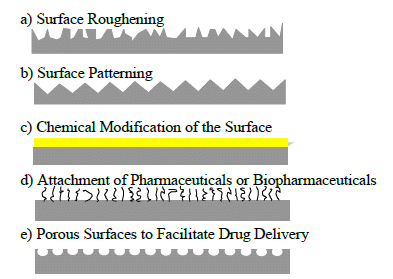
5. Methods for Surface Modification

Surface modifications should generally be thin, affecting only the topmost layer of the surface; thick layers may undesirably alter the bulk properties and have difficulty adhering to the surface, while overly thin layers are subject to erosion; despite these requirements, however, there are a number of ways to modify the surface of a material to enhance its functionality [31,38]. For polymer-derived stents, methods for modifying surfaces with the end goal of achieving improved blood compatibility, re-endothelialization, or both can be grouped into six major categories. These categories are surface roughening, surface patterning, chemical modification of the surface, surface coatings and films, attachment of pharmaceuticals or biopharmaceuticals to the surface, and the formation of porous surfaces to facilitate drug delivery, many of which are represented in Figure 1. Multiple techniques may be used to achieve the desired properties [1].
Figure 1. Surface modification techniques commonly used to enhance endothelialization and/or reduce thrombosis.
Polymers 06 02309 g001

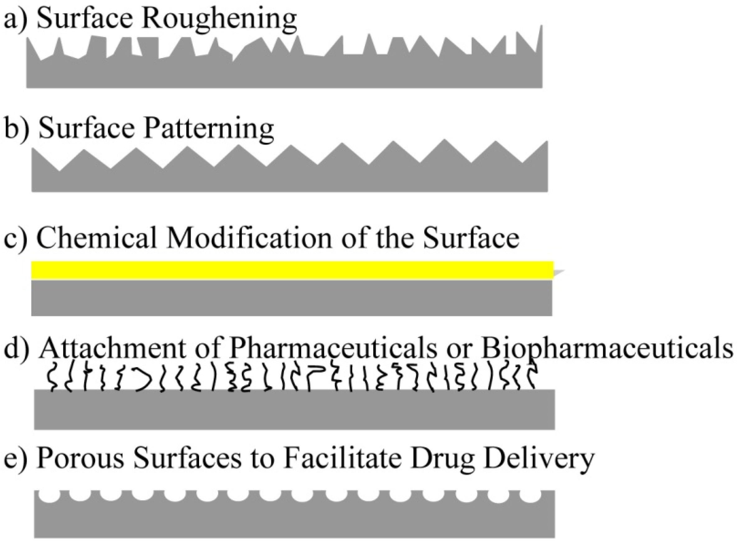
6. Surface Roughening

In general, surface roughening aims not only to increase the surface area of the material, but also to restrict cell movement, which contributes to enhanced cell attachment [21,39,40,41]. Cells are still able to migrate on roughened surfaces, but no significant increases or decreases in migration have been noted compared to smooth surfaces [42]. In addition, surface roughening modifies the topology of the surface without chemical alteration, which may have benefits, depending upon the material and its desired use [43].
For metals, roughening techniques such as sputtering with TiN or TiO2 have been used to successfully enhance endothelial cell attachment. However, these cells express less endothelial nitric oxide synthase (eNOS), which may lead to increased endothelial cell dysfunction; this reduced eNOS activity has actually been shown to be characteristic of metals in general, modified or bare, presenting a reason for further research into non-metal implant materials [44]. Microblasting followed by reactive ion etching on metal surfaces also produces roughened, high energy surfaces that may potentially improve cell attachment [20].
For polymers, oxygen or argon plasma deposition increases surface roughness as well as hydrophilicity, both of which have been shown to enhance cell attachment; application of plasma deposition towards SMP-based stents may allow for enhanced wound healing and biocompatibility [21,25]. Plasma processing alters the surface topography through melting and recrystallization processes, resulting in more ridges compared to the original surface, as displayed in Figure 2 [30,45]. Etching and sanding, both plasma- or chemical-based, as well as polishing and/or microblasting also serve to improve surface roughness [1,46].
Figure 2. Scanning electron microscope (SEM) image of RIE textured silicon surface using plasma consisting of Cl2, CF4 and O2 gases (scale bar = 200 nm). Reprinted with permission from Elsevier, 2001 [30].
Polymers 06 02309 g002
Shadpour et al., roughened polymer surfaces using a slurry of alumina particles, with the intention of enhancing endothelial cell attachment without altering the chemical make-up of the polymer surface. This process, in addition to being used to roughen the surface and increase surface area, can also be used for patterning purposes, both of which encourage cell and biomolecule attachment [43]. This method, which has been shown to increase cell attachment while modifying the polymer surface without disturbing the bulk, may be worth investigating for next-generation SMP stents due to the potential for increased biocompatibility.
Plasma- and chemical-based etching occurs when a surface is exposed to etching gas, which is often a type of plasma, and the top layer of the surface is changed through chain scission processes where old bonds are broken and new ones formed; more simply, etching degrades the polymer surface [24,47,48]. This process also modifies the surface topography and affects surface wettability, potentially driving the surface to become more biocompatible [47,48]. Etching can also be performed prior to coating a material, to ensure that the coating adheres [48]. Treatment with particular acids, which has an “etching effect”, may also encourage attachment and migration of endothelial cells, especially in polymeric hydrogels [49].
Grafting of different length polymer chains can alter the surface roughness, particularly on a nanometer-scale. Roughening at this scale has been shown to enhance cell attachment and improve biocompatibility [50].
Transfer printing, a common technique used for patterning, may also be used to roughen the polymer surface. The mold that houses the polymer during curing transfers the roughened features onto the surface during polymerization, as seen in Figure 3 [51].
Although many of these techniques have not yet been applied to SMPs, their use on polymers shows promise for the successful application to SMPs, granted that the methods continue to modify only the topmost layers of the material.
Figure 3. Atomic force microscopy (AFM) images of (a) untreated, (b) micro-roughened and (c) nano-roughened polydimethylsiloxane (PDMS) films. Reprinted with permission from IOP, 2009 [51].
Polymers 06 02309 g003

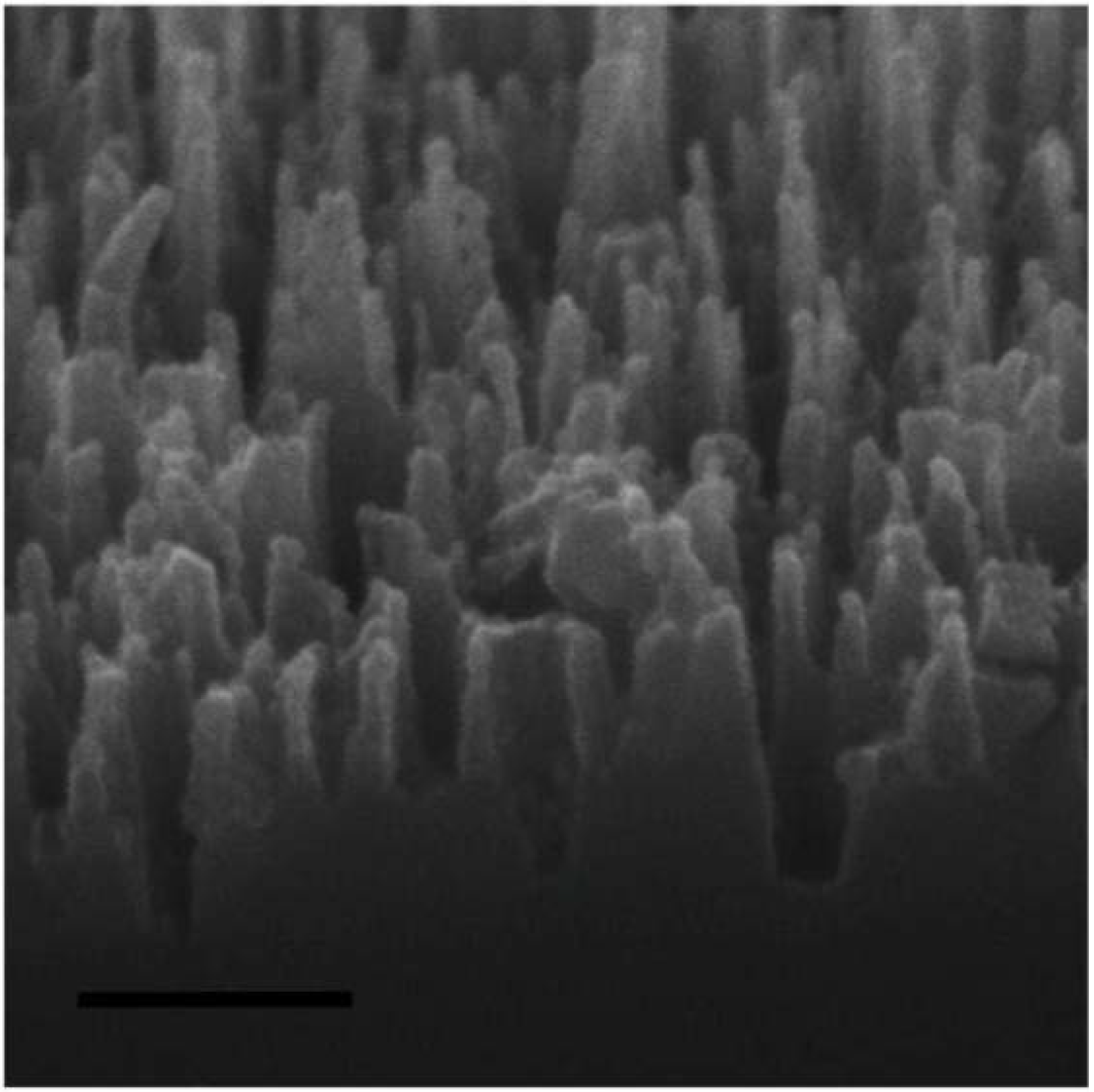
7. Surface Patterning

Surface patterning offers a more organized means of roughening to alter the surface of a material. Patterning may quell non-specific protein-surface interactions, as these effects often lead to device failure [52]. Such patterning techniques are often used to enhance endothelial cell attachment, which in turn encourages vessel wall healing and promotes an anti-thrombotic environment.
Nanopillar arrays, formed by plasma processing as shown in Figure 4, provide a scaffold for cell proliferation or drug delivery [30]. Patterning on metal surfaces, primarily on the nanometer scale, has been shown to promote more endothelial cell attachment compared to random nanopatterning or even patterning on the micron scale [1,53]. These nanopatterned surfaces also encourage more endothelial cell attachment compared to smooth cell attachment which is desirable in vessel healing, support greater cell densities on the surface, and even enhance spreading of these endothelial cells [1,51]. Cells in their native environment come into contact with features on the nano-scale, which could be the reason for enhanced cell attachment [50,54,55]. Some patterning methods strive to mimic native endothelium for a biomimetic effect, in hopes of encouraging more rapid endothelialization and vessel healing, without the presence of plasma proteins or extracellular matrix [1,2,52,54]. Biomimetic patterning may have major implications for SMP stents in that increased biocompatibility can be obtained simply by polymerizing the stent inside of a native blood vessel, directly transferring native endothelial pattern onto the stent surface.
Figure 4. SEM image of Silicon pillars formed via plasma processing (scale bar = 20 μm). Reprinted with permission from Elsevier, 2001 [30].
Polymers 06 02309 g004
Patterning can also be achieved through diblock copolymer grafts, which form nanometer-sized patterns on solid surfaces. Diblock copolymers can be either physically or chemically attached and form nano-sized domains when they undergo microphase separation. These patterns either encourage or discourage protein adsorption and/or cell adhesion, depending on the polymers involved. For this reason, diblock copolymers have been investigated with regard to surface energy or topography, and are being explored for their potential uses in reference to bioactivity [56].
Polymers that undergo phase separation such as the mixture of polystyrene and poly(4-bromostyrene) can produce a range of surface topographies just by varying the polymer concentrations and proportions [57,58]. Changes in polymer ratio can yield variations in shape, such as pits, islands and ribbons for example, whereas changing the concentration of the polymer may alter the feature sizes. Cell spreading and proliferation differ based on feature height, with shorter feature heights producing promising results in enhanced cell spreading and proliferation [58].
With regard to polymer surface patterning, lithography is one of the more frequently used techniques, a technique common in the electronics field, mainly for patterning of silicon wafers [32]. Patterns can include anything from dots and pillars to grooves and ridges, where grooves and ridges are the most studied, often due to the increased tendency of cells to attach and spread along those features [32,59]. Lithography may even be used to create hierarchical patterns or tilted patterns, if desired [60]. A few theories have attempted to predict why cells prefer to align along grooves and ridges, but different cells have different preferences with regard to size and shape of the formed pattern [32]. Photolithography is commonly used on polymer surfaces and this technique selectively exposes surfaces to photoirradiation, creating a pattern on the surface [61,62,63]. This allows for controlled topographical features, directing cell attachment [61,64]. Lithographic techniques continue to be a prominent surface modification method for polymers and applying these methods to SMPs, particularly SMP-based stents, may also prove beneficial.
Microfluidic channels offer another means to direct cell adhesion via patterning. Proteins adsorb onto the surface after passing through elastomeric channels in solution form, and once adhered, these proteins, such as fibronectin and collagen, are used for selective cell adhesion. This method can also be used to produce a patterned cell co-culture, if two different types of cells need to adhere to the same surface [65].
Self-Assembled Monolayers (SAMs), a common chemical-based surface modification technique, have also been explored in creating patterns on biomaterial surfaces [32,66]. SAMs encourage cell adhesion and orientation, qualities that are advantageous to stent biocompatibility, by controlling protein adsorption onto the surface [67]. SAMs are also used for microcontact printing, another method for patterning that is commonly used to encourage cell attachment [30].
With regard to SMP-specific patterning techniques, methods in which balls (steel or lime glass) that make indentations on the surface have been explored. Different sized indentations can be made using different sized balls [68]. In addition, wrinkling patterns on top of SMPs can be formed using the shape memory capabilities of the polymer itself and if the wrinkling is controlled, a number of surface properties that improve biocompatibility can be manipulated, including roughness, wetting, and bonding among others [69].
Transfer printing involves the transfer of a pattern from a mold to a polymer substrate, resulting in a thin patterned film on the surface of the polymer [60]. These films are usually polymers themselves, and have the potential to encourage cell adhesion by introducing nanoscale patterns that favor cell attachment. Transfer printing can also create surfaces with hydrophobic and hydrophilic characteristics, directing cell attachment to certain areas [70]. Zhao et al., determined that microtransfer molding using a PDMS mold creates micron-sized patterns, which may once again increase endothelial cell attachment [71].
Similar to transfer printing, stencil-assisted printing involves using a stencil to imprint a desired pattern or structure onto the polymer surface. The patterns develop on the surface that is left uncovered by the stencil, thus directing cell attachment to these exposed areas. This technique does not require any chemical modification after the stencil has been manufactured, making it an appealing method to enhance material biocompatibility as well as a potential technique for surface patterning of SMPs [52].
Nanopatterning through dip-pen lithography uses the tip of an atomic force microscope (AFM) to create a pattern on a material’s surface. This tip is dipped into a polymer solution and touched to the surface of a material, altering the chemical makeup of that surface in an organized manner, creating a pattern [72]. Depending upon the polymer that is applied to the surface, enhancement in blood compatibility and/or cell attachment can be achieved. The use of a heated tip to create patterns on the surface of SMPs has been explored, and may provide an avenue for patterning SMPs to encourage cell attachment [73]. Indentations can also be made using a scanning force microscope (SFM), generally for analytical purposes, but there may be potential for surface modification here as well [74,75].
In an effort to physically mimic the patterns found in native vessels, pre-polymer solutions were polymerized inside of a harvested, native blood vessel [60]. The polymer adopts the surface features of the blood vessel on its own surface, but the main limitation of this method is that the vessel tissue had to be dissolved to remove the polymer, rendering reproducibility difficult. However, since SMPs acquire their permanent shapes during initial polymerization, this method applied to SMPs may be worth further investigation.
Although SMPs have been treated with only a few patterning techniques, the success associated with patterning polymer materials suggests that patterning SMPs with these techniques may have positive outcomes.

8. Chemical Modification of the Surface

Chemical modification techniques chemically alter the surface of a material without significantly affecting its bulk properties. Some examples of chemical modifications include chemical vapor deposition (CVD), plasma vapor deposition (PVD), grafting techniques, self-assembled monolayers (SAMs), among others [1,76].
For metals, many chemical modification techniques, such as plasma immersion ion implantation (PIII) using acetylene, nitrogen or oxygen, are used to reduce corrosion, wear and metal leaching into the surrounding environment and even increase hardness of the material [16]. PIII treatment of polymers has been shown to reduce thrombus formation and platelet aggregation by increasing hydrophilicity and protein adsorption onto the surface, as displayed in Figure 5 [77].
Figure 5. Isolated platelets in buffer adhering to both surfaces, but platelets in plasma do not adhere to ion treated polymer surface (scale bar = 20 μm). Reprinted with permission from PNAS, 2011 [77].
Polymers 06 02309 g005
Chemical vapor deposition (CVD) utilizes plasma or other reactive chemicals to deposit thin films onto the surface of the material, slightly altering the surface to allow for deposition of the film [64,78]. Due to the non-fouling properties associated with the deposited film, plasma-driven CVD techniques are popular for blood compatibility [78]. One form of CVD has been used on stents and other blood contacting devices commercially, and goes by the name parylene. Parylene aids in biocompatibility as well as providing a means for sustained drug release from a porous matrix [79]. However, this coating does not have functional groups to attach biomolecules, so further treatment with plasma or chemicals to introduce tethering molecules would be required for biomolecule attachment.
Low pressure plasma treatments that use electrons, ions, radicals, metastables or ultraviolet rays (UV) radiation elicit reactions at the surface of polymers [48]. For plasma-based treatments, ammonia plasma treatment may encourage cell attachment more through the interaction of acidic groups on the plasma membrane surface and amine/amide groups on the surface of the polymer, which play an important role in endothelial cell adhesion and growth [21,80]. Some studies have shown that cornea cells showed enhanced attachment and growth on plasma-treated surfaces vs. untreated surfaces, and exploration into whether this applies to other types of cells may have merit [48]. Studies prepared by Ho et al. found that polymer samples that undergo water vapor plasma treatment may elicit enhanced cell attachment compared to untreated samples, potentially due to the formation of hydroxyl groups on the surface, allowing for hydrogen bond formation between the surface and the cells [81,82].
While research has been generally inconclusive about which surfaces are best for supporting cell adhesion and growth, surfaces that are mildly hydrophobic or mildly hydrophilic appear to support optimal cell development; these mild conditions may be achieved through plasma treatment using reaction gases containing organic compounds [32,65,83,84,85]. Plasma treatment of polymer materials has positive effects on cell adhesion and development on the material surface, mainly by enhancement of hydrophilicity and wettability [38,49,83,86]. Plasma deposition may even be used to reduce thrombogenicity [38].
Plasma vapor deposition (PVD) techniques such as matrix-assisted pulsed laser evaporation, deposit organic and biological materials onto the surface of blood-contacting devices, altering the surface [1]. Ionic plasma deposition has been shown to increase endothelial cell adhesion [87]. Certain polymer surfaces exposed to N2 and O2 in Helium display enhanced attachment properties, with the extent of surface modification depending upon the polymer surface itself [21,47]. Other surfaces exposed to nitrogen gases have been known to exhibit reduced thrombotic properties [3,88]. As with etching, plasma processes cause the formation of free radicals at the material surface, causing the formation of cross-links [25]. These reactive surfaces can be used to encourage coverage with a thin film or can facilitate the attachment of (bio)molecules.
Photografting of polymers using high energy electrons, gamma radiation, ultraviolet (UV) light and visible light can change the surface of polymers to improve blood compatibility and enhance endothelialization [1,21]. Bilek et al., found that treatment of a polymer surface with ions to create a free radical surface encourages protein immobilization while retaining protein structure, potentially enhancing biocompatibility [77]. Photo-oxidation, a method to introduce hydrophilic groups to polymer surfaces in a controlled manner through the manipulation of photo-oxidation time and grafting time, has also been shown to be beneficial to endothelial cell development on the material surface [32].
Chemical grafting methods, such as the grafting of polyethylene glycol (PEG) monoacrylates to the surface of a biomaterial, can reduce the attachment of erythrocytes through steric repulsion, thus decreasing the risk of thrombosis [89,90]. PEG is largely hydrophilic and has a large exclusion volume, contributing to this effect and it is also non-toxic and non-immunogenic which are important components of a biocompatible material [89]. Grafting copolymerization methods that graft hydrophilic polymers onto hydrophobic surfaces in an effort to neutralize hydrophobicity may encourage cell adhesion [32]. Plasma and ultraviolet grafting on polymer surfaces may also promote anticoagulation and antibacterial properties [6].
Self-Assembled Monolayers (SAMs) modify the surfaces of materials to enhance hydrophobicity/hydrophilicity or to add reactive or functional groups to the surface. SAMs change the surface energy or wettability of the polymer surface through careful selection of the functional groups used for the monolayer, potentially increasing biocompatibility of the material within the vessel [91]. SAMs offer the benefit of ease of fabrication, and the ability to control order and orientation, allowing for the exposure of a select group on the modified surface, creating the ability to cater the biocompatibility of the material to suit specific needs [38].
Chemical modification techniques strive to alter the surface of the material in order to enhance the functionality of that material. Polymer substrates undergo exposure to these different techniques, resulting in a material with an improved surface and mostly unchanged bulk properties. If performed properly, these chemical modification techniques can be applied to SMPs allowing for a better surface without affecting the bulk.

9. Surface Coatings and Films

Surface coatings and films are additional ways to modify surfaces of both metals and polymers in an effort to increase biocompatibility. These techniques often do not involve direct attachment of chemical groups or chemical alteration of the surface the way conventional chemical modification techniques do, but still alter the surfaces for increased biocompatibility. A few coating and film techniques that have been shown to increase endothelial cell attachment or reduce blood coagulation and thrombosis are discussed.
With regard to wet coating/solvent coating of stents, dimethyl sulfoxide (DMSO) has been shown to prevent vascular smooth muscle cell activity on the stent surface, reducing chances for restenosis while also preventing tissue factor activity, thus discouraging thrombosis [19]. Studies show that DMSO does not exert toxicity to human vascular endothelial cells, further solidifying this technique as a potentially viable option for polymers and SMPs [92]. Dip coating, used to form nanostructures on the surface of medical-grade polymers, creates superhydrophobic surfaces that prevent blood coagulation [93]. Coating polymers with polyelectrolyte multilayers provides a good platform for endothelial cells on polymer surfaces [6].
Langmuir-Blodgett (LB) films, consisting of highly ordered, densely packed structures of known thickness deposited and crosslinked to the surface of the polymer, also allow for cell adhesion, decreased platelet adhesion and enhanced hemocompatibility [21,94]. These LB films can be deposited on polymer surfaces by chemically treating the polymer to attract the LB film and introducing the polymer to a Langmuir-Blodgett trough, allowing a monolayer to form prior to endothelial cell exposure [95]. These LB films have not yet been studied extensively on three dimensional scaffolds, but implementing these films on three dimensional structures may be worth further investigation due to the increased biocompatibility offered by this technique. Layer-by-Layer (LbL) polymer films have been shown to reduce platelet adhesion on nitinol, a commonly used stent material [1]. LbL deposition of chitosan on the surface of the polymer poly-l-lactide (PLLA), showed improved cell compatibility [96]. Studies with diamond-like carbon (DLC) films have also displayed successful attempts to improve blood compatibility on polymer surfaces [8].

10. Attachment of Pharmaceuticals, Biopharmaceuticals or Biomolecules to the Surface

The ability to attach a substance to the surface of a material while retaining its bulk properties is an appealing method of delivery for pharmaceuticals or biomolecules [26]. Polymers usually have inert surfaces, so in those instances, the surface must be functionalized prior to attaching the bioactive molecule to the surface. As a surface technique, these methods can be applied to either polymers or SMPs, and while most of these techniques have been tested on polymers, there may be benefits to applying these techniques on SMPs. The bioactive compound can be attached through electrostatic interactions, ligand-receptor interactions, or covalent attachment, where covalent linkages are most common as this linkage is often the most stable [26].
Chemical Vapor Deposition (CVD) is not only used to enhance biocompatibility, but is also used to create tethering groups on the surface of a polymer for proteins and other biomolecules to attach via covalent bonding [1,78,79]. Some of these biomolecules help create a less thrombotic environment by immobilizing on the surface of polymers in the blood vessel. Plasma deposition techniques produce stable films that can aid in corrosion resistance and functionalization sites for the attachment of (bio)pharmaceuticals onto the surfaces of both metals and polymers [1].
Wet chemical surface modification methods require chemical reagents to create reactive functional groups on the surface of a polymer, often without expensive equipment or methods, and can be done easily in a laboratory setting. Wet chemicals are able to accomplish deeper penetration of porous surfaces compared to energy-source-based modification techniques, creating a more stable and noncorrosive functionalized surface. If repeatability is desired however, this method may not be the ideal choice, as a wide range of reactive groups are generated and the orientation of the biomolecule can be crucial for attachment. However, to promote specificity, it may be possible to block some functional groups, allowing for the specific molecule, whether it be a molecule to enhance endothelialization or a protein to reduce thrombus formation, to attach successfully [26].
Plasma treatment methods can introduce reactive groups to the surface of a normally inert polymer, allowing for the attachment of a desired bioactive compound. These methods do not require hazardous chemicals, yet still have the capability to modify the surface while imparting less degradation and roughness compared to wet chemical surface modification techniques [26]. In addition, the film deposited on the polymer surface can be manipulated by changing the deposition rate, energy range and surface topography [97]. Plasma pre-treatment has also been used prior to attaching collagen to a polymer nanofiber mesh, a method that showed increased cell attachment, spreading and viability [98]. Thus, if a less corrosive method for biomolecule attachment is required, plasma treatment may be a favorable option, but great care must be taken to avoid contamination of the sample. Once the surface has been functionalized, the desired bioactive agent can be attached for purposes of enhanced cell attachment or thrombosis prevention.
Nitric oxide (NO) or thrombomodulin, both of which are integral to maintaining homeostasis in the blood vessel, can be released from the polymer backbone itself or attached to the surface [21]. Gene-eluting stents are capable of delivering biologically active, therapeutic genes in an effort to reduce restenosis, accelerate re-endothelialization and reduce thrombosis. Another set of stents, termed biologically active stents, incorporate antibodies or proteins, such as CD34 antibodies, onto the surface to attract endothelial progenitor cells, or the Arg-Gly-Asp peptide sequence, which also attracts endothelial progenitor cells, speeding up the re-endothelialization process [17,99].
In order to mimic naturally occurring conditions in the blood vessel, there has been some work in functionalizing the surface of the polymer with the arginine-glycine-aspartate (RGD) sequence, a protein commonly found in the native extracellular matrix (ECM) [100,101]. Hegemann et al., determined that this environment promotes endothelial cell attachment and growth [102]. Similarly, other cell-adhesion peptides, such as glycine-arginine-glycine-aspartate (GRGD), immobilized on the surface of biomaterials have displayed enhanced endothelial cell attachment [103,104,105,106]. Pre-absorbed proteins, such as fibronectin, laminin and gelatin, present on polymer surfaces have been shown to increase cell attachment, but may reduce cell proliferation [107,108]. Endothelial cells may be seeded directly onto the material prior to implantation to ensure biocompatibility [94]. To combat thrombotic events directly, adding lysine to the surface of a material has been shown to perform clot lysis, preventing blood coagulation [109].
Layer-by-Layer (LbL) polymer films have also been used to effectively deliver nitric oxide (NO) donor to the site of vessel injury and can house DNA to be delivered to the vessel wall from the covering [1]. This technique deposits both positively and negatively charged biomacromolecules as well, and can even be used for biodegradable polymers, due its mild preparation environment [32].
Polypyrrole composites, an electrically-conducting polymer, containing heparin or sodium nitrate, have the ability to switch between oxidized and reduced states, and this switching ability controls the release of biological signaling agents such as growth factors, thus directing cell growth on a surface [110].
Biomolecules attached to the surface of polymeric materials in a patterned manner may help control cell behavior or direct cell signaling [111]. A range of biomolecules have been immobilized on material surfaces, where the selection of biomolecule(s) is dictated by the nature of cells to be deposited on the surface.
The desire to attach (bio)pharmaceuticals and (bio)molecules to material surfaces is driven by the need provide localized delivery of the molecule or drug without changing the bulk properties of the delivery vehicle. Although many of these techniques require reactive surface groups, further investigation into functionalization of SMPs for (bio)molecule and (bio)pharmaceutical may be desirable, especially for localized drug and molecule delivery.

11. Porous Surfaces to Facilitate Drug Delivery

As mentioned before, stents are commonly used as drug delivery vehicles to stimulate vessel healing and allow for better incorporation of the stent into the body without the use of oral anticoagulant drugs. The drugs used with the stents can be attached directly to the surface of the stent, as was discussed briefly above, or they can be incorporated into the surface of the stent using pores to house the drug until delivery.
Porous stents allow for drug incorporation without an additional polymer coating that is commonly found in drug eluting stents [1]. A variety of surface modification techniques have been used to adjust surfaces for the purpose of housing drug for delivery. Etching of the polymer surface for long periods of time may cause pores to form, which can be used to house drug for localized delivery [47,112]. The use of photolithography or soft lithography to create pores in polymer sample surfaces or to fabricate porous micro- or nano-particles for embedding onto a polymer surface for drug delivery has also been under investigation [113]. Sandblasting has been shown to effectively create porous surfaces on metal stents [1]. Aluminum coatings exposed to acidic solutions form ceramic aluminum oxide, resulting in nanoscale pores on that film for drug delivery [1,114]. Acidic treatment of stainless steel stents has also been successful in creating porous surfaces for drug elution [114]. Stents with a porous hydroxyapatite coating have exhibited promising results for drug elution as well [1,115].
A porous surface formed by carbon nanoparticles embedded in polymer has displayed promise as a means for localized drug delivery [116]. Similarly, cobalt-chromium alloy stents covered with a porous carbon-carbon coating also showed potential in the arena of drug elution and enhanced cell attachment [116].
Drug delivery from pores is not solely limited to the surface; research efforts have also looked into loading drug components into the bulk of SMPs and using shape memory capabilities for drug elution [117,118]. This is particularly applicable to SMP stents, since loading a SMP stent with drug components allows for sustained and localized drug delivery [117]. Drug release can be controlled by altering the co-monomer ratio, but efforts to maintain the shape memory effect must also be considered [118]. Hydrogels, a class of polymer with swelling capabilities, have also been used for slow-release, drug delivery using a diffusion mechanism through pores that often penetrate the bulk material of the hydrogel [119,120]. Porous surfaces may allow for localized delivery of a drug or molecule without the need for prior functionalization of the surface. If functionalization for molecular tethering is not an option, forming pores in the surface for drug incorporation may prove beneficial for a range of surfaces, and might soon be extended to SMPs.

12. Conclusions

Although many surface modification techniques have been evaluated in an effort to create the “better” stent, the ideal surface modification remains elusive, reinforcing the research need in this particular area. SMPs, with their capability for better and minimally-invasive delivery, have the potential to become a commonly used material for cardiovascular stents. Surface modification of such polymer systems through the application of many of these techniques should provide an important added benefit to the many aforementioned benefits of SMP devices. In addition, many of these techniques have not yet been used widely on bioabsorbable or biodegradable stent materials, which are becoming more prevalent as the desire for the stent as a “temporary scaffold” continues to grow. Additional research on surface modification of bioabsorbable materials will therefore become more important.

Acknowledgments

This work was supported in part by grants from the National Institutes of Health (NIH) (NIH T32 HL072738, NIH K24 HL081506).

Author Contributions

Tina Govindarajan took the lead in writing the review manuscript. Robin Shandas provided guidance throughout the writing process and provided editing, suggestions, and funding.

Conflicts of Interest

The authors declare no conflict of interest.

References

  1. Martinez, A.W.; Chaikof, E.L. Microfabrication and nanotechnology in stent design. WIREs Nanomed. Nanobiotechnol. 2011, 3, 256–268. [Google Scholar] [CrossRef]
  2. Miller, D.C.; Thapa, A.; Haberstroh, K.M.; Webster, T.J. Endothelial and vascular smooth muscle cell function on poly(lactic-co-glycolic acid) with nano-structured surface features. Biomaterials 2004, 25, 53–61. [Google Scholar] [CrossRef]
  3. Chandy, T.; Das, G.S.; Wilson, R.F.; Rao, G.H.R. Use of plasma glow for surface engineering biomolecules to enhance bloodcompatibility of Dacron and PTFE vascular prosthesis. Biomaterials 2000, 21, 699–712. [Google Scholar] [CrossRef]
  4. Reape, T.J.; Groot, P.H.E. Chemokines and atherosclerosis. Atherosclerosis 1999, 147, 213–225. [Google Scholar] [CrossRef]
  5. Grabow, N.; Martin, D.P.; Schmitz, K.P.; Sternberg, K. Absorbable polymer stent technologies for vascular regeneration. J. Chem. Technol. Biotechnol. 2010, 85, 744–751. [Google Scholar] [CrossRef]
  6. Boura, C.; Menu, P.; Payan, E.; Picart, C.; Voegel, J.C.; Muller, S.; Stoltz, J.F. Endothelial cells grown on thin polyelectrolyte mutlilayered films: An evaluation of a new versatile surface modification. Biomaterials 2003, 24, 3521–3530. [Google Scholar] [CrossRef]
  7. Tran, H.S.; Puc, M.M.; Hewitt, C.W.; Soll, D.B.; Marra, S.W.; Simonetti, V.A.; Cilley, J.H.; DelRossi, A.J. Diamond-like carbon coating and plasma or glow discharge treatment of mechanical heart valves. J. Investig. Surg. 1999, 12, 133–140. [Google Scholar] [CrossRef]
  8. Alanazi, A.; Nojiri, C.; Noguchi, T.; Kido, T.; Komatsu, Y.; Hirakuri, K.; Funakubo, A.; Sakai, K.; Fukui, Y. Improved blood compatibility of DLC coated polymeric material. ASAIO 2000, 46, 440–443. [Google Scholar] [CrossRef]
  9. Peng, T.; Gibula, P.; Yao, K.; Goosen, M.F.A. Role of polymers in improving the results of stenting in coronary arteries. Biomaterials 1996, 17, 685–694. [Google Scholar] [CrossRef]
  10. Gopinath, M.; Feldman, M.D.; Patel, D.; Agrawal, C.M. Coronary stents: A materials perspective. Biomaterials 2007, 28, 1689–1710. [Google Scholar] [CrossRef]
  11. Kahn, W.; Farah, S.; Domb, A.J. Drug eluting stents: Developments and current stents. J. Control. Release 2012, 161, 703–712. [Google Scholar] [CrossRef]
  12. Kolodgie, F.; Nakazawa, G.; Sangiorgi, G.; Ladich, E.; Burke, A.; Virmani, R. Pathology of atherosclerosis and stenting. Neuroimaging Clin. N. Am. 2007, 17, 285–301. [Google Scholar] [CrossRef]
  13. Yakacki, C.M.; Shandas, R.; Lanning, C.; Rech, B.; Eckstein, A.; Gall, K. Unconstrained recovery characterization of shape-memory polymer networks for cardiovascular applications. Biomaterials 2007, 28, 2255–2263. [Google Scholar] [CrossRef]
  14. Van der Hoeven, B.; Pires, N.; Warda, H.; Oemrawsingh, P.; Vanvlijmen, B.; Quax, P.; Schalij, M.; Vanderwall, E.; Jukema, J. Drug-eluting stents: Results, promises and problems. Int. J. Cardiol. 2005, 99, 9–17. [Google Scholar]
  15. Joner, M.; Finn, A.V.; Farb, A.; Mont, E.K.; Kolodgie, F.D.; Ladich, E.; Kutys, R.; Skorija, K.; Gold, H.K.; Virmani, R. Pathology of drug-eluting stents in humans: Delayed healing and late thrombotic risk. J. Am. Coll. Cardiol. 2006, 48, 193–202. [Google Scholar] [CrossRef]
  16. Chu, P. Enhancement of surface properties of biomaterials using plasma-based technologies. Surf. Coat. Technol. 2007, 201, 8076–8082. [Google Scholar] [CrossRef]
  17. Costa, M.A. Molecular basis of restenosis and drug-eluting stents. Circulation 2005, 111, 2257–2273. [Google Scholar] [CrossRef]
  18. Huang, N.; Yang, P.; Leng, Y.X.; Chen, J.Y.; Wang, J.; Wan, G.J.; Sun, H.; Wu, X.; Zhao, A.S. Improving blood compatibility of cardiovascular devices by surface modification. Key Eng. Mater. 2007, 342–343, 801–804. [Google Scholar]
  19. Luscher, T.F.; Steffel, J.; Eberli, F.R.; Joner, M.; Nakazawa, G.; Tanner, F.C.; Virmani, R. Drug-eluting stent and coronary thrombosis: Biological mechanisms and clinical implications. Circulation 2007, 115, 1051–1058. [Google Scholar] [CrossRef]
  20. Pypen, C.M.J.M.; Plenk, H.; Ebel, M.F.; Svagera, R.; Werisch, J. Characterization of microblasted and reactive ion etched surfaces on the commercially pure metals Niobium, Tantalum and Titanium. J. Mater. Sci. Mater. Med. 1997, 8, 781–784. [Google Scholar] [CrossRef]
  21. Pavithra, D.; Doble, M. Biofilm formation, bacterial adhesion and host response on polymeric implants—Issues and prevention. Biomed. Mater. 2008, 3, 1–11. [Google Scholar]
  22. Ormiston, J.A.; Serruys, P.W.; Regar, E.; Dudek, D.; Thuesen, L.; Webster, M.W.I.; Onuma, Y.; Garcia, H.M.; McGreevy, R.; Veldhof, S. A bioabsorbable everolimus-eluting coronary stent system for patients with single de-novo coronary artery lesions (ABSORB): A prospective open-label trial. Lancet 2008, 371, 899–907. [Google Scholar] [CrossRef]
  23. Di Mario, C.; Ferrante, G. Biodegradable drug-eluting stents: Promises and pitfalls. Lancet 2008, 371, 873–874. [Google Scholar]
  24. Hegemann, D.; Brunner, H.; Oehr, C. Plasma treatment of polymers for surface and adhesion improvement. Nucl. Instrum. Methods B 2003, 208, 281–286. [Google Scholar] [CrossRef]
  25. Fare, S.; Valtulina, V.; Petrini, P.; Alessandrini, E.; Pietrocola, G.; Tanzi, M.C.; Speziale, P.; Visai, L. In vitro interaction of human fibroblasts and platelets with a shape-memory polyurethane. J. Biomed. Mater. Res. A 2005, 73, 1–11. [Google Scholar] [CrossRef]
  26. Goddard, J.; Hotchkiss, J. Polymer surface modification for the attachment of bioactive compounds. Prog. Polym. Sci. 2007, 32, 698–725. [Google Scholar] [CrossRef]
  27. Angelova, N.; Hunkeler, D. Rationalizing the design of polymeric biomaterials. Trends Biotechnol. 1999, 17, 409–421. [Google Scholar] [CrossRef]
  28. Lendlein, A.; Behl, M.; Hiebl, B.; Wischke, C. Shape-memory polymers as a technology platform for biomedical applications. Expert Rev. Med. Devices 2010, 7, 357–379. [Google Scholar] [CrossRef]
  29. Hersel, U.; Dahmen, C.; Kessler, H. RGD modified polymers: Biomaterials for stimulated cell adhesion and beyond. Biomaterials 2003, 24, 4385–4415. [Google Scholar] [CrossRef]
  30. Craighead, H.G.; James, C.D.; Turner, A.M.P. Chemical and topographical patterning for directed cell attachment. Curr. Opin. Solid Sate Mater. Sci. 2001, 5, 177–184. [Google Scholar] [CrossRef]
  31. Chu, P.K.; Chen, J.Y.; Wang, L.P.; Huang, N. Plasma-surface modification of biomaterials. Mater. Sci. Eng. 2002, 36, 143–206. [Google Scholar] [CrossRef]
  32. Ma, Z.; Mao, Z.; Gao, C. Surface modification and property analysis of biomedical polymers used for tissue engineering. Colloid Surf. B 2007, 60, 137–157. [Google Scholar] [CrossRef]
  33. Prasad, C.K.; Muraleedharan, C.V.; Krishnan, L.K. Bio-mimetic composite matrix that promotes endothelial cell growth for modification of biomaterial surface. J. Biomed. Mater. Res. A 2006, 80, 644–654. [Google Scholar]
  34. Helmus, M.N.; Gibbons, D.F.; Cebon, D. Biocompatibility: Meeting a key functional requirement of next-generation medical devices. Toxicol. Pathol. 2008, 36, 70–80. [Google Scholar] [CrossRef]
  35. Arima, Y.; Iwata, H. Effect of wettability and surface functional groups on protein adsorption and cell adhesion using well-defined mixed self-assembled monolayers. Biomaterials 2007, 28, 3074–3082. [Google Scholar] [CrossRef]
  36. Van Wachem, P.; Hogt, A.; Beugeling, T.; Feijen, J.; Bantjes, A.; Detmers, J.; van Aken, W. Adhesion of cultured human endothelial cells onto methacrylate polymers with varying surface wettability and charge. Biomaterials 1987, 8, 323–328. [Google Scholar]
  37. Van Wachem, P.; Beugeling, T.; Feijen, J.; Bantjes, A.; Detmers, J.; van Aken, W. Interaction of cultured human endothelial cells with polymeric surfaces of different wettabilities. Biomaterials 1985, 6, 403–408. [Google Scholar]
  38. Ratner, B.D. Surface modification of polymers: Chemical, biological and surface analytical challenges. Biosens. Bioelectron. 1995, 10, 797–804. [Google Scholar] [CrossRef]
  39. Lyu, S.P.; Cernohous, J.J.; Bates, F.S.; Macosko, C.W. Interfacial reaction induced roughening in polymer blends. Macromolecules 1999, 32, 106–110. [Google Scholar] [CrossRef]
  40. Curtis, A.; Wilkinson, C. Topographical control of cells. Biomaterials 1997, 18, 1573–1583. [Google Scholar] [CrossRef]
  41. Meredith, J.C.; Sormana, J.L.; Keselowsky, B.G.; Garcia, A.J.; Tona, A.; Karim, A.; Amis, E.J. Combinatorial characterization of cell interactions with polymer surfaces. J. Biomed. Mater. Res. A 2003, 66, 483–490. [Google Scholar] [CrossRef]
  42. Lampin, M.; Warocquier-Clerout, R.; Legris, C.; Degrange, M.; Sigot-Luizard, M.F. Correlation between substratum roughness and wettability, cell adhesion, and cell migration. J. Biomed. Mater. Res. 1997, 36, 99–108. [Google Scholar] [CrossRef]
  43. Shadpour, H.; Allbritton, N.L. In-situ roughening of polymeric microstructures. ACS Appl. Mater. Interfaces 2010, 2, 1086–1093. [Google Scholar] [CrossRef]
  44. Yeh, H.I.; Lu, S.K.; Tian, T.Y.; Hong, R.C.; Lee, W.H.; Tsai, C.H. Comparison of endothelial cells grown on different stent materials. J. Biomed. Mater. Res. A 2006, 76, 835–841. [Google Scholar] [CrossRef]
  45. Noeske, M.; Degenhardt, J.; Strudthoff, S.; Lommatzsch, U. Plasma jet treatment of five polymers at atmospheric pressure: Surface modifications and the relevance for adhesion. Int. J. Adhes. Adhes. 2004, 24, 171–177. [Google Scholar] [CrossRef]
  46. Ikada, Y. Surface modification of polymers for medical applications. Biomaterials 1994, 15, 725–736. [Google Scholar] [CrossRef]
  47. Avram, M.; Avram, A.M.; Bragaru, A.; Ghui, A.; Iliescu, C. Plasma surface modification for selective hydrophobic control. Roman J. Inf. Sci. Technol. 2008, 11, 409–422. [Google Scholar]
  48. Oehr, C. Plasma surface modification of polymers for biomedical use. Nucl. Instrum. Methods B 2003, 208, 40–47. [Google Scholar] [CrossRef]
  49. McAuslan, B.R.; Johnson, G. Cell responses to biomaterials I: Adhesion and growth of vascular endothelial cells on poly(hydroxyethyl methacrylate) following surface modification by hydrolytic etching. J. Biomed. Mater. Res. 1987, 21, 921–935. [Google Scholar] [CrossRef]
  50. Chung, T.W.; Liu, D.Z.; Wang, S.Y.; Wang, S.S. Enhancement of the growth of human endothelial cells by surface roughness at nanometer scale. Biomaterials 2003, 24, 4655–4661. [Google Scholar] [CrossRef]
  51. Ranjan, A.; Webster, T.J. Increased endothelial cell adhesion and elongation on micron-patterned nano-rough poly(dimethylsiloxane) films. Nanotechnology 2009, 20, 1–11. [Google Scholar]
  52. Falconnet, D.; Csucs, G.; Grandin, H.M.; Textor, M. Surface engineering approaches to micropattern surfaces for cell-based assays. Biomaterials 2006, 27, 3044–3063. [Google Scholar] [CrossRef]
  53. Khang, D.; Lu, J.; Yao, C.; Haberstroh, K.; Webster, T. The role of nanometer and sub-micron surface features on vascular and bone cell adhesion on titanium. Biomaterials 2008, 29, 970–983. [Google Scholar] [CrossRef]
  54. Flemming, R.G.; Murphy, C.J.; Abrams, G.A.; Goodman, S.L.; Nealey, P.F. Effects of artificial micro and nano structured surfaces on cell behavior. Biomaterials 1999, 20, 573–588. [Google Scholar] [CrossRef]
  55. Yim, E.K.F.; Reano, R.M.; Pang, S.W.; Yee, A.F.; Chen, C.S.; Leong, K.W. Nanopattern-induced changes in morphology and motility of smooth muscle cells. Biomaterials 2005, 26, 5405–5413. [Google Scholar] [CrossRef]
  56. Yu, Q.; Zhang, Y.; Chen, H.; Chou, Z.; Wu, Z.; Huang, H.; Brash, J.L. Protein adsorption and cell adhesion/detachment behavior on dual-responsive silicon surfaces modified with poly(N-isopropylacrylamide)-block-polystyrene copolymer. Langmuir 2010, 26, 8582–8588. [Google Scholar] [CrossRef]
  57. Karim, A.; Slawecki, T.M.; Kumar, S.K.; Douglas, J.F.; Satija, S.K.; Han, C.C.; Russell, T.P.; Liu, Y.; Overney, R.; Sokolov, J.; et al. Phase-separation-induced surface patterns in thin polymer blend films. Macromolecules 1998, 31, 857–862. [Google Scholar] [CrossRef]
  58. Dalby, M.J.; Yarwood, S.J.; Riehle, M.O.; Johnstone, H.J.H.; Affrossman, S.; Curtis, A.S.G. Increasing fibroblast response to materials using nanotopography: morphological and genetic measurements of cell response to 13-nm-high polymer demixed islands. Exp. Cell Res. 2002, 276, 1–9. [Google Scholar] [CrossRef]
  59. Wilkinson, C.D.W.; Riehle, M.; Wood, M.; Gallagher, J.; Curtis, A.S.G. The use of materials patterned on a nano- and micro-metric scale in cellular engineering. Mater. Sci. Eng. C 2002, 9, 263–269. [Google Scholar] [CrossRef]
  60. Del Campo, A.; Arzt, E. Fabrication approaches for generating complex micro- and nanopatterns on polymeric surfaces. Chem. Rev. 2008, 108, 911–945. [Google Scholar]
  61. Nie, Z.; Kumacheva, E. Patterning surfaces with functional polymers. Nat. Mater. 2008, 7, 277–290. [Google Scholar] [CrossRef]
  62. Xu, C.; Yang, F.; Wang, S.; Ramakrishna, S. In Vitro study of human vascular endothelial cell function on materials with various surface roughness. J. Biomed. Mater. Res. A 2004, 71, 154–161. [Google Scholar] [CrossRef]
  63. Kane, R.S.; Takayama, S.; Ostuni, E.; Ingber, D.E.; Whitesides, G.M. Patterning proteins and cells using soft lithography. Biomaterials 1999, 20, 2363–2376. [Google Scholar] [CrossRef]
  64. Lahann, J.; Balcells, M.; Rodon, T.; Lee, J.; Choi, I.S.; Jensen, K.F.; Langer, R. Reactive polymer coatings: A platform for patterning proteins and mammalian cells onto a broad range of materials. Langmuir 2002, 18, 3632–3638. [Google Scholar] [CrossRef]
  65. Yamato, M.; Konno, C.; Utsumi, M.; Kikuchi, A.; Okano, T. Thermally responsive polymer-grafted surfaces facilitate patterned cell seeding and co-culture. Biomaterials 2002, 23, 561–567. [Google Scholar] [CrossRef]
  66. Zhang, M.; Desai, T.; Ferrari, M. Proteins and cells on PEG immobilized silicon surfaces. Biomaterials 1998, 19, 953–960. [Google Scholar] [CrossRef]
  67. Mrksich, M.; Whitesides, G.M. Using self-assembled monolayers to understand the interactions of man-made surfaces with proteins and cells. Annu. Rev. Biophys. Biomol. Struct. 1996, 25, 55–78. [Google Scholar] [CrossRef]
  68. Liu, N.; Xie, Q.; Huang, W.M.; Phee, S.J.; Guo, N.Q. Formation of micro protrusion arrays atop shape memory polymer. J. Micromech. Microeng. 2008, 18. [Google Scholar] [CrossRef]
  69. Zhong, Y.; Huang, W.M.; Fu, Y.Q. Formation of micro/nano-scale wrinkling patterns atop shape memory polymers. J. Micromech. Microeng. 2011, 21. [Google Scholar] [CrossRef]
  70. Zheng, Z.; Azzaroni, O.; Zhou, F.; Huck, W.T.S. Topography printing to locally control wettability. J. Am. Chem. Soc. 2006, 128, 7730–7731. [Google Scholar] [CrossRef]
  71. Zhao, X.M.; Xia, Y.; Whitesides, G.M. Fabrication of three-dimensional micro-structures: Microtransfer molding. Adv. Mater. 1996, 8, 837–840. [Google Scholar] [CrossRef]
  72. Jinxia, S.H.I.; Heng, L.I.; Pingsheng, H.E. Micro- and nano-patterning of polymers. Chin. Sci. Bull. 2004, 49, 1431–1436. [Google Scholar] [CrossRef]
  73. Yang, F.; Wornyo, E.; Gall, K.; King, W.P. Nanoscale indent formation in shape memory polymers using a heated probe tip. Nanotechnology 2007, 18, 285–302. [Google Scholar]
  74. Wornyo, E.; Gall, K.; Yang, F.; King, W. Nanoindentation of shape memory polymer networks. Polymer 2007, 48, 3213–3225. [Google Scholar] [CrossRef]
  75. Hinz, M.; Kleiner, A.; Hild, S.; Marti, O.; Durig, U.; Gotsmann, B.; Drechsler, U.; Albrecht, T.R.; Vettiger, P. Temperature dependent nano indentation of thin polymer films with the scanning force microscope. Eur. Polym. J. 2004, 40, 957–964. [Google Scholar] [CrossRef]
  76. Lee, H.; Dellatore, S.M.; Miller, W.M.; Messersmith, P.B. Mussel-Inspired Surface Chemistry for Multifunctional Coatings. Science 2007, 318, 426–430. [Google Scholar] [CrossRef]
  77. Bilek, M.M.M.; Bax, D.V.; Kondyurin, A.; Yin, Y.; Nosworthy, N.J.; Fisher, K.; Waterhouse, A.; Weiss, A.S.; dos Remedios, C.G.; McKenzie, D.R. Free radical functionalization of surfaces to prevent adverse responses to biomedical devices. Proc. Natl. Acad. Sci. USA 2011, 108, 14405–14410. [Google Scholar] [CrossRef]
  78. Favia, P.; Dagostino, R. Plasma treatments and plasma deposition of polymers for biomedical applications. Surf. Coat. Technol. 1998, 98, 1102–1106. [Google Scholar] [CrossRef]
  79. Sharif, F.; Hynes, S.O.; Cooney, R.; Howard, L.; McMahon, J.; Daly, K.; Crowley, J.; Barry, F.; O’Brien, T. Gene-eluting stents: Adenovirus-mediated delivery of ENOS to the blood vessel wall accelerates re-endothelialization and inhibits restenosis. Mol. Ther. 2008, 16, 1674–1680. [Google Scholar] [CrossRef]
  80. Lu, A.; Sipehia, R. Antithrombotic and fibrinolytic system of human endothelial cells seeded on PTFE: The effects of surface modification of PTFE by ammonia plasma treatment and ECM protein coatings. Biomaterials 2000, 22, 1439–1446. [Google Scholar] [CrossRef]
  81. Lee, J.H.; Park, J.W.; Lee, H.B. Cell adhesion and growth on polymer surfaces with hydroxyl groups prepared by water vapour plasma treatment. Biomaterials 1991, 12, 443–448. [Google Scholar] [CrossRef]
  82. Lee, J.H.; Jung, H.W.; Kang, I.K.; Lee, H.B. Cell behavior on polymer surfaces with different functional groups. Biomaterials 1994, 15, 705–711. [Google Scholar] [CrossRef]
  83. Dekker, A.; Reitsma, K.; Beugeling, T.; Bantjes, A.; Feijen, J.; Kirkpatrick, C.; Vanaken, W. Surface modification of hydrophobic polymers for improvement of endothelial cell-surface interactions. Clin. Mater. 1992, 11, 157–162. [Google Scholar] [CrossRef]
  84. Tsuda, Y.; Kikuchi, A.; Yamato, M.; Umezu, M.; Okano, T. Control of cell adhesion and detachment using temperature and themoresponsive copolymer grafted culture surfaces. J. Biomed. Mater. Res. A 2004, 69, 70–78. [Google Scholar] [CrossRef]
  85. Vladkova, T.G. Surface engineered polymeric biomaterials with improved biocontact properties. Int. J. Polym. Sci. 2010, 2010. [Google Scholar] [CrossRef]
  86. Egitto, F.D.; Matienzo, L.J. Plasma modification of polymer surfaces for adhesion improvement. IBM J. Res. Dev. 1994, 38, 423–439. [Google Scholar] [CrossRef]
  87. Pareta, R.A.; Reising, A.B.; Miller, T.; Storey, D.; Webster, T. Increased endothelial cell adhesion on plasma modified nanostructured polymeric and metallic surfaces for vascular stent applications. Biotechnol. Bioeng. 2009, 103, 459–471. [Google Scholar] [CrossRef]
  88. Chu, C.F.L.; Lu, A.; Liszkowski, M.; Sipehia, R. Enhanced growth of animal and human endothelial cells on biodegradable polymers. BBA Gen. Subj. 1999, 1472, 479–485. [Google Scholar] [CrossRef]
  89. Feng, Y.; Zhao, H.; Behl, M.; Lendlein, A.; Guo, J.; Yang, D. Grafting of poly(ethylene glycol) monoacrylates on polycarbonateurethane by UV initiated polymerization for improving hemocompatibility. J. Mater. Sci. Mater. Med. 2013, 24, 61–70. [Google Scholar] [CrossRef]
  90. Liang, C.; Li, L.; Mao, C.; Zhang, J.; Shen, J. Synthesis and characterization of shape-memory polyurethane films with blood compatibility. In Proceedings of the Second International Conference on Smart Materials and Nanotechnology in Engineering, Weihai, China, 8 July 2009.
  91. Chaudhury, M. Self-assembled monolayers on polymer surfaces. Biosens. Bioelectron. 1995, 10, 785–788. [Google Scholar] [CrossRef]
  92. Camici, G.G.; Steffel, J.; Akhmedov, A.; Schafer, N.; Baldinger, J.; Schulz, U.; Shojaati, K.; Matter, C.M.; Yag, Z.; Luscher, T.F.; et al. Dimethyl sulfoxide inhibits tissue factor expreesion, thrombus formation, and vascular smooth muscle cell activation: A potential strategy for drug-eluting stents. Circulation 2006, 114, 1512–1521. [Google Scholar] [CrossRef]
  93. Sun, T.; Tan, H.; Han, D.; Fu, Q.; Jiang, L. No platelet can adhere—Largely improved blood compatibility on nanostructured superhydrophobic surfaces. Small 2005, 1, 959–963. [Google Scholar] [CrossRef]
  94. Tirrell, M.; Kokkoli, E.; Biesalski, M. The role of surface science in bioengineered materials. Surf. Sci. 2002, 500, 61–83. [Google Scholar] [CrossRef]
  95. Pakalms, T.; Haverstick, K.L.; Fields, G.B.; McCarthy, J.B.; Mooradian, D.L.; Tirrell, M. Cellular recognition of synthetic peptide amphiphiles in self-assembled monolayer films. Biomaterials 1999, 20, 2265–2279. [Google Scholar] [CrossRef]
  96. Zhu, Y.; Gao, C.; He, T.; Liu, X.; Shen, J. Layer-by-layer assembly to modify poly(l-lactic acid) surface toward improving its cytocompatibility to human endothelial cells. Biomacromolecules 2003, 4, 446–452. [Google Scholar] [CrossRef]
  97. Wang, Y.X.; Robertson, J.L.; Spillman, W.B.; Claus, R.O. Effects of the chemical structure and the surface properties of polymeric biomaterials on their biocompatibility. Pharm. Res. 2004, 21, 1362–1373. [Google Scholar] [CrossRef]
  98. Mao, C.; Qiu, Y.; Sang, H.; Mei, H.; Zhu, A.; Shen, J.; Lin, S. Various approaches to modify biomaterial surfaces for improving hemocompatibility. Adv. Colloid Interface 2004, 110, 5–17. [Google Scholar] [CrossRef]
  99. Xu, H.; Nguyen, K.T.; Brilakis, E.S.; Yang, J.; Fuh, E.; Banerjee, S. Enhanced endothelialization of a new stent polymer through surface enhancement and incorporation of growth factor-delivering microparticles. J. Cardiovasc. Transl. Res. 2012, 5, 519–527. [Google Scholar] [CrossRef]
  100. Mann, B.K.; West, J.L. Cell adhesion peptides alter smooth muscle cell adhesion, proliferation, migration, and matrix protein synthesis on modified surfaces and in polymer scaffolds. J. Biomed. Mater. Res. 2002, 60, 86–93. [Google Scholar] [CrossRef]
  101. Larsen, C.C.; Kligman, F.; Kottke-Marchant, K.; Marchant, R.E. The effect of RGD fluorosurfactant polymer modification of EPTFE on endothelial cell adhesion, growth, and function. Biomaterials 2006, 27, 4846–4855. [Google Scholar] [CrossRef]
  102. Hegemann, D.; Brunner, H.; Oehr, C. Plasma Treatment of polymers to generate stable, hydrophobic surfaces. Plasmas Polym. 2002, 6, 221–235. [Google Scholar] [CrossRef]
  103. McMillan, R.; Meeks, B.; Bensebaa, F.; Deslandes, Y.; Sheardown, H. Cell adhesion peptide modification of gold-coated polyurethanes for vascular endothelial cell adhesion. J. Biomed. Mater. Res. 2001, 54, 272–283. [Google Scholar] [CrossRef]
  104. Kouvroukoglu, S.; Dee, K.C.; Bizios, R.; McIntire, L.; Zygouakis, K. Endothelial cell migration on surfaces modified with immobilized adhesive peptides. Biomaterials 2000, 21, 1725–1733. [Google Scholar] [CrossRef]
  105. Massia, S.P.; Hubbell, J.A. Covalently attached GRGD on polymer surfaces promotes biospecific adhesion of mammalian cells. Ann. N. Y. Acad. Sci. 1990, 589, 261–270. [Google Scholar] [CrossRef]
  106. Zhu, Y.; Gao, C.; He, T.; Shen, J. Endothelium regeneration on luminal surface of polyurethane vascular scaffold modified with diamine and covalently grafted with gelatin. Biomaterials 2004, 25, 423–430. [Google Scholar] [CrossRef]
  107. He, W.; Ma, Z.W.; Yong, T.; Teo, W.E.; Ramakrishna, S. Fabrication of collagen-coated biodegradable polymer nanofiber mesh and its potential for endothelial cells growth. Biomaterials 2005, 26, 7606–7615. [Google Scholar] [CrossRef]
  108. Balcells, M.; Edelman, E.R. Effect of pre-adsorbed proteins on attachment, proliferation, and function of endothelial cells. J. Cell Phys. 2002, 191, 155–161. [Google Scholar] [CrossRef]
  109. Chen, H.; Yuan, L.; Song, W.; Wu, Z.; Li, D. Biocompatible polymer materials: Role of protein-surface interactions. Prog. Polym. Sci. 2008, 33, 1059–1087. [Google Scholar] [CrossRef]
  110. Garner, B.; Hodgson, A.J.; Wallace, G.G.; Underwood, P.A. Human endothelial cell attachment to and growth on polypyrrole-heparin is vitronectin dependent. J. Mater. Sci. 1999, 10, 19–27. [Google Scholar]
  111. Ito, Y. Surface micropatterning to regulate cell functions. Biomaterials 1999, 20, 2333–2342. [Google Scholar] [CrossRef]
  112. Ye, Y.W.; Landau, C.; Willard, J.E.; Rajasubramanian, G.; Moskowitz, A.; Aziz, S.; Meidell, R.S.; Eberhart, R.C. Bioresorbable microporous stents deliver recombinant adenovirus gene transfer vectors to the arterial wall. Ann. Biomed. Eng. 1998, 26, 398–408. [Google Scholar] [CrossRef]
  113. Kim, S.; Kim, J.; Jeon, O.; Kwon, I.C.; Park, K. Engineered polymers for advanced drug delivery. Eur. J. Parm. BioPharm. 2009, 71, 420–430. [Google Scholar] [CrossRef]
  114. Wieneke, H.; Dirsch, O.; Sawitowski, T.; Gu, T.C.; Brauer, H.; Dahmen, H.; Fischer, A.; Wnendt, S.; Erbel, R. Synergistic effects of a novel nanoporous stent coating and tacrolimus on intima proliferation in rabbits. Catheter. Cardiovasc. Interv. 2003, 60, 399–407. [Google Scholar] [CrossRef]
  115. Costa, J.R., Jr.; Abizaid, A.; Costa, R.; Feres, F.; Tanajura, L.F.; Abizaid, A.; Maldonado, G.; Staico, R.; Siqueira, D.; Sousa, A.G.M.R. 1-Year results of the hydroxyapatite polymer-free sirolimus-eluting stent for the treatment of single De Novo Coronary Lesions: The VESTASYNC I trial. JACC Cardiovasc. Interv. 2009, 2, 422–427. [Google Scholar] [CrossRef]
  116. Bhargava, B.; Reddy, N.K.; Karthikeyan, G.; Raju, R.; Mishra, S.; Singh, S.; Waksman, R.; Virmani, R.; Somaraju, R. A novel paclitaxel-eluting porous carbon–carbon nanoparticle coated, nonpolymeric cobalt–chromium stent: Evaluation in a porcine model. Catheter. Cardiovasc. Interv. 2006, 67, 698–702. [Google Scholar] [CrossRef]
  117. Wache, H.M.; Tartakowska, D.J.; Hentrich, A.; Wagner, M.H. Development of a polymer stent with shape memory effect as a drug delivery system. J. Mater. Sci. Mater. Med. 2003, 14, 109–112. [Google Scholar] [CrossRef]
  118. Wischke, C.; Behl, M.; Lendlein, A. Drug-releasing shape-memory polymers—The role of morphology, processing effects and matrix degradation. Expert Opin. Drug Deliv. 2013, 10, 1193–1205. [Google Scholar] [CrossRef]
  119. Langer, R. Polymer-controlled drug delivery systems. Acc. Chem. Res. 1993, 26, 537–542. [Google Scholar] [CrossRef]
  120. Pillai, O.; Panchagnula, R. Polymers in drug delivery. Curr. Opin. Chem. Biol. 2001, 5, 447–451. [Google Scholar] [CrossRef]

