Review

The Role of Specialized Photoreceptors in the  Protection of Energy‐Rich Tissues

by
Ian T. Major
1,†,
Marcelo L. Campos
2,† and
Javier E.  Moreno
3,*
1
Michigan State University, 220 Trowbridge Rd, East Lansing, MI 48824, USA
2
Departamento de Botânica, Instituto de Ciências Biológicas, Universidade de Brasília, Brasília, Distrito Federal 70910‐900, Brazil
3
Instituto de Agrobiotecnología del Litoral, UNL‐Conicet, Facultad de Bioquímica y Ciencias Biológicas, Santa Fe 3000, Argentina
*
Author to whom correspondence should be addressed.
These authors contributed equally to this work.
Agronomy 2017, 7(1), 23; https://doi.org/10.3390/agronomy7010023
Submission received: 30 January 2017 / Accepted: 5 March 2017 / Published: 8 March 2017
(This article belongs to the Special Issue Impacts of Photoreceptors on Plant Growth and Development)

Abstract

:
The perception and absorption of light by plants is a driving force in plant evolutionary history, as plants have evolved multiple photoreceptors to perceive different light attributes including duration, intensity, direction and quality. Plant photoreceptors interpret these signals from the light environment and mold plant architecture to maximize foliar light capture. As active sites of the production and accumulation of energy-rich products, leaves are targets of pests and pathogens, which have driven the selection of physiological processes to protect these energy-rich tissues. In the last ten years, several research groups have accumulated evidence showing that plant photoreceptors control specific molecular programs that define plant growth and immune processes. Here, we discuss recent knowledge addressing these roles in Arabidopsis and show that (1) plant immune responses affect energy acquisition and partitioning; (2) plant photoreceptors interpret the light environment and control growth and immune processes; and finally; (3) defense and light signaling pathways can be genetically manipulated to obtain plants able to grow and defend at the same time. This basic knowledge from Arabidopsis plants should lead new lines of applied research in crops.

1. Introduction

As primary producers of the food chain, plants face constant attack from pests and pathogens that feed on their energy-rich tissues, while competing for sunlight with neighboring plants. Consequently, plants evolved strategies to protect their energy-rich tissues from attackers by initiating defense responses and to outperform competitors by reconfiguring plant architecture for rapid, extensile growth. However, both altered architecture and investment in defenses restricts the generation of new leaf tissues. Thus, plant strategies for competitive production and protection of biomass have future costs associated with reduced capacity for energy capture. It is therefore crucial for plants to perceive and integrate environmental cues and properly execute growth and defense programs when conditions warrant competitive growth or immunity from pests and pathogens.
Plant growth and health relies on the ability to perceive, absorb and transform solar radiation into photosynthates. Most of the absorbed light is funneled through chlorophyll and accessory pigments, but a reduced proportion of the light spectra is reflected and transmitted by green tissues. Plants perceive different attributes of the light environment using specialized photoreceptors, including phytochromes, cryptochromes, phototropins, and the ultraviolet-B photoreceptor. These photoreceptors, in turn, regulate morphological programs that shape plant architecture and defense responses to ensure efficient light absorption and tissue protection that maximize biomass production. In this review, we discuss the role of these specialized photoreceptors as key modulators of physiological responses that ultimately drive plant fitness through regulation of growth and defense strategies. We highlight recent evidence indicating that some photoreceptors are a fundamental component of a complex signaling hub that integrates growth and defense transcriptional modules to maximize fitness. In this sense, we present a model where light signaling cascades can be rewired to achieve robust growth concomitantly with strong defense.

2. Specialized Photoreceptors Perceive the Light Environment

Light is the main source of external energy for plants and is also an important source of information. Using specialized photoreceptors, plants perceive the duration, quality, direction and intensity of sunlight, enabling more efficient “foraging for light”. In this respect, neighboring plants are competitors for light and trigger an adaptive response to escape shade commonly referred to as the shade avoidance syndrome (SAS) [1]. This morphological response is characterized by elongation of hypocotyls, stems, and leaf petioles, as well as upward bending of leaves (hyponasty), reduced branching, and early flowering upon extended shading. SAS also includes reduced biomass, decreased leaf number, thinner leaves, lower leaf mass per area, and reduced chlorophyll and photosynthetic rates [2]. Overall, SAS expression reconfigures plant architecture to maximize future light absorption.
Chlorophyll-containing tissues strongly absorb photosynthetically active radiation (PAR) in the 400–700 nm range, preferentially in the blue (B, 480 nm) and red (R, 660 nm) bands. This causes a significant drop in the total amount of photons, as well as a change in light quality. Light reflected and transmitted by leaves is characterized by reduced PAR intensity, no ultraviolet-B (UV-B, 280–315 nm) photons and enrichment of far-red (FR, 730 nm). In crowded stands or under a plant canopy, these changes in light attributes elicit SAS. Interestingly, several of the photoreceptors involved with SAS expression are also associated with the protection of plant biomass through the modulation of defense responses. Here, we will focus on those photoreceptors that have been shown to modulate the plant immune system and consequently the protection of plant biomass: (1) UV-B absorbing UVR8 (UV RESISTANCE LOCUS 8); (2) B-absorbing cryptochromes (CRY); and (3) R- and FR-absorbing phytochromes (PHY).
Solar UV-B radiation is perceived by the photoreceptor UVR8 [3,4]. Upon UV-B perception, inactive dimers of UVR8 proteins undergo a conformational change, releasing monomers that accumulate in the nucleus. The nuclear-localized UVR8 monomers form a complex with and stabilize the E3 ubiquitin-ligase COP1 (CONSTITUTIVELY PHOTOMORPHOGENIC 1) [5], which induces expression of HY5 (ELONGATED HYPOCOTYL 5), a master transcription factor for several UV-B responsive genes required for seedling UV-B acclimation. Attenuation of solar UV-B radiation can significantly promote leaf and stem elongation and reduce the load of DNA damage [5,6,7].
There is an important drop of B photons in crowded stands or under a plant canopy that elicits SAS [8,9,10], which is promoted by the cryptochromes CRY1 and CRY2. In the case of low B, the transcription factors PIF4 and PIF5 (PHYTOCHROME INTERACTING FACTOR 4 and 5) are required for a full SAS response, since the single pif4 and pif5 mutants have a reduced expression of SAS while the pif4 pif5 double mutant is unresponsive to B attenuation [8]. This photosensory pathway was further delineated by the recent finding that CRY1/2 physically interacts with PIF4/5 and that CRY2 shares similar DNA-regulatory target regions as PIF4/5 [11]. It thus appears that the CRYs modulate PIF4/5 activity to promote SAS under low B [11].
The phytochrome photoreceptors oscillate between an active and inactive state depending on the R:FR ratio of the light environment [2]. Even subtle changes in the R:FR ratio by neighboring, non-shading plants changes the state of the phytochrome pool and triggers a SAS response, a feature that allows plants to detect the presence of future competitors. Phytochrome B (phyB) is the main photoreceptor of shading from neighboring competitors, as demonstrated by the strong and constitutive SAS phenotype of the single phyB mutant. phyB inactivation leads to enhanced stability of PIF3 and PIF4 [12], triggering cell elongation responses through the induction of genes involved in AUX [13,14] and GA synthesis [15], and of cell wall degrading enzymes [16,17]. The Arabidopsis genome encodes five phytochrome genes: PHYA, B, C, D and E. Recent studies are shedding light on the relative contribution of each phytochrome to the different developmental and physiological responses in Arabidopsis [18,19].

3. The Jasmonate and Salicylate Pathways Drive Inducible Defenses

Plants have an important diversity of defense responses, many of which are activated only after stress. These inducible defenses minimize the costs of synthesizing defensive metabolites and limit detrimental effects on plant health. In general, plant inducible defenses against chewing insects and necrotrophic pathogens are controlled by the lipid-derived hormone jasmonic acid (JA), whereas the shikimate-derived hormone salicylic acid (SA) controls inducible defenses against sucking arthropods and biotrophic pathogens [20]. In both cases, hormone synthesis is initiated by perception of specific elicitors associated with attack. These “danger” signals include plant-derived signals produced by cellular damage (i.e., damaged self), so-called damage-associated molecular patterns (DAMPs), or specific elicitors produced by foreign organisms, known as herbivore-associated molecular patterns (HAMPs) from feeding arthropod herbivores, and as microbe-associated molecular patterns (MAMPs) from invading microbes [21]. In an example of co-evolution between host and attacker, herbivores and microbes secrete effectors that target host signaling to neutralize defenses, while plants deploy receptors that recognize specific effectors to elicit immunity.
Studies over the past decade have dramatically improved our understanding of the molecular controls of plant defense responses against pests and pathogens. Immune responses are maintained in a repressed “off” state in healthy plants by key transcriptional regulators, the JAZ (JASMONATE ZIM-DOMAIN) and NPR (NONEXPRESSOR OF PR GENES) proteins. These regulators perceive elevated JA or SA levels in plants challenged by pests or pathogens and activate appropriate responses. For JA signaling, JA promotes direct binding of JAZ proteins with the F-Box protein COI1 (CORONATINE INSENSITIVE 1), and this interaction targets the JAZs for degradation by the proteasome [22,23,24,25,26]. The Arabidopsis thaliana genome encodes 13 JAZ proteins with overlapping functions. Functional specificity of individual JAZ proteins is conferred by the diversity of their conserved domains, which is expanded by alternative splicing [27,28,29]. Differences in the conserved domains of JAZs influence their interaction with other proteins, including altered affinity for COI1 that determines JAZ degradation rate. Together, these differences fine-tune JA signaling and preference for target transcription factors that defines immune and developmental responses [28,30]. For SA signaling, NPR proteins may perceive SA as a multireceptor complex, where SA sensitivity would depend on the NPR protein in the receptor [31]. SA binding converts NPR1 into a transcriptional activator that initiates immune responses [32], while NPR3 and NPR4 proteins integrate SA levels by binding SA with different affinities to regulate turnover of NPR1 by the proteasome [33]. A high turnover of NPR1 is critical to plant immune responses [34] and also relies on other protein modifications including phosphorylation and SUMOylation [35].
Numerous laboratory and field experiments using different plant crops have shown a conserved antagonism between JA- and SA-defense responses, whereby the activation of JA-related responses is associated with suppression of SA-related responses and vice-versa [36]. For example, insect-damaged or JA-induced plants were more susceptible to infection by pathogens that trigger SA-dependent defense responses [37,38]. Similarly, Arabidopsis plants infected with the biotrophic pathogen Pseudomonas syringae, which elicits SA responses, were more susceptible to the necrotrophic pathogen Alternaria brassicicola by suppression of JA responses [39]. Interestingly, attackers have evolved to manipulate JA-SA antagonism; several biotrophic pathogens deploy effectors that hijack JA signaling to suppress SA-mediated immunity, while insects have strategies that hijack SA signaling to interfere with JA defenses [40]. Several points of crosstalk have been reported that contribute to JA-SA antagonism [41], including recent reports showing that NPR3 and NPR4 interact directly with several JAZ proteins to trigger JAZ degradation in a COI1-independent manner [42] and that JA activates a signaling cascade that inhibits SA accumulation [43]. It is becoming evident that SA-JA antagonism allows plants to tailor immune responses against specific attackers [36], which may mitigate the costs associated with immune responses.

4. Plant Immunity Constrains Growth and Development

Activation of defense processes can have negative consequences on plant growth and development [44], including reduced growth of shoots and roots, delayed development, and ultimately reduced biomass. The clearest examples of these trade-offs are constitutive defense mutants in which growth is severely hindered [45,46,47,48,49,50]. Photosynthesis is often reported to be decreased during defense responses, which would reduce growth and biomass if resources are limiting [51,52]. There is good evidence that wounding actively suppresses photosynthesis, as the decrease in photosynthesis is greater than the amount of tissue damaged and requires intact JA signaling [52,53]. Recently, the key defense regulator MYC2 was shown to directly trigger expression of genes involved in chlorophyll degradation, suggesting a mechanism of JA-mediated suppression of photosynthesis [54,55]. However, other evidence questions whether photosynthesis is genuinely suppressed during defense responses. Chlorophyll degradation itself may be a defensive strategy, as degradation products have anti-insect activity [56]. Other studies have shown that photosynthetic rates are not perturbed when defense responses are activated in the absence of physical damage, suggesting that some reports of decreased photosynthesis could be a consequence of physical damage (e.g., localized water loss from disrupted cells) [48,49,57]. Importantly, active defense responses trigger reduced growth without a corresponding reduction in photosynthesis, demonstrating that decreased photosynthesis is not the main cause of reduced growth during defense responses [48,49].
Decreased biomass is also common during defense responses [58], and may be ascribed to tissue loss (i.e., damage or removal) and suppression of plant growth. However, the partitioning of resources to defense may play an important role, since the relationship between leaf area and leaf mass is determined by the partitioning of carbon among different physiological processes [59]. Interestingly, leaves treated with JA showed a delayed reduction in biomass relative to leaf area and these leaves had a higher ratio of leaf dry mass to area [60]. These data are consistent with a hypothesis that activation of defense responses prioritizes investment of resources in leaf defense and structural integrity, representing a strategy to protect energy-rich tissues. However, reduced investment in additional photosynthetic tissues is a lost opportunity, the consequence of which is compounding future losses in whole-plant photosynthesis and growth [60].
Several studies are now revealing that the processes to invest in growth and defense are determined by a complex signaling network, with cross-talk between hormone and light signaling pathways. Several pathogens perturb these pathways by manipulating growth-promoting levels of the hormone auxin (AUX) as a strategy to promote disease [61]. Correspondingly, plant immune responses stabilize the AUX/IAA repressor proteins to suppress auxin signaling [62]. Growth–defense trade-offs are also determined by competitive binding of JAZ proteins with DELLA proteins, which are repressors of growth responses promoted by the gibberellin (GA) hormone [63,64]. Growth cues promote GA-dependent degradation of DELLAs to allow transcription factors such as PIFs to activate growth traits, while stress cues trigger JA-mediated degradation of JAZs to allow transcription factors such as MYCs to activate defense responses. Interestingly, recent data suggests that the MYC transcription factors, which are associated primarily with defense processes, may also be repressors of growth themselves. MYC2 negatively regulates root growth via the PLT1 and PLT2 transcription factors [65], while gain-of-function mutations of MYC2 (myc2-322B) and MYC3 (atr2D), which have reduced interaction with JAZ repressors, exhibit reduced shoot growth [66,67,68].

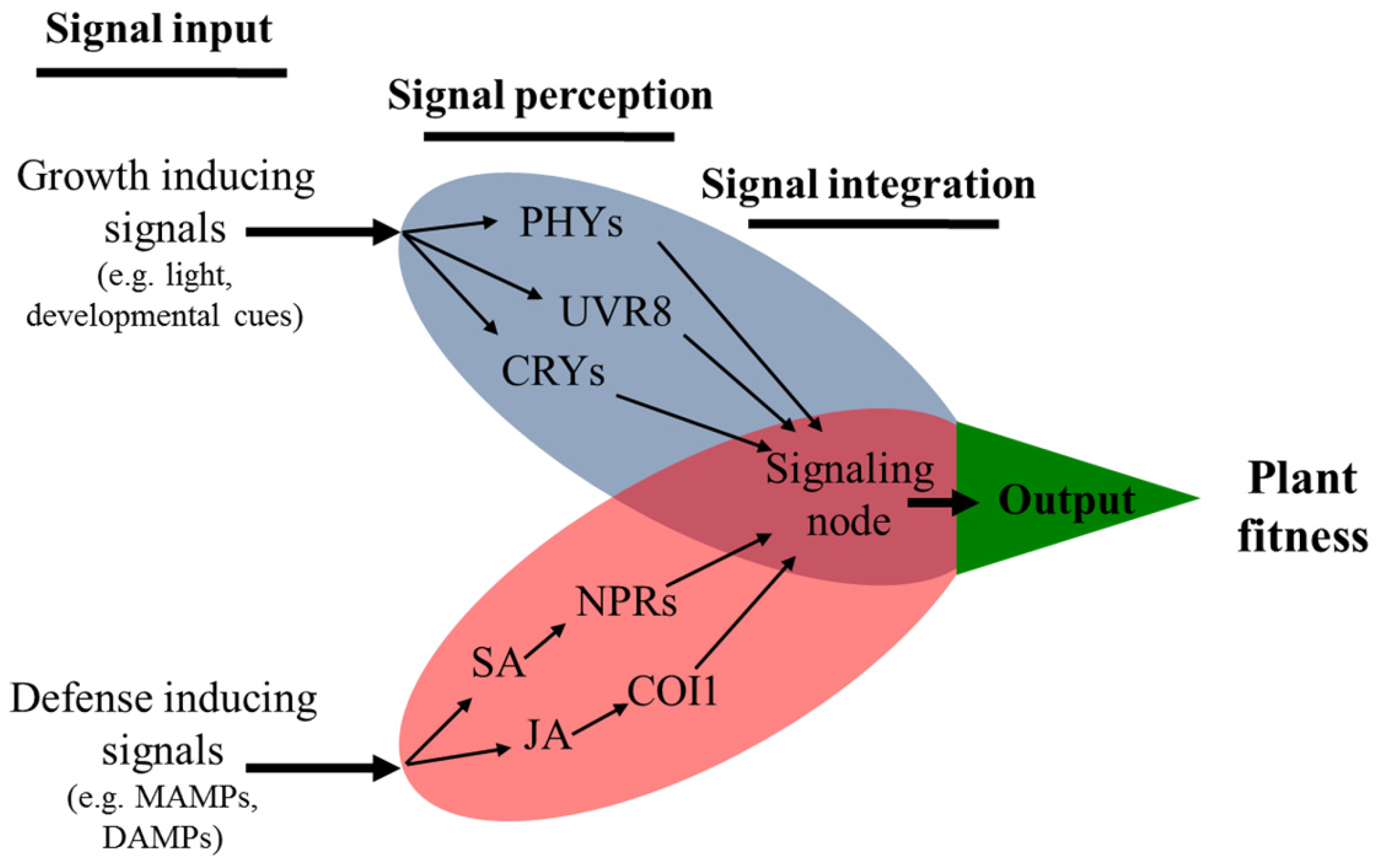
5. Specialized Photoreceptors Inform Growth–Defense Trade-offs

Increasing evidence supports a positive correlation between higher crop densities and the occurrence of pests [69]. These observations in agricultural and ecological systems led to the proposed “dilemma of plants”; that is whether to invest limited resources for competitive growth with neighboring plants or for robust defense against insects and pathogens [70]. The high susceptibility of densely-cropped plants is partly related to changes in the light environment. Crowded stands combine lower PAR, less UV-B radiation, less B, and a lower R:FR ratio. In recent years, the use of Arabidopsis photoreceptor mutants and light-manipulation experiments has generated important evidence showing that these light components contribute to the immune-suppressed state of plants growing in crowded stands.
UV-B and inducible defenses: UV-B attenuation experiments in the field have clearly shown that natural herbivory is more severe on plants growing with attenuated levels of UV-B compared to those exposed to full sunlight [7,71,72,73]. Most of the plants increase the accumulation of soluble phenolic compounds in green tissues in response to UV-B, where some of these compounds were shown to have a direct role on insect herbivore performance [51,74,75]. In addition, UV-B altered Plutella xylostella oviposition behavior in a JA-dependent manner, since adult moths laid more eggs on plants growing under attenuated UV-B [72]. Conversely, plants supplemented with UV-B experienced less herbivory and disease symptoms than control plants [51,76]. Resistance to Botrytis cinerea infection was significantly increased in plants grown with supplemental UV-B, an effect that required the UVR8 photoreceptor but not JA signaling [76]. This UV-B effect on B. cinerea resistance was mediated by the accumulation of sinapate in Arabidopsis [76].
Blue light and inducible defenses: B depletion does not appear to affect immunity. Arabidopsis plants expressing SAS, either due to B depletion or mutation of the CRY1 photoreceptor, were not more susceptible to B. cinerea infection [77]. Moreover, cry1 mutants showed similar resistance to infection by P. syringae [78,79]. However, the cry1 mutant has also been reported more susceptible to P. syringae [80]. B may also indirectly influence defense responses since B controls stomatal openings, which are the main points of entry for pathogens [81,82]. B signals also synchronize the circadian clock, which regulates plant growth and defense processes. Immune responses against both herbivores and pathogens are ‘gated’ by the plant circadian clock such that defense is strongest in the morning when biotic attack is more likely [83,84,85]. Interestingly, NPR1 also contributes to synchronizing the circadian clock, allowing SA to reinforce the circadian clock and gate immune responses [86]. Altogether, B signals contribute to circadian gating of immunity, which allows plants to reinforce morning defenses in ‘anticipation’ of challenge from herbivores and pathogens, while also minimizing the negative side effects on growth responses that take place at night.
R:FR ratio and inducible defenses: Plants expressing SAS, from either high-density planting or supplemental FR, displayed weak defense phenotypes against insect herbivores, necrotrophic fungus, and pathogenic bacteria [77,87,88]. The levels of defense metabolites, including phenolics, aliphatic glucosinolates, and camalexin, were lower in SAS-expressing plants upon B. cinerea infection [77,87,89]. In addition, growth in FR-rich environments suppressed both JA- and SA-dependent expression of defense genes [88]. Together, these results demonstrated that phytochrome inactivation by FR reduces JA and SA sensitivity. phyB is the principle phytochrome receptor responsible for FR-mediated perturbation of immune responses, as phyB mutants are less sensitive to JA and SA, and are more susceptible to insect herbivory and to infection by B. cinerea and P. syringae [77,87,88,90]. The molecular mechanisms behind these growth–defense trade-offs include multiple layers of regulation by FR: (1) JA-mediated degradation of JAZ10 is significantly reduced in seedlings exposed to FR radiation and in the phyB mutant background [91]; (2) FR destabilizes MYC2, MYC3 and MYC4 proteins, key transcription factors of JA responses, in a phyB-dependent manner [92]; (3) FR triggers turnover of DELLA proteins [91], which releases repression of PIFs to activate growth processes, as well as the release of JAZ proteins from DELLA–JAZ interactions to enhance repression of JA responses (see Section 3) [62,63]; and (4) FR interferes with NPR1 phosphorylation, contributing to reduced NPR1-mediated immune responses [88].

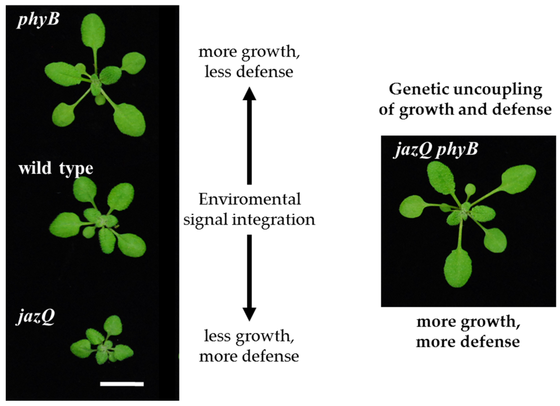
6. Growth–Defense Decisions Are Controlled by the Phytochrome B Signaling Pathway

Several studies are now revealing that the classic and simple view that growth and defense processes are limited by resource constraints is too simplistic; instead, growth and defense are determined by signaling programs. For example, studies have shown that JA-mediated growth repression involves reduced cell division and expansion by perturbation of the cell cycle [58,93,94]. Moreover, Arabidopsis mutants that no longer produced glucosinolates (the main defense compound in brassica plants) still exhibited growth suppression, demonstrating that growth and defense are not strictly coupled genetically [50,95]. Similarly, the “decision” to prioritize growth over defense during SAS appears to be a specific physiological response mediated by phyB, rather than the limitation of plant energy constraints [87,91]. As such, several studies are revealing that SAS-mediated growth promotion and defense suppression can be processes that are uncoupled in various mutants. For example, the cpr1 and cev1 mutants, which have constitutively active SA- and JA-dependent defense responses, respectively, both express partial SAS in response to FR, suggesting that these mutants can direct resources to growth and defense [88]. In the sav3-2 mutant, FR exposure does not trigger expression of SAS but does suppress immune responses, demonstrating that the morphological investment of energy during SAS expression cannot per se explain the increased susceptibility to insects or B. cinerea [77,87]. Defense suppression is also uncoupled from SAS in the jaz10 phyB double mutant, which maintains constitutive SAS but with wild type JA sensitivity [91]. These points of cross-talk among growth and defense signaling pathways are establishing how complex networks of regulators govern these processes to properly respond to external conditions [49,95,96]. An emerging question is whether we can use the knowledge of these networks to manipulate growth–defense trade-offs (see Section 7 below).

7. Manipulating Repressors of Defense and Light Signaling for Improved Protection and Growth of Energy-Rich Tissues

The molecular basis of how the JA signaling pathway integrates growth and defense remained obscured by the genetic redundancy among the JAZ transcriptional repressors. As a consequence, the majority of single jaz mutants showed no clear phenotype [25]. In an effort to overcome this situation, Campos et al. (2016) developed a higher-order mutant that lacks five of the thirteen JAZ repressors present in the Arabidopsis genome. Removal of multiple JAZ genes in this jaz quintuple (jazQ) mutant led to constitutive activation of JA responses, causing hypersensitivity to exogenous JA treatment, upregulation of defense-related genes, increased production of secondary metabolites and higher resistance to insect herbivory attack [49]. On the other hand, rosette expansion was restricted in this higher-order mutant, as jazQ exhibited stunted rosettes and overall delayed development when compared to wild type plants (Figure 1). In addition, jazQ was ~50% lighter than wild type in terms of rosette dry weight, indicating strong down-regulation of plant biomass accumulation. Taken together, these results support the premise that JA modulates a transcriptional route whose activation (through removal of multiple JAZ transcriptional repressors) culminates in strong promotion of defense along with a reduction in growth-related processes (see Section 3).
The evident shift observed in jazQ growth–defense parameters can be explained in light of its genetic lesions. The five JAZ impaired in jazQ (JAZ1, JAZ3, JAZ4, JAZ9 and JAZ10) were shown to physically interact with DELLA proteins [64]. The concept that plant hormones integrate a regulatory network that determines growth–defense outputs was further confirmed by Campos et al. (2016), who genetically rewired crosstalk among defense and growth signaling pathways to obtain an Arabidopsis genotype where growth and defense are transcriptionally and physiologically uncoupled. The authors showed that the combination of the “more defenses, less growth” jazQ with the “more growth, less defense” phyB genotypes produced a higher-order mutant (named jazQ phyB) where growth and defense processes were simultaneously activated (Figure 1). These results are consistent with, and greatly expand upon, previous work showing that suppression of defenses is uncoupled from shade avoidance traits in the jaz10 phyB double mutant [91,97]. The jazQ phyB plants showed increased expression of defense-related genes, higher production of secondary metabolites and enhanced resistance against insect herbivory, as well as growth promotion and faster development, even when compared with wild type Arabidopsis plants [49]. Full transcriptome sequencing indicated that the jazQ phyB phenotype is the result of an additive combination of the immune program activated in jazQ and the growth program activated in phyB. Those results indicate that depletion of the JAZ and DELLA repressors caused by jazQ and phyB mutations respectively leads to concomitant de-repression of growth and defense transcriptional cascades in the jazQ phyB mutant [49,91]. Specific traits found in jazQ phyB were not observed in the jazQ or phyB mutants. For instance, the biomass of both jazQ and phyB was about 50% of that observed for wild type plants, yet the combination of these mutations restored plant biomass to similar levels as wild type plants. Projected leaf area and rosette diameter were also larger in jazQ phyB compared to the parental mutants, as well as wild type plants. At the transcriptional level, more than a thousand genes were differentially expressed in jazQ phyB but not in its parental mutants. Taken together, these results indicate, at one side, a strong modular effect of defense and growth transcriptional programs mainly dependent on MYC and DELLA transcriptional regulators and, at the other side, demonstrate that growth and photomorphogenic signaling pathways show a synergistic control of transcriptional changes to achieve a balanced physiological response to the dilemma of plants.

8. Conclusions

Recently, a wealth of studies have begun to unveil the molecular basis of the trade-offs between growth and defense, indicating the presence of a complex signaling hub where hormone and light signaling pathways converge to optimize fitness. In light of this recent evidence, we propose a model showing that growth and defense trade-offs are not just the result of metabolic constraints (such as resource limitation), but rather a consequence of a transcriptional network that evolved to maximize plant fitness (Figure 2) [49,95]. As such, growth and defense environmental cues are initially perceived by receptors, which in turn, activate transcriptional cascades responsible for the activation of specialized responses. A signaling node integrates these transcriptional programs to precisely control the elicitation of growth and defense traits in order to optimize plant performance. The proposed interaction of regulatory modules is prone to evolve in natural settings, where plants are constantly facing a blend of environmental signals that must be integrated and properly translated into growth and defense traits to maximize fitness. A relevant application of this premise is the use of genetic rewiring to achieve concomitant activation of growth and defense [49,91]. This strategy could lead, in a relative short time, to the development of new crop cultivars where high biomass production is combined with lower demand for pesticides.

Acknowledgments

This work was supported by grants to J.E.M. Consejo Nacional de Investigaciones Científicas y Técnicas (PIP2014-2016 11220130100267CO), Universidad Nacional del Litoral (50220140100011LI), and the Agencia Nacional de Promoción Científica y Tecnológica (PICT2013-3285). MLC is supported by a fellowship from the Conselho Nacional de Desenvolvimento Cientifico e Tecnológico (CNPq-Brazil).

Conflicts of Interest

The authors declare no conflict of interest.

References

  1. Ballaré, C.L.; Scopel, A.L.; Sanchez, R.A. Far-red radiation reflected from adjacent leaves: An early signal of competition in plant canopies. Science 1990, 247, 329–332. [Google Scholar] [CrossRef] [PubMed]
  2. Casal, J.J. Photoreceptor signaling networks in plant responses to shade. Ann. Rev. Plant Biol. 2013, 64, 403–427. [Google Scholar] [CrossRef] [PubMed]
  3. Rizzini, L.; Favory, J.J.; Cloix, C.; Faggionato, D.; O’Hara, A.; Kaiserli, E.; Baumeister, R.; Schafer, E.; Nagy, F.; Jenkins, G.I.; et al. Perception of UV-B by the Arabidopsis UVR8 protein. Science 2011, 332, 103–106. [Google Scholar] [CrossRef] [PubMed]
  4. Heijde, M.; Ulm, R. UV-B photoreceptor-mediated signalling in plants. Trends Plant Sci. 2012, 17, 230–237. [Google Scholar] [CrossRef] [PubMed]
  5. Favory, J.J.; Stec, A.; Gruber, H.; Rizzini, L.; Oravecz, A.; Funk, M.; Albert, A.; Cloix, C.; Jenkins, G.I.; Oakeley, E.J.; et al. Interaction of COP1 and UVR8 regulates UV-B-induced photomorphogenesis and stress acclimation in Arabidopsis. EMBO J. 2009, 28, 591–601. [Google Scholar] [CrossRef] [PubMed]
  6. Mazza, C.A.; Ballaré, C.L. Photoreceptors UVR8 and phytochrome B cooperate to optimize plant growth and defense in patchy canopies. New Phytol. 2015, 207, 4–9. [Google Scholar] [CrossRef] [PubMed]
  7. Mazza, C.A.; Gimenez, P.I.; Kantolic, A.G.; Ballaré, C.L. Beneficial effects of solar UV-B radiation on soybean yield mediated by reduced insect herbivory under field conditions. Physiol. Plant. 2013, 147, 307–315. [Google Scholar] [CrossRef] [PubMed]
  8. Keller, M.M.; Jaillais, Y.; Pedmale, U.V.; Moreno, J.E.; Chory, J.; Ballaré, C.L. Cryptochrome 1 and phytochrome B control shade-avoidance responses in Arabidopsis via partially independent hormonal cascades. Plant J. 2011, 67, 195–207. [Google Scholar] [CrossRef] [PubMed]
  9. Keuskamp, D.H.; Keller, M.M.; Ballaré, C.L.; Pierik, R. Blue light regulated shade avoidance. Plant. Signal. Behav. 2012, 7, 514–517. [Google Scholar] [CrossRef] [PubMed]
  10. Keuskamp, D.H.; Sasidharan, R.; Vos, I.; Peeters, A.J.; Voesenek, L.A.; Pierik, R. Blue-light-mediated shade avoidance requires combined auxin and brassinosteroid action in Arabidopsis seedlings. Plant J. 2011, 67, 208–217. [Google Scholar] [CrossRef] [PubMed]
  11. Pedmale, U.V.; Huang, S.S.; Zander, M.; Cole, B.J.; Hetzel, J.; Ljung, K.; Reis, P.A.; Sridevi, P.; Nito, K.; Nery, J.R.; et al. Cryptochromes interact directly with PIFs to control plant growth in limiting blue light. Cell 2016, 164, 233–245. [Google Scholar] [CrossRef] [PubMed]
  12. De Lucas, M.; Daviere, J.M.; Rodriguez-Falcon, M.; Pontin, M.; Iglesias-Pedraz, J.M.; Lorrain, S.; Fankhauser, C.; Blazquez, M.A.; Titarenko, E.; Prat, S. A molecular framework for light and gibberellin control of cell elongation. Nature 2008, 451, 480–484. [Google Scholar] [CrossRef] [PubMed]
  13. Hornitschek, P.; Kohnen, M.V.; Lorrain, S.; Rougemont, J.; Ljung, K.; Lopez-Vidriero, I.; Franco-Zorrilla, J.M.; Solano, R.; Trevisan, M.; Pradervand, S.; et al. Phytochrome interacting factors 4 and 5 control seedling growth in changing light conditions by directly controlling auxin signaling. Plant J. 2012, 71, 699–711. [Google Scholar] [CrossRef] [PubMed]
  14. Tao, Y.; Ferrer, J.L.; Ljung, K.; Pojer, F.; Hong, F.; Long, J.A.; Li, L.; Moreno, J.E.; Bowman, M.E.; Ivans, L.J.; et al. Rapid synthesis of auxin via a new tryptophan-dependent pathway is required for shade avoidance in plants. Cell 2008, 133, 164–176. [Google Scholar] [CrossRef] [PubMed]
  15. Feng, S.; Martinez, C.; Gusmaroli, G.; Wang, Y.; Zhou, J.; Wang, F.; Chen, L.; Yu, L.; Iglesias-Pedraz, J.M.; Kircher, S.; et al. Coordinated regulation of Arabidopsis thaliana development by light and gibberellins. Nature 2008, 451, 475–479. [Google Scholar] [CrossRef] [PubMed]
  16. Sasidharan, R.; Chinnappa, C.C.; Staal, M.; Elzenga, J.T.; Yokoyama, R.; Nishitani, K.; Voesenek, L.A.; Pierik, R. Light quality-mediated petiole elongation in Arabidopsis during shade avoidance involves cell wall modification by xyloglucan endotransglucosylase/hydrolases. Plant Physiol. 2010, 154, 978–990. [Google Scholar] [CrossRef] [PubMed]
  17. Li, L.; Ljung, K.; Breton, G.; Schmitz, R.J.; Pruneda-Paz, J.; Cowing-Zitron, C.; Cole, B.J.; Ivans, L.J.; Pedmale, U.V.; Jung, H.S.; et al. Linking photoreceptor excitation to changes in plant architecture. Genes Dev. 2012, 26, 785–790. [Google Scholar] [CrossRef]
  18. Sanchez-Lamas, M.; Lorenzo, C.D.; Cerdán, P.D. Bottom-up assembly of the phytochrome network. PLoS Genet. 2016, 12, e1006413. [Google Scholar] [CrossRef] [PubMed]
  19. Strasser, B.; Sanchez-Lamas, M.; Yanovsky, M.J.; Casal, J.J.; Cerdán, P.D. Arabidopsis thaliana life without phytochromes. Proc. Natl. Acad. Sci. USA 2010, 107, 4776–4781. [Google Scholar] [CrossRef] [PubMed]
  20. Pieterse, C.M.; van der Does, D.; Zamioudis, C.; Leon-Reyes, A.; Van Wees, S.C. Hormonal modulation of plant immunity. Annu. Rev. Cell Dev. Biol. 2012, 28, 489–521. [Google Scholar] [CrossRef] [PubMed]
  21. Campos, M.L.; Kang, J.H.; Howe, G.A. Jasmonate-triggered plant immunity. J. Chem. Ecol. 2014, 40, 657–675. [Google Scholar] [CrossRef] [PubMed]
  22. Katsir, L.; Schilmiller, A.L.; Staswick, P.E.; He, S.Y.; Howe, G.A. COI1 is a critical component of a receptor for jasmonate and the bacterial virulence factor coronatine. Proc. Natl. Acad. Sci. USA 2008, 105, 7100–7105. [Google Scholar] [CrossRef] [PubMed]
  23. Sheard, L.B.; Tan, X.; Mao, H.; Withers, J.; Ben-Nissan, G.; Hinds, T.R.; Kobayashi, Y.; Hsu, F.F.; Sharon, M.; Browse, J.; et al. Jasmonate perception by inositol-phosphate-potentiated COI1-JAZ co-receptor. Nature 2010, 468, 400–405. [Google Scholar] [CrossRef] [PubMed]
  24. Chini, A.; Fonseca, S.; Fernandez, G.; Adie, B.; Chico, J.M.; Lorenzo, O.; Garcia-Casado, G.; Lopez-Vidriero, I.; Lozano, F.M.; Ponce, M.R.; et al. The JAZ family of repressors is the missing link in jasmonate signalling. Nature 2007, 448, 666–671. [Google Scholar] [CrossRef] [PubMed]
  25. Thines, B.; Katsir, L.; Melotto, M.; Niu, Y.; Mandaokar, A.; Liu, G.; Nomura, K.; He, S.Y.; Howe, G.A.; Browse, J. JAZ repressor proteins are targets of the SCFcoi1 complex during jasmonate signalling. Nature 2007, 448, 661–665. [Google Scholar] [CrossRef] [PubMed]
  26. Yan, Y.; Stolz, S.; Chetelat, A.; Reymond, P.; Pagni, M.; Dubugnon, L.; Farmer, E.E. A downstream mediator in the growth repression limb of the jasmonate pathway. Plant Cell 2007, 19, 2470–2483. [Google Scholar] [CrossRef] [PubMed]
  27. Chung, H.S.; Cooke, T.F.; Depew, C.L.; Patel, L.C.; Ogawa, N.; Kobayashi, Y.; Howe, G.A. Alternative splicing expands the repertoire of dominant JAZ repressors of jasmonate signaling. Plant J. 2010, 63, 613–622. [Google Scholar] [CrossRef] [PubMed]
  28. Moreno, J.E.; Shyu, C.; Campos, M.L.; Patel, L.C.; Chung, H.S.; Yao, J.; He, S.Y.; Howe, G.A. Negative feedback control of jasmonate signaling by an alternative splice variant of JAZ10. Plant Physiol. 2013, 162, 1006–1017. [Google Scholar] [CrossRef] [PubMed]
  29. Chung, H.S.; Howe, G.A. A critical role for the TIFY motif in repression of jasmonate signaling by a stabilized splice variant of the JASMONATE ZIM-domain protein JAZ10 in Arabidopsis. Plant Cell 2009, 21, 131–145. [Google Scholar] [CrossRef] [PubMed]
  30. Chini, A.; Gimenez-Ibanez, S.; Goossens, A.; Solano, R. Redundancy and specificity in jasmonate signalling. Curr. Opin. Plant Biol. 2016, 33, 147–156. [Google Scholar] [CrossRef] [PubMed]
  31. Pajerowska-Mukhtar, K.M.; Emerine, D.K.; Mukhtar, M.S. Tell me more: Roles of NPRs in plant immunity. Trends Plant Sci. 2013, 18, 402–411. [Google Scholar] [CrossRef] [PubMed]
  32. Wu, Y.; Zhang, D.; Chu, J.Y.; Boyle, P.; Wang, Y.; Brindle, I.D.; de Luca, V.; Despres, C. The Arabidopsis NPR1 protein is a receptor for the plant defense hormone salicylic acid. Cell Rep. 2012, 1, 639–647. [Google Scholar] [CrossRef] [PubMed]
  33. Fu, Z.Q.; Yan, S.; Saleh, A.; Wang, W.; Ruble, J.; Oka, N.; Mohan, R.; Spoel, S.H.; Tada, Y.; Zheng, N.; et al. NPR3 and NPR4 are receptors for the immune signal salicylic acid in plants. Nature 2012, 486, 228–232. [Google Scholar] [CrossRef] [PubMed] [Green Version]
  34. Spoel, S.H.; Mou, Z.; Tada, Y.; Spivey, N.W.; Genschik, P.; Dong, X. Proteasome-mediated turnover of the transcription coactivator NPR1 plays dual roles in regulating plant immunity. Cell 2009, 137, 860–872. [Google Scholar] [CrossRef] [PubMed]
  35. Saleh, A.; Withers, J.; Mohan, R.; Marques, J.; Gu, Y.; Yan, S.; Zavaliev, R.; Nomoto, M.; Tada, Y.; Dong, X. Posttranslational modifications of the master transcriptional regulator NPR1 enable dynamic but tight control of plant immune responses. Cell Host Microbe 2015, 18, 169–182. [Google Scholar] [CrossRef] [PubMed]
  36. Thaler, J.S.; Humphrey, P.T.; Whiteman, N.K. Evolution of jasmonate and salicylate signal crosstalk. Trends Plant Sci. 2012, 17, 260–270. [Google Scholar] [CrossRef] [PubMed]
  37. Thaler, J.S.; Fidantsef, A.L.; Bostock, R.M. Antagonism between jasmonate- and salicylate-mediated induced plant resistance: Effects of concentration and timing of elicitation on defense-related proteins, herbivore, and pathogen performance in tomato. J. Chem. Ecol. 2002, 28, 1131–1159. [Google Scholar] [CrossRef] [PubMed]
  38. Cipollini, D. Costs and benefits of induced resistance to herbivores and pathogens in plants. CAB Rev. Perspect. Agric. Vet. Sci. Nutr. Nat. Res. 2010, 5. [Google Scholar] [CrossRef]
  39. Spoel, S.H.; Johnson, J.S.; Dong, X. Regulation of tradeoffs between plant defenses against pathogens with different lifestyles. Proc. Natl. Acad. Sci. USA 2007, 104, 18842–18847. [Google Scholar] [CrossRef] [PubMed] [Green Version]
  40. Zhang, L.; Zhang, F.; Melotto, M.; Yao, J.; He, S.Y. Jasmonate signaling and manipulation by pathogens and insects. J. Exp. Bot. 2017. [Google Scholar] [CrossRef] [PubMed]
  41. Gimenez-Ibanez, S.; Solano, R. Nuclear jasmonate and salicylate signaling and crosstalk in defense against pathogens. Front. Plant Sci. 2013, 4, 72. [Google Scholar] [CrossRef] [PubMed]
  42. Liu, L.; Sonbol, F.M.; Huot, B.; Gu, Y.; Withers, J.; Mwimba, M.; Yao, J.; He, S.Y.; Dong, X. Salicylic acid receptors activate jasmonic acid signalling through a non-canonical pathway to promote effector-triggered immunity. Nat. Commun. 2016, 7, 13099. [Google Scholar] [CrossRef] [PubMed]
  43. Zheng, X.Y.; Spivey, N.W.; Zeng, W.; Liu, P.P.; Fu, Z.Q.; Klessig, D.F.; He, S.Y.; Dong, X. Coronatine promotes Pseudomonas syringae virulence in plants by activating a signaling cascade that inhibits salicylic acid accumulation. Cell Host Microbe 2012, 11, 587–596. [Google Scholar] [CrossRef] [PubMed]
  44. Agrawal, A.A.; Hastings, A.P.; Johnson, M.T.; Maron, J.L.; Salminen, J.P. Insect herbivores drive real-time ecological and evolutionary change in plant populations. Science 2012, 338, 113–116. [Google Scholar] [CrossRef] [PubMed]
  45. Gomez-Gomez, L.; Felix, G.; Boller, T. A single locus determines sensitivity to bacterial flagellin in Arabidopsis thaliana. Plant J. 1999, 18, 277–284. [Google Scholar] [CrossRef] [PubMed]
  46. Clarke, J.D.; Volko, S.M.; Ledford, H.; Ausubel, F.M.; Dong, X. Roles of salicylic acid, jasmonic acid, and ethylene in cpr-induced resistance in Arabidopsis. Plant Cell 2000, 12, 2175–2190. [Google Scholar] [CrossRef] [PubMed]
  47. Zhang, Y.; Goritschnig, S.; Dong, X.; Li, X. A gain-of-function mutation in a plant disease resistance gene leads to constitutive activation of downstream signal transduction pathways in suppressor of npr1–1, constitutive 1. Plant Cell 2003, 15, 2636–2646. [Google Scholar] [CrossRef] [PubMed]
  48. Attaran, E.; Major, I.T.; Cruz, J.A.; Rosa, B.A.; Koo, A.J.; Chen, J.; Kramer, D.M.; He, S.Y.; Howe, G.A. Temporal dynamics of growth and photosynthesis suppression in response to jasmonate signaling. Plant Physiol. 2014, 165, 1302–1314. [Google Scholar] [CrossRef] [PubMed]
  49. Campos, M.L.; Yoshida, Y.; Major, I.T.; de Oliveira Ferreira, D.; Weraduwage, S.M.; Froehlich, J.E.; Johnson, B.F.; Kramer, D.M.; Jander, G.; Sharkey, T.D.; et al. Rewiring of jasmonate and phytochrome B signalling uncouples plant growth-defense tradeoffs. Nat. Commun. 2016, 7, 12570. [Google Scholar] [CrossRef] [PubMed]
  50. Zust, T.; Joseph, B.; Shimizu, K.K.; Kliebenstein, D.J.; Turnbull, L.A. Using knockout mutants to reveal the growth costs of defensive traits. Proc. Biol. Sci. 2011, 278, 2598–2603. [Google Scholar] [CrossRef] [PubMed]
  51. Izaguirre, M.M.; Scopel, A.L.; Baldwin, I.T.; Ballaré, C.L. Convergent responses to stress. Solar ultraviolet-B radiation and Manduca sexta herbivory elicit overlapping transcriptional responses in field-grown plants of Nicotiana longiflora. Plant Physiol. 2003, 132, 1755–1767. [Google Scholar] [CrossRef] [PubMed]
  52. Nabity, P.D.; Zavala, J.A.; DeLucia, E.H. Herbivore induction of jasmonic acid and chemical defences reduce photosynthesis in Nicotiana attenuata. J. Exp. Bot. 2013, 64, 685–694. [Google Scholar] [CrossRef] [PubMed]
  53. Zangerl, A.R.; Hamilton, J.G.; Miller, T.J.; Crofts, A.R.; Oxborough, K.; Berenbaum, M.R.; de Lucia, E.H. Impact of folivory on photosynthesis is greater than the sum of its holes. Proc. Natl. Acad. Sci. USA 2002, 99, 1088–1091. [Google Scholar] [CrossRef] [PubMed]
  54. Qi, T.; Song, S.; Ren, Q.; Wu, D.; Huang, H.; Chen, Y.; Fan, M.; Peng, W.; Ren, C.; Xie, D. The Jasmonate-ZIM-domain proteins interact with the WD-repeat/bHLH/MYB complexes to regulate jasmonate-mediated anthocyanin accumulation and trichome initiation in Arabidopsis thaliana. Plant Cell 2011, 23, 1795–1814. [Google Scholar] [CrossRef] [PubMed]
  55. Zhu, X.; Chen, J.; Xie, Z.; Gao, J.; Ren, G.; Gao, S.; Zhou, X.; Kuai, B. Jasmonic acid promotes degreening via MYC2/3/4- and ANAC019/055/072-mediated regulation of major chlorophyll catabolic genes. Plant J. 2015, 84, 597–610. [Google Scholar] [CrossRef] [PubMed]
  56. Hu, X.; Makita, S.; Schelbert, S.; Sano, S.; Ochiai, M.; Tsuchiya, T.; Hasegawa, S.F.; Hortensteiner, S.; Tanaka, A.; Tanaka, R. Reexamination of chlorophyllase function implies its involvement in defense against chewing herbivores. Plant Physiol. 2015, 167, 660–670. [Google Scholar] [CrossRef] [PubMed]
  57. Bilgin, D.D.; Zavala, J.A.; Zhu, J.; Clough, S.J.; Ort, D.R.; DeLucia, E.H. Biotic stress globally downregulates photosynthesis genes. Plant Cell Environ. 2010, 33, 1597–1613. [Google Scholar] [CrossRef] [PubMed]
  58. Zhang, Y.; Turner, J.G. Wound-induced endogenous jasmonates stunt plant growth by inhibiting mitosis. PLoS ONE 2008, 3, e3699. [Google Scholar] [CrossRef] [PubMed]
  59. Weraduwage, S.M.; Chen, J.; Anozie, F.C.; Morales, A.; Weise, S.E.; Sharkey, T.D. The relationship between leaf area growth and biomass accumulation in Arabidopsis thaliana. Front. Plant Sci. 2015, 6, 167. [Google Scholar] [CrossRef] [PubMed]
  60. Havko, N.E.; Major, I.T.; Jewell, J.B.; Attaran, E.; Browse, J.; Howe, G.A. Control of carbon assimilation and partitioning by jasmonate: An accounting of growth-defense tradeoffs. Plants 2016, 5, 7. [Google Scholar] [CrossRef] [PubMed]
  61. Huot, B.; Yao, J.; Montgomery, B.L.; He, S.Y. Growth-defense tradeoffs in plants: A balancing act to optimize fitness. Mol. Plant 2014, 7, 1267–1287. [Google Scholar] [CrossRef] [PubMed]
  62. Navarro, L.; Bari, R.; Achard, P.; Lison, P.; Nemri, A.; Harberd, N.P.; Jones, J.D. DELLAs control plant immune responses by modulating the balance of jasmonic acid and salicylic acid signaling. Curr. Biol. 2008, 18, 650–655. [Google Scholar] [CrossRef] [PubMed]
  63. Hou, X.; Lee, L.Y.; Xia, K.; Yan, Y.; Yu, H. DELLAs modulate jasmonate signaling via competitive binding to JAZs. Dev. Cell 2010, 19, 884–894. [Google Scholar] [CrossRef] [PubMed]
  64. Yang, D.L.; Yao, J.; Mei, C.S.; Tong, X.H.; Zeng, L.J.; Li, Q.; Xiao, L.T.; Sun, T.P.; Li, J.; Deng, X.W.; et al. Plant hormone jasmonate prioritizes defense over growth by interfering with gibberellin signaling cascade. Proc. Natl. Acad. Sci. USA 2012, 109, E1192–E1200. [Google Scholar] [CrossRef] [PubMed]
  65. Chen, Q.; Sun, J.; Zhai, Q.; Zhou, W.; Qi, L.; Xu, L.; Wang, B.; Chen, R.; Jiang, H.; Qi, J.; et al. The basic helix-loop-helix transcription factor MYC2 directly represses PLETHORA expression during jasmonate-mediated modulation of the root stem cell niche in Arabidopsis. Plant Cell 2011, 23, 3335–3352. [Google Scholar] [CrossRef] [PubMed]
  66. Smolen, G.A.; Pawlowski, L.; Wilensky, S.E.; Bender, J. Dominant alleles of the basic helix-loop-helix transcription factor ATR2 activate stress-responsive genes in Arabidopsis. Genetics 2002, 161, 1235–1246. [Google Scholar] [PubMed]
  67. Gasperini, D.; Chetelat, A.; Acosta, I.F.; Goossens, J.; Pauwels, L.; Goossens, A.; Dreos, R.; Alfonso, E.; Farmer, E.E. Multilayered organization of jasmonate signalling in the regulation of root growth. PLoS Genet. 2015, 11, e1005300. [Google Scholar] [CrossRef] [PubMed] [Green Version]
  68. Goossens, J.; Swinnen, G.; Vanden Bossche, R.; Pauwels, L.; Goossens, A. Change of a conserved amino acid in the MYC2 and MYC3 transcription factors leads to release of JAZ repression and increased activity. New Phytol. 2015, 206, 1229–1237. [Google Scholar] [CrossRef] [PubMed]
  69. Moreno, J.E.; Ballaré, C.L. Phytochrome regulation of plant immunity in vegetation canopies. J. Chem. Ecol. 2014, 40, 848–857. [Google Scholar] [CrossRef] [PubMed]
  70. Herms, D.A.; Mattson, W.J. The dilemma of plants: To grow or defend. Q. Rev. Biol. 1992, 67, 283–335. [Google Scholar] [CrossRef]
  71. Mazza, C.A.; Zavala, J.; Scopel, A.L.; Ballaré, C.L. Perception of solar UVB radiation by phytophagous insects: Behavioral responses and ecosystem implications. Proc. Natl. Acad. Sci. USA 1999, 96, 980–985. [Google Scholar] [CrossRef] [PubMed]
  72. Caputo, C.; Rutitzky, M.; Ballaré, C.L. Solar ultraviolet-B radiation alters the attractiveness of Arabidopsis plants to diamondback moths (Plutella xylostella L.): Impacts on oviposition and involvement of the jasmonic acid pathway. Oecologia 2006, 149, 81–90. [Google Scholar] [CrossRef] [PubMed]
  73. Zavala, J.A.; Mazza, C.A.; Dillon, F.M.; Chludil, H.D.; Ballaré, C.L. Soybean resistance to stink bugs (Nezara viridula and Piezodorus guildinii) increases with exposure to solar UV-B radiation and correlates with isoflavonoid content in pods under field conditions. Plant Cell Environ. 2015, 38, 920–928. [Google Scholar] [CrossRef] [PubMed]
  74. Mazza, C.A.; Boccalandro, H.E.; Giordano, C.V.; Battista, D.; Scopel, A.L.; Ballaré, C.L. Functional significance and induction by solar radiation of ultraviolet-absorbing sunscreens in field-grown soybean crops. Plant Physiol. 2000, 122, 117–126. [Google Scholar] [CrossRef] [PubMed]
  75. Izaguirre, M.M.; Mazza, C.A.; Svatos, A.; Baldwin, I.T.; Ballaré, C.L. Solar ultraviolet-B radiation and insect herbivory trigger partially overlapping phenolic responses in Nicotiana attenuata and Nicotiana longiflora. Ann. Bot. 2007, 99, 103–109. [Google Scholar] [CrossRef] [PubMed]
  76. Demkura, P.V.; Ballaré, C.L. UVR8 mediates UV-B-induced Arabidopsis defense responses against Botrytis cinerea by controlling sinapate accumulation. Mol. Plant 2012, 5, 642–652. [Google Scholar] [CrossRef] [PubMed]
  77. Cerrudo, I.; Keller, M.M.; Cargnel, M.D.; Demkura, P.V.; de Wit, M.; Patitucci, M.S.; Pierik, R.; Pieterse, C.M.; Ballaré, C.L. Low red/far-red ratios reduce Arabidopsis resistance to Botrytis cinerea and jasmonate responses via a COI1-JAZ10-dependent, salicylic acid-independent mechanism. Plant Physiol. 2012, 158, 2042–2052. [Google Scholar] [CrossRef] [PubMed]
  78. Griebel, T.; Zeier, J. Light regulation and daytime dependency of inducible plant defenses in Arabidopsis: Phytochrome signaling controls systemic acquired resistance rather than local defense. Plant Physiol. 2008, 147, 790–801. [Google Scholar] [CrossRef] [PubMed]
  79. Jeong, R.D.; Chandra-Shekara, A.C.; Barman, S.R.; Navarre, D.; Klessig, D.F.; Kachroo, A.; Kachroo, P. Cryptochrome 2 and phototropin 2 regulate resistance protein-mediated viral defense by negatively regulating an E3 ubiquitin ligase. Proc. Natl. Acad. Sci. USA 2010, 107, 13538–13543. [Google Scholar] [CrossRef] [PubMed]
  80. Wu, L.; Yang, H.Q. Cryptochrome 1 is implicated in promoting R protein-mediated plant resistance to Pseudomonas syringae in Arabidopsis. Mol. Plant 2010, 3, 539–548. [Google Scholar] [CrossRef] [PubMed]
  81. Zeiger, E.; Iino, M.; Shimazaki, K.I.; Ogawa, T. The blue-light response of stomata: Mechanism and function. In Stomatal Function; Zeiger, E., Farquhar, G.D., Cowan, I.R., Eds.; Stanford University Press: Stanford, CA, USA, 1981; pp. 209–227. [Google Scholar]
  82. Zhang, C.; Xie, Q.; Anderson, R.G.; Ng, G.; Seitz, N.C.; Peterson, T.; McClung, C.R.; McDowell, J.M.; Kong, D.; Kwak, J.M.; et al. Crosstalk between the circadian clock and innate immunity in Arabidopsis. PLoS Pathog. 2013, 9, e1003370. [Google Scholar] [CrossRef] [PubMed]
  83. Wang, W.; Barnaby, J.Y.; Tada, Y.; Li, H.; Tor, M.; Caldelari, D.; Lee, D.U.; Fu, X.D.; Dong, X. Timing of plant immune responses by a central circadian regulator. Nature 2011, 470, 110–114. [Google Scholar] [CrossRef] [PubMed]
  84. Goodspeed, D.; Chehab, E.W.; Covington, M.F.; Braam, J. Circadian control of jasmonates and salicylates: The clock role in plant defense. Plant. Signal. Behav. 2013, 8, e23123. [Google Scholar] [CrossRef] [PubMed]
  85. Goodspeed, D.; Chehab, E.W.; Min-Venditti, A.; Braam, J.; Covington, M.F. Arabidopsis synchronizes jasmonate-mediated defense with insect circadian behavior. Proc. Natl. Acad. Sci. USA 2012, 109, 4674–4677. [Google Scholar] [CrossRef] [PubMed]
  86. Zhou, M.; Wang, W.; Karapetyan, S.; Mwimba, M.; Marques, J.; Buchler, N.E.; Dong, X. Redox rhythm reinforces the circadian clock to gate immune response. Nature 2015, 523, 472–476. [Google Scholar] [CrossRef] [PubMed]
  87. Moreno, J.E.; Tao, Y.; Chory, J.; Ballaré, C.L. Ecological modulation of plant defense via phytochrome control of jasmonate sensitivity. Proc. Natl. Acad. Sci. USA 2009, 106, 4935–4940. [Google Scholar] [CrossRef] [PubMed]
  88. de Wit, M.; Spoel, S.H.; Sanchez-Perez, G.F.; Gommers, C.M.; Pieterse, C.M.; Voesenek, L.A.; Pierik, R. Perception of low red:far-red ratio compromises both salicylic acid- and jasmonic acid-dependent pathogen defences in Arabidopsis. Plant J. 2013, 75, 90–103. [Google Scholar] [CrossRef] [PubMed]
  89. Cargnel, M.D.; Demkura, P.V.; Ballaré, C.L. Linking phytochrome to plant immunity: Low red : Far-red ratios increase Arabidopsis susceptibility to Botrytis cinerea by reducing the biosynthesis of indolic glucosinolates and camalexin. New Phytol. 2014, 204, 342–354. [Google Scholar] [CrossRef] [PubMed]
  90. Gangappa, S.N.; Berriri, S.; Kumar, S.V. PIF4 coordinates thermosensory growth and immunity in Arabidopsis. Curr. Biol. CB 2017, 27, 243–249. [Google Scholar] [CrossRef] [PubMed]
  91. Leone, M.; Keller, M.M.; Cerrudo, I.; Ballaré, C.L. To grow or defend? Low red : Far-red ratios reduce jasmonate sensitivity in Arabidopsis seedlings by promoting DELLA degradation and increasing JAZ10 stability. New Phytol. 2014, 204, 355–367. [Google Scholar] [CrossRef] [PubMed]
  92. Chico, J.M.; Fernandez-Barbero, G.; Chini, A.; Fernandez-Calvo, P.; Diez-Diaz, M.; Solano, R. Repression of jasmonate-dependent defenses by shade involves differential regulation of protein stability of MYC transcription factors and their JAZ repressors in Arabidopsis. Plant Cell 2014, 26, 1967–1980. [Google Scholar] [CrossRef] [PubMed]
  93. Noir, S.; Bomer, M.; Takahashi, N.; Ishida, T.; Tsui, T.L.; Balbi, V.; Shanahan, H.; Sugimoto, K.; Devoto, A. Jasmonate controls leaf growth by repressing cell proliferation and the onset of endoreduplication while maintaining a potential stand-by mode. Plant Physiol. 2013, 161, 1930–1951. [Google Scholar] [CrossRef] [PubMed]
  94. Pauwels, L.; Morreel, K.; De Witte, E.; Lammertyn, F.; van Montagu, M.; Boerjan, W.; Inze, D.; Goossens, A. Mapping methyl jasmonate-mediated transcriptional reprogramming of metabolism and cell cycle progression in cultured Arabidopsis cells. Proc. Natl. Acad. Sci. USA 2008, 105, 1380–1385. [Google Scholar] [CrossRef] [PubMed]
  95. Kliebenstein, D.J. False idolatry of the mythical growth versus immunity tradeoff in molecular systems plant pathology. Physiol. Mol. Plant Pathol. 2016, 95, 55–59. [Google Scholar] [CrossRef]
  96. Ballaré, C.L. Light regulation of plant defense. Ann. Rev. Plant Biol. 2014, 65, 335–363. [Google Scholar] [CrossRef] [PubMed]
  97. Cerrudo, I.; Caliri-Ortiz, M.E.; Keller, M.M.; Degano, M.E.; Demkura, P.V.; Ballaré, C.L. Exploring growth-defense tradeoffs in Arabidopsis. Phytochrome B inactivation requires JAZ10 to suppress plant immunity but not to trigger shade avoidance responses. Plant Cell Environ. 2016. [Google Scholar] [CrossRef] [PubMed]
Figure 1. Genetic uncoupling of growth and defense in Arabidopsis. The jazQ phyB mutant combines the large rosette of phyB with elevated defenses of jazQ. Photograph of 3 weeks old wild type, jazQ phyB, phyB and jazQ rosettes. Plants were grown in soil and maintained at 20 °C (±1 °C) under 16 h at a light intensity of 120 μmol m−2·s−1 and 8 h dark. Bar = 1 cm.
Agronomy 07 00023 g001
Figure 2. Environmental cues are perceived and integrated through light and defense signaling to appropriately drive plant growth and defense processes. Proposed model of the integration of growth and defense signals, whereby a signaling node integrates multiple signals perceived from the environment to define the plant phenotype (output), which will ultimately determine plant fitness. The nature of the signal elicits different receptors (signal perception) to trigger signaling cascades that are integrated by molecular players acting as nodes of integration. The final plant phenotype is determined by the degree of activation and repression of specific molecular pathways.
Agronomy 07 00023 g002

