Review

TRPV1 and Endocannabinoids: Emerging Molecular Signals that Modulate Mammalian Vision

by
Daniel A. Ryskamp
1,2,*,†,
Sarah Redmon
1,2,†,
Andrew O. Jo
1,† and
David Križaj
1,2,3,4,*
1
Department of Ophthalmology & Visual Sciences, Moran Eye Institute, University of Utah School of Medicine, Salt Lake City, UT 84132, USA
2
Interdepartmental Program in Neuroscience, University of Utah School of Medicine, Salt Lake City, UT 84132, USA
3
Department of Neurobiology & Anatomy, University of Utah School of Medicine, Salt Lake City, UT 84132, USA
4
Center for Translational Medicine, University of Utah School of Medicine, Salt Lake City, UT 84132, USA
*
Authors to whom correspondence should be addressed.
Those authors contributed equally to this work.
Cells 2014, 3(3), 914-938; https://doi.org/10.3390/cells3030914
Submission received: 1 July 2014 / Revised: 27 August 2014 / Accepted: 5 September 2014 / Published: 12 September 2014
(This article belongs to the Special Issue Transient Receptor Potential (TRP) Channels)

Abstract

:
Transient Receptor Potential Vanilloid 1 (TRPV1) subunits form a polymodal cation channel responsive to capsaicin, heat, acidity and endogenous metabolites of polyunsaturated fatty acids. While originally reported to serve as a pain and heat detector in the peripheral nervous system, TRPV1 has been implicated in the modulation of blood flow and osmoregulation but also neurotransmission, postsynaptic neuronal excitability and synaptic plasticity within the central nervous system. In addition to its central role in nociception, evidence is accumulating that TRPV1 contributes to stimulus transduction and/or processing in other sensory modalities, including thermosensation, mechanotransduction and vision. For example, TRPV1, in conjunction with intrinsic cannabinoid signaling, might contribute to retinal ganglion cell (RGC) axonal transport and excitability, cytokine release from microglial cells and regulation of retinal vasculature. While excessive TRPV1 activity was proposed to induce RGC excitotoxicity, physiological TRPV1 activity might serve a neuroprotective function within the complex context of retinal endocannabinoid signaling. In this review we evaluate the current evidence for localization and function of TRPV1 channels within the mammalian retina and explore the potential interaction of this intriguing nociceptor with endogenous agonists and modulators.

1. Introduction

Transient Receptor Potential (TRP) channels form a superfamily of non-selective cation channels that provide cells and organs within the vertebrate body with information about the external and internal environment. The channels participate in the sensory transduction of light, pain, touch, temperature, osmolarity, taste, pheromones, acidity, inflammation, oxidation, metabolic energy and polyunsaturated fatty acids [1,2,3,4,5,6,7], whereas loss- and gain-of-function TRP channel mutations cause diseases that range from visceral organ failure (TRPP2/polycystic kidney disease; TRPC6/focal and segmental glomerulosclerosis) to skeletomuscular dysplasias (TRPV4/Charcot-Marie-Tooth disease type 2C), locomotor dysfunction (TRPC3; cerebellar ataxia) and blindness (TRPM1; congenital stationary night blindness) [8]. Within the brain, TRP channels have increasingly been shown to play essential and supportive roles in neuronal and glial signaling [9,10] through monitoring of systemic osmo- and thermoregulation [11,12], neuro-glial-vascular coupling [13,14], initiation of neural stress responses [15], neuroprotection [16] and neurodegeneration [17]. Despite the accumulated knowledge, the primary functions for most TRP isoforms expressed in a cell type-specific manner across the vertebrate central nervous system (CNS) remain poorly understood. Functional information on TRP signaling is particularly scarce for the retina, which, however, expresses the transcripts of most, if not all, known TRP channel isoforms [18]. Localization of TRPs in the retina has been difficult given the non-specificity of most available antibodies [18,19,20]; however, evidence suggests that expression of some channels (e.g., TRPM1 & 3, TRPC6, TRPC7 and TRPV4) may be confined to specific subsets of retinal cells [10,17,21,22,23], whereas others (e.g., TRPC1, TRPC3, TRPML1, TRPM7 and TRPP2) appear to be expressed across multiple retinal layers [18,19]. The absence of functional information on TRP signaling in vertebrate retinas appears ironic given that the original dTRP channel was discovered as a light-activated photochannel in Drosophila photoreceptors [1,24]. Nonetheless, amongst the important discoveries from recent years is the identification of TRPM1 as the mGluR6-gated cation channel that is required for transmission of the light signal in ON bipolar cells [10,25], discovery that the Trpm3 gene and its intronically hosted micro-RNA gene (miR-204) are localized to cells residing within the inner nuclear layer [18,26] and the demonstration of the central role of TRPC6/7 heteromers in phototransduction by melanopsin-expressing RGCs [22]. These studies showed that TRP isoforms play fundamental and irreplaceable functions in vertebrate vision. Here, we review potential roles for the vanilloid isoform 1 (TRPV1), which while arguably one of the most thoroughly studied TRP channels within the PNS and CNS, has remained understudied within the context of retinal physiology and visual signaling.
TRPV1 was first identified by Julius and coworkers when a single clone of cDNA conferred responsiveness to the spicy ingredient from hot chili peppers, capsaicin [2]. With six transmembrane domains, a pore between segments 5 and 6 and large intracellular N- and C-termini, this tetrameric ligand-gated channel shares a common structure with the other 27 mammalian TRPs as well as with the voltage-activated potassium (Kv) channel family [27]. The 6 ankyrin repeats within the N-terminus are likely to mediate protein-protein, protein-cytoskeleton and heteromeric interactions as well as trafficking, ligand binding and modulation by ATP and calmodulin [7,28,29,30]. The channel, at −60 mV, conducts a slowly developing nonselective cation current with a PCa/PNa of 9.6 and a single channel conductance of ~−80 pS at positive and −40 pS at negative membrane potentials. TRPV1 desensitizes in response to calmodulin binding to N- and C-termini but may change its cation permeability during prolonged agonist stimulation, following exposure to protons and/or phosphorylation [2,31,32]. Indeed, Ca2+ entry through TRPV1 can be large enough to cause a self-impacting negative feedback on channel permeability and downregulation of voltage-operated Ca2+ channels [33]. The responses of the channel are additionally determined by splice variations [12], heteromultimerization with other TRP channel subunits including TRPV2-4 [34], phosphatidylinositol 4,5-biphosphate (PIP2) and other membrane-delimited lipids [35,36,37] and insertion/internalization [38,39]. A pivotal role for TRPV1, validated by genetic ablation, small interfering RNA knockdown and pharmacological experiments [15,40] is dynamic modulation of the neuronal response to injury that leads to nociception and hyperalgesia. The channel also contributes to pain transduction through polymodal integration of stimuli such as chemicals (capsaicin, resiniferatoxin or gingerol), pain, temperature (>42 °C), acidity, shrinking, endocannabinoids (eCBs) and eicosanoids [2,36,41,42].
TRPV1 permeability and gating are fine-tuned by a multitude of direct and indirect mechanisms. The channel is indirectly sensitized by inflammatory mediators such as bradykinin, leukotriene B4, histamines and prostaglandins that impact it in part through heteromeric G proteins and tyrosine kinase pathways, whereas certain stimuli (heat, protons, voltage) sensitize the channel to the other agonists [43,44]. TRPV1 also contains consensus sites for protein kinases A and C and src tyrosine kinases that regulate its inactivation properties through phosphorylation [45,46,47,48] and should therefore be viewed as a complex, highly modulatable sensory switch which can be flipped on or off by combinatorial action of modulators, agonists and the intra-/extracellular context [42,43,49]. Although functional data on TRPV1 obtained for any particular tissue, cell type, or condition are not automatically generalizable, it may be helpful to consider studies of TRPV1 in the brain to appreciate the state of our understanding of TRPV1 in its visual extension, the retina.

2. TRPV1 in the Brain

2.1. TRPV1 Expression in the Brain

Although TRPV1 expression is ~20-30-fold lower in the CNS compared to the dorsal root ganglion [50,51,52], a combination of pharmacological, genetic and biochemical data suggest widespread distribution across the neural axis from the spinal cord to brain areas that may include the hippocampus, hypothalamus, thalamus, cerebellum, cortex and limbic system [53,54,55]. Accordingly, functional studies have implicated the channel in drug addiction, anxiety/fear behavior and long-term memory formation [56,57,58]. Injection of capsaicin induced changes in thermogenic behavior [59] and locomotion [60], presumably by acting on hypothalamic and striatal circuits. Because antibodies, radioligand binding and reverse transcription (RT)-PCR assays have provided highly variable localization evidence for TRPV1 expression across the brain, and because studies that used similar assays often reached different conclusions regarding the contribution of the channel to neuronal function [20,30,61,62,63], there seems to be no uniform consensus on the distribution of TRPV1 channels in the CNS. Analysis of TRPV1 distribution is further complicated by the heterogeneous expression of TRPV1 splice variants, compensatory effects in KO mice, differences in age-dependent phenotypes/expression and differential expression across mouse strains/backgrounds [12,50]. Functional studies have led to variable conclusions regarding the role of the channel in synaptic plasticity [56,57,64]. A paradigmatic example that challenges much of the previous work is the recent analysis of Trpv1PLAP–nlacZ reporter mice which suggests that TRPV1 expression within the brain is highly restricted, with the most pronounced reporter signals observed within the caudal hypothalamus [51], which shares with the retina the susceptibility to high doses of systemic capsaicin [65,66]. It is possible, however, that use of reporter mice might have missed alternate TRPV1 isoforms and/or resulted in an artificial decrease in endogenous TRPV1 levels [55]. Thus, although many details with respect to the cellular/regional distribution of TRPV1 remain to be resolved, it is likely that the channel has a broader distribution and range of functions within the CNS than generally thought.

3. TRPV1 in the Retina

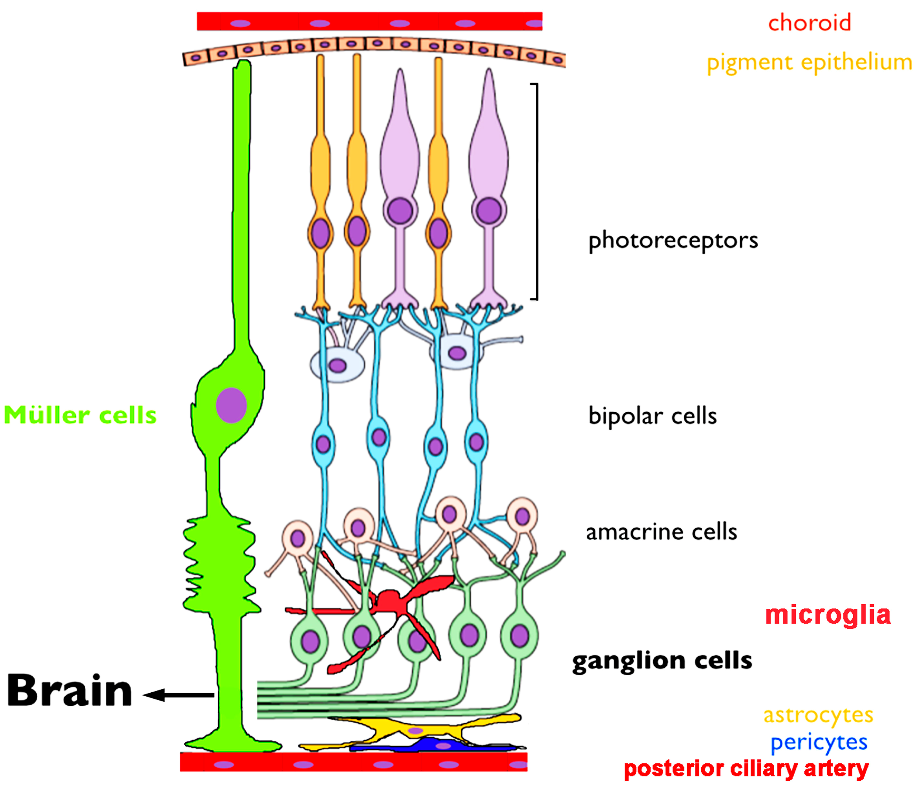
3.1. Overview of Retinal Anatomy and Early Visual Information Processing

When light enters the eye, it is focused onto the retina by the lens and transduced by photoreceptors into electrical and chemical signals that are processed as they are passed through retinal circuits towards the brain. In a nutshell, the flow of visual signal consists of a vertical glutamatergic signals mediated by excitatory photoreceptor, bipolar and ganglion cell synapses and inhibitory/modulatory contributions from horizontal networks mediated by horizontal and amacrine cells, respectively (Figure 1).
Absorption of photons results in the reduction of tonic release of glutamate to postsynaptic horizontal cells and bipolar cells, respectively. Bipolar cells transmit this information to RGCs through two opposing yet complementary pathways. A non-inverting synapse to OFF bipolar cells mediates the excitatory response to decrements of light through ionotropic AMPA/KA receptors. Conversely, the ON pathway is based on a metabotropic mGluR6- TRPM1 mechanism whereby glutamate tonically suppresses bipolar cells and thus cells become disinhibited by light-induced suppression of glutamate release from rods and cones. As bipolar cells forward feed visual information, it is processed through parallel and serial pathways that involve feedback interactions with amacrine cells that contribute to lateral inhibition that sculpts the spatiotemporal organization of RGC receptive fields. As shown in Figure 1, RGCs represent the final output pathway that conveys the visual signal to the midbrain [67]. RGC function and output can be influenced by retinal glia that include Müller cells that span the tissue (and provide critical ionic, metabolic and modulatory of support for neurons, [68]), protoplasmic astrocytes that line the inner limiting membrane (ILM) and (in vascularized retinas) control the permeability of the blood-retina barrier, and microglia, which represent the resident immune cells [69]. Whilst organization of retinal circuits represents a key determinant of visual information processing, the physiological state of every cell type is dynamically altered through activity-dependent and neurodegeneration-driven changes in calcium homeostasis, functional and structural connectivity across and between retinal laminae [67,69,70]. TRP channels represent new players that play increasingly visible roles in retinal neuronal and glial Ca homeostasis by transducing the RGC response to light and driving plasma membrane Ca influx as well as through interactions with intracellular Ca stores, heteromerization and activation of purinergic receptors and pannexin hemichannels [17,18,19,21,22,25,71,72,73,74,75].
Figure 1. A schematic of the retina showing overall arrangement of retinal layers and relationship to the critical vascular and pigment layers. An excitatory vertical chain (photoreceptors, bipolar cells, ganglion cells) provides a direct route for transmitting visual information to the midbrain. Lateral inputs from horizontal cells and amacrine cells provide luminance gain control and organization spatiotemporal receptive fields of retinal neurons. Müller glia provide most of the functions performed by astrocytes in the brain. Astrocytes form the blood-retina barrier together with pericytes and vascular endothelial cells. Microglia play a role in developmental pruning and the retinal immune response whereas the choroid and posterior ciliary artery feed outer and inner retinal neurons, respectively. Adapted from [70].
Cells 03 00914 g001

3.2. TRPV1 Channel Distribution in the Retina

The first evidence suggestive of TRPV1 expression in the retina was obtained by Ritter and Dinh [66,76], years before the seminal cloning of TRPV1 in dorsal root ganglia (DRG) [2]. The investigators used cupric silver staining to reveal capsaicin-induced neurodegeneration in the rat retina. This effect was observed in the IPL and RGCL and was also associated with impaired anterograde axonal transport [77]. Interestingly, unilateral transection of the optic nerve prior to capsaicin administration prevented contralateral spread of neurodegeneration to RGC projection sites within the midbrain [66]. In some cases, capsaicin treatment appeared to damage RGC projections without destroying the soma [76], consistent with the documented ability of capsaicin to act on axons in peripheral nerves [78]. Subsequently, Szallasi et al. [61], used autoradiography of whole-head cryosections to confirm the retina as a binding site for the TRPV1 agonists capsaicin and its ultrapotent analog, the Euphorbia resinifera-derived resiniferatoxin.
Some of these early studies may need to be re-evaluated. Similar to the limitations of approaches that exclusively rely on antibody staining, results from studies confined to the use of pharmacological agents such as capsaicin (agonist) and capsazepine (antagonist) should take into account non-specific and/or mis-targeting effects. TRPV1 was originally called the ‘capsaicin receptor’ based on the active ingredient in Capsaicum chili peppers; however, capsaicin has been subsequently shown to also affect a range of TRPV1-independent mechanisms that include activation of voltage-gated inward and outward currents, impairment of mitochondrial function, inhibition of prostaglandin E2 production and importantly, binding to the cannabinoid CB1 receptor [79,80,81,82]. The use of the agonist is particularly complex within the retinal context, as capsaicin induces large inward currents in ON-bipolar cells that mediate an excitatory, inverted, photoreceptor response towards the inner retina. Capsaicin-evoked responses within rod and cone ON bipolar cells at first appeared to be indistinguishable from the effects mediated by light and mGluR6 antagonists and therefore pointed at a “TRPV1-like” identity of the long-sought transduction channel. However, it was soon discovered that ON bipolar light responses are unaffected in Trpv1−/− mice, whereas they are eliminated by deletion of another TRP channel, the melastatin receptor TRPM1 [10]. While this suggested that capsaicin sensitivity is mediated through TRPM1, only partial reduction in the amplitude of capsaicin responses was observed in Trpm1−/− mice [25], indicating that TRPM1 might not represent the exclusive capsaicin sensor in ON bipolars. Consistent with this, Xu et al. [83] recently reported that the capsaicin response is eliminated in mice lacking the mGluR6 receptor, suggesting that the compound also acts on the metabotropic glutamate receptor or its downstream Go protein heteromers. Irrespective of the precise mechanism, it is especially important to consider potential non-TRPV1-mediated effects when capsaicin is used to analyze retinal function or pathology. The results obtained by using the popular competitive antagonist capsazepine should likewise be interpreted with caution, as the drug can produce effects in Trpv1−/− mice. These include antagonism of voltage-activated Ca2+ channels [84], acetylcholine receptors [85], hyperpolarization-activated cation channels (Ih) [86] and stimulation of amiloride-sensitive ENaC channels [87].
Nonetheless, numerous studies demonstrated the presence of TRPV1 mRNA and protein in the mammalian retina as well as in teleosts, amphibians and avians [10,18,71,72,88,89], leading Zimov and Yazulla to suggest that the role of the channel in visual signaling might be conserved across vertebrates [90,91]. A recent study, using probes and antibodies that detected TRPV1 signals in DRG, suggested that TRPV1 mRNA and protein levels in the mouse retina are unusually low [18]. The differences between studies are likely to reflect use of different antibodies and staining protocols, but also underscore the need for using Trpv1−/− mice as controls for semi-quantitative expression studies.
A potential role for TRPV1 in ontogeny was suggested by Leonelli et al. [92], who detected stable TRPV1 protein expression in embryonic and early postnatal (E19 through P14) rat retinas, followed by a marked increase in expression during the transition from eye opening to adulthood (at ~P60). Exposure of neonatal rats to capsazepine increased the prevalence of subsets of parvalbumin+ amacrine-like cells and increased immunoreactivity (ir) for the presynaptic amacrine marker synapsin 1b. Capsazepine also reduced the normal, developmentally driven, apoptosis in the isolated neonatal rat retina but did not affect retinal layering in the adults; thus, it was proposed that TRPV1 plays a role in the wiring of retinal circuits [93].
Similar to studies in the brain and non-excitable tissues [51,63], immunohistochemical (IHC) staining of mammalian retinas has yielded variable, antibody-specific patterns. The most commonly reported observation from studies that rely mainly on IHC is prominent TRPV1-ir in RGCs and astrocytes. In rat, TRPV1 labeling was confined to a subset of cells in the GCL and within the optic nerve head, but was weak within the optic nerve itself [92]. Further consistent with RGC localization, axotomy of the optic nerve resulted in decreased TRPV1 expression that was mirrored by an injury-induced decline in the population of TRPV1-expressing cells [88,94] and TRPV1-r was also detected within retinocollicular projections within the superior colliculus [95]. Suggestive of potential presynaptic function, TRPV1-ir puncta in the inner plexiform layer (IPL) colocalized with the synaptic vesicle marker synaptophysin. Increased density of labeled axon bundles near the optic nerve head was also observed together with the labeling of distinct cell types in the GCL that included large SMI32+ cell bodies, axons and dendrites and smaller Thy-1.1+ cells. Consistent with the labeling in retinal sections, TRPV1-ir was also observed in somata/dendrites/neurites of immunopanned and cultured RGCs [72]. Sappington et al. [72] reported that retinal TRPV1 expression increases with intraocular pressure (IOP) and suggested that mechanical activation of the channel induced by chronic increases in IOP may drive progressive degeneration of RGCs.
Trpv1 mRNA signals in immunomagnetically separated postnatal and adult rat RGCs are weaker compared to the whole retina [72], indicating the transcripts are likely to be generated in other cell types in addition to RGCs. Accordingly, immunolocalization and pharmacological assays implicated photoreceptors, bipolar cells, amacrine cells and glia as potential TRPV1 expressors in rodents, teleosts and primates [92,96,97,98]. TRPV1-ir was observed in synaptic terminals of salamander and rat photoreceptors [92,98,99], suggesting that the channel could modulate the transmission of the response to postsynaptic bipolar and horizontal cells. However, arguing against a major role in phototransduction and neurotransmission in the outer retina, photopic and scotopic ERG a- and b- waves in Trpv1−/− mice are indistinguishable from field potentials in wild type animals [10]. It is possible, however, that the diffuse TRPV1-ir observed in the OPL [72,100] corresponds to Müller glial processes and/or resident microglia. Consistent with the microglial locus, TRPV1-ir is present in Iba1+ and OX-42+ cells whereas cultured retinal microglia respond to capsaicin [71,92]. TRPV1 expression in Müller cells is less clear. Leonelli et al. [92] did not detect TRPV1-ir in rat Müller glia whereas Martinez-Garcia et al. [89] reported TRPV1-ir in rabbit Müller cells. Thus, localization to photoreceptors, Müller glia, bipolar cells and/or amacrine cells may be species-specific and the identity of TRPV1 signals in the mammalian retina remains a subject for further studies. Leonelli et al. [92] detected weak TRPV1-ir puncta in astrocytes that line the vasculature at the inner limiting membrane (Figure 1). This was recently confirmed in the functional study by Ho et al. [101] who provided evidence that the channel plays a role in guiding astrocyte migration following wound injury. Finally, TRPV1-ir was also reported in the retinal pigment epithelium in rabbits and humans [89], indicating a yet-to-be-defined role in the ion transport function of these cells that are critically important for photoreceptor function and survival.
The variable results obtained with TRPV1 antibodies and pharmacological agents suggest the need for additional functional assays as well as control experiments based on the widely available mice with the genetically ablated channel. One example of such an auxiliary method is optical imaging, which indeed shows robust, desensitizing, capsaicin-induced [Ca2+]i elevations in mouse RGC somata and microglial cells, but not in Müller astroglia or photoreceptors (Figure 2). The identity of the channel was additionally confirmed by blocking capsaicin-evoked [Ca2+]i elevations with capsazepine and by showing their absence in Trpv1−/− cells [102]. Functional expression of TRPV1 in RGCs and microglial cells, together with their absence from photoreceptors and bipolar cells suggests that the channel may play a more significant function within the inner retina.
Figure 2. An acutely isolated and dissociated mouse RGC (blue) and microglial cell (µG; green) loaded with fura-2 respond to capsaicin (40 µM) with transient [Ca2+]i elevations that inactivate during stimulus application, whereas a representative photoreceptor (PR; purple) and Müller cell (MC; red) do not respond to the TRPV1 agonist (see [19,103] for methods).
Cells 03 00914 g002

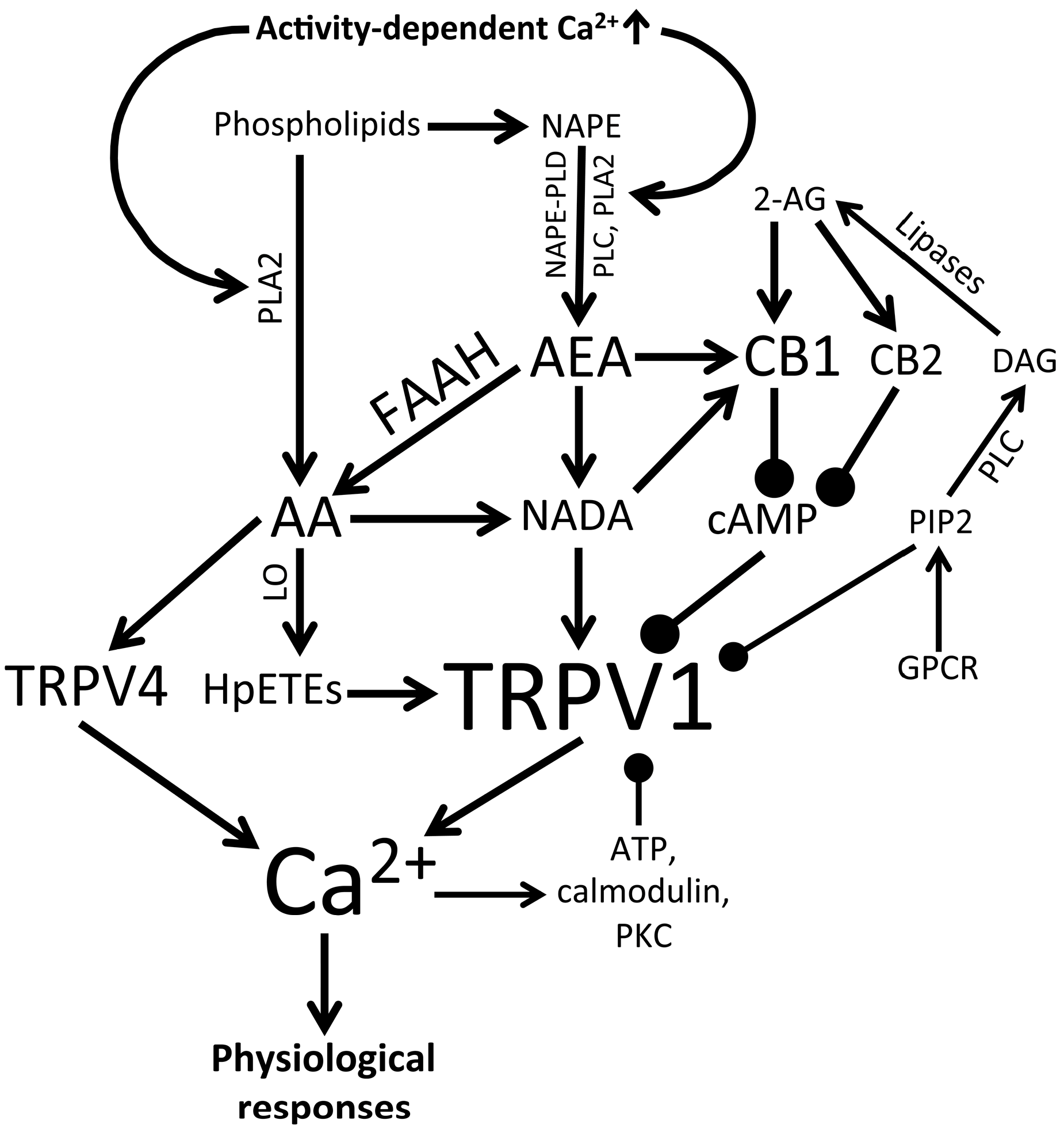
3.3. Endogenous and Synthetic TRPV1 Agonists and Retinal Signaling

Because TRPV1 receptors are unlikely to experience conspicuous nociceptive, mechanical and noxious thermal stimuli in the healthy retina, their primary activators of are likely to be endogenous polyunsaturated fatty acids. TRPV1 is gated by precursors and derivatives of arachidonic acid commonly lumped together into the “endovanilloid/endocannabinoid” (eCB) category of signaling metabolites that are ubiquitous across all retinal layers [98]. These may include anandamide (Arachidonoyl ethanolamide; AEA), unsaturated N-acyl-dopamines (e.g., N-Arachidonoyl dopamine; NADA), 2-arachidonoglycerol (2-AG, an ester formed from arachidonic acid and glycerol) and lipoxygenase metabolites of arachidonic acid (hydroperoxyeicosatetraenoic acids; HpETEs) [36] (Figure 2).
eCBs affect TRPV1 activity directly and indirectly via activation of cannabinoid receptors (CBRs), a 2-isoform family of G protein-coupled receptors with ~44% amino acid similarity that are associated with intracellular cAMP metabolism and downstream activation of the MAPK-ERK pathway. Type I CB receptors (CB1Rs) are most abundant GPCR within the CNS and all TRPV1-expressing primary sensory neurons express CB1R in vitro [104]. The complexity of eCB composition translates to their biological effects. Thus, AEA activates TRPV1 and CB1Rs, whereas 2-AG is a full agonist of CB1Rs and CB2Rs but does not affect TRPV1 [105]. eCB biosynthesis from phospholipid precursors is primarily activity-dependent and follows activation of Ca2+-dependent phospholipases (including phospholipases A2 and C). For example, 2-AG (which, unlike anandamide is expressed at high levels in the CNS) is primarily synthesized by phospholipase C and by diacylglycerol lipase (DAGL) acting on the second messenger diacylglycerol (DAG) [106] (Figure 3). Both AEA and 2-AG freely diffuse across the membrane but can be released into extracellular space to affect neural activity via presynaptic and/or postsynaptic binding sites. eCBs target pre- and/or postsynaptic TRPV1 channels and were reported to bind the cognate TRPV2-4 isoforms with moderate-to-high efficacy and potency (EC50 ~3.7 µM) [107]. The EC50 values for binding of cannabinoid compounds to CBs and TRPV1 are typically in the micromolar range, although some (such as NADA) can bind TRPV1 in the nanomolar range [82]. Thus, eCB release will exert a multimodal effect through separate CBR and vanilloid receptor mechanisms, but may also trigger reciprocal interactions between the two. Interestingly, coactivation of excitatory (TRPV1) and inhibitory (CB1R) eCB-dependent mechanisms can be concentration-dependent whereby CB1R-mediated effects predominate at low (nM) levels of AEA and TRPV1-mediated effects are more pronounced at higher (~µM) concentrations [104]. Alternatively, the two effectors of AEA might be activated sequentially, with stimulation of high-affinity CB1Rs preceding subsequent activation of TRPV1 [108]. Moreover, reciprocal interaction between the GPCR and the ion channel may involve CB1R-mediated sensitization or desensitization of TRPV1 through PKA/PKC-dependent regulation of its phosphorylation state [109]. With respect to the retina, CB1R transcripts were found in both plexiform layers [99,110,111], whereas DAG lipases were localized to OFF bipolar cells [112]. Given the prominent localization of TRPV1 in the inner retina, it is plausible that Ca2+-dependent eCB release, associated with periods of intense ON- and OFF RGC activation results in indirect (CBR-mediated) and direct TRPV1 modulation. As if this is not complicated enough, arachidonic acid and its downstream eicosanoid metabolites also activate TRPV4, a polymodal vanilloid receptor homolog that acts as a RGC osmosensor [17]. The properties of natural stimuli (light) that lead to endocannabinoid concentrations high enough for retinal TRPV1 and/or CB1R activation remain to be identified.
Enzymatic synthesis and degradation of endovanilloid/eCBs is a likely rate-limiting step in TRPV1 activation and inhibition. AEA is degraded by the fatty acid amide hydrolase (FAAH), resulting in the production of ethanolamine and arachidonic acid (AA), whereas 2-AG can be hydrolyzed by monoacylglycerol lipase (MAGL), α/β-hydrolase domain 6, 12 (ABHD6/ABHD12), and/or cyclooxygenase-2 (COX-2) [for retinal distributions see [112,113]. FAAH is ubiquitously expressed across the brain and the retina, with expression reported in subsets of photoreceptors, RGCs, dopaminergic and cholinergic amacrine cells and horizontal cells [98,112,114]. IHC studies in fish colocalized FAAH with CB1Rs and TRPV1 within presynaptic terminals of the IPL and OPL [90,91]. FAAH expression in photoreceptors is consistent with the observed AEA uptake [100] and localization of CB1 receptors [99]. The NMDA inhibitor MK801 reduced retinal FAAH activity [88], possibly by downregulating the Ca2+-dependent modulation of the enzyme. FAAH-mediated catabolism of anandamide may be suppressed by PEA, which is present rat, cat and monkey retinas, potentiating eCB signaling in vivo [115]. The augmentation of eCB signaling by inhibition of FAAH can produce effects that markedly differ from those elicited by CB1 agonists [116], consistent with synergistic action on TRP channels. Likewise, the breakdown of the FAAH metabolite arachidonic acid by 12-lipoxygenase (12-LO) and 15-lipoxygenase (15-LO) generates 12(S)-HpETE and 15(S)-HpETE, which also bind and activate TRPV1 [117,118]. It would be interesting to see whether the retinas of FAAH−/− mice favor eCB signaling over that of endovanilloids and how this affects the responses of retinas to light/darkness.
Figure 3. Cellular activity initiates synthesis of endocannabinoids/endovanilloids, which activate (arrows) or modulate (closed circles) TRPV1 and TRPV4 activity and thereby regulate physiological responses. 2-AG (2-arachidonoylglycerol), AA (arachidonic acid), AEA (anandamide), CB1 (cannabinoid receptor type I), CB2 (cannabinoid receptor type I), DAG (diacylglycerol), FAAH (fatty acid amide hydrolase), GPCR (G protein-coupled receptor), HpETEs (hydroperoxyeicosatetraenoic acids), LO (lipoxygenase), NADA (N-arachidonoyl dopamine), NAPE (N-acylphosphatidylethanolamine), NAPE-PLD (NAPE-specific phospholipase D), PIP2 (phosphatidylinositol 4,5-bisphosphate), PKC (protein kinase C), PLA2 (phospholipase A2), PLC (phospholipase C).
Cells 03 00914 g003
To understand TRPV1 function in the retina one must differentiate the stimulatory effects of endogenous and synthetic cannabinoids on visual signaling and their protective effects in pathological paradigms. Systemic exposure to marijuana produces a dazzling variety of visual experiences that include increased photosensitivity [119], changes in color discrimination [120] and double/blurred vision [96,121], with a mechanism of action that is likely to involve both retinal and cerebral targets. The psychoactive ingredient in marijuana, Δ9-tetrahydrocannibinol (THC), mimics eCBs through its stimulation of CB1 receptors but does not activate TRPV1. Cannabinoid effects within the retina were also shown to be complex under experimentally tractable conditions. Amongst the observed effects are modulation of intracellular signaling in most retinal cell types including potassium and calcium currents in amphibian and fish photoreceptors [96,122], voltage-operated currents in rodent RGCs [123] and transmitter release from amacrine and bipolar processes in teleosts and mice [97,124].
Data from exogenous CB1 agonists, eCB measurements, and FAAH inhibition has suggested an important role for eCB signaling in neuroprotection. THC lowers IOP and protects the retina from excitotoxic damage [96,125,126]. Analogously, synthetic cannabinoids evinced remarkable protection for photoreceptors and bipolar cells in a rat model of retinitis pigmentosa [127] and were suggested to reduce retinal pigment epithelium (RPE) and RGC damage in models of ischemia, glaucoma, diabetic retinopathy, oxidative damage and/or traumatic ocular injury. Suggestive of lost protection, decreased levels of 2-AG have been reported in ocular tissues from glaucoma patients [128]. FAAH inhibition elevates AEA in young, but not old, rat retinas, an effect that was associated with CB1-dependent RGC neuroprotection following axotomy [129]. In a study of ischemic-reperfusion (I-R), Nucci et al. [88] used high IOP (~120 mmHg) to block blood flow within and induce RGC loss in the rat retina. The cytotoxic effect of I-R was antagonized by systemic FAAH inhibition or intravitreal injection of MetAEA, a stable analogue of anandamide. MetAEA-mediated neuroprotection was blocked by inhibition of either CB1 or TRPV1, indicating that both receptors might be involved in neuroprotective effects of anandamide. Other studies showed that cells exposed to excitotoxic conditions can be rescued with eCBs, possibly by suppressing excessive release of glutamate, depolarization-mediated Ca2+ overloads and/or excessive firing within the retina [123,124,130]. It is unclear if TRPV1 and CB1Rs regulate the same neuroprotective pathway [but see 102]. Moreover, it remains to be seen whether CB1 and/or TRPV1 inhibition or genetic deletion exacerbate RGC loss in I-R. While eCBs act via CB1Rs [98], the consequences of parallel activation of TRPV1 were not assessed. Thus, it is not known whether CB1R activation protects retinal neurons synergistically with TRPV1 and/or CB1Rs provide protection by antagonizing Ca2+ overloads mediated by TPRV1. In either case, CB1Rs and TRPV1 represent novel potential targets that might confer neuroprotection within the retina without causing excitotoxicity. A bottleneck in investigating these potentially ubiquitous signaling pathways in the retina has been that, in contrast to the effects of synthetic cannabinoids applied to (isolated, cultured) retinal cells, the biological functions of eCBs, TRPV1 and their interactions across retinal circuits remain almost entirely unknown.

3.4. Is TRPV1 Neurodegenerative or Neuroprotective?

The expression of TRPV1 in the IPL and GCL [66,72,92] implies a potential role for pathological enhancement of RGC excitability, post-synaptic voltage-operated Ca2+ influx and glutamate receptor-mediated currents that lead to cell injury via Ca2+ overload. Consistent with this, maximal TRPV1 activation has been shown to cause neuronal and glial excitotoxicity in other areas of the brain [131,132,133,134]. Expanding the results from early studies in the retina [66,77], a recent study reported that capsaicin exposure is sufficient to induce apoptosis in cultured RGCs [72]. Intravitreal injection of capsaicin likewise caused retinal thinning and increased the number of Fluoro-Jade B positive cells in the GCL and INL, particularly in axotomized retinas, an effect that was blocked by capsazepine [94]. Because TRPV1 channels contribute to mechanosensation in some cell types [42,135], but see [136], exposure to hydrostatic pressure was employed to test whether TRPV1 might represent the intrinsic pressure-sensitive pathway through which RGCs respond to elevated IOP. Cultured RGCs and microglia were placed into a pressure chamber and the authors reported increased [Ca2+]i when the pressure was raised by 70 mm Hg [71,72]. The observation that excessive TRPV1 activity is excitotoxic for RGCs, potentially due to Ca2+ overload [72,137], is a valuable step forward in our understanding of TRPV1 pathophysiology within the retina. By providing evidence that the channel might be intrinsically sensitive to pressure, these data provide an attractive model for mechanically-induced Ca2+ dysregulation and RGC remodeling/apoptosis in IOP-induced retinal disease. However, the experimental paradigm has recently been questioned within the context of glaucoma [138], as intended pressure application within the standard laboratory incubator may not have reached the intended pressure differential across the plasma membrane due to non-compressibility of water. Moreover, loss of TRPV1 channels is not protective in mouse glaucoma models [17]. Nonetheless, the mechanobiology of IOP and distribution of mechanical forces across retinal tissue under pathological conditions remains a key challenge in retinal research. This includes characterization of the pressure gradient between the inner and outer eye, which might result in the expansion of the orbit [73] and the optic nerve head [139]. TRPV1, which may sense mechanical stress under certain conditions (such as mechanical hyperalgesia or cell shrinking) [42] might therefore play an accentuated role in retinal pathology.
While TRPV1 activation might be deleterious under some conditions, it has been associated with neuroprotection in other contexts [140]. Recent findings [16], which show that IOP-induced damage to RGCs is augmented in Trpv1−/− animals, suggest that a novel neuroprotective function for TRPV1 channels. RGC injury in Trpv1−/− mice and capsazepine-treated rats was associated with inhibited anterograde axonal transport of a fluorescently tagged marker, axonal loss and astrogliosis in the microbead occlusion glaucoma model. TRPV1 ablation also suppressed a post-IOP exposure-dependent increase in RGC firing and increased the amount of depolarizing current needed to reach the RGC firing threshold. The authors suggest that TRPV1 activity rescues RGCs by promoting their excitability during retinal stress. Another protective mechanism might involve TRPV1-dependent release of the protective interleukin 6 (IL-6) from activated microglia [141], as TRPV1 inhibition by iodo-resiniferatoxin (specific) and Ruthenium Red (non-specific) prevented hydrostatic pressure-induced release of IL-6 and NFκB translocation into the nucleus of microglia [71]. A recent in vivo study by Sakamoto et al. [142] showed that TRPV1 agonists (capsaicin or SA13353) might evince a paradoxical suppression of NMDA-mediated excitotoxicity of rat RGCs. This effect was prevented by capsazepine, CGRP (8–37) (calcitonin gene-related peptide receptor antagonist) or RP67580 (tachykinin NK1 receptor antagonist). CGRP and substance P, which are increased by electrical stimulation of the retina [143] and metabolic stress [144], were protective [142]. Low levels of systemic capsaicin were reported to elevate the CGRP concentration and reduce apoptosis in the GCL of rats with STZ-induced diabetic retinopathy [144]. Taken together, the results from capsaicin exposure studies appear contradictory and unresolved. It appears that TRPV1 activity can be beneficial or detrimental to RGCs depending on the disease model, the extent of TRPV1 activation and activation of auxiliary calcium effectors, however, there is also the serious concern of concomitant overstimulation of capsaicin-sensitive mechanisms such as TRPM1 that needs to be addressed in future work.

3.5. TRPV1 and Retinal Vasoregulation

TRPV1 is expressed in vascular endothelial cells and arteriolar smooth muscle cells [20,51,145,146] and may modulate vascular tone and permeability by serving as a source of Ca2+ for NO synthesis. Donnerer and Lembeck [147] first showed that capsaicin evokes vascular responses that are independent of sensory innervation. TRPV1’s contribution to the regulation of blood distribution appears to be tissue-specific, as TRPV1 agonists can cause neurogenic vasodilation (in the skin) and vasoconstriction (in skeletal muscle) [148,149,150]. In the retina, TRPV1-ir colocalized with vascular endothelial cells at the inner limiting membrane [92], suggesting that the channel might also modulate vascular flow and/or permeability in this tissue, possibly via Ca2+-dependent upregulation of inducible and endothelial NO synthase isoforms [151] and/or production of CGRP [145,152]. eCB analogs reduced proliferation of human retinal vascular endothelial cells [153], suggesting additional roles in the regulation of vascular leakage and integrity of the blood-retina-barrier.
TRPV1 expression in astrocytes lining blood vessels within the inner retina is also likely to modulate glial control of vascular tone and/or Ca2+-dependent astrogliosis [101]. Inflammation that accompanies reactive gliosis of protoplasmic astrocytes and Müller glia can itself affect blood-retina-barrier permeability, vascular leakage and oxidative damage [73]. As these events are particularly hazardous to RGCs, both pathological and neuroprotective aspects of TRPV1 and eCB activity may offer potential benefits for treating traumatic, inflammatory and ischemic optic neuropathies in diabetic retinopathy, ischemia, optic neuritis and glaucoma.

4. Conclusions

Although TRPV1 expression has been documented across many cell types and in species across phyla ranging from fish to nonhuman primates, at present remarkably little definitive information is available about what the channel is doing in visual processing, retinal development and homeostatic functions of retinal neurons, glia and vasculature. At the very least, it seems that the network comprised of TRPV1 channels, CB1 receptors, eCBs /eicosanoids, kinases and phosphatases (Figure 3) represents a novel neuromodulatory system that could tune the excitability of retinal neurons through amplification or dampening of retinal output and/or contribute to activity-dependent refinement and retinal patterning of visual circuits. The polymodal properties of the TRPV1 channel suggest that, similar to its sibling TRPV4 [17,73], eCB-dependent modulation of Ca2+ entry could drive numerous Ca2+-dependent processes associated with normal and pathological retinal physiology. The prominent functional expression of TRPV1 channels in RGCs and microglia (Figure 2) and the stupendous complexity of endovanilloid/endocannabinoid signaling (Figure 3) suggest that the channels could modulate the diverse presynaptic inputs to RGCs as well as participate in nonretrograde, light-dependent, modulation of retinal output. It will be important to determine how TRPV1 activators and modulators orchestrate channel properties and its interactions with voltage and calcium effectors such as voltage-operated Ca2+ channels, NMDA receptors and Ca2+-dependent transcription factors (e.g., NFAT, NFkB, c-fos and DREAM). A particularly interesting area of research will be to evaluate the role of CB1R–TRPV1 interactions, especially under pathological conditions where cannabinoids can provide significant neuroprotection [98]. Given that TRPV1 was identified as a potential intracellular Ca2+ release channel [154] studies are also needed to evaluate its relationship to Ca2+ release from intracellular stores and activation of store-operated currents, which have been shown to play an increasingly prominent function in retinal Ca2+ homeostasis [74,103,155,156]. Because light-dependent changes in [Ca2+]i levels are the main driver of tonic neurotransmission that characterizes retinal signaling in light and darkness [157], Ca2+ influx and clearance in retinal neurons are under tight control. A clear example of such exquisite control are photoreceptors where µV changes in presynaptic membrane potential translate into discernable differences in [Ca2+]i within photoreceptor terminals and visual perception [158,159]. Thus, even modest [Ca2+]i elevations mediated by physiological TRPV1 activation could have significant repercussion for downstream visual signaling. Finally, taking into account the reported roles for TRPV1 in hypertonic transduction and mechanosensation [160], it will be of interest whether the channel also contributes to retinal osmoregulation and intrinsic responsiveness to IOP and/or mechanical trauma. This would implicate this polymodal channel in key aspects of retinal homeostasis and response to stress. In summary, the determination of the role that TRP channels play in invertebrate vision together with the elucidation of TRPV1 function in acute and chronic pain represent two of the pinnacles of modern neuroscience. In comparison, TRPV1 research in the mammalian visual system is in its infancy and more studies are needed to better define the involvement of this channel in sensory transduction at the single cell level within the retina, function of visual circuits and the pathogenesis/etiology of blinding diseases.

Acknowledgments

This work was supported by the (University of Utah) Undergraduate Research Opportunity Program (A.J.), HHMI Med into Grad scholarship (D.R.), NIH (T32DC008553, D.R.; RO1EY13870, RO1EY022076, P30EY014800 Vision Core grant), Department of Defense W81XWH-12-1-0244, State of Utah TCIP, Glaucoma Research Foundation and unrestricted grants from Research to Prevent Blindness to the Moran Eye Institute.

Author Contributions

D.A.R., S.R., A.O.J. and D.K. wrote the review. D.A.R., S.R. and A.O.J. collected and analyzed the data.

Conflicts of Interest

The authors declare no conflict of interest.

References

  1. Minke, B. Light-induced reduction in excitation efficiency in the trp mutant of Drosophila. J. Gen. Physiol. 1982, 79, 361–385. [Google Scholar] [CrossRef]
  2. Caterina, M.J.; Schumacher, M.A.; Tominaga, M.; Rosen, T.A.; Levine, J.D.; Julius, D. The capsaicin receptor: A heat-activated ion channel in the pain pathway. Nature 1997, 389, 816–824. [Google Scholar] [CrossRef]
  3. Clapham, D.E. TRP channels as cellular sensors. Nature 2003, 426, 517–524. [Google Scholar] [CrossRef]
  4. Suzuki, M.; Mizuno, A.; Kodaira, K.; Imai, M. Impaired pressure sensation in mice lacking TRPV4. J. Biol. Chem. 2003, 278, 22664–22668. [Google Scholar] [CrossRef]
  5. Watanabe, H.; Vriens, J.; Prenen, J.; Droogmans, G.; Voets, T.; Nilius, B. Anandamide and arachidonic acid use epoxyeicosatrienoic acid to activate TRPV4 channels. Nature 2003, 424, 434–438. [Google Scholar] [CrossRef]
  6. Kraft, R.; Harteneck, C. The mammalian melastatin-related transient receptor potential cation channels: An overview. Pflugers Arch. 2005, 451, 204–211. [Google Scholar] [CrossRef]
  7. Phelps, C.B.; Wang, R.R.; Choo, S.S.; Gaudet, R. Differential regulation of TRPV1, TRPV3, and TRPV4 sensitivity through a conserved binding site on the ankyrin repeat domain. J. Biol. Chem. 2010, 285, 731–740. [Google Scholar] [CrossRef]
  8. Nilius, B.; Owsianik, G. Transient receptor potential channelopathies. Pflugers Arch. 2010, 460, 437–450. [Google Scholar] [CrossRef]
  9. Kraft, R.; Grimm, C.; Grosse, K.; Hoffmann, A.; Sauerbruch, S.; Kettenmann, H.; Schultz, G.; Harteneck, C. Hydrogen peroxide and ADP-ribose induce TRPM2-mediated calcium influx and cation currents in microglia. Am. J. Physiol. Cell Physiol. 2004, 286, 129–137. [Google Scholar]
  10. Shen, Y.; Heimel, J.A.; Kamermans, M.; Peachey, N.S.; Gregg, R.G.; Nawy, S. A transient receptor potential-like channel mediates synaptic transmission in rod bipolar cells. J. Neurosci. 2009, 29, 6088–6093. [Google Scholar] [CrossRef]
  11. Gavva, N.R.; Bannon, A.W.; Surapaneni, S.; Hovland, D.N.; Lehto, S.G.; Gore, A.; Juan, T.; Deng, H.; Han, B.; Klionsky, L.; et al. The vanilloid receptor TRPV1 is tonically activated in vivo and involved in body temperature regulation. J. Neurosci. 2007, 27, 3366–3374. [Google Scholar] [CrossRef]
  12. Sudbury, J.R.; Ciura, S.; Sharif-Naeini, R.; Bourque, C.W. Osmotic and thermal control of magnocellular neurosecretory neurons-role of an N-terminal variant of TRPV1. Eur. J. Neurosci. 2010, 32, 2022–2030. [Google Scholar] [CrossRef]
  13. Dunn, K.M.; Hill-Eubanks, D.C.; Liedtke, W.B.; Nelson, M.T. TRPV4 channels stimulate Ca2+-induced Ca2+ release in astrocytic endfeet and amplify neurovascular coupling responses. Proc. Natl. Acad. Sci. U.S.A. 2013, 110, 6157–6162. [Google Scholar] [CrossRef]
  14. Filosa, J.A.; Yao, X.; Rath, G. TRPV4 and the regulation of vascular tone. J. Cardiovasc. Pharmacol. 2013, 61, 113–119. [Google Scholar] [CrossRef]
  15. Caterina, M.J.; Leffler, A.; Malmberg, A.B.; Martin, W.J.; Trafton, J.; Petersen-Zeitz, K.R.; Koltzenburg, M.; Basbaum, A.I.; Julius, D. Impaired nociception and pain sensation in mice lacking the capsaicin receptor. Sci. 2000, 288, 306–313. [Google Scholar] [CrossRef]
  16. Ward, N.J.; Ho, K.W.; Lambert, W.S.; Weitlauf, C.; Calkins, D.J. Absence of transient receptor potential vanilloid-1 accelerates stress-induced axonopathy in the optic projection. J. Neurosci. 2014, 34, 3161–3170. [Google Scholar] [CrossRef]
  17. Ryskamp, D.A.; Witkovsky, P.; Barabas, P.; Huang, W.; Koehler, C.; Akimov, N.P.; Lee, S.H.; Chauhan, S.; Xing, W.; Renteria, R.C.; et al. The polymodal ion channel transient receptor potential vanilloid 4 modulates calcium flux, spiking rate, and apoptosis of mouse retinal ganglion cells. J. Neurosci. 2011, 31, 7089–7101. [Google Scholar] [CrossRef]
  18. Gilliam, J.C.; Wensel, T.G. TRP channel gene expression in the mouse retina. Vis. Res. 2011, 51, 2440–2452. [Google Scholar] [CrossRef]
  19. Molnar, T.; Barabas, P.; Birnbaumer, L.; Punzo, C.; Kefalov, V.; Križaj, D. Store-operated channels regulate intracellular calcium in mammalian rods. J. Physiol. 2012, 590, 3465–3481. [Google Scholar] [CrossRef]
  20. Tóth, A.; Czikora, Á.; Pásztor, E.T.; Dienes, B.; Bai, P.; Csernoch, L.; Rutkai, I.; Csató, V.; Mányiné, I.S.; Pórszász, R.; et al. Vanilloid receptor-1 (TRPV1) expression and function in the vasculature of the rat. J. Histochem. Cytochem. 2014, 62, 129–144. [Google Scholar] [CrossRef]
  21. Sekaran, S.; Lall, G.S.; Ralphs, K.L.; Wolstenholme, A.J.; Lucas, R.J.; Foster, R.G.; Hankins, M.W. 2-Aminoethoxydiphenylborane is an acute inhibitor of directly photosensitive retinal ganglion cell activity in vitro and in vivo. J. Neurosci. 2007, 27, 3981–3986. [Google Scholar]
  22. Xue, T.; Do, M.T.H.; Riccio, A.; Jiang, Z.; Hsieh, J.; Wang, H.C.; Merbs, S.L.; Welsbie, D.S.; Yoshioka, T.; Weissgerber, P.; et al. Melanopsin signaling in mammalian iris and retina. Nature 2011, 479, 67–73. [Google Scholar] [CrossRef]
  23. Albert, E.S.; Bec, J.M.; Desmadryl, G.; Chekroud, K.; Travo, C.; Gaboyard, S.; Bardin, F.; Marc, I.; Dumas, M.; Lenaers, G.; et al. TRPV4 channels mediate the infared laser-evoked response in sensory neurons. J. Neurophysiol. 2012, 107, 3227–3234. [Google Scholar] [CrossRef] [Green Version]
  24. Montell, C.; Jones, K.; Hafen, E.; Rubin, G. Rescue of the Drosophila phototransduction mutation trp by germline transformation. Science 1985, 230, 1040–1043. [Google Scholar]
  25. Morgans, C.W.; Zhang, J.; Jeffrey, B.G.; Nelson, S.M.; Burke, N.S.; Duvoisin, R.M.; Brown, R.L. TRPM1 is required for the depolarizing light response in retinal ON-bipolar cells. Proc. Natl. Acad. Sci. U.S.A. 2009, 106, 19174–19178. [Google Scholar]
  26. Krol, J.; Busskamp, V.; Markiewicz, I.; Stadler, M.B.; Ribi, S.; Richter, J.; Duebel, J.; Bicker, S.; Fehling, H.J.; Schübeler, D.; et al. Characterizing light-regulated retinal microRNAs reveals rapid turnover as a common property of neuronal microRNAs. Cell 2010, 141, 618–631. [Google Scholar] [CrossRef]
  27. Moiseenkova-Bell, V.Y.; Stanciu, L.A.; Serysheva, I.I.; Tobe, B.J.; Wensel, T.G. Structure of TRPV1 channel revealed by electron cryomicroscopy. Proc. Natl. Acad. Sci. U.S.A. 2008, 105, 7451–7455. [Google Scholar] [CrossRef]
  28. Arniges, M.; Fernández-Fernández, J.M.; Albrecht, N.; Schaefer, M.; Valverde, M.A. Human TRPV4 channel splice variants revealed a key role of ankyrin domains in multimerization and trafficking. J. Biol. Chem. 2006, 281, 1580–1586. [Google Scholar] [CrossRef]
  29. Lishko, P.V.; Procko, E.; Jin, X.; Phelps, C.B.; Gaudet, R. The ankyrin repeats of TRPV1 bind multiple ligands and modulate channel sensitivity. Neuron 2007, 54, 905–918. [Google Scholar]
  30. Everaerts, W.; Nilius, B.; Owsianik, G. The vanilloid transient receptor potential channel TRPV4: From structure to disease. Prog. Biophys. Mol. Biol. 2010, 103, 2–17. [Google Scholar] [CrossRef]
  31. Tominaga, M.; Caterina, M.J.; Malmberg, A.B.; Rosen, T.A.; Gilbert, H.; Skinner, K.; Raumann, B.E.; Basbaum, A.I.; Julius, D. The cloned capsaicin receptor integrates multiple pain-producing stimuli. Neuron 1998, 21, 531–543. [Google Scholar] [CrossRef]
  32. Numazaki, M.; Tominaga, T.; Takeuchi, K.; Murayama, N.; Toyooka, H.; Tominaga, M. Structural determinant of TRPV1 desensitization interacts with calmodulin. Proc. Natl. Acad. Sci. U.S.A. 2003, 100, 8002–8006. [Google Scholar] [CrossRef]
  33. Wu, Z.Z.; Chen, S.R.; Pan, H.L. Transient receptor potential vanilloid type 1 activation down-regulates voltage-gated calcium channels through calcium-dependent calcineurin in sensory neurons. J. Biol. Chem. 2005, 280, 18142–18151. [Google Scholar] [CrossRef]
  34. Hellwig, N.; Albrecht, N.; Harteneck, C.; Schultz, G.; Schaefer, M. Homo- and heteromeric assembly of TRPV channel subunits. J. Cell Sci. 2005, 118, 917–928. [Google Scholar] [CrossRef]
  35. Prescott, E.D.; Julius, D. A modular PIP2 binding site as a determinant of capsaicin receptor. Science 2003, 300, 1284–1288. [Google Scholar] [CrossRef]
  36. Van der Stelt, M.; Di Marzo, V. Endovanilloids. Eur. J. Biochem. 2004, 271, 1827–1834. [Google Scholar]
  37. Woo, D.H.; Jung, S.J.; Zhu, M.H.; Park, C.K.; Kim, Y.H.; Oh, S.B.; Lee, C.J. Direct activation of transient receptor potential vanilloid 1 (TRPV1) by diacylglycerol (DAG). Mol. Pain 2008, 4, 1744–8069. [Google Scholar]
  38. Zhang, X.; Huang, J.; McNaughton, P.A. NGF rapidly increases membrane expression of TRPV1 heat-gated ion channels. Neuromol. Med. 2005, 24, 4211–4223. [Google Scholar]
  39. Sanz-Salvador, L.; Andrés-Borderia, A.; Ferrer-Montiel, A.; Planells-Cases, R. Agonist- and Ca2+-dependent desensitization of TRPV1 channel targets the receptor to lysosomes for degradation. J. Biol. Chem. 2012, 287, 19462–19471. [Google Scholar]
  40. Christoph, T.; Grünweller, A.; Mika, J.; Schäfer, M.K.; Wade, E.J.; Weihe, E.; Erdmann, V.A.; Frank, R.; Gillen, C.; Kurreck, J. Silencing of vanilloid receptor TRPV1 by RNAi reduces neuropathic and visceral pain in vivo. Biochem. Biophys. Res. Commun. 2006, 350, 238–243. [Google Scholar] [CrossRef]
  41. McNamara, F.N.; Randall, A.; Gunthorpe, M.J. Effects of piperine, the pungent component of black pepper, at the human vanilloid receptor (TRPV1). Br. J. Pharmacol. 2005, 144, 781–790. [Google Scholar] [CrossRef]
  42. Prager-Khoutorsky, M.; Khoutorsky, A.; Bourque, C.W. Unique Interweaved Microtubule Scaffold Mediates Osmosensory Transduction via Physical Interaction with TRPV1. Neuron 2014, 83, 866–878. [Google Scholar] [CrossRef]
  43. Immke, D.C.; Gavva, N.R. The TRPV1 receptor and nociception. Semin. Cell Dev. Biol. 2006, 17, 582–591. [Google Scholar] [CrossRef]
  44. Vigna, S.R.; Shahid, R.A.; Nathan, J.D.; McVey, D.C.; Liddle, R.A. Leukotriene B4 mediates inflammation via TRPV1 in duct obstruction-induced pancreatitis in rats. Pancreas 2011, 40, 708–714. [Google Scholar] [CrossRef]
  45. Bhave, G.; Hu, H.J.; Glauner, K.S.; Zhu, W.; Wang, H.; Brasier, D.J.; Oxford, G.S.; Gereau, R.W. Protein kinase C phosphorylation sensitizes but does not activate the capsaicin receptor transient receptor potential vanilloid 1 (TRPV1). Proc. Natl. Acad. Sci. U.S.A. 2003, 100, 12480–12485. [Google Scholar]
  46. Mandadi, S.; Tominaga, T.; Numazaki, M.; Murayama, N.; Saito, N.; Armati, P.J.; Roufogalis, B.D.; Tominaga, M. Increased sensitivity of desensitized TRPV1 by PMA occurs through PKCε-mediated phosphorylation at S800. Pain 2006, 123, 106–116. [Google Scholar] [CrossRef]
  47. Tominaga, M.; Wada, M.; Masu, M. Potentiation of capsaicin receptor activity by metabotropic ATP receptors as a possible mechanism for ATP-evoked pain and hyperalgesia. Proc. Natl. Acad. Sci. U.S.A. 2001, 98, 6951–6956. [Google Scholar] [CrossRef]
  48. Puntambekar, P.; Van Buren, J.; Raisinghani, M.; Premkumar, L.S.; Ramkumar, V. Direct interaction of adenosine with the TRPV1 channel protein. J. Neurosci. 2004, 29, 153–158. [Google Scholar]
  49. Nagy, I.; Sántha, P.; Jancsó, G.; Urbán, L. The role of the vanilloid (capsaicin) receptor (TRPV1) in physiology and pathology. Eur. J. Pharmacol. 2004, 500, 351–369. [Google Scholar]
  50. Sanchez, J.F.; Krause, J.E.; Cortright, D.N. The distribution and regulation of vanilloid receptor VR1 and VR1 5' splice variant RNA expression in rat. Neurosci. 2001, 107, 373–381. [Google Scholar]
  51. Cavanaugh, D.J.; Chesler, A.T.; Jackson, A.C.; Sigal, Y.M.; Yamanaka, H.; Grant, R.; O'Donnell, D.; Nicoll, R.A.; Shah, N.M.; Julius, D.; et al. TRPV1 reporter mice reveal highly restricted brain distribution and functional expression in arteriolar smooth muscle cells. J. Neurosci. 2011, 31, 5067–5077. [Google Scholar] [CrossRef]
  52. Han, L.; Ma, C.; Liu, Q.; Weng, H.-J.; Cui, Y.; Tang, Z.; Kim, Y.; Nie, H.; Qu, L.; Patel, K.N.; et al. A subpopulation of nociceptors specifically linked to itch. Nat. Neurosci. 2013, 16, 174–182. [Google Scholar]
  53. Di Marzo, V. Targeting the endocannabinoid system: To enhance or reduce? Nat. Rev. 2008, 7, 438–455. [Google Scholar]
  54. Kauer, J.A.; Gibson, H.E. Body-temperature maintenance as the predominant function of the vanilloid receptor TRPV1. Trends in Neurosci. 2009, 32, 215–224. [Google Scholar] [CrossRef]
  55. Martins, D.; Tavares, I.; Morgado, C. "Hotheaded": The role of TRPV1 in brain functions. Neuropharmacol. 2014, 85, 151–157. [Google Scholar] [CrossRef]
  56. Marsch, R.; Foeller, E.; Rammes, G.; Bunck, M.; Kössl, M.; Holsboer, F.; Zieglgänsberger, W.; Landgraf, R.; Lutz, B.; Wotjak, C.T. Reduced anxiety, conditioned fear, and hippocampal long-term potentiation in transient receptor potential vanilloid type 1 receptor-deficient mice. J. Neurosci. 2007, 27, 832–839. [Google Scholar] [CrossRef]
  57. Gibson, H.E.; Edwards, J.G.; Page, R.S.; Van Hook, M.J.; Kauer, J.A. TRPV1 channels mediate long-term depression at synapses on hippocampal interneurons. Neuron 2008, 57, 746–759. [Google Scholar]
  58. Adamczyk, P.; Miszkiel, J.; McCreary, A.C.; Filip, M.; Papp, M.; Przegaliński, E. The effects of cannabinoid CB1, CB2 and vanilloid TRPV1 receptor antagonists on cocaine addictive behavior in rats. Brain Res. 2012, 1444, 45–54. [Google Scholar]
  59. Osaka, T.; Kobayashi, A.; Lee, T.H.; Namba, Y.; Inoue, S.; Kimura, S. Lack of integrative control of heat production and heat loss after capsaicin administration. Pflugers Arch. 2000, 440, 440–445. [Google Scholar] [CrossRef]
  60. Dawbarn, D.; Harmar, A.J.; Pycock, C.J. Intranigral injection of capsaicin enhances motor activity and depletes nigral 5-hydroxytrptamine but not substance P. Neuropharmacol. 1981, 20, 341–346. [Google Scholar] [CrossRef]
  61. Szallasi, A.; Nilsson, S.; Farkas-Szallasi, T.; Blumberg, P.M.; Hökfelt, T.; Lundberg, J.M. Vanilloid (capsaicin) receptors in the rat: Distribution in the brain, regional differences in the spinal cord, axonal transport to the periphery, and depletion by systemic vanilloid treatment. Brain Res. 1995, 703, 175–183. [Google Scholar]
  62. Benninger, F.; Freund, T.F.; Hájos, N. Control of excitatory synaptic transmission by capsaicin is unaltered in TRPV1 vanilloid receptor knockout mice. Neurochem. Int. 2008, 52, 89–94. [Google Scholar] [CrossRef]
  63. Everaerts, W.; Sepúlveda, M.R.; Gevaert, T.; Roskams, T.; Nilius, B.; De Ridder, D. Where is TRPV1 expressed in the bladder, do we see the real channel? Naunyn Schmiedebergs Arch Pharmacol. 2009, 379, 421–425. [Google Scholar] [CrossRef]
  64. Brown, T.E.; Chirila, A.M.; Schrank, B.R.; Kauer, J.A. Loss of interneuron LTD and attenuated pyramidal cell LTP in TRPV1 and TRPV3 KO mice. Hippocampus 2013, 23, 662–671. [Google Scholar] [CrossRef]
  65. Ritter, S.; Dinh, T.T. Capsaicin-induced neuronal degeneration: Silver impregnation of cell bodies, axons, and terminals in the central nervous system of the adult rat. J. Comp. Neurol. 1988, 271, 79–90. [Google Scholar] [CrossRef]
  66. Ritter, S.; Dinh, T. Capsaicin-induced neuronal degeneration in the brain and retina of preweanling rats. J. Comp. Neurol. 1990, 296, 447–461. [Google Scholar] [CrossRef]
  67. Marc, R.E.; Jones, B.W.; Watt, C.B.; Anderson, J.R.; Sigulinsky, C.; Lauritzen, S. Retinal connectomics: Towards complete, accurate networks. Prog. Retin. Eye Res. 2013, 37, 141–162. [Google Scholar] [CrossRef]
  68. Bringmann, A.; Iandiev, I.; Pannicke, T.; Wurm, A.; Hollborn, M.; Wiedemann, P.; Osborne, N.N.; Reichenbach, A. Cellular signaling and factors involved in Müller cell gliosis: Neuroprotective and detrimental effects. Prog. Retin. Eye Res. 2009, 28, 423–451. [Google Scholar] [CrossRef]
  69. Cuenca, N.; Fernández-Sánchez, L.; Campello, L.; Maneu, V.; De la Villa, P.; Lax, P.; Pinilla, I. Cellular responses following retinal injuries and therapeutic approaches for neurodegenerative diseases. Prog. Retin. Eye Res. 2014, in press. [Google Scholar]
  70. Chalupa, L.M.; Werner, J.S. The Visual Neurosciences; MIT Press: Cambridge, MA, USA, 2003. [Google Scholar]
  71. Sappington, R.M.; Calkins, D.J. Contribution of TRPV1 to microglia-derived IL-6 and NFκB translocation with elevated hydrostatic pressure. Investig. Ophthalmol. Vis. Sci. 2008, 49, 3004–3017. [Google Scholar] [CrossRef]
  72. Sappington, R.M.; Sidorova, T.; Long, D.J.; Calkins, D.J. TRPV1: Contribution to retinal ganglion cell apoptosis and increased intracellular Ca2+ with exposure to hydrostatic pressure. Investig. Ophthalmol. Vis. Sci. 2009, 50, 717–728. [Google Scholar]
  73. Križaj, D.; Ryskamp, D.A.; Tian, N.; Tezel, G.; Mitchell, C.H.; Slepak, V.Z.; Shestopalov, V.I. From mechanosensitivity to inflammatory responses: New players in the pathology of glaucoma. Curr. Eye Res. 2014, 39, 105–119. [Google Scholar] [CrossRef]
  74. Szikra, T.; Cusato, K.; Thoreson, W.B.; Barabas, P.; Bartoletti, T.M.; Križaj, D. Depletion of calcium stores regulates calcium influx and signal transmission in rod photoreceptors. J. Physiol. 2008, 586, 4859–4875. [Google Scholar] [CrossRef]
  75. Beckel, J.M.; Argall, A.J.; Lim, J.C.; Xia, J.; Lu, W.; Coffey, E.E.; Macarak, E.J.; Shahidullah, M.; Delamere, N.A.; Zode, G.S.; et al. Mechanosensitive release of adenosine 5'-triphosphate through pannexin channels and mechanosensitive upregulation of pannexin channels in optic nerve head astrocytes: A mechanism for purinergic involvement in chronic strain. Glia 2014, 62, 1486–1501. [Google Scholar] [CrossRef]
  76. Ritter, S.; Dinh, T. Age-related changes in capsaicin-induced degeneration in rat brain. J. Comp. Neurol. 1992, 318, 103–116. [Google Scholar] [CrossRef]
  77. Ritter, S.; Dinh, T.T. Prior optic nerve transection reduces capsaicin-induced degeneration in rat subcortical visual structures. J. Comp. Neurol. 1991, 308, 79–90. [Google Scholar] [CrossRef]
  78. Jancsó, G.; Király, E.; Jancsó-Gábor, A. Direct evidence for an axonal site of action of capsaicin. Naunyn Schmiedebergs Arch. Pharmacol. 1980, 313, 91–94. [Google Scholar] [CrossRef]
  79. Kim, C.S.; Kawada, T.; Kim, B.S.; Han, I.S.; Choe, S.Y.; Kurata, T.; Yu, R. Capsaicin exhibits anti-inflammatory property by inhibiting IκB-a degradation in LPS-stimulated peritoneal macrophages. Cell Signal. 2003, 15, 299–306. [Google Scholar] [CrossRef]
  80. Costa, R.M.; Liu, L.; Nicolelis, M.A.L.; Simon, S.A. Gustatory effects of capsaicin that are independent of TRPV1 receptors. Chem. Senses 2005, 30, i198–i200. [Google Scholar] [CrossRef]
  81. Athanasiou, A.; Smith, P.A.; Vakilpour, S.; Kumaran, N.M.; Turner, A.E.; Bagiokou, D.; Layfield, R.; Ray, D.E.; Westwell, A.D.; Alexander, S.P.; et al. Vanilloid receptor agonists and antagonists are mitochondrial inhibitors: How vanilloids cause non-vanilloid receptor mediated cell death. Biochem. Biophys. Res. Commun. 2007, 354, 50–55. [Google Scholar]
  82. Di Marzo, V.; De Petrocellis, L.; Fezza, F.; Ligresti, A.; Bisogno, T. Anandamide receptors. Prostaglandins Leukot. Essent. Fat. Acids 2002, 66, 377–391. [Google Scholar] [CrossRef]
  83. Xu, Y.; Dhingra, A.; Fina, M.E.; Koike, C.; Furukawa, T.; Vardi, N. mGluR6 deletion renders the TRPM1 channel in retina inactive. J. Neurophysiol. 2012, 107, 948–957. [Google Scholar]
  84. Docherty, R.J.; Yeat, J.C.; Piper, A.S. Capsazepine block of voltage-activated calcium channels in adult rat dorsal root ganglion neurones in culture. Br. J. Pharmacol. 1997, 121, 1461–1467. [Google Scholar]
  85. Liu, L.; Simon, S.A. Capsazepine, a vanilloid receptor antagonist, inhibits nicotinic acetylcholine receptors in rat trigeminal ganglia. Neurosci. Lett. 1997, 228, 29–32. [Google Scholar] [CrossRef]
  86. Ray, A.M.; Benham, C.D.; Roberts, J.C.; Gill, C.H.; Lanneau, C.; Gitterman, D.P.; Harries, M.; Davis, J.B.; Davies, C.H. Capsazepine protects against neuronal injury caused by oxygen glucose deprivation by inhibition I(h). J. Neurosci. 2003, 23, 10146–10153. [Google Scholar]
  87. Yamamura, H.; Ugawa, S.; Ueda, T.; Nagao, M.; Shimada, S. Capsazepine is a novel activator of the δ subunit of the human epithelial Na+ channel. J. Biol. Chem. 2004, 279, 44483–44489. [Google Scholar]
  88. Nucci, C.; Gasperi, V.; Tartaglione, R.; Cerulli, A.; Terrinoni, A.; Bari, M.; De Simone, C.; Agrò, A.F.; Morrone, L.A.; Corasaniti, M.T.; et al. Involvement of the endocannabinoid system in retinal damage after high intraocular pressure-induced ischemia in rats. Investig. Ophthalmol. Vis. Sci. 2007, 48, 2997–3004. [Google Scholar] [CrossRef]
  89. Martinez-Garcia, M.C.; Martinez, T.; Pañeda, C.; Gallego, P.; Jimenez, A.I.; Merayo, J. Differential expression and localization of transient receptor potential vanilloid 1 in rabbit and human eyes. Histol. Histopathol. 2013, 28, 1507–1516. [Google Scholar]
  90. Zimov, S.; Yazulla, S. Localization of vanilloid receptor 1 (TRPV1/VR1)-like immunoreactivity in goldfish and zebrafish retinas: Restriction to photoreceptor synaptic ribbons. J. Neurocytol. 2004, 33, 441–452. [Google Scholar] [CrossRef]
  91. Zimov, S.; Yazulla, S. Vanilloid receptor 1 (TRPV1/VR1) co-localizes with fatty acid amide hydrolase (FAAH) in retinal amacrine cells. Vis. Neurosci. 2007, 24, 581–591. [Google Scholar]
  92. Leonelli, M.; Martins, D.O.; Kihara, A.H.; Britto, L.R. Ontogenetic expression of the vanilloid receptors TRPV1 and TRPV2 in the rat retina. Int. J. Dev. Neurosci. 2009, 27, 709–718. [Google Scholar]
  93. Leonelli, M.; Martins, D.O.; Britto, L.R. TRPV1 receptors modulate retinal development. Int. J. Dev. Neurosci. 2011, 29, 405–413. [Google Scholar] [CrossRef]
  94. Leonelli, M.; Martins, D.O.; Britto, L.R. TRPV1 receptors are involved in protein nitration and Müller cell reaction in the acutely axotomized rat retina. Exp. Eye Res. 2010, 91, 755–768. [Google Scholar] [CrossRef]
  95. Maione, S.; Cristino, L.; Migliozzi, A.L.; Georgiou, A.L.; Starowicz, K.; Salt, T.E.; Di Marzo, V. TRPV1 channels control synaptic plasticity in the developing superior colliculus. J. Physiol. 2009, 587, 2521–2535. [Google Scholar]
  96. Fan, S.F.; Yazulla, S. Biphasic modulation of voltage-dependent currents of retinal cones by cannabinoid CB1 receptor agonist WIN 55212-2. Vis. Neurosci. 2003, 20, 177–188. [Google Scholar]
  97. Warrier, A.; Wilson, M. Endocannabinoid signaling regulates spontaneous transmitter release from embryonic retinal amacrine cells. Vis. Neurosci. 2007, 24, 25–35. [Google Scholar]
  98. Yazulla, S. Endocannabinoids in the retina: From marijuana to neuroprotection. Prog. Retin. Eye Res. 2008, 27, 501–526. [Google Scholar] [CrossRef]
  99. Straiker, A.; Stella, N.; Piomelli, D.; Mackie, K.; Karten, H.J.; Maguire, G. Cannabinoid CB1 receptors and ligands in vertebrate retina: Localization and function of an endogenous signaling system. Proc. natl. Acad. Sci. U.S.A. 1999, 96, 14565–14570. [Google Scholar] [CrossRef]
  100. Glaser, S.T.; Deutsch, D.G.; Studholme, K.M.; Zimov, S.; Yazulla, S. Endocannabinoids in the intact retina: 3 H-anandamide uptake, fatty acid amide hydrolase immunoreactivity and hydrolysis of anandamide. Vis. Neurosci. 2005, 22, 693–705. [Google Scholar]
  101. Ho, K.W.; Lambert, W.S.; Calkins, D.J. Activation of TRPV1 cation channel contributes to stress-induced astrocyte migration. Glia 2014. Epub ahead of print. [Google Scholar]
  102. Jo, A.O.; Ryskamp, D.A.; Redmon, S.; Barabas, P.; Križaj, D. Nonretrograde endocannabinoid signaling modulates retinal ganglion cell calcium homeostasis through the TRPV1cation channel. Investig. Ophtalmol. Vis. Sci. 2014, 55. E-Abstract 3021. [Google Scholar]
  103. Szikra, T.; Barabas, P.; Bartoletti, T.M.; Huang, W.; Akopian, A.; Thoreson, W.B.; Križaj, D. Calcium homeostasis and cone signaling are regulated by interactions between calcium stores and plasma membrane ion channels. PLoS One 2009, 4, e6723. [Google Scholar]
  104. Ahluwalia, J.; Urban, L.; Bevan, S.; Nagy, I. Anandamide regulates neuropeptide release from capsaicin-sensitive primary sensory neurons by activating both the cannabinoid 1 receptor and the vanilloid receptor 1 in vitro. Eur. J. Neurosci. 2003, 12, 2611–2618. [Google Scholar] [CrossRef]
  105. Kishimoto, Y.; Kano, M. Endogenous cannabinoid signaling through the CB1 receptor is essential for cerebellum-dependent discrete motor learning. J. Neurosci. 2006, 26, 8829–8837. [Google Scholar] [CrossRef]
  106. Cadas, H.; Gaillet, S.; Beltramo, M.; Venance, L.; Piomelli, D. Biosynthesis of an endogenous cannabinoid precursor in neurons and its control by calcium and cAMP. J. Neurosci. 1996, 16, 3934–3942. [Google Scholar]
  107. De Petrocellis, L.; Schiano, M.A.; Imperatore, R.; Cristino, L.; Starowicz, K.; Di Marzo, V. A re-evaluation of 9-HODE activity at TRPV1 channels in comparison with anandamide: Enantioselectivity and effects at other TRP channels and in sensory neurons. Br. J. Pharmacol. 2012, 167, 1643–1651. [Google Scholar] [CrossRef]
  108. Tóth, B.I.; Dobrosi, N.; Dajnoki, A.; Czifra, G.; Oláh, A.; Szöllosi, A.G.; Juhász, I.; Sugawara, K.; Paus, R.; Bíró, T. Endocannabinoids modulate human epidermal keratinocyte proliferation and survival via the sequential engagement of cannabinoid receptor-1 and transient receptor potential vanilloid-1. J. Invest. Dermatol. 2011, 131, 1095–1104. [Google Scholar] [CrossRef]
  109. Jeske, N.A.; Patwardhan, A.M.; Gamper, N.; Price, T.J.; Akopian, A.N.; Hargreaves, K.M. Cannabinoid WIN 55,212–2 regulates TRPV1 phosphorylation in sensory neurons. J. Biol. Chem. 2006, 281, 32789–32890. [Google Scholar]
  110. Buckley, N.E.; Hansson, S.; Harta, G.; Mezey, E. Expression of the CB1 and CB2 receptor messenger RNAs during embryonic development in the rat. Neuroscience 1998, 82, 1131–1149. [Google Scholar]
  111. Porcella, A.; Maxia, C.; Gessa, G.L.; Pani, L. The human eye expresses high levels of CB1 cannabinoid receptor mRNA and protein. Eur. J. Neurosci. 2000, 12, 1123–1127. [Google Scholar] [CrossRef]
  112. Hu, S.S.J.; Arnold, A.; Hutchens, J.M.; Radicke, J.; Cravatt, B.F.; Wager-Miller, J.; Mackie, K.; Straiker, A. Architecture of cannabinoid signaling in mouse retina. J. Comp. Neurol. 2010, 518, 3848–3866. [Google Scholar] [CrossRef]
  113. Wilkinson-Berka, J.L.; Alousis, N.S.; Kelly, D.J.; Gilbert, R.E. COX-2 inhibition and retinal angiogenesis in a mouse model of retinopathy of prematurity. Invest. Ophthalmol. Vis. Sci. 2003, 44, 974–979. [Google Scholar] [CrossRef]
  114. Struik, M.L.; Yazulla, S.; Kamermans, M. Cannabinoid agonist WIN 55212-2 speeds up the cone response to light offset in goldfish retina. Vis. Neurosci. 2006, 23, 285–293. [Google Scholar]
  115. Matias, I.; Wang, J.W.; Moriello, A.S.; Nieves, A.; Woodward, D.F.; Di Marzo, V. Changes in endocannabinoid and palmitoylethanolamide levels in eye tissues of patients with diabetic retinopathy and age-related macular degeneration. Prostaglandins Leukot. Essent. Fat. Acids 2006, 75, 413–418. [Google Scholar] [CrossRef]
  116. Kumar, R.N.; Chambers, W.A.; Pertwee, R.G. Pharmacological actions and therapeutic uses of cannabis and cannabinoids. Anaesthesia 2001, 56, 1059–1068. [Google Scholar] [CrossRef]
  117. Hwang, S.W.; Cho, H.; Kwak, J.; Lee, S.Y.; Kang, C.J.; Jung, J.; Cho, S.; Min, K.H.; Suh, Y.G.; Kim, D.; et al. Direct activation of capsaicin receptors by products of lipoxygenases: Endogenous capsaicin-like substances. Proc. Natl. Acad. Sci. U.S.A. 2000, 97, 6155–6160. [Google Scholar] [CrossRef]
  118. Piomelli, D. The ligand that came from within. Trends in Pharmacol. Sci. 2001, 22, 17–19. [Google Scholar] [CrossRef]
  119. Russo, E.B.; Merzouki, A.; Mesa, J.M.; Frey, K.A.; Bach, P.J. Cannabis improves night vision: A case study of dark adaptometry and scotopic sensitivity in kif smokers of the Rif mountains of northern Morocco. J. Ethnopharmacol. 2004, 93, 99–104. [Google Scholar] [CrossRef]
  120. Adams, A.J.; Brown, B.; Haegerstrom-Portnoy, G.; Flom, M.C.; Jones, R.T. Evidence for acute effects of alcohol and marijuana on color discrimination. Percept. Psychophys. 1976, 20, 119–124. [Google Scholar] [CrossRef]
  121. Dawson, W.W.; Jimenez-Antillon, C.F.; Perez, J.M.; Zeskind, J.A. Marijuana and vision—after ten years’ use in Costa Rica. Investig. Ophtalmol. Vis. Sci. 1977, 16, 689–699. [Google Scholar]
  122. Straiker, A.; Sullivan, J.M. Cannabinoid receptor activation differentially modulates ion channels in photoreceptors of the tiger salamander. J. Neurophysiol. 2003, 89, 2647–2654. [Google Scholar] [CrossRef]
  123. Lalonde, M.R.; Jollimore, C.A.B.; Stevens, K.; Barnes, S.; Kelly, M.E.M. Cannabinoid receptor-mediated inhibition of calcium signaling in rat retinal ganglion cells. Mol. Vis. 2006, 12, 1160–1166. [Google Scholar]
  124. Middleton, T.P.; Protti, D.A. Cannabinoids modulate spontaneous synaptic activity in retinal ganglion cells. Vis. Neurosci. 2011, 28, 393–402. [Google Scholar] [CrossRef]
  125. El-Remessy, A.B.; Khalil, I.E.; Matragoon, S.; Abou-Mohamed, G.; Tsai, N.J.; Roon, P.; Caldwell, R.B.; Caldwell, R.W.; Green, K.; Liou, G.I. Neuroprotective effect of (-)Δ9-tetrahydrocannabinol and cannabidiol in N-Methyl-d-Aspartate-induced retinal neurotoxicity. Am. J. Pathol. 2003, 163, 1997–2008. [Google Scholar] [CrossRef]
  126. Opere, C.A.; Zheng, W.D.; Zhao, M.; Lee, J.S.; Kulkarni, K.H.; Ohia, S.E. Inhibition of potassium- and ischemia-evoked [3H] D-aspartate release from isolated bovine retina by cannabinoids. Curr. Eye Res. 2006, 31, 645–653. [Google Scholar] [CrossRef]
  127. Lax, P.; Esquiva, G.; Altavilla, C.; Cuenca, N. Neuroprotective effect of the cannabinoid agonist HU210 on retinal degeneration. Exp. Eye Res. 2014, 120, 175–185. [Google Scholar] [CrossRef]
  128. Chen, J.; Matias, I.; Dinh, T.; Lu, T.; Venezia, S.; Nieves, A.; Woodward, D.F.; Di Marzo, V. Finding of endocannabinoids in human eye tissues: Implications for glaucoma. Biochem. Biophys. Res. Commun. 2005, 330, 1062–1067. [Google Scholar] [CrossRef]
  129. Slusar, J.E.; Cairns, E.A.; Szczesniak, A.M.; Bradshaw, H.B.; Di Polo, A.; Kelly, M.E. The fatty acid amide hydrolase inhibitor, URB597, promotes retinal ganglion cell neuroprotection in a rat model of optic nerve axotomy. Neuropharmacology 2013, 72, 116–125. [Google Scholar] [CrossRef]
  130. Millns, P.J.; Chimenti, M.; Ali, N.; Ryland, E.; de Lago, E.; Fernandez-Ruiz, J.; Chapman, V.; Kendall, D.A. Effects of inhibition of fatty acid amide hydrolase vs the anandamide membrane transporter on TRPV1-mediated calcium responses in adult DRG neurons; the role of CB receptors. Eur. J. Neurosci. 2006, 24, 3489–3495. [Google Scholar] [CrossRef]
  131. Shin, C.Y.; Shin, J.; Kim, B.M.; Wang, M.H.; Jang, J.H.; Surh, Y.J.; Oh, U. Essential role of mitochondrial permeability transition in vanilloid receptor 1-dependent cell death of sensory neurons. Mol. Cell Neurosci. 2003, 24, 57–68. [Google Scholar] [CrossRef]
  132. Kim, S.R.; Lee, D.Y.; Chung, E.S.; Oh, U.; Kim, S.U.; Jin, B.K. Transient receptor potential vanilloid subtype 1 mediates cell death of mesencephalic dopaminergic neurons in vivo and in vitro. J. Neurosci. 2005, 25, 662–671. [Google Scholar] [CrossRef]
  133. Kim, S.R.; Kim, S.U.; Oh, U.; Jin, B.K. Transient receptor potential vanilloid subtype 1 mediates microglial cell death in vivo and in vitro via Ca2+-mediated mitochondrial damage and cytochrome c release. J. Immunol. 2006, 177, 4322–4329. [Google Scholar] [CrossRef]
  134. Shirakawa, H.; Yamaoka, T.; Sanpei, K.; Sasaoka, H.; Nakagawa, T.; Kaneko, S. TRPV1 stimulation triggers apoptotic cell death of rat cortical neurons. Biochem. Biophys. Res. Commun. 2008, 377, 1211–1215. [Google Scholar] [CrossRef]
  135. Birder, L.A.; Nakamura, N.Y.; Kiss, S.; Nealen, M.L.; Barrick, S.; Kanai, A.J.; Wang, E.; Ruiz, G.; de Groat, W.C.; Apodaca, G.; et al. Altered urinary bladder function in mice lacking the vanilloid receptor TRPV1. Nat. Neurosci. 2002, 5, 856–860. [Google Scholar] [CrossRef]
  136. Eijkelkamp, N.; Quick, K.; Wood, J.N. Transient receptor potential channels and mechanosensation. Neurosci. 2013, 36, 519–546. [Google Scholar]
  137. Ho, K.W.; Ward, N.J.; Calkins, D.J. TRPV1: A stress response protein in the central nervous system. Am. J. Neurodegener. Dis. 2012, 1, 1–14. [Google Scholar]
  138. Sanderson, J.; Rhodes, J.; Osborne, A.; Broadway, D. Increased hydrostatic pressure does not cause loss of retinal ganglion cell viability in human organotypic retinal cultures. Acta Ophthalmol. 2011, 89. [Google Scholar] [CrossRef]
  139. Burgoyne, C.F. A biomechanical paradigm for axonal insult within the optic nerve head in aging and glaucoma. Exp. Eye Res. 2011, 93, 120–132. [Google Scholar] [CrossRef]
  140. Veldhuis, W.B.; van der Stelt, M.; Wadman, M.W.; van Zadelhoff, G.; Maccarrone, M.; Fezza, F.; Veldink, G.A.; Vliegenthart, J.F.G.; Bär, P.R.; Nicolay, K.; et al. Neuroprotection by the endogenous cannabinoid anandamide and arvanil against in vivo excitotoxicity in the rat: Role of vanilloid receptors and lipoxygenases. J. Neurosci. 2003, 23, 4127–4133. [Google Scholar]
  141. Sappington, R.M.; Chan, M.; Calkins, D.J. Interleukin-6 protects retinal ganglion cells from pressure-induced death. Investig. Ophthalmol. Vis. Sci. 2006, 47, 2932–2942. [Google Scholar] [CrossRef]
  142. Sakamoto, K.; Kuroki, T.; Okuno, Y.; Sekiya, H.; Watanabe, A.; Sagawa, T.; Ito, H.; Mizuta, A.; Mori, A.; Nakahara, T.; et al. Activation of the TRPV1 channel attenuates N-methyl-D-aspartic acid-induced neuronal injury in the rat retina. Eur. J. Pharmacol. 2014, 733, 13–22. [Google Scholar] [CrossRef]
  143. Bronzetti, E.; Artico, M.; Koyacs, I.; Felici, L.M.; Magliulo, G.; Vignone, D.; D'Ambrosio, A.; Forte, F.; De Liddo, R.; Feher, J. Expression of neurotransmitters and neurotrophins in neurogenic inflammation of the rat retina. Eur. J. Histochem. 2007, 51, 251–260. [Google Scholar]
  144. Yang, J.H.; Guo, Z.; Zhang, T.; Meng, X.X.; Sun, T.; Wu, J. STZ treatment induced apoptosis of retinal cells and effect of up-regulation of calcitonin gene related peptide in rats. J. Diabetes Complicat. 2013, 27, 531–537. [Google Scholar] [CrossRef]
  145. Ching, L.C.; Kou, Y.R.; Shyue, S.K.; Su, K.H.; Wei, J.; Cheng, L.C.; Yu, Y.B.; Pan, C.C.; Lee, T.S. Molecular mechanisms of activation of endothelial nitric oxide synthase mediated by transient receptor potential vanilloid type 1. Cardiovasc. Res. 2011, 91, 492–501. [Google Scholar] [CrossRef]
  146. Martin, E.; Dahan, D.; Cardouat, G.; Gillibert-Duplantier, J.; Marthan, R.; Savineau, J.P.; Ducret, T. Involvement of TRPV1 and TRPV4 channels in migration of rat pulmonary arterial smooth muscle cells. Pflugers Arch. 2012, 464, 261–272. [Google Scholar] [CrossRef]
  147. Donnerer, J.; Lembeck, F. Analysis of the effects of intravenously injected capsaicin in the rat. Naunyn Schmiedebergs Arch. Pharmacol. 1982, 320, 54–57. [Google Scholar] [CrossRef]
  148. Wang, L.; Wang, D.H. TRPV1 gene knockout impairs post ischemic recovery in isolated perfused heart in mice. Circ. 2005, 112, 3617–3623. [Google Scholar] [CrossRef]
  149. Kark, T.; Bagi, Z.; Lizanecz, E.; Pásztor, E.T.; Erdei, N.; Czikora, A.; Papp, Z.; Edes, I.; Pórszász, R.; Tóth, A. Tissue-specific regulation of microvascular diameter: Opposite functional roles of neuronal and smooth muscle located vanilloid receptor-1. Mol. Pharmacol. 2008, 73, 1405–1412. [Google Scholar] [CrossRef]
  150. Guarini, G.; Ohanyan, V.A.; Kmetz, J.G.; DelloStritto, D.J.; Thoppil, R.J.; Thodeti, C.K.; Meszaros, J.G.; Damron, D.S.; Bratz, I.N. Disruption of TRPV1-mediated coupling of coronary blood flow to cardiac metabolism in diabetic mice: Role of nitric oxide and BK channels. Am. J. Physiol. Heart Circ. Physiol. 2012, 303, H216–H223. [Google Scholar] [CrossRef]
  151. Leonelli, M.; Martins, D.O.; Britto, L.R. Retinal cell death induced by TRPV1 activation involves NMDA signaling and upregulation of nitric oxide synthases. Cell Mol. Neurobiol. 2013, 33, 379–392. [Google Scholar] [CrossRef]
  152. Prieto, D.; Benedito, S.; Nielsen, P.J.; Nyborg, N.C.B. Calcitonin gene-related peptide is a potent vasodilator of bovine retinal arteries in vitro. Exp. Eye Res. 1991, 53, 399–405. [Google Scholar] [CrossRef]
  153. Samudre, S.S.; Nicholls, M.; Williams, P.B.; Lattanzio, F.A. Endocannabinoid analogs reduce human retinal vascular endothelial cell proliferation. FASEB J. 2009, 23. E-Abstract LB404. [Google Scholar]
  154. Turner, H.; Fleig, A.; Stokes, A.; Kinet, J.P.; Penner, R. Discrimination of intracellular calcium store subcompartments using TRPV1 (transient receptor potential channel, vanilloid subfamily member 1) release channel activity. Biochem. J. 2003, 371, 341–350. [Google Scholar] [CrossRef]
  155. Križaj, D.; Bao, J.X.; Schmitz, Y.; Witkovsky, P.; Copenhagen, D.R. Caffeine-sensitive calcium stores regulate synaptic transmission from retinal rod photoreceptors. J. Neurosci. 1999, 19, 7249–7261. [Google Scholar]
  156. Chen, M.; Križaj, D.; Thoreson, W.B. Intracellular calcium stores drive slow non-ribbon vesicle release from rod photoreceptors. Front. Cell. Neurosci. 2014, 8. [Google Scholar] [CrossRef]
  157. Križaj, D.; Copenhagen, D.R. Calcium regulation in photoreceptors. Front. Biosci. 2002, 7, d2023–d2044. [Google Scholar] [CrossRef]
  158. Fain, G.L.; Granda, A.M.; Maxwell, J.M. Voltage signal of photoreceptors at visual threshold. Nature 1977, 265, 181–183. [Google Scholar]
  159. Thoreson, W.B.; Rabi, K.; Townes-Anderson, E.; Heidelberger, R. A highly Ca2+-sensitive pool of vesicles contributes to linearity at the rod photoreceptor ribbon synapse. Neuron 2004, 42, 595–605. [Google Scholar]
  160. Ciura, S.; Liedtke, W.; Bourque, C.W. Hypertonicity sensing in organum vasculosum lamina terminalis neurons: A mechanical process involving TRPV1 but not TRPV4. J. Neurosci. 2011, 31, 14669–14676. [Google Scholar] [CrossRef]

