Next Issue
Volume 6, September
Previous Issue
Volume 6, March


Cells, Volume 6, Issue 2 (June 2017) – 7 articles

Cover Story (view full-size image): 
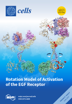
Ligand binding rearranges conformation of the extracellular domains of an EGFR dimer from tethered to untethered. This rearrangement induces a rotation/twist of the transmembrane domain of the receptor parallel to the plane of the cell membrane, resulting in the reorientation of the intracellular kinase domain dimer from a symmetric inactive configuration (left) to an asymmetric active form (right). Oncogenic mutations, shown by red balls, may also induce the asymmetric active form of the kinase dimer without ligand binding. View this paper
  • Issues are regarded as officially published after their release is announced to the table of contents alert mailing list.
  • You may sign up for e-mail alerts to receive table of contents of newly released issues.
  • PDF is the official format for papers published in both, html and pdf forms. To view the papers in pdf format, click on the "PDF Full-text" link, and use the free Adobe Reader to open them.
Order results
Result details
Section
Select all
Export citation of selected articles as:
937 KiB  
Review
Telomere Biology—Insights into an Intriguing Phenomenon
by Shriram Venkatesan, Aik Kia Khaw and Manoor Prakash Hande
Cells 2017, 6(2), 15; https://doi.org/10.3390/cells6020015 - 19 Jun 2017
Cited by 19 | Viewed by 6639
Abstract
Bacteria and viruses possess circular DNA, whereas eukaryotes with typically very large DNA molecules have had to evolve into linear chromosomes to circumvent the problem of supercoiling circular DNA of that size. Consequently, such organisms possess telomeres to cap chromosome ends. Telomeres are [...] Read more.
Bacteria and viruses possess circular DNA, whereas eukaryotes with typically very large DNA molecules have had to evolve into linear chromosomes to circumvent the problem of supercoiling circular DNA of that size. Consequently, such organisms possess telomeres to cap chromosome ends. Telomeres are essentially tandem repeats of any DNA sequence that are present at the ends of chromosomes. Their biology has been an enigmatic one, involving various molecules interacting dynamically in an evolutionarily well-trimmed fashion. Telomeres range from canonical hexameric repeats in most eukaryotes to unimaginably random retrotransposons, which attach to chromosome ends and reverse-transcribe to DNA in some plants and insects. Telomeres invariably associate with specialised protein complexes that envelop it, also regulating access of the ends to legitimate enzymes involved in telomere metabolism. They also transcribe into repetitive RNA which also seems to be playing significant roles in telomere maintenance. Telomeres thus form the intersection of DNA, protein, and RNA molecules acting in concert to maintain chromosome integrity. Telomere biology is emerging to appear ever more complex than previously envisaged, with the continual discovery of more molecules and interplays at the telomeres. This review also includes a section dedicated to the history of telomere biology, and intends to target the scientific audience new to the field by rendering an understanding of the phenomenon of chromosome end protection at large, with more emphasis on the biology of human telomeres. The review provides an update on the field and mentions the questions that need to be addressed. Full article
(This article belongs to the Special Issue DNA Repair Defects and Telomere Dysfunction in Diseases)
Show Figures

Figure 1

257 KiB  
Review
Methods for Measuring Autophagy in Mice
by Manon Moulis and Cécile Vindis
Cells 2017, 6(2), 14; https://doi.org/10.3390/cells6020014 - 08 Jun 2017
Cited by 49 | Viewed by 7908
Abstract
Autophagy is a dynamic intracellular process that mediates the degradation of damaged cytoplasmic components by the lysosome. This process plays important roles in maintaining normal cellular homeostasis and energy balance. Measuring autophagy activity is critical and although the determination of autophagic flux in [...] Read more.
Autophagy is a dynamic intracellular process that mediates the degradation of damaged cytoplasmic components by the lysosome. This process plays important roles in maintaining normal cellular homeostasis and energy balance. Measuring autophagy activity is critical and although the determination of autophagic flux in isolated cells is well documented, there is a need to have reliable and quantitative assays to evaluate autophagy in whole organisms. Because mouse models have been precious in establishing the functional significance of autophagy under physiological or pathological conditions, we present in this chapter a compendium of the current available methods to measure autophagy in mice, and discuss their advantages and limitations. Full article
(This article belongs to the Special Issue Assays to Monitor Autophagy in Model Systems)
1510 KiB  
Review
Activation of the EGF Receptor by Ligand Binding and Oncogenic Mutations: The “Rotation Model”
by Endang R. Purba, Ei-ichiro Saita and Ichiro N. Maruyama
Cells 2017, 6(2), 13; https://doi.org/10.3390/cells6020013 - 02 Jun 2017
Cited by 114 | Viewed by 15021
Abstract
The epidermal growth factor receptor (EGFR) plays vital roles in cellular processes including cell proliferation, survival, motility, and differentiation. The dysregulated activation of the receptor is often implicated in human cancers. EGFR is synthesized as a single-pass transmembrane protein, which consists of an [...] Read more.
The epidermal growth factor receptor (EGFR) plays vital roles in cellular processes including cell proliferation, survival, motility, and differentiation. The dysregulated activation of the receptor is often implicated in human cancers. EGFR is synthesized as a single-pass transmembrane protein, which consists of an extracellular ligand-binding domain and an intracellular kinase domain separated by a single transmembrane domain. The receptor is activated by a variety of polypeptide ligands such as epidermal growth factor and transforming growth factor α. It has long been thought that EGFR is activated by ligand-induced dimerization of the receptor monomer, which brings intracellular kinase domains into close proximity for trans-autophosphorylation. An increasing number of diverse studies, however, demonstrate that EGFR is present as a pre-formed, yet inactive, dimer prior to ligand binding. Furthermore, recent progress in structural studies has provided insight into conformational changes during the activation of a pre-formed EGFR dimer. Upon ligand binding to the extracellular domain of EGFR, its transmembrane domains rotate or twist parallel to the plane of the cell membrane, resulting in the reorientation of the intracellular kinase domain dimer from a symmetric inactive configuration to an asymmetric active form (the “rotation model”). This model is also able to explain how oncogenic mutations activate the receptor in the absence of the ligand, without assuming that the mutations induce receptor dimerization. In this review, we discuss the mechanisms underlying the ligand-induced activation of the preformed EGFR dimer, as well as how oncogenic mutations constitutively activate the receptor dimer, based on the rotation model. Full article
(This article belongs to the Special Issue Receptor Tyrosine Kinases in Health and Disease)
Show Figures

Figure 1

1910 KiB  
Review
Major Tumor Suppressor and Oncogenic Non-Coding RNAs: Clinical Relevance in Lung Cancer
by Kentaro Inamura
Cells 2017, 6(2), 12; https://doi.org/10.3390/cells6020012 - 09 May 2017
Cited by 80 | Viewed by 11612
Abstract
Lung cancer is the leading cause of cancer deaths worldwide, yet there remains a lack of specific and sensitive tools for early diagnosis and targeted therapies. High-throughput sequencing techniques revealed that non-coding RNAs (ncRNAs), e.g., microRNAs and long ncRNAs (lncRNAs), represent more than [...] Read more.
Lung cancer is the leading cause of cancer deaths worldwide, yet there remains a lack of specific and sensitive tools for early diagnosis and targeted therapies. High-throughput sequencing techniques revealed that non-coding RNAs (ncRNAs), e.g., microRNAs and long ncRNAs (lncRNAs), represent more than 80% of the transcribed human genome. Emerging evidence suggests that microRNAs and lncRNAs regulate target genes and play an important role in biological processes and signaling pathways in malignancies, including lung cancer. In lung cancer, several tumor suppressor/oncogenic microRNAs and lncRNAs function as biomarkers for metastasis and prognosis, and thus may serve as therapeutic tools. In this review, recent work on microRNAs and lncRNAs is introduced and briefly summarized with a focus on potential biological and therapeutic applications. Full article
Show Figures

Figure 1

2238 KiB  
Review
Taking a Bad Turn: Compromised DNA Damage Response in Leukemia
by Nadine Nilles and Birthe Fahrenkrog
Cells 2017, 6(2), 11; https://doi.org/10.3390/cells6020011 - 04 May 2017
Cited by 15 | Viewed by 6635
Abstract
Genomic integrity is of outmost importance for the survival at the cellular and the organismal level and key to human health. To ensure the integrity of their DNA, cells have evolved maintenance programs collectively known as the DNA damage response. Particularly challenging for [...] Read more.
Genomic integrity is of outmost importance for the survival at the cellular and the organismal level and key to human health. To ensure the integrity of their DNA, cells have evolved maintenance programs collectively known as the DNA damage response. Particularly challenging for genome integrity are DNA double-strand breaks (DSB) and defects in their repair are often associated with human disease, including leukemia. Defective DSB repair may not only be disease-causing, but further contribute to poor treatment outcome and poor prognosis in leukemia. Here, we review current insight into altered DSB repair mechanisms identified in leukemia. While DSB repair is somewhat compromised in all leukemic subtypes, certain key players of DSB repair are particularly targeted: DNA-dependent protein kinase (DNA-PK) and Ku70/80 in the non-homologous end-joining pathway, as well as Rad51 and breast cancer 1/2 (BRCA1/2), key players in homologous recombination. Defects in leukemia-related DSB repair may not only arise from dysfunctional repair components, but also indirectly from mutations in key regulators of gene expression and/or chromatin structure, such as p53, the Kirsten ras oncogene (K-RAS), and isocitrate dehydrogenase 1 and 2 (IDH1/2). A detailed understanding of the basis for defective DNA damage response (DDR) mechanisms for each leukemia subtype may allow to further develop new treatment methods to improve treatment outcome and prognosis for patients. Full article
(This article belongs to the Special Issue DNA Repair Defects and Telomere Dysfunction in Diseases)
Show Figures

Figure 1

4932 KiB  
Article
Distinct Fiber Type Signature in Mouse Muscles Expressing a Mutant Lamin A Responsible for Congenital Muscular Dystrophy in a Patient
by Alice Barateau, Nathalie Vadrot, Onnik Agbulut, Patrick Vicart, Sabrina Batonnet-Pichon and Brigitte Buendia
Cells 2017, 6(2), 10; https://doi.org/10.3390/cells6020010 - 24 Apr 2017
Cited by 4 | Viewed by 6265
Abstract
Specific mutations in LMNA, which encodes nuclear intermediate filament proteins lamins A/C, affect skeletal muscle tissues. Early-onset LMNA myopathies reveal different alterations of muscle fibers, including fiber type disproportion or prominent dystrophic and/or inflammatory changes. Recently, we identified the p.R388P LMNA mutation [...] Read more.
Specific mutations in LMNA, which encodes nuclear intermediate filament proteins lamins A/C, affect skeletal muscle tissues. Early-onset LMNA myopathies reveal different alterations of muscle fibers, including fiber type disproportion or prominent dystrophic and/or inflammatory changes. Recently, we identified the p.R388P LMNA mutation as responsible for congenital muscular dystrophy (L-CMD) and lipodystrophy. Here, we asked whether viral-mediated expression of mutant lamin A in murine skeletal muscles would be a pertinent model to reveal specific muscle alterations. We found that the total amount and size of muscle fibers as well as the extent of either inflammation or muscle regeneration were similar to wildtype or mutant lamin A. In contrast, the amount of fast oxidative muscle fibers containing myosin heavy chain IIA was lower upon expression of mutant lamin A, in correlation with lower expression of genes encoding transcription factors MEF2C and MyoD. These data validate this in vivo model for highlighting distinct muscle phenotypes associated with different lamin contexts. Additionally, the data suggest that alteration of muscle fiber type identity may contribute to the mechanisms underlying physiopathology of L-CMD related to R388P mutant lamin A. Full article
(This article belongs to the Collection Lamins and Laminopathies)
Show Figures

Figure 1

16850 KiB  
Article
Microinjection of Antibodies Targeting the Lamin A/C Histone-Binding Site Blocks Mitotic Entry and Reveals Separate Chromatin Interactions with HP1, CenpB and PML
by Charles R. Dixon, Melpomeni Platani, Alexandr A. Makarov and Eric C. Schirmer
Cells 2017, 6(2), 9; https://doi.org/10.3390/cells6020009 - 25 Mar 2017
Cited by 8 | Viewed by 7117
Abstract
Lamins form a scaffold lining the nucleus that binds chromatin and contributes to spatial genome organization; however, due to the many other functions of lamins, studies knocking out or altering the lamin polymer cannot clearly distinguish between direct and indirect effects. To overcome [...] Read more.
Lamins form a scaffold lining the nucleus that binds chromatin and contributes to spatial genome organization; however, due to the many other functions of lamins, studies knocking out or altering the lamin polymer cannot clearly distinguish between direct and indirect effects. To overcome this obstacle, we specifically targeted the mapped histone-binding site of A/C lamins by microinjecting antibodies specific to this region predicting that this would make the genome more mobile. No increase in chromatin mobility was observed; however, interestingly, injected cells failed to go through mitosis, while control antibody-injected cells did. This effect was not due to crosslinking of the lamin polymer, as Fab fragments also blocked mitosis. The lack of genome mobility suggested other lamin-chromatin interactions. To determine what these might be, mini-lamin A constructs were expressed with or without the histone-binding site that assembled into independent intranuclear structures. HP1, CenpB and PML proteins accumulated at these structures for both constructs, indicating that other sites supporting chromatin interactions exist on lamin A. Together, these results indicate that lamin A-chromatin interactions are highly redundant and more diverse than generally acknowledged and highlight the importance of trying to experimentally separate their individual functions. Full article
(This article belongs to the Collection Lamins and Laminopathies)
Show Figures

Figure 1

Previous Issue
Next Issue
Back to TopTop