Article

Antibiotic Resistance Profiling and Genotyping of Vancomycin-Resistant Enterococci Collected from an Urban River Basin in the Provincial City of Miyazaki, Japan

1
Department of Environment and Resource Science, Interdisciplinary Graduate School of Agriculture and Engineering, University of Miyazaki, Gakuen Kibanadai-Nishi 1-1, Miyazaki 889-2192, Japan
2
Department of Bacteriology, Faculty of Medical Sciences, Kyushu University, Maidashi Higashi-ku 3-1-1, Fukuoka 812-8582, Japan
3
Department of Civil and Environmental Engineering, Faculty of Engineering, University of Miyazaki, Gakuen Kibanadai-Nishi 1-1, Miyazaki 889-2192, Japan
*
Author to whom correspondence should be addressed.
Water 2017, 9(2), 79; https://doi.org/10.3390/w9020079
Submission received: 6 November 2016 / Accepted: 19 January 2017 / Published: 30 January 2017
(This article belongs to the Special Issue Water Quality and Health)

Abstract

:
The distribution characteristics of vancomycin-resistant enterococci (VRE) and the resistance of enterococcus isolates to various antibiotics were investigated in Yae River, which flows through Miyazaki city, Japan. The prevalence of VRE among specimens collected from the urban river basin using mEI agar was 0.9% (2 of 226 enterococcal isolates). In the 333 enterococcal isolates obtained using mEI agar or vancomycin-supplemented mEI agar, the possession of the vancomycin-resistant genes (vanA, vanB, vanC1, and vanC2/C3) was examined using multiplex PCR analysis. Although VRE possessing vanA and vanB were not detected in any isolates, isolates possessing vanC2/C3 were detected at all sampling sites and on all days. All isolates (101 strains) possessing vanC2/C3 that were obtained on vancomycin-supplemented mEI agar were identified as E. casseliflavus and analyzed for genotypes using pulse-field gel electrophoresis (PFGE) analysis. These E. casseliflavus isolates revealed them to be genetically highly divergent strains, suggesting that many contamination sources were present in this study area. Many of the enterococcal isolates obtained were resistant to erythromycin, ciprofloxacin, and tetracycline; enterococci distributed in the studied urban river basin are resistant to universally applicable antibiotics. These results indicate that VRE carrying vanC2/C3 are distributed in Yae River, and the sources of VRE are scattered across the river basin.

1. Introduction

With the use of antimicrobials for infectious disease therapy and growth-promoting agents for livestock, antibiotic-resistant bacteria have emerged, and infections caused by such bacteria are a global problem [1]. Each year in the United States, a medically advanced nation, 2 million people acquire serious infections caused by bacteria that are resistant to one or more antimicrobials, and at least 23,000 people die each year as a direct result of these infections [2]. On surveillance of the clinical facilities of 30 European countries, a number of bacteria resistant to clinically important antibiotics, such as fluoroquinolones, third-generation cephalosporins, and aminoglycosides, were identified [3]. The presence of antimicrobial-resistant bacteria has been observed extensively in hospitals, clinical settings, fish farms, and livestock. However, antimicrobial-resistant bacteria have also been detected in soil, sewage, rivers, and coastal areas [4,5,6,7,8].
Enterococci are gram-positive bacteria that form a part of the natural flora in the intestinal tract of humans and animals [9]. Because of their ubiquity in the feces of warm-blooded animals and persistence in the environment, enterococci have been traditionally used as indicators of fecal pollution of recreational waters [10]. In a previous study, the Enterococcus genus consists of 35 recognized species, and some enterococci can infect humans [9]. Particularly, E. faecalis and E. faecium are the two species most frequently associated with a range of enterococcal diseases in clinical settings, being responsible for one-third of all nosocomial infections worldwide [11]. Enterococci have multiple intrinsic mechanisms that confer resistance to different antimicrobials such as penicillin, monobactam, and low levels of aminoglycosides [12]. However, with the increased use of glycopeptide antibiotics, mainly vancomycin, in medical institutions, vancomycin-resistant enterococci (VRE) have arisen. Vancomycin is used for the treatment of serious infections caused by gram-positive bacteria such as methicillin-resistant Staphylococcus aureus and enterococci when treatment with other antibiotics has failed. VRE have been detected in regions such as Europe, US, and South Asia [11,13,14,15]. Although the occurrence of VRE in Japan is much lower in comparison with that in these regions, expansion of VRE nosocomial infections is considered a public health concern [16]. With the emergence of VRE, these enterococci can be present in a same area, and it is possible that the bacteria including enterococci are capable of sharing antibiotic resistance genes among members of the microbial communities that exist in the natural environment, such as soil and water [17].
There is little information concerning the presence of VRE and enterococci resistant to various antibiotics in aquatic environments, although a survey result has been reported. In the US, where VRE are a major cause of nosocomial infections, the percentage of VRE was 3% of all enterococci isolated from municipal wastewaters [18]. VRE have also been detected in soil and rivers in Europe [19]. In Japan, although VRE have been detected in sewage and rivers, their prevalence appears lower than in US and European countries [20]. However, there are few data concerning the surveillance of antimicrobial resistance for enterococci in aquatic environments and little long-term monitoring data on the distribution of VRE in river basins in Japan. In particular, the distribution of VRE in river basins flowing through provincial cities has yet to be determined. When enterococci resistant to clinically important antimicrobials exist in water environments close to human communities, the risk of opportunistic infection by these bacteria increases both in medical institutions and in the community at large.
In the current study, the distribution of VRE and the resistances of enterococci to various antimicrobials were investigated in the basin of an urban river, Yae River, which flows through to the provincial city of Miyazaki, Japan. We further performed genotyping of the isolates identified as VRE and investigated the difference in the prevalence of VRE according to their genotypes and sampling sites in the river basin.

2. Materials and Methods
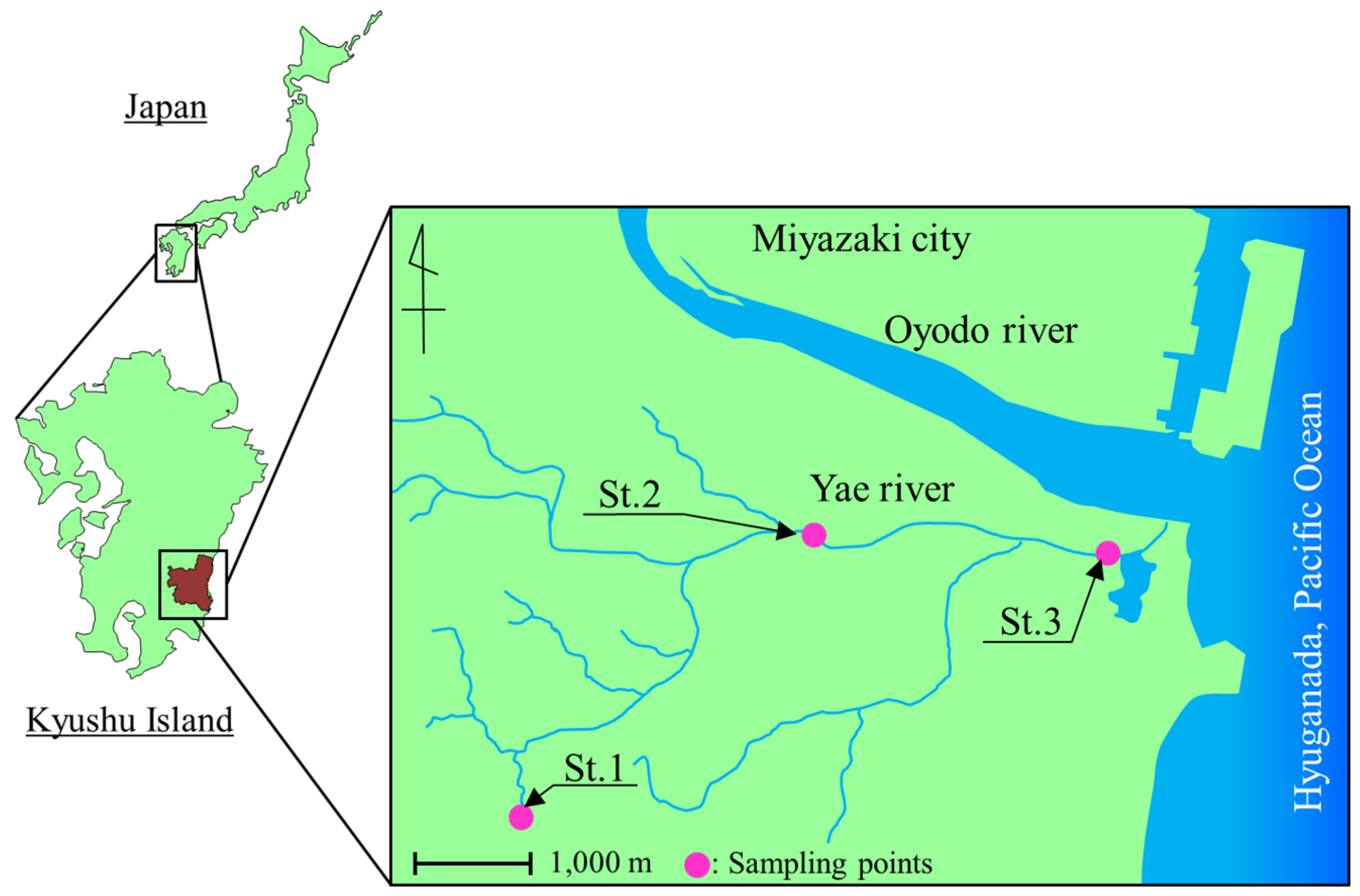
2.1. Sampling

River water samples were collected from Yae River, which flows through Miyazaki city (Figure 1). Miyazaki city has an approximate population of 400,000, and considered a provincial city in this study. Miyazaki city has a developed infrastructure with a sewer system and septic tanks which collected wastewater for treatment. The sampling location for Yae River is shown in Figure 1. St.1 is regions in the upper basin of Yae River and has a densely populated area, Kiyotake: approximate population of 29,000. Although the sewer system of Kiyotake has been completed, some districts and houses are not connected to the sewer system. Therefore, in the upper basin in Yae River, the percentage of domestic wastewater flow to the river is high. St.2 locates midstream in Yae River, which flows through an urban area and is surrounded by some marshy area. St.3 is located downstream of St.2 in an estuarine environment; it flows into the Hyuga-nada Sea, which is part of the Pacific Ocean in Japan. Sampling was performed a total of four times: December 2013 and May, July, and September 2014. Water samples were collected in sterile 1-L polyethylene bottles and transported to the laboratory for microbial and water quality analysis. Microbial analysis and water quality tests were started within 4 h of sampling. Turbidity, pH, and electrical conductivity were also determined using a turbidity meter (SEP-PT-706D; Mitsubishi Kagaku, Tokyo, Japan), a pH meter (HM-30G; TOA DKK, Tokyo, Japan), and a conductivity meter (CM30S; TOA DKK, Tokyo, Japan), respectively.

2.2. Enumeration and Isolation of Bacteria

River water samples were analyzed for total coliform, Escherichia coli, and enterococci. Total coliform and E. coli were tested by a Colilert®-18 kit (IDEXX Laboratories, Westbrook, ME, USA), according to the manufacturer’s instruction. In brief, 1 or 10 mL of each river water samples were diluted with sterile distilled water (total volume: 100 mL), and the Colilert®-18 kit reagents were added directly to one pack to a 100 mL sample in a sterile vessel. After shaking to dissolve reagents, the sample mixture was poured into a Quanti-Tray and incubated for 18 h at 37 ± 1.0 °C. This kit calculates as the most probable number (MPN) value on the basis of the presence or absence of fluorescence in Quanti-Tray.
Enterococci were enumerated on membrane-Enterococcus indoxyl-β-d-glucoside (mEI) agar plates, unmodified or supplemented with vancomycin using the membrane filtration method (MF) [21]. River water samples were filtered through a membrane filter (0.45-µm pore, 47-mm diameter, sterile, mixed cellulose ester; Advantec, Tokyo, Japan) and incubated on membrane-Enterococcus indoxyl-β-d-glucoside (mEI) agar plates for 24 h at 41 ± 1.0 °C. To screen for vancomycin-resistant strains in the river water, the mEI agar was supplemented with 4 μg·mL−1 vancomycin (Wako Pure Chemical Industries, Ltd., Osaka, Japan) [20]. After incubation, colonies on the filter that had blue halos were regarded as enterococci. The enterococcal count isolated by the MF method was expressed as CFU 100 mL−1 of water. The number of bacteria for each of the river water samples isolated using MF was determined from the mean CFU on three replicate mEI agar plates.
There were 60 single colonies randomly isolated from each of the conventional mEI agar (hereafter referred to as mEI agar) and vancomycin-supplemented mEI agar plates (referred to as VCM-mEI agar) and streaked on a Todd–Hewitt agar plate (TH agar, 1.5% agar; Bacto; Becton, Dickinson, NJ, USA). In the case of <60 isolates being available, all single colonies were isolated. These plates were then incubated for 24 h at 37 °C.

2.3. Identification of Enterococci and Detection of Vancomycin-Resistant Genes by Polymerase Chain Reaction (PCR) Analysis

The identification of enterococcus species and detection of vancomycin-resistant genes were performed using multiplex PCR analysis [22]. Species identification was conducted for four species, E. faecalis, E. faecium, E. gallinarum, and E. casseliflavus. The presence of four vancomycin-resistant genes, vanA, vanB, vanC1, and vanC2/C3 was examined. Genomic DNA was extracted from a single colony on Todd–Hewitt agar plates using InstaGene Matrix (Bio-Rad Laboratories, Hercules, CA, USA) according to the manufacturer’s instructions. The enzyme KAPA Taq Extra (Kapa Biosystems, Nippon Genetics Co, Ltd., Tokyo, Japan) was used for gene amplification. The PCR amplification program was as follows: initial denaturation at 94 °C for 5 min, 30 amplification cycles of denaturation at 94 °C for 1 min, annealing at 54 °C for 1 min, and elongation at 72 °C for 1 min, and final extension at 72 °C for 10 min. PCR products were confirmed by electrophoretic analysis of 5 μL of the reaction mixture on a 1.5% agarose gel. A 100-bp DNA ladder (Takara, Otsu, Japan) was used as the molecular size marker. E. faecalis NBRC100481, E. faecium NBRC 100486, E. gallinarum NBRC 100478, and E. casseliflavus NBRC 100675 were used as positive controls in all PCR experiments.
Subsequently, the isolates containing vancomycin-resistant genes from strains grown on VCM-mEI agar were identified using 16S rRNA gene sequence analysis according to a previously described method [20].

2.4. Determination of Minimum Inhibitory Concentration (MIC)

The MIC against each antibiotic was determined on Mueller Hinton agar using the agar dilution method according to the Clinical and Laboratory Standards Institute (CLSI) guidelines [23]. The enterococcal isolates were cultured for 24 h in Mueller Hinton broth (Becton Dickinson, Spark, MD, USA) and then diluted to a final concentration of approximately 1 × 10⁵ CFU·mL−1 with fresh Mueller Hinton broth. Inocula were then applied to the surface of Mueller Hinton agar (1.7% agar) plates containing various concentrations (0.25–128 μg·mL−1) of each antibiotic, which were prepared using microplates (Sakuma Co., Tokyo, Japan). The plates were incubated at 37 °C for 20 h, and MICs were determined. MIC breakpoints for intermediate and complete resistance were based on CLSI criteria [24].
The antibiotics used in the current study included penicillin G (PCG, Wako Pure Chemical Industries), ampicillin (ABPC, Wako Pure Chemical Industries, Ltd., Osaka, Japan), chloramphenicol (CP, Sigma-Aldrich, St. Louis, MO, USA), ciprofloxacin (CPFX, LKT Laboratories, Inc., St. Paul, MN, USA), erythromycin (EM, Wako Pure Chemical Industries), high-level gentamycin (GEM, 500 μg·mL−1, Wako Pure Chemical Industries), high-level streptomycin (STM, 2,000 μg·mL−1, Wako Pure Chemical Industries), imipenem (IPM, LKT Laboratories), tetracycline (TC, Wako Pure Chemical Industries), and vancomycin (VCM). All of the tested agents were dissolved in distilled water or other appropriate solvents according to the recommendations of the CLSI. A reference strain of E. faecalis ATCC 29212 was used as a quality control.

2.5. Pulse-Field Gel Electrophoresis (PFGE) Typing

To elucidate the genetic relationships among the Enterococcus strains isolated using VCM-mEI agar plates, PFGE was performed using a CHEF Bacterial genomic DNA plug kit (Bio-Rad) according to the manufacturer’s protocol with slight modifications [24]. In brief, each strain was cultivated on TH agar for 24 h at 37 °C, and 8–10 colonies were suspended in 1.0 mL of sterilized physiological saline in a 1.5-mL microcentrifuge tube. The tube was centrifuged for 3 min at 13,523 ×g (centrifuge 5424; Eppendorf, Hamburg, Germany), and the supernatant was discarded. The pellet was resuspended in 150 μL of cell suspension buffer. The suspension was mixed with 3.0 μL of lysozyme (25 mg·mL−1; Sigma-Aldrich, St. Louis, MO, USA), 3.0 μL of lysostaphin (2.0 mg·mL−1; Wako Pure Chemical Industries), and 3.0 μL of mutanolysin (≥4000 U·mg−1; Sigma-Aldrich, St. Louis, MO, USA), followed by incubation for 10 min at 37 °C. Next, a 100 μL suspension was mixed with an equal volume of liquid 2% CleanCut agarose and poured into plug moulds. The sample plugs were then incubated for 4 h at 37 °C in 500 μL of lysozyme buffer containing 10 μL each of the lysozyme, lysostaphin, and mutanolysin solutions. The plug washed with 1× wash buffer was incubated in 1 mL of proteinase K buffer (containing 1 mg proteinase K·mL−1; Sigma-Aldrich) for 24 h at 50 °C. After incubation, the buffer was removed from the microcentrifuge tube, and the plug was washed five times using 1 mL 1× wash buffer for 8 h with rotation in a microtube rotator (MTR-103; As One, Osaka, Japan). The DNA embedded in each plug was digested with the restriction enzyme SmaI (25 U·plug−1; Takara, Otsu, Japan) in 300 μL of the SmaI buffer for 20 h at 25 °C after treatment with 1 mL of 10-fold-diluted SmaI wash buffer.
DNA fragments were separated for 20 h at 14 °C on 1% pulse-field certified agarose gel (Bio-Rad) in the 0.5× Tris/Borate/EDTA buffer, with a switch ramp time from 5.3 to 34.9 s at a 120° angle, using a CHEF DRII system (Bio-Rad Laboratories). The sizing ladder used for PFGE was a lambda DNA ladder with a range of 48.5 kb–1.0 MB (Lonza, Rockland, ME, USA).

2.6. Dendrogram Analysis of PFGE Patterns

Dendrogram analysis of band-based PFGE patterns was performed using a gene profiler (Scanalytics, Buckinghamshire, UK). Levels of similarity between fingerprints were expressed as Dice coefficients, which were calculated by determining the ratio of twice the number of bands shared by two patterns to the total number of bands in both patterns. PFGE patterns were clustered using the unweighted pair group method with arithmetic means [25]. In the dendrogram analysis of PFGE fingerprints, isolates that belonged to the same cluster with a 0.8 similarity level were considered as genetically related strains (strains showing >80% similarity were considered as being genetically related).

3. Results

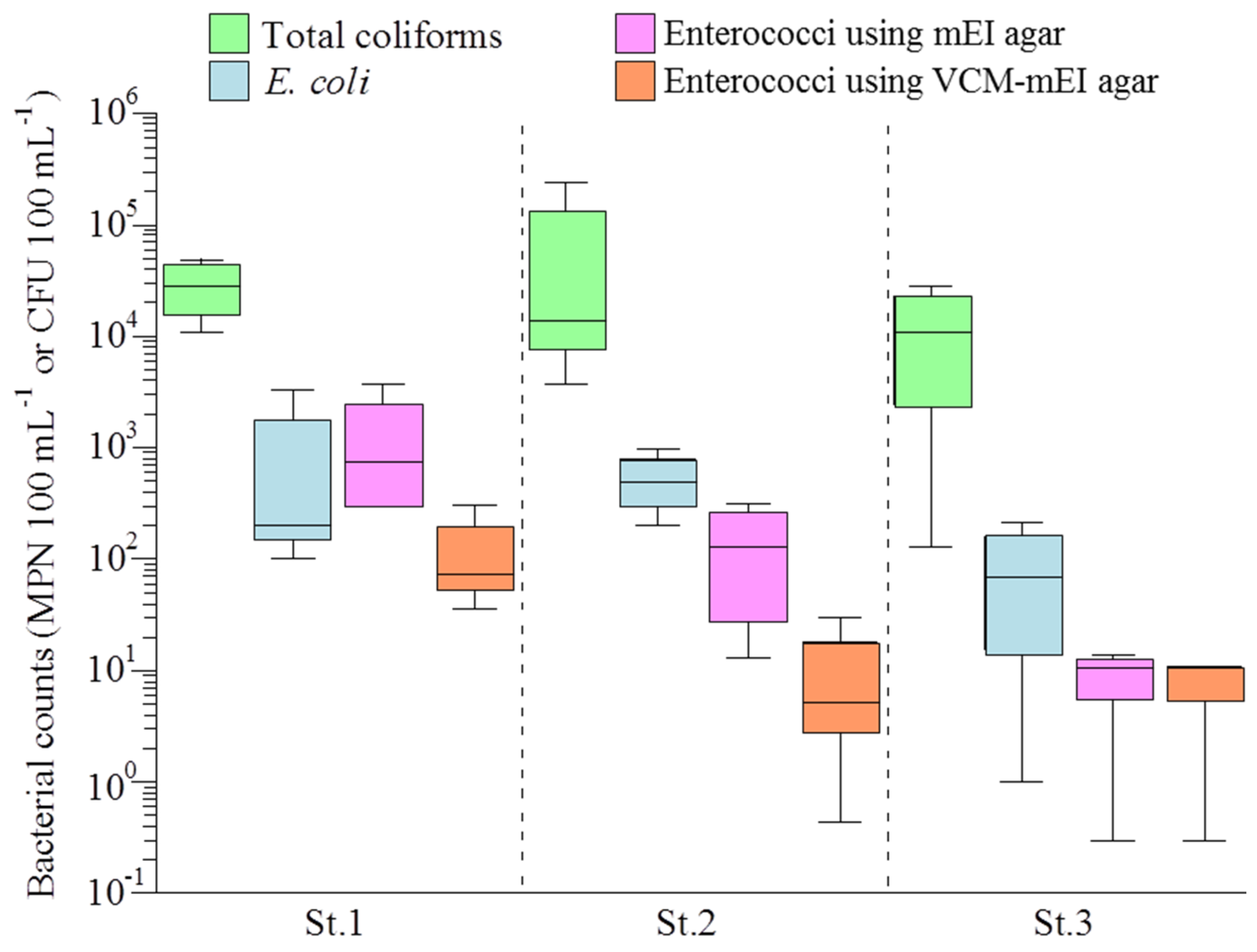
3.1. Bacterial Counts and Water Quality of the Yae River Basin

The bacterial counts at three sampling points (St.1–St.3) in the Yae River basin are shown in Figure 2. The water quality parameters are shown in Table S1. The number of total coliforms was detected at a concentration range of 1.2 × 10² to 2.4 × 10⁵ MPN 100 mL−1. The average counts of total coliforms and enterococci were the highest at St.1, 3.0 × 10⁵ MPN 100 mL−1 and 1.4 × 10³ CFU 100 mL−1, respectively. The median densities of total coliforms were greater at St.1 than at St.2 and St.3. Both fecal indicator bacteria tended to be more abundant at St.1 than at St.2 and St.3. It was assumed that because St.1 is located in the upper basin of Yae River, an area where a greater percentage of properties are not connected to the sewer system, the count of each fecal indicator bacterium was increased by the inflow of septic tank-aerated water and domestic wastewater. The turbidity and TOC tended to be higher at St.1 (turbidity: 0.40–27.8 kaolin units, TOC: 1.94–7.70 mg-C L−1) than St.2 and St.3 (turbidity: 0.25–11.3 kaolin units, TOC: 1.19–4.32 mg-C L−1). At St.3, the values of electric conductivity were the highest (9.78–30.5 mS·cm−1) among the three sites (St.1: 0.51–0.69 mS·cm−1, St.2: 0.21–0.39 mS·cm−1) throughout the study period, suggesting that the bacterial counts were decreased by the dilution effect of river water due to the inflow of seawater and a lot of tributaries, or influence of sea salt. Noticeable differences were observed in the number of total coliforms, E. coli, and enterococci in each sampling period.

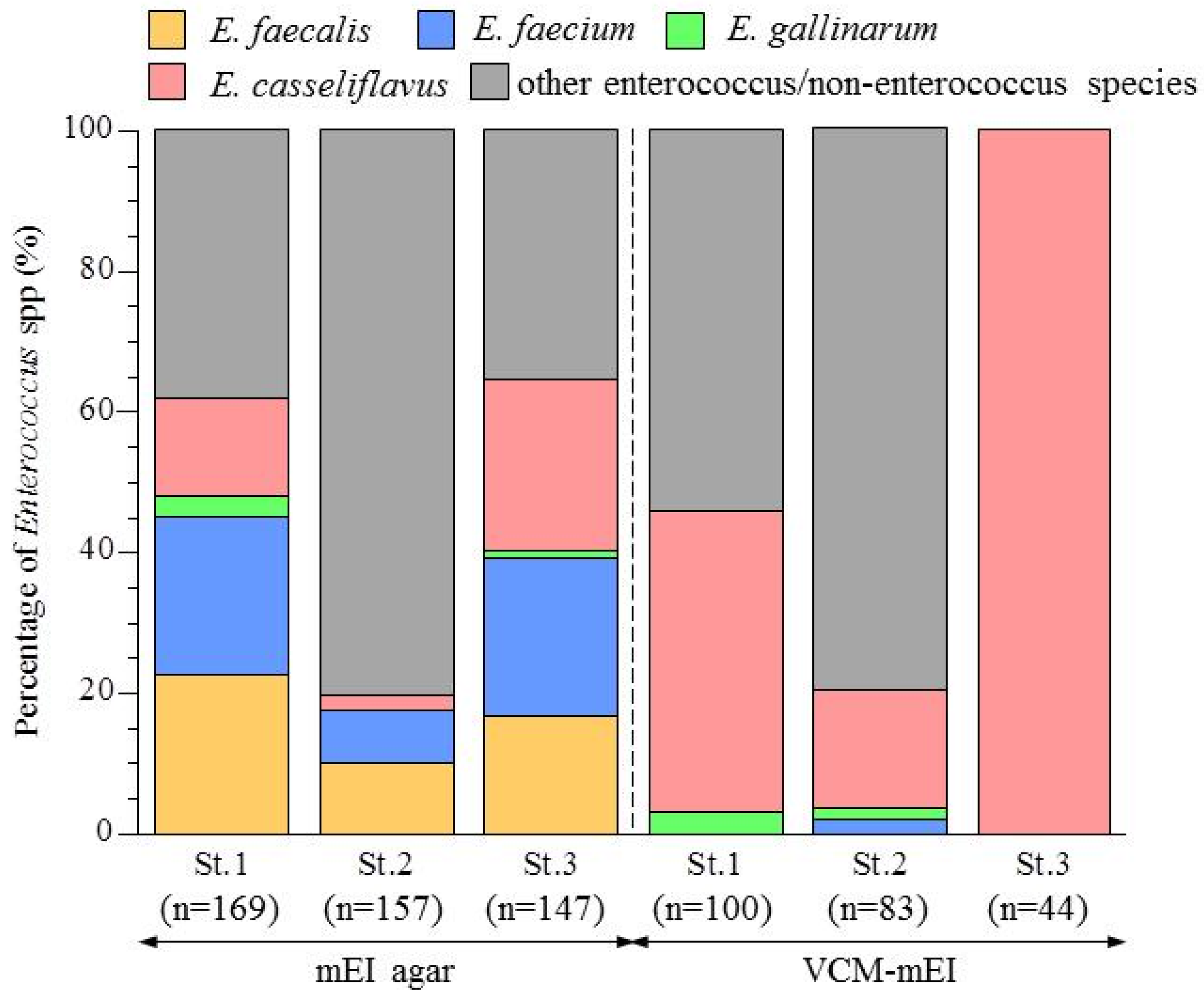
3.2. Abundance of Enterococcus Species in the Yae River Basin

All presumptive enterococci isolated from each sampling site using the mEI agar and VCM-mEI agar plates were subjected to species identification using PCR analysis. Proportions of confirmed enterococcus species among the isolated strains are shown in Figure 3.
The 473 isolates obtained using the mEI agar plates were identified as E. faecalis (78 isolates, 16.5%), E. faecium (79 isolates, 16.7%), E. gallinarum (6 isolates, 1.3%), E. casseliflavus (63 isolates, 13.3%), or “other enterococcus/non-enterococcus species” (247 isolates, 52.2%). Notably, 80.3% of the strains isolated from St.2 were “other enterococcus/non-enterococcus species”, which was remarkably different from the findings at St.1 and St.3, where these species occupied 40.8% and 35.4%, respectively.
By contrast, the 227 isolates obtained using the VCM-mEI agar plates were identified as E. faecalis (two isolates, 0.88%), E. gallinarum (four isolates, 1.8%), E. casseliflavus (101 isolates, 44%), and other enterococcus/non-enterococcus species (120 isolates, 53%); thus, most of the confirmed enterococcus strains were E. casseliflavus. Notably, however, the proportions of E. casseliflavus as well as those of “other enterococcus/non-enterococcus species” differed clearly between the three sampling sites. At St.1 and St.2, 50% and 80% of the strains were “other enterococcus/non-enterococcus species”, respectively. All strains isolated at St.3 were E. casseliflavus while a smaller number of enterococci were isolated at St.3 than other sampling sites because enterococcal concentrations were lowest (0.3–11 CFU 100 mL−1).

3.3. Antimicrobial Susceptibility of Enterococcal Isolates from the Yae River Basin

Among the 700 strains isolated using mEI agar or VCM-mEI agar, 47.6% (333 isolates) were Enterococcus, as described above. These confirmed enterococcal strains were examined for their MICs against ten antibiotics, representing eight major clinically important groups of antibiotics. Table 1 shows the numbers and percentages of the isolates collected from mEI agar plate at each sampling point classified as susceptible, intermediately resistant, and resistant to each antibiotic. Among the 226 enterococcal isolates, only two strains isolated at St.2 exhibited intermediate resistance to VCM. The percentages of enterococcal isolates resistant or intermediately resistant to EM, CPFX, and TC among all isolates were 54% (122 isolates), 53% (120 isolates), and 14% (31 isolates), respectively. Notably, in a comparison among the three sampling stations, MIC90 of TC was 32 μg·mL−1 at downstream (St.3), which were four- and two-fold larger than that at upstream (St.1) and midstream (St.2), respectively.
The results of MIC tests for the 107 enterococcal isolates obtained using the VCM-mEI agar plates are shown in Table 2. Among these, 11 isolates exhibited intermediate resistance to VCM, ten of which were isolated from the upper river basin (St.1). Compared to the isolates obtained using the mEI agar plates (Table 1), apparently higher proportions of isolates were resistant or intermediately resistant to EM (85 isolates, 80%) and CPFX (68 isolates, 64%), while those resistant or intermediately resistant to TC (ten isolates, 9%) were in a similar range using VCM-mEI agar plates.
The percentages of isolates resistant (either resistant or intermediately resistant) to each antibiotic among each of the four Enterococcus species were summarized in Table 3. VCM-resistant isolates included E. casseliflavus (nine isolates), E. gallinarum (three isolates), and E. faecium (one isolate). TC- and CPFX-resistant strains were found in all four species, while EM-resistant strains were found in E. faecalis, E. faecium, and E. casseliflavus but not in E. gallinarum. Most notably, as much as 82% and 81% of E. faecium strains were EM- and CPFX-resistant, respectively, and most of the E. faecium strains (50 out of the 79 strains) were resistant to both antibiotics. One E. faecium isolate was resistant to PCG and this strain was resistant to EM, TC, and CPFX as well (see the next paragraph). Another notable finding may be the higher percentage of E. gallinarum isolates resistant to TC; 60% in E. gallinarum and 6.1%–23% in the other three enterococcus species.

3.4. Determination of Vancomycin-Resistant Genes

In the 333 enterococcal isolates obtained using mEI agar or Van-mEI agar, the possession of the vancomycin-resistant genes (vanA, vanB, vanC1, and vanC2/C3) was examined using PCR analysis (Table 4). Although vanA and vanB were not detected in any isolates, vanC was detected in approximately half of the isolates; 3% (10 isolates) and 49% (164 isolates) of the isolates carried vanC1 and vanC2/C3, respectively, and isolates possessing vanC2/C3 were detected at all sampling sites and sampling days. In particular, at St.3 in September 2014, all 70 isolates were vanC2/C3-positive. Isolates possessing vanC1 were detected at St.1 (eight isolates) and St.2 (one isolate) but not detected at St.3.
Among the enterococcal isolates possessing vanC1 or vanC2/C3, vancomycin-resistance (resistant or intermediately resistant) was observed in 30% (3/10 isolates) and 5.5% (9/164 isolates) of the isolates, respectively. Therefore, isolates possessing vanC1 were less frequently detected than those possessing vanC2/C3, but the percentage of VCM-resistant isolates was high (MIC 90: 8 μg·mL−1). On the contrary, enterococcal isolates carrying vanC2/C3 were detected at all sampling sites, but the proportion of VCM-resistant isolates was low (MIC 90: 4 μg·mL−1). In one vancomycin-resistant E. faecalis isolate (MIC: 8 μg·mL−1), none of the four vancomycin-resistant genes was detected, suggesting that it may contain other vancomycin-resistant genes such as vanE and vanG [26] or variant.

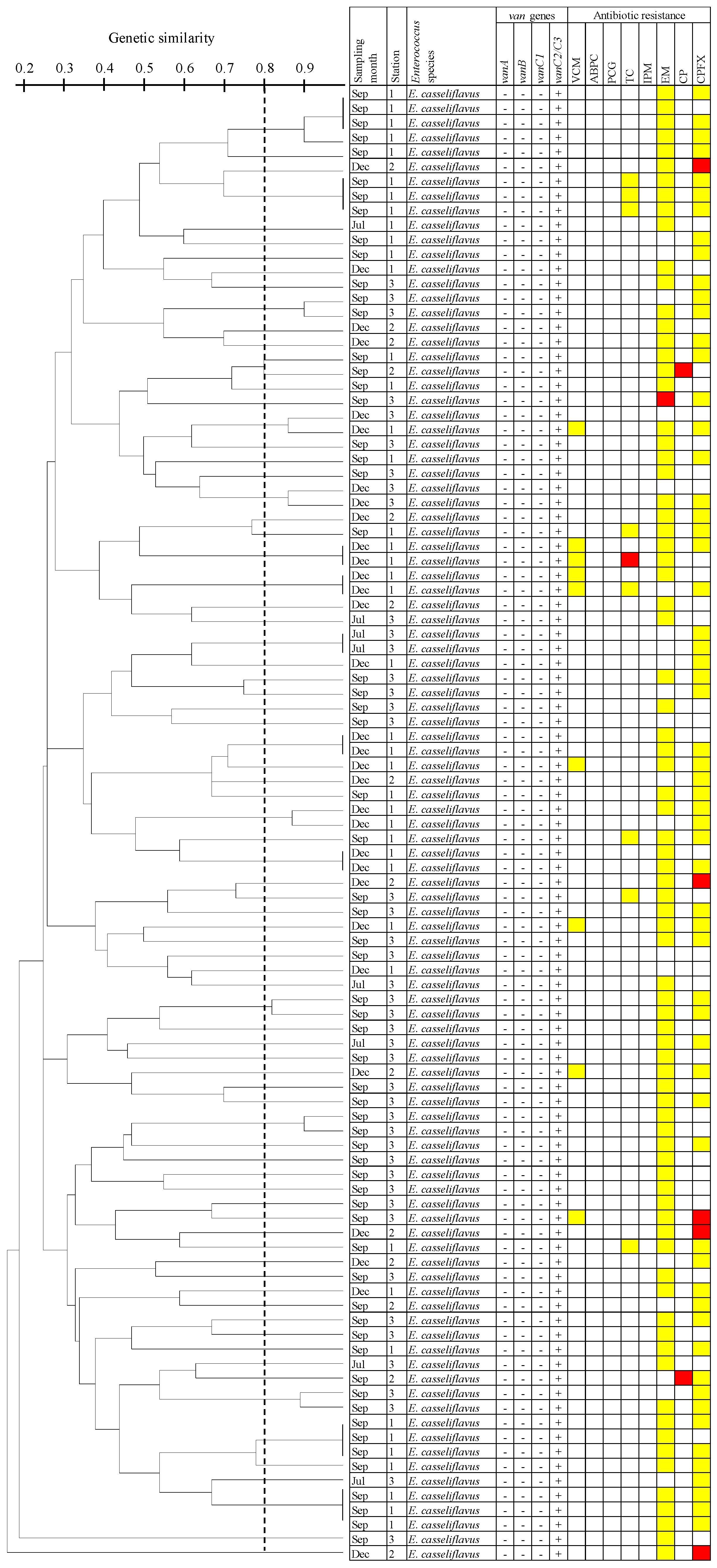
3.5. Dendrogram Analysis of E. Casseliflavus Isolates Carrying the vanC2/C3 Gene Using PFGE

Strains possessing vanC2/C3 isolated from Van-mEI agar (except strains isolated from conventional mEI agar) were identified using 16S rRNA gene sequence analysis. All 101 isolates possessing vanC2/C3were identified as E. casseliflavus. All of the 101 E. casseliflavus isolates possessing vanC2/C3 were analyzed by PFGE using SmaI, and the result of their dendrogram analysis is shown in Figure 4 along with the antibiotic resistance profiles of each isolate (Figure 4). By this analysis, 88 PFGE types were detected, indicating the significant diversity of vanC2/C3-carrying E. casseliflavus isolates. For example, 34 isolates obtained at three sampling sites in September 2014 were separated into distinct 34 PFGE types. Several sets of strains having the same PFGE type were detected, and these strains were isolated at the same sampling site on the same sampling day. Importantly, however, two sets of strains that are closely related (defined as showing a similarity level of >0.8) were identified—strains isolated at St.1 and St.3 in December 2013 and those isolated at St.1 and St.2 in September 2014.

4. Discussion

In this study, we investigated the distribution of VRE and the resistance of enterococci to various antibiotics and revealed that enterococci were distributed throughout the Yae River basin. Enterococci are used as indicators of fecal contamination in aquatic environments. As indicators of fecal contamination, they have many advantages, including resistance to multiple environmental factors, such as sodium chloride concentration, alkaline, increased temperature and pH, in comparison to other fecal indicator bacteria [27]. In particular, long-term persistence and non-growth in water environments are extremely important elements for properly evaluating fecal contamination [28]. In addition, long-term survival of enterococci in the water environment may enable the efficient transmission of their antibiotic-resistant genes to other bacteria. Therefore, information on the distribution of VRE and enterococci resistance to clinical antimicrobials in aquatic environment is important and these data may further support identifying the hypothesized transmission route for antibiotic-resistant bacteria and reveal bacterial dynamics in water environments.
The number of enterococci upstream (St.1) was the highest among the sampling sites. Because St.1 is located in the upper basin of the Yae River where there is no centralized sewer system connected to households, enterococci and E. coli counts had increased by the inflow of septic tank-aerated water. The enterococci counts tended to decrease at St.2 and St.3 rather than St.1, suggesting that the number of enterococci were decreased by the dilution effect of river water due to the inflow of seawater and a lot of tributaries, or influence of salt. In the Miyazaki city study area, E. faecalis and E. faecium were previously detected in river, estuarine, and coastal areas [29], suggesting that enterococci are ubiquitously present in the water environments of Miyazaki, a provincial seaside city. This study confirmed these findings.
VCM-resistant enterococcal isolates (MIC > 32 μg·mL−1) were not detected in Yae River. Among enterococcal isolates obtained using mEI agar, the percentage of isolates with intermediate VCM resistance was only 0.9% (2/226 isolates). Even among the isolates obtained using Van-mEI agar, only 10% (11/107 isolates) were intermediately VCM resistant. In our previous study, VCM-resistant enterococci from sewage and river water were confirmed using mEI agar supplemented with the same concentration (4 μg·mL−1) of VCM [20], and found only 12% (sewage) or 24% (river water) of isolates from the agar showed resistance to VCM. To screen for VCM-resistant enterococci in water samples more efficiently, it is necessary to increase the VCM concentration. Another screening method, the membrane filtration method with the Slanetz and Bartley agar containing 16 μg·mL−1 of VCM, has been used to investigate VCM-resistant enterococci in treated effluent of wastewater treatment plants and in surface water in the Netherlands [30]. However, agar supplemented with VCM at an excessively high concentration (32 μg·mL−1) allows the growth of Pediococcus spp., Leuconostoc spp. and Lactobacillus spp., which are intrinsically resistant to glycopeptides [20,30]. These results suggested that an appropriate concentration of VCM for screening VCM-resistant enterococci in water environment is in the range of 4–16μg·mL−1.
VRE infections have become a serious problem in the US and Europe, and VRE have been detected in aquatic environments. It has been reported that VRE (MIC of VCM > 256 μg·mL−1) constituted 1.6% of enterococcal strains isolated from the rural drinking water supply in Ireland [31]. Using VCM-supplemented agar (VCM concentration 18 μg·mL−1), it was also reported that VRE represents 8% (18 of the 227 enterococcus strains) of isolates located in public beaches in Washington and California [32]. The results of this study suggest that the proportion of VRE in aquatic environments is lower in Japan than in other countries, which is consistent with the lower incidence of VRE infections in Japan than in the US and European countries. The low detection rates of VRE in Japan may be due to a shorter history of the use of avoparcin, a growth promoter for livestock, compared to other countries [33] and the use of lower dosages of VCM clinically than in the US [34]. In European countries, VRE are prevalent in the environment; they were detected in livestock and wastewater treatment plants [35,36]. Several studies indicated that the intensive use of avoparcin as a growth promoter in food animals, such as broilers and pigs has contributed to an increase in VRE [37,38]. Despite the ban on the use of the avoparcin for livestock in European countries since 1997, VRE are still being frequently detected in broilers [39]. In an investigation of eight livestock farms in Greece, the prevalence of VRE carrying vanA in broilers was 14.4% [40]. VRE have also been detected during the wastewater treatment process and in hospital effluents in the US [41,42]. It is likely that the excessive use of glycopeptide antibiotics such as vancomycin in healthcare facilities has resulted in the selective increase in VRE in the human intestine.
All VRE isolates from Yae River possessed vanC. vanC-expressing VRE were detected in all sampling sites of the Yae River basin. Several reports on the presence of vanC-expressing VRE in river water and coastal areas have been published [43,44]. The predominant distribution of VRE carrying vanC1 and vanC2C/3 was also observed in three main rivers of Korea, with their concentrations ranging from 1 to 23 CFU 100 mL−1 [43]. In northern Greece, vanC2/C3 type VRE has also been isolated from coastal waters [44]. These results indicated that vanC type VRE may be widely distributed in aquatic environments, such as rivers and coastal areas. By contrast, VRE carrying vanA and vanB, which are the most important genotype regarding nosocomial infections [45], were not detected in Yae River. It has been reported that the isolates carrying vanA and vanB have been detected in rivers in Ireland and recreational beaches in the US [31,32]. The lower prevalence of VRE in the Yae River than in other countries, including US and Europe, may be related to the absence of strains carrying vanA and vanB in water environment.
Among the 473 strains isolated using mEI agar, 47.8% (226 isolates) were identified as the four tested Enterococcus species. The percentage of Enterococcus at St.1 and St.3 were 59.2% and 64.6%, respectively. Regarding the percentage of Enterococcus spp. in each sampling site, the percentage of other enterococcus/non-enterococcus species at St.2 was higher than those at St.1 and St.3. In a previous report, among the Enterococcus isolates collected from river and soil environments, E. hirae [46] and E. mundtii [47] were detected in addition to E. faecalis, E. faecium, E. casseliflavus, and E. gallinarum. The presence of E. casseliflavus, E. mundtii [48], and E. sulfureus [49] are reported to be associated with plant. At St.2, the bottom sediment of the river is muddy, while vegetation and habitats of fish and waterfowl were identified at St.1 and St.3. These facts assumed that the compositions of Enterococcus spp. at these sites may be affected by plants or animals. As expected, the ratios of Enterococcus spp. isolates obtained using VCM-mEI agar (VCM-supplemented media) greatly differed from those obtained using mEI agar (non-supplemented media). The ratio of E. casseliflavus was significantly higher in the enterococcal isolates obtained using VCM-mEI agar; 44% of the isolates were E. casseliflavus. This is consistent with the fact that E. casseliflavus has a low level of intrinsic resistance to glycopeptides, which is thought to be attributable to the chromosomally encoded vanC2/C3 gene [45].
In fact, all isolates (101 strains isolated from VCM-mEI agar) carrying vanC2/C3 that were obtained in this study were E. casseliflavus. PFGE analysis of these E. casseliflavus isolates revealed they are genetically highly diverse strains yielding 88 PFGE types. All of the 101 E. casseliflavus isolates possessing vanC2/C3 obtained from the St.1 (43 isolates), St.2 (14 isolates), and St.3(44 isolates) produced 31, 14, and 43 PFGE types, respectively, indicating the St.2 and St.3 were more genetically diverse than St.1. In addition, all the isolates (34 isolates) collected at three sampling sites in September 2014 were separated into distinct PFGE types. Furukawa and Suzuki analyzed 155 E. faecium and 138 E. faecalis isolates collected from river water and recreational beach in Miyazaki, Japan by PFGE [25]. By the analysis, 63 and 93 PFGE types were detected, respectively, indicating significant diversity of Enterococcus isolates. Another important finding from this analysis may be that the closely related E. casseliflavus strains were obtained at different sampling sites on the same day. This pattern of bacterial spread may be at least partly attributable to the distribution of enterococci in the downstream region of the river basin where the sewer system has been fully developed. Notably, the isolates with identical genotypes, which were obtained at the same sampling site, often exhibited different antibiotic resistance profiles (Figure 4). This result highlights the highly variable antibiotic resistance profiles of enterococci, which may have resulted from gain or loss of antibiotic resistance genes. Such genetic changes may occur not only within hosts but also in the aquatic environment through the transfer of antibiotic-resistant genes, which are driven by various genetic elements, such as conjugative plasmids or transposons.
Finally, our data regarding the resistance of environmental enterococcal isolates against antimicrobials other than VCM may also be important. Many of the enterococcal isolates obtained in this study were resistant to EM and CPFX as well as to TC, albeit to a lesser extent. The high proportions of resistance to CPFX are expected because this agent is ineffective against enterococci; additionally, most clinical enterococcal isolates are resistant [50]. However, the high rates of TC and EM resistance are noteworthy. These antibiotics are widely used not only for humans but also on livestock and fish farms [51,52]. Such widespread use of these antibiotics is most likely linked to the frequent isolation of strains resistant to TC and EM. Similar high rates of resistance to these antibiotics have also been reported for enterococcal strains isolated from rivers in other countries [53,54]. It was revealed that enterococci distributed in the studied urban river basin in this study are resistant to universally applicable antibiotics.

5. Conclusions

This study investigated the distribution of vancomycin-resistant enterococci (VRE) in Yae River, which flows through Miyazaki city, a provincial city in Japan. Species compositions and antibiotic-resistant profiles of enterococcal isolates distributed in Yae River were also examined. The prevalence of VRE in this urban river was 0.9%, in line with previous findings indicating that Japan has a lower prevalence of VRE than other countries. Although VRE possessing vanA and vanB were not detected in any isolates, vanC was detected in approximately half of the isolates, and isolates possessing vanC2/C3 were detected at all sampling sites and sampling days. Among the 700 strains isolated using mEI agar or vancomycin (VCM)-membrane-Enterococcus indoxyl-β-d-glucoside (mEI) agar, 47.6% (333 isolates) were identified as four tested Enterococcus species; the dominant species was E. casseliflavus (164 isolates). All isolates (101 strains) possessing vanC2/C3 that were obtained on VCM-mEI agar were E. casseliflavus and were analyzed for genotypes using pulse-field gel electrophoresis (PFGE) analysis. Accordingly, these E. casseliflavus isolates revealed that they are genetically highly divergent strains and indicated that the sources of E. casseliflavus possessing vanC2/C3 are scattered throughout the Yae River basin. The resistance of environmental enterococcal isolates against antimicrobials other than VCM was evaluated. VRE and enterococci resistant to erythromycin (EM), ciprofloxacin (CPFX), and tetracycline (TC) from an urban river were detected in this study, and the risk of infection due to antibiotic-resistant bacteria in public water systems was identified. It is important to reduce the risk of infection caused by antibiotic-resistant bacteria thorough social infrastructure development in the river basin. More investigations on the presence of VRE in this aquatic environment should be gathered immediately.

Supplementary Materials

The following are available online at www.mdpi.com/2073-4441/9/2/79/s1. Table S1: Water quality of samples collected from the survey points.

Acknowledgments

This work was partly supported by JSPS KAKENHI Grant Number 26340085 from Grants-in-Aid for Scientific Research (C) to Y.S. and by JSPS KAKENHI Grant Number 15J11464 from JSPS Research Fellowships for Young Scientists to M.N. The authors would like to thank Enago (www.enago.jp) for the English language review

Author Contributions

Masateru Nishiyama: designed and performed the experiments, analyzed the data, wrote the paper and revised it. Yoshitoshi Oura: conceived the experiment, wrote the paper and revised it. Tetsuya Hayashi: conceived the experiment, wrote the paper and revised it. Yoshihiro Suzuki conceived and designed the experiment, wrote the paper and revised it.

Conflicts of Interest

The authors declare no conflict of interest.

References

  1. World Health Organization (WHO). Antimicrobial Resistance: Global Report on Surveillance 2014. Available online: http://www.who.int/drugresistance/documents/surveillancereport/en/ (accessed on 4 May 2014).
  2. Center for Disease Control and Prevention (CDC). Antibiotic Resistance Threats in the United States, 2013; Centers for Disease Control and Prevention: Atlanta, GA, USA, 2013.
  3. European Center for Disease Prevention and Control. Antimicrobial Resistance Surveillance in Europe 2013. European Antimicrobial Resistance Surveillance Network (EARS-Net). ECDC: Stockholm, 2014. Available online: http://ecdc.europa.eu/en/publications/Documents/antibiotic-resistance-in-EU-summary.pdf (accessed on 4 November 2015).
  4. Alm, E.W.; Zimbler, D.; Callahan, E.; Plomaritis, E. Patterns and persistence of antibiotic resistance in faecal indicator bacteria from freshwater recreational beaches. J. Appl. Microbiol. 2014, 117, 273–285. [Google Scholar] [CrossRef] [PubMed]
  5. Kumarasamy, K.K.; Toleman, M.A.; Walsh, T.R.; Bagaria, J.; Butt, F.; Balakrishnan, R.; Chaudhary, U.; Doumith, M.; Giske, C.G.; Irfan, S.; et al. Emergence of a new antibiotic resistance mechanism in India, Pakistan, and the UK: A molecular, biological, and epidemiological study. Lancet Infect. Dis. 2010, 10, 597–602. [Google Scholar] [CrossRef]
  6. Mesa, R.J.; Blanc, V.; Blanch, A.R.; Cortés, P.; González, J.J.; Lavilla, S.; Miró, E.; Muniesa, M.; Saco, M.; Tórtola, M.T.; et al. Extended-spectrum beta-lactamase-producing Enterobacteriaceae in different environments (humans, food, animal farms and sewage). J. Antimicrob. Chemother. 2006, 58, 211–215. [Google Scholar] [CrossRef] [PubMed]
  7. Suzuki, Y.; Kajii, S.; Nishiyama, M.; Iguchi, A. Susceptibility of Pseudomonas aeruginosa isolates collected from river water in Japan to antipseudomonal agents. Sci. Total. Environ. 2013, 450–451, 148–154. [Google Scholar] [CrossRef] [PubMed]
  8. Leonard, A.F.; Zhang, L.; Balfour, A.J.; Garside, R.; Gaze, W.H. Human recreational exposure to antibiotic resistant bacteria in coastal bating waters. Environ. Int. 2015, 82, 92–100. [Google Scholar] [CrossRef] [PubMed]
  9. Facklam, R.R.; Carvalho, M.S.; Teixeira, L.M. History, taxonomy, biochemical characteristics, and antibiotic susceptibility testing of enterococci. In The Enterococci: Pathogenesis, Molecular Biology, and Antibiotic Resistance; Gilmore, M.S., Ed.; ASM Press: Washington, DC, USA, 2002; pp. 1–54. [Google Scholar]
  10. U.S. Environmental Protection Agency. Ambient Water-Quality Criteria for Bacteria-1986; Office of Water Regulations and Standards, U.S. Environmental Protection Agency: Washington, DC, USA, 1986; Volume 17.
  11. Werner, G.; Coque, T.M.; Hammerum, A.M.; Hope, R.; Hryniewicz, W.; Johnson, A.; Klare, I.; Kristinsson, K.G.; Leclercq, R.; Lester, C.H.; et al. Emergence and spread of vancomycin resistance among enterococci in Europe. Eur. Surveill. 2008, 13, 19046. [Google Scholar]
  12. Huycke, M.M.; Sahm, D.F.; Gilmore, M.S. Multiple-drug resistant enterococci: The nature of the problem and an agenda for future. Emerg. Infect. Dis. 1998, 4, 239–249. [Google Scholar] [CrossRef] [PubMed]
  13. Moscoso, M.; Domenech, M.; García, E. Vancomycin tolerance in Gram-positive cocci. Environ. Microbiol. Rep. 2011, 3, 640–650. [Google Scholar] [CrossRef] [PubMed]
  14. Deshpande, L.M.; Fritsche, T.R.; Moet, G.J.; Biedenbach, D.J.; Jones, R.N. Antimicrobial resistance and molecular epidemiology of vancomycin-resistant enterococci from North American and Europe: A report from the SENTRY antimicrobial surveillance program. Diagn. Microbiol. Infect. Dis. 2007, 15, 838–839. [Google Scholar]
  15. Hsueh, P.R.; Chen, M.L.; Sun, C.C.; Chen, W.H.; Pan, H.J.; Yang, L.S.; Chang, S.C.; Ho, S.W.; Lee, C.Y.; Hsieh, W.C.; et al. Antimicrobial drug resistance in pathogens causing nosocomial infections at a university hospital in Taiwan, 1981–1999. Emerg. Infect. Dis. 2002, 8, 63–68. [Google Scholar] [CrossRef] [PubMed]
  16. Matsushima, A.; Takakura, S.; Yamamoto, M.; Matsumura, Y.; Shirano, M.; Nagano, M.; Ito, Y.; Iinuma, Y.; Shimizu, T.; Fujita, N.; et al. Regional spread and control of vancomycin-resistant Enterococcus faecium and Enterococcus faecalis in Kyoto, Japan. Eur. J. Clin. Microbiol. Infect. Dis. 2012, 31, 1095–1100. [Google Scholar] [CrossRef] [PubMed]
  17. Taylor, N.G.; Vemer-Jeffreys, D.W.; Baker-Austin, C. Aquatic systems: Maintaining, mixing and mobilizing antimicrobial resistance? Trends Ecol. Evol. 2011, 26, 278–284. [Google Scholar] [CrossRef] [PubMed]
  18. Rosenberg Goldstein, R.E.; Micallef, S.A.; Gibbs, S.G.; George, A.; Claye, E.; Sapkota, A.; Joseph, S.W.; Sapkota, A.R. Detection of vancomycin-resistant enterococci (VRE) at four U. S. wastewater treatment plants that provide effluent for reuse. Sci. Total Environ. 2014, 466–467, 404–411. [Google Scholar] [CrossRef] [PubMed]
  19. Iversen, A.; Kuhn, I.; Franklin, A.; Mollby, R. High prevalence of vancomycin-resistant enterococci in Swedish sewage. Appl. Environ. Microbiol. 2002, 68, 2838–2842. [Google Scholar] [CrossRef] [PubMed]
  20. Nishiyama, M.; Iguchi, A.; Suzuki, Y. Identification of Enterococcus faecium and Enterococcus faecalis as vanC-type vancomycin-resistant enterococci (VRE) from sewage and river water in the provincial city of Miyazaki, Japan. J. Environ. Sci. Health A 2015, 50, 16–25. [Google Scholar] [CrossRef] [PubMed]
  21. United States Environmental Protection Agency. Method 1600: Enterococci in Water by Membrane Filtration Using Membrane-Enterococcus Indoxyl-β-D-Glucoside Agar (mEI), EPA-821-R-02-022; U.S. Environmental Protection Agency: Washington, DC, USA, 2002.
  22. Kariyama, R.; Mitsuhata, R.; Chow, J.W.; Clewell, D.B.; Kumon, H. Simple and reliable multiplex PCR assay for surveillance isolates of vancomycin-resistant enterococci. J. Clin. Microbiol. 2000, 38, 3092–3095. [Google Scholar] [PubMed]
  23. Nagayama, A.; Yamaguchi, K.; Watanabe, K.; Tanaka, M.; Kobayashi, I.; Nagasawa, Z. Final report from the committee on antimicrobial susceptibility testing, Japanese Society of Chemotherapy, on the agar dilution method (2007). J. Infect. Chemother. 2008, 14, 383–392. [Google Scholar] [CrossRef] [PubMed]
  24. Clinical Laboratory Standards Institute. Performance Standards for Antimicrobial Susceptibility Testing. Twenty-second Informational Supplement, M100-S22. 2012, 27, 90–92. Available online: http://mazums.ac.ir/dorsapax/userfiles/file/moavenat%20darman/M100-S22.pdf (accessed on 26 January 2017). [Google Scholar]
  25. Furukawa, T.; Suzuki, Y. A proposal for source tracking of fecal pollution in recreational water by pulsed-field gel electrophoresis. Microbes Environ. 2013, 28, 444–449. [Google Scholar] [CrossRef] [PubMed]
  26. Gholizadeh, M.; Courvalin, P. Acquired and intrinsic glycopeptide resistance in enterococci. Int. J. Antimicrob. Agents 2000, 16, S11–S17. [Google Scholar] [CrossRef]
  27. Murray, B.E. The life and times of the Enterococcus. Clin. Microbiol. Rev. 1990, 3, 46–65. [Google Scholar] [CrossRef] [PubMed]
  28. Lleó, M.M.; Bonato, B.; Benedetti, D.; Canepari, P. Survival of enterococcal species in aquatic environments. FEMS Microbiol. Ecol. 2005, 54, 189–196. [Google Scholar] [CrossRef] [PubMed]
  29. Furukawa, T.; Yoshida, T.; Suzuki, Y. Application of PFGE to source tracking of fecal pollution in coastal recreation area: A case study in Aoshima Beach, Japan. J. Appl. Microbiol. 2011, 110, 688–696. [Google Scholar] [CrossRef] [PubMed]
  30. Taučer-Kapteijn, M.; Hoogenboezem, W.; Heiliegers, L.; de Bolster, D.; Medema, G. Screening municipal wastewater effluent and surface water used for drinking water production for the presence of ampicillin and vancomycin resistant enterococci. Int. J. Hyg. Environ. Health 2016, 219, 437–442. [Google Scholar] [CrossRef] [PubMed]
  31. Morris, D.; Galvin, S.; Boyle, F.; Hickey, P.; Mulligan, M.; Cormican, M. Enterococcus faecium of the vanA genotype in rural drinking water, effluent, and the aqueous environment. Appl. Environ. Microbiol. 2012, 78, 596–598. [Google Scholar] [CrossRef] [PubMed]
  32. Roberts, M.C.; Soge, O.O.; Giardino, M.A.; Mazengia, E.; Ma, G.; Meschke, J.S. Vancomycin-resistant Enterococcus spp. in marine environments from the West Coast of the USA. J. Appl. Microbiol. 2009, 107, 300–307. [Google Scholar] [CrossRef] [PubMed]
  33. Yoshimura, H.; Ishimaru, M.; Endoh, Y.S.; Suginaka, M.; Yamatani, S. Isolation of glycopeptide-resistant enterococci from chickens in Japan. Antimicrob. Agents Chemother. 1998, 42, 3333. [Google Scholar] [PubMed]
  34. Kirst, H.A.; Thompson, D.G.; Nicas, T.I. Historical yearly usage of vancomycin. Antimicrob. Agents Chemother 1998, 42, 1303–1304. [Google Scholar] [PubMed]
  35. Garcia-Migura, L.; Liebana, E.; Jensen, L.B.; Barnes, S.; Pleydell, E. A longitudinal study to assess the persistence of vancomycin-resistant Enterococcus faecium (VREF) on an intensive broiler farm in the United Kingdom. FEMS Microbiol. Lett. 2007, 275, 319–325. [Google Scholar] [CrossRef] [PubMed]
  36. Kotzamanidis, C.; Zdraags, A.; Kourelis, A.; Moraitou, E.; Papa, A.; Yiantzi, V.; Pantelidou, C.; Yiangou, M. Characterization of vanA-type Enterococcus faecium isolates from urban and hospital wastewater and pig. J. Appl. Microbiol. 2009, 107, 997–1005. [Google Scholar] [CrossRef] [PubMed]
  37. Bustamante, W.; Alpízar, A.; Hernández, S.; Pacheco, A.; Vargas, N.; Herrera, M.L.; Vargas, Á.; Caballero, M.; García, F. Predominance of vanA genotype among vancomycin-resistant Enterococcus isolates frim poultry and swine in Costa Rica. Appl. Environ. Microbiol. 2003, 69, 7414–7419. [Google Scholar] [CrossRef] [PubMed]
  38. Manson, J.M.; Smith, J.M.B.; Cook, G.M. Persistence of vancomycin resistant enterococci in New Zealand broilers after discontinuation of avoparcin use. Appl. Environ. Microbiol. 2004, 70, 5764–5768. [Google Scholar] [CrossRef] [PubMed]
  39. Bortolaia, V.; Mander, M.; Jensen, L.B.; Olsen, J.E.; Guardabassi, L. Persistence of vancomycin resistance in multiple clones of Enterococcus faecium isolated from Danish broilers 15 years after the bans of avoparcin. Antimicrob. Agents Chemother. 2015, 59, 2926–2929. [Google Scholar] [CrossRef] [PubMed]
  40. Tzavaras, I.; Siarkou, V.I.; Zdragas, A.; Kotzamanidis, C.; Vafeas, G.; Bourtzi-Hatzopoulou, E.; Poumaras, S.; Sofianou, D. Diversity of vanA-type vancomycin-resistant Enterococcus faecium isolated from broilers, poultry slaughterers and hospitalized humans in Greece. J. Antimicrob. Chemother. 2012, 67, 1811–1818. [Google Scholar] [CrossRef] [PubMed]
  41. Nagulapally, S.R.; Ahmad, A.; Henry, A.; Marchin, G.L.; Zurek, L.; Bhandari, A. Occurrence of ciprofloxacin-, trimethoprim-sulfamethoxazole-, and vancomycin-resistant bacteria in a municipal wastewater treatment plant. Water Environ. Res. 2009, 81, 82–90. [Google Scholar] [CrossRef] [PubMed]
  42. Varela, A.R.; Ferro, G.; Vredenburg, J.; Yanik, M.; Vieira, L.; Rizzo, L.; Lameiras, C.; Manaia, C.M. Vancomycin resistant enterococci: From the hospital effluent to the urban wastewater treatment plant. Sci. Total Environ. 2013, 450–451, 155–161. [Google Scholar] [CrossRef] [PubMed]
  43. Nam, S.; Kim, M.J.; Park, C.; Park, J.G.; Maeng, P.J.; Lee, G.C. Detection and genotyping of vancomycin-resistant Enterococcus spp. by multiplex polymerase chain reaction in Korean aquatic environmental samples. Int. J. Hyg. Environ. Health 2013, 216, 421–427. [Google Scholar] [CrossRef] [PubMed]
  44. Zdragas, A.; Partheniou, P.; Kotzamanidis, C.; Psoni, L.; Koutita, O.; Moraitou, E.; Tzanetakis, N.; Yiangou, M. Molecular characterization of low-level vancomycin-resistant enterococci found in coastal water of Thermaikos Gulf, Northern Greece. Water Res. 2008, 42, 1274–1280. [Google Scholar] [CrossRef] [PubMed]
  45. Kak, V.; Chow, J.W. Acquired antibiotic resistances in enterococci. In The Enterococci: Pathogenesis, Molecular Biology, and Antibiotic Resistance; Gilmore, M.S., Ed.; ASM Press: Washington, DC, USA, 2002; pp. 355–383. [Google Scholar]
  46. Łuczkiewicz, A.; Jankowska, K.; Kurlenda, J.; Olańczuk-Neyman, K. Identification and antimicrobial resistance of Enterococcus spp. isolated from surface water. Water Sci. Technol. 2010, 62, 466–473. [Google Scholar] [CrossRef] [PubMed]
  47. Collins, M.D.; Farrow, J.A.E.; Jones, D. Enterococcus mundtii sp. nov. Int. J. Syst. Bacteriol. 1986, 36, 8–12. [Google Scholar] [CrossRef]
  48. Ulrich, A.; Müller, T. Heterogeneity of plant associated streptococci as characterized by phenotypic features and restriction analysis of PCR amplified 16S rDNA. J. Appl. Microbiol. 1998, 84, 293–303. [Google Scholar] [CrossRef] [PubMed]
  49. Martinez-Murcia, A.J.; Collins, M.D. Enterococcus sulfureus, a new yellow pigmented Enterococcus species. FEMS Microbiol. Lett. 1991, 80, 69–74. [Google Scholar] [CrossRef]
  50. Courvalin, P. Vancomycin resistance in gram-positive cocci. Clin. Infect. Dis. 2006, 42, 25–34. [Google Scholar] [CrossRef] [PubMed]
  51. Beuckers, A.G.; Zaheer, R.; Cook, S.R.; Stanford, K.; Chaves, A.V.; Ward, M.P.; McAllister, T.A. Effect of in-feed administration and withdrawal of tylosin phosphate on antibiotic resistance in enterococci isolated from feedlot steers. Front. Microbiol. 2015, 6. [Google Scholar] [CrossRef] [PubMed]
  52. Harnisz, M.; Korzeniewska, E.; Gołaś, I. The impact of a freshwater fish warm on the community of tetracycline-resistant bacteria and the structure of tetracycline resistance genes in river water. Chemosphere 2015, 128, 134–141. [Google Scholar] [CrossRef] [PubMed]
  53. Munir, M.; Wong, K.; Xagoraraki, I. Release of antibiotic resistant bacteria and genes in the effluent and biosolids of five wastewater utilities in Michigan. Water Res. 2011, 45, 681–693. [Google Scholar] [CrossRef] [PubMed]
  54. Gao, P.; Mao, D.; Luo, Y.; Wang, L.; Xu, B.; Xu, L. Occurrence of sulphonamide and tetracycline-resistant bacteria and resistance genes in aquaculture environment. Water Res. 2012, 46, 2355–2364. [Google Scholar] [CrossRef] [PubMed]
Figure 1. The locations of sampling points in Yae River in Miyazaki city, Japan. St.1: upstream, St.2: midstream, and St.3: downstream.
Water 09 00079 g001
Figure 2. Bacterial counts at each sampling site during the study period. The green bar indicates the total coliform, the blue bar indicates the E. coli, the pink bar indicates the enterococci using mEI agar, and the orange bar indicated the enterococci using VCM-mEI agar. The symbols indicate the following: box, 25–75th percentile; horizontal line, median; and whiskers, minimum-maximum value.
Water 09 00079 g002
Figure 3. Diversity of Enterococcus species composition in Yae River.
Water 09 00079 g003
Figure 4. Dendrograms of pulsed-field gel electrophoresis types and antibiotic resistance profiles for E. casseliflavus isolates from each sampling site in Yae River. Concerning antibiotic resistance phenotypes, red indicates resistance, yellow indicates intermediate resistance, and white indicates susceptibility. VCM, Vancomycin; ABPC, Ampicillin; PCG, Penicillin G; TC, Tetracycline; IPM, Imipenem; EM, Erythromycin; CP, Chloramphenicol; CPFX, Ciprofloxacin; +, Positive; −, Negative.
Water 09 00079 g004
Table 1. Antibiotic susceptibilities of enterococci isolated in Yae River using mEI agar.
Antimicrobial agentMIC test range (μg·mL−1)St.1 (100 isolates)MIC50 a (μg·mL−1)MIC90 b (μg·mL−1)St.2 (31 isolates)MIC50 (μg·mL−1)MIC90 (μg·mL−1)St.3 (95 isolates)MIC50 (μg·mL−1)MIC90 (μg·mL−1)
SusceptibleIntermediateResistantSusceptibleIntermediateResistantSusceptibleIntermediateResistant
No. isolates (% isolates)No. isolates (% isolates)No. isolates (% isolates)
VCM0.25-256100 (100%)0 (0%)0 (0%)2429 (94%)2 (6%)0 (0%)1495 (100%)0 (0%)0 (0%)0.52
PCG0.25-128100 (100%)0 (0%)0 (0%)1230 (97%)0 (0%)1 (3%)2295 (100%)0 (0%)0 (0%)22
ABPC0.25-128100 (100%)0 (0%)0 (0%)0.5131 (100%)0 (0%)0 (0%)0.5195 (100%)0 (0%)0 (0%)0.51
IPM0.25-128100 (100%)0 (0%)0 (0%)1231 (100%)0 (0%)0 (0%)1295 (100%)0 (0%)0 (0%)24
EM0.25-12825 (25%)72 (72%)3 (3%)1214 (45%)16 (52%)1 (3%)1265 (68%)29 (31%)1 (1%)0.51
TC0.25-12888 (88%)3 (3%)9 (9%)2827 (87%)0 (0%)4 (13%)21680 (84%)1 (1%)14 (15%)232
CPFX0.0625-3222 (22%)64 (64%)14 (14%)2413 (42%)13 (42%)5 (16%)4871 (75%)20 (21%)4 (4%)0.52
CP0.25-128100 (100%)0 (0%)0 (0%)8831 (100%)0 (0%)0 (0%)8895 (100%)0 (0%)0 (0%)88
GEN500100 (100%)0 (0%)0 (0%)c 28 (100%)0 (0%)0 (0%) 95 (100%)0 (0%)0 (0%)
STM2,000100 (100%)0 (0%)0 (0%) 28 (100%)0 (0%)0 (0%) 95 (100%)0 (0%)0 (0%)
MIC: minimum inhibitory concentration; a MIC50: minimum concentration at which growth of 50% the isolates is inhibited; b MIC90: minimum concentration at which growth of 90% the isolates is inhibited; c gentamaycin and streptomycin were only tested high-level resistance, because MICs were not calculated; VCM vancomycin, PCG penicillin G, ABPC ampicillin, IPM imipenem, EM erythromycin, TC tetracycline, CPFX ciprofloxacin, CP chloramphenicol, GEN gentamycin, STM streptomycin.
Table 2. Antibiotic susceptibilities of enterococci isolated in Yae River using Van-mEI agar.
Antimicrobial agentMIC test range (μg·mL−1)St.1 (46 isolates)MIC50 a (μg·mL−1)MIC90 b (μg·mL−1)St.2 (17 isolates)MIC50 (μg·mL−1)MIC90 (μg·mL−1)St.3 (44 isolates)MIC50 (μg·mL−1)MIC90 (μg·mL−1)
SusceptibleIntermediateResistantSusceptibleIntermediateResistantSusceptibleIntermediateResistant
No. isolates (% isolates)No. isolates (% isolates)No. isolates (% isolates)
VCM0.25-25637 (80%)9 (20%)0 (0%)4416 (94%)1 (6%)0 (0%)4443 (98%)1 (2%)0 (0%)44
PCG0.25-12846 (100%)0 (0%)0 (0%)0.250.517 (100%)0 (0%)0 (0%)0.5244 (100%)0 (0%)0 (0%)0.50.5
ABPC0.25-12846 (100%)0 (0%)0 (0%)0.250.517 (100%)0 (0%)0 (0%)0.250.544 (100%)0 (0%)0 (0%)0.50.5
IPM0.25-12846 (100%)0 (0%)0 (0%)0.5117 (100%)0 (0%)0 (0%)0.25244 (100%)0 (0%)0 (0%)0.250.5
EM0.25-1288 (17%)38 (83%)0 (0%)224 (24%)12 (71%)1 (6%)2210 (23%)33 (75%)1 (2%)12
TC0.25-12837 (80%)7 (15%)2 (4%)4817 (100%)0 (0%)0 (0%)1143 (98%)1 (2%)0 (0%)44
CPFX0.0625-3212 (46%)34 (74%)0 (0%)225 (29%)7 (41%)5 (29%)2422 (50%)20 (45%)2 (5%)12
CP0.25-12846 (100%)0 (0%)0 (0%)8817 (100%)0 (0%)0 (0%)4844 (100%)0 (0%)0 (0%)22
GEN50046 (100%)0 (0%)0 (0%)c 17 (100%)0 (0%)0 (0%) 44 (100%)0 (0%)0 (0%)
STM2,00046 (100%)0 (0%)0 (0%) 17 (100%)0 (0%)0 (0%) 44 (100%)0 (0%)0 (0%)
MIC: minimum inhibitory concentration; a MIC50: minimum concentration at which growth of 50% the isolates is inhibited; b MIC90: minimum concentration at which growth of 90% the isolates is inhibited; c gentamaycin and streptomycin were only tested high-level resistance, because MICs were not calculated; VCM vancomycin, PCG penicillin G, ABPC ampicillin, IPM imipenem, EM erythromycin, TC tetracycline, CPFX ciprofloxacin, CP chloramphenicol, GEN gentamycin, STM streptomycin.
Table 3. Antibiotic resistance of 333 Enterococcus spp. from Yae River.
Antimicrobial AgentNo. of Resistant or Intermediately Resistant Isolates (% Isolates)
E. faecalisE. faeciumE. gallinarumE. casseliflavus
(n = 80)(n = 79)(n = 10)(n = 164)
VCM0 (0%)1 (1.3%)3 (30%)9 (5.5%)
PCG0 (0%)1 (1.3%)0 (0%)0 (0%)
ABPC0 (0%)0 (0%)0 (0%)0 (0%)
IPM0 (0%)0 (0%)0 (0%)0 (0%)
EM36 (45%)65 (82%)0 (0%)106 (65%)
TC7 (8.8%)18 (23%)6 (60%)10 (6.1%)
CPFX33 (41%)64 (81%)2 (20%)89 (54%)
CP0 (0%)0 (0%)0 (0%)0 (0%)
GEN0 (0%)0 (0%)0 (0%)0 (0%)
STM0 (0%)0 (0%)0 (0%)0 (0%)
MDR a5 (6.3%)7 (8.9%)1 (10%)15 (9.1%)
VCM vancomycin, PCG penicillin G, ABPC ampicillin, IPM imipenem, EM erythromycin, TC tetracycline, CPFX ciprofloxacin, CP chloramphenicol, GEN gentamycin, STM streptomycin a MDR, multidrug resistance which was defined as being resistant or intermediately resistant to ≥3 chemical groups of antimicrobial agents.
Table 4. Detection of vancomycin-resistant genes in enterococcal isolates in Yae River.
DateStationTotal IsolatesvanAvanBvanC1vanC2/C3Negative
No. IsolatesNo. Isolates (% Isolates)
13-DecemberSt.1490 (0%)0 (0%)3 (6%)17 (35%)29 (59%)
St.2250 (0%)0 (0%)0 (0%)12 (48%)13 (52%)
St.330 (0%)0 (0%)0 (0%)3 (100%)0 (0%)
14-MaySt.1570 (0%)0 (0%)5 (9%)24 (42%)28 (49%)
St.260 (0%)0 (0%)0 (0%)2 (33%)4 (67%)
St.3320 (0%)0 (0%)1 (3%)0 (0%)31 (97%)
14-JulySt.1130 (0%)0 (0%)0 (0%)1 (8%)12 (92%)
St.250 (0%)0 (0%)1 (20%)0 (0%)4 (80%)
St.3340 (0%)0 (0%)0 (0%)7 (21%)27 (79%)
14-SeptemberSt.1270 (0%)0 (0%)0 (0%)25 (93%)2 (7%)
St.2120 (0%)0 (0%)0 (0%)3 (25%)9 (75%)
St.3700 (0%)0 (0%)0 (0%)70 (100%)0 (0%)
All isolates3330 (0%)0 (0%)10 (3%)164 (49%)159 (48%)

