Article

Photodynamic Action against Wastewater Microorganisms and Chemical Pollutants: An Effective Approach with Low Environmental Impact

by
Maria Bartolomeu
1,
Sílvia Reis
1,
Milton Fontes
2,
Maria Graça P. M. S. Neves
3,
Maria Amparo F. Faustino
3,* and
Adelaide Almeida
1,*
1
Department of Biology and CESAM, University of Aveiro, 3810-193 Aveiro, Portugal
2
Águas do Centro Litoral (AdCL), 3030-410 Coimbra, Portugal
3
Department of Chemistry and QOPNA, University of Aveiro, 3810-193 Aveiro, Portugal
*
Authors to whom correspondence should be addressed.
Water 2017, 9(9), 630; https://doi.org/10.3390/w9090630
Submission received: 21 June 2017 / Revised: 12 August 2017 / Accepted: 12 August 2017 / Published: 23 August 2017
(This article belongs to the Special Issue New Advances in Disinfection of Wastewater)

Abstract

:
Wastewater (WW) from urban and industrial activities is often contaminated with microorganisms and chemical pollutants. To reduce the concentration of microorganisms in WW to levels comparable to those found in natural waters, the sewage effluent is usually subjected to disinfection with chlorine, ozone, or ultraviolet light, which may lead to the formation of toxic products and contribute to the selection of resistant genes. Moreover, the changing patterns of infectious diseases and the emerging of multidrug resistant microbial strains entail the development of new technologies for WW decontamination. Microbial photodynamic inactivation (PDI) with photosensitizers, oxygen, and visible light has demonstrated to be effective in the inactivation of microorganisms via photogeneration of reactive oxygen species able to induce microbial damage at the external structures level. The promising results of PDI suggest that this principle can be applied to WW treatment to inactivate microorganisms but also to photodegrade chemical pollutants. The aim of this study was to assess the applicability of PDI for the microbial and chemical decontamination of secondarily treated WW. To evaluate the efficiency of bacterial inactivation in WW, experiments were done in both phosphate buffer saline (PBS) and filtered WW with the bioluminescent Escherichia coli, using small and large volumes of WW. The potential of PDI to inactivate the native bacteria (E. coli and Enterococcus) present in WW was tested and assays without the adding of bacteria to the WW were performed. It was also tested if the same PDI protocol was able to induce phototransformation of phenol. The cationic porphyrin 5,10,15,20-tetrakis(1-methylpyridinium-4-yl)porphyrin tetra-iodide (Tetra-Py+-Me) was shown to be effective against both bacterial groups representing both Gram-negative and Gram-positive bacteria used as microbiological parameters to instigate water quality and even showing the power to photooxidate organic compounds. As the photosensitizer when immobilized on solid matrixes can be easily removed, recovered, and reused, an effective, less-expensive, easy-applicable, and environmentally friendly technology can be applied to treat WW, inactivating microorganisms and degrading chemical contaminants at the same time.

1. Introduction

In general, wastewater (WW) are secondarily treated and launched into rivers and seawater. This effluent contains high concentrations of microorganisms (MO), but water dilution makes it acceptable in terms of quality indicators. However, the emerging of multidrug resistant strains (MDR), brought serious risks when WW is not properly treated, particularly if it contains hospital effluents, where MDR bacteria are commonly found [1,2,3], contributing to a widespread of emerging MO and consequent contamination of natural waters [1,4,5,6]. Hospital WW is discharged in municipal sewage system without prior treatment, contributing to a widespread contamination of natural waters with emerging pathogenic bacteria that can carry MDR genes, such as Vibrio, Streptococcus, Staphylococcus, Pseudomonas, and Enterobacteriaceae [5,7]. In addition, many chemical contaminants from pharmaceutical and personal care products (PPCP), such as disinfectants, antiseptics, antibiotics, and organic solvents, are not completely eliminated by secondary treatment [8], which utilizes bacterial biological degradation, producing soluble microbial products [9] that are dangerous for aquatic organisms.
Tertiary disinfection treatments may be expensive, toxic to aquatic species, and induce genetic damages to MO [10]. The development of new water treatment technologies for the inactivation of MO and degradation of chemical contaminants must be considered [11,12].
Microbial photodynamic inactivation (PDI) with photosensitizers (PS) (e.g., porphyrins) and visible light demonstrated to be effective for destruction of MO, namely Gram-positive and Gram-negative bacteria, as well as viruses, fungi, and parasites [8,9,10,11,12] by photogeneration of reactive oxygen species (ROS) such as singlet oxygen (1O2) and/or reactive oxygen radicals [13,14,15,16,17,18,19], inducing microbial damage at the external structures level (lipids and proteins) [20,21].
It is known that PDI is a process that occurs within a short time frame and that only takes place when a PS, light with appropriate wavelength (preferentially coincident with one of the PS maximum absorption peaks) [22], and molecular oxygen are present [23]. Briefly, when the PS absorbs light, an electronic transition from the ground state to a triplet state via a short-lived excited singlet state occurs [23,24]. At this energy level, the PS has the ability to transfer energy to molecular oxygen (O2) originating 1O2, or to surrounding substrates leading to the formation of radical species or peroxides [19,23,24], which are responsible for the oxidation of microbial constituents and chemical contaminants.
In the EU Directive 2006/7/EC, it was established, based on scientific evidences, that the microbiological analysis to investigate water quality of bathing water areas, which, due to their location, can be affected by secondarily treated WW discharges, must be focused on two robust and relevant microbiological indicators for human health: Escherichia coli and intestinal Enterococci [25]. Escherichia coli is a Gram-negative bacterium abundant in human and animal feces and its presence in water is indicative of potential dangerous contamination. Enterococcus is a group of Gram-positive cocci that occurs as a commensal MO in human intestines and is also a stable indicator of water quality [26].
Among the chemical pollutant phenol is the most abundant contaminant in industrial wastewater [27,28]. Phenol is commonly used as an antiseptic and disinfectant, being also used in the preparation of cosmetics, in pesticides, and in the pharmaceutical industry [29]. The concentration of phenol compounds in wastewater plants depends on the origin of the water influent. The waste discharges of, for example, the paper, plastic, and glue manufacturing industries, can be as high as 20 mg L−1; for those of petrochemical industries, values can reach within a range of 20–200 mg L−1 [30]. The contamination of natural waters with phenol is a problem in terms of environmental considerations owing to its high toxicity [27,28].
The aim of this study was to evaluate if the PDI efficiency of the cationic 5,10,15,20-tetrakis(1-methylpyridinium-4-yl)porphyrin tetra-iodide (Tetra-Py+-Me) is effective in inactivating MO in secondarily treated WW (from now on, it will be referred just by WW) and if the same protocol is able to photodegrade contaminants, turning the approach an effective, less-expensive, easy-applicable, and environmentally friendly technology. For this, two bacterial groups (Escherichia coli and Enterococcus) and a chemical pollutant (phenol) were used.
The selection of Tetra-Py+-Me as PS was related with two important aspects—easily accessibility and its recognized efficiency to photoinactivate both Gram-positive and Gram-negative bacteria and several other types of microorganisms [13]. For Gram-negative bacteria, the presence of positive charges in the macrocycle core is a key feature for the PS efficiency, which is believed to be due to the increased interaction between the positive charge of these derivatives and the negative sites of lipopolysaccharides that constitute the external bacterial membrane of Gram-negative bacteria. In general, Gram-negative bacteria are less susceptible to photodynamic inactivation than are Gram-positive bacteria. The difference in susceptibility between the two types of bacteria, Gram-negative and Gram-positive, is explained on the basis of the structural features of their cell wall [31,32]. The selection of phenol was related with its high concentration in wastewater and to its high toxicity for aquatic organisms when discharged in natural waters [27,28].

2. Materials and Methods

2.1. Wastewater Samples

Wastewater composite samples for this study were collected at the wastewater treatment plant ETAR de Ílhavo (Águas do Centro Litoral, AdCL), Gafanha da Encarnação, Ílhavo, Aveiro, Portugal (40°36′16.1″ N 8°42′33.5″ W). ETAR de Ílhavo serves an extensive geographic area: the southern part of Aveiro, Ílhavo, Mira, Vagos, and part of Cantanhede. In these areas, there are several industrial zones served by a sanitary network, so the wastewater that flows to this wastewater treatment plant is a mixture of domestic and industrial tributaries. The composite samples were representative of a period of 24 h and were collected on different days (14, 15, 22, and 30 January; 6, 20, and 26 February; 24 April; 8 and 20 May), encompassing a period of four months. WW samples were collected at early morning. The samples were protected from light and refrigerated at 4 °C. Some of the collected WW samples were filtered using a sterile 0.45 µm pore-sized membrane (to eliminate the residual bacteria).

2.2. Bacterial Culture Conditions

Recombinant bioluminescent E. coli was used as bacterial model in this study, as a faster method to monitor the viability of E. coli cells during the photoinactivation process, since the light output from the bioluminescent bacteria is a highly sensitive reporter of their metabolic activity [33]. The transformation of this bacteria with luxCD-ABE genes from marine bioluminescent bacteria Vibrio fischeri was prepared in our laboratory as described in the literature [33]. A colony of the bacterial model used was isolated from a culture earlier made in tryptic soy agar (TSA, Liofilchem, Roseto Degli Abruzzi, Italy) and transferred to 30 mL of TSB. Before each assay, the bacterial cells were grown in tryptic soy broth (TSB, Liofilchem, Roseto Degli Abruzzi, Italy) at 25 °C for 18 h. After that procedure, an aliquot was transferred to fresh medium and allowed to grow under the same conditions. This procedure was repeated twice.
The native E. coli and Enterococcus present in secondarily treated WW were quantified by the pour-plate method using specific growth media, m-FC agar (Merck, Darmstadt, Germany) for E. coli and m-KF agar (Merck, Darmstadt, Germany) for Enterococcus.

2.3. Photosensitizer

The photosensitizer 5,10,15,20-tetrakis(1-methylpyridinium-4-yl)porphyrin tetra-iodide (Tetra-Py+-Me) used in this study was prepared according to the literature [16]. Their 1H NMR and UV–Vis spectra were consistent with the literature data. Their purity was confirmed by thin layer chromatography and 1H NMR. 1H NMR (DMSO-d6): −3.12 (s, 2H, NH), 4.73 (s, 12H, CH3), 9.00 (d, J = 6.5 Hz, 8H, Py-o-H), 9.22 (s, 8H, β-H), 9.49 (d, J = 6.5 Hz, 8H, Py-m-H). UV–Vis (DMSO) λmax (log ε): 425 (5.43), 516 (4.29), 549 (3.77), 588 (3.84), 642 (3.30) nm. The stock solutions (500 µM) of this porphyrin were prepared using the polar aprotic solvent dimethyl sulfoxide (DMSO).

2.4. Irradiation Conditions

Following the pre-irradiation incubation period, the bacteria samples were exposed, under stirring (100 rpm) for 90 min, to artificial white light (PAR radiation, 13 OSRAM 21 lamps of 18 W each, 380–700 nm) with an irradiance of 40 W m−2 (measured with a power meter Coherent FieldMaxII).
The experiments of photodegradation of phenol were also carried out using the same artificial white light, but also with solar irradiation outside the laboratory. Samples were exposed to solar light on sunny winter/spring days, in the Littoral Centre of Portugal, where the average irradiance light was ~790 W m−2 (measured with a power meter Coherent FieldMaxII), with irradiance variances between 389 and 1206 W m−2.

2.5. Antimicrobial Photodynamic Therapy (PDI) Treatments

The effectiveness of PDI was evaluated using three different types of microcosms: (i) wastewater posteriorly filtered where it was added E. coli bacteria; (ii) wastewater, without filtration, where the native bacteria were present; (iii) phosphate buffered saline (PBS) solution where it was added E. coli bacteria. This last microcosm (PBS) was considered just for comparison. In all experiments (samples and dark controls) the % of dimethyl sulfoxide (DMSO) used in the bacterial suspension medium was 2%. At this concentration, the DMSO does not affect the bacterial cells viability. The bacterial concentration in the dark controls was constant during all the experimental procedure.

2.5.1. PDI Assays Performed in Filtered WW and in PBS

For the PDI assays made in filtered WW, fresh bacterial cultures of bioluminescent E. coli were tenfold diluted in WW, and the resulting bacterial suspensions were distributed in sterilized glass beakers. The appropriate volume of Tetra-Py+-Me from stock solution was added to achieve a final concentration of 10 µM. In order to evaluate if the initial concentration of bacteria had an influence on the effectiveness of the PDI procedure, different assays were performed using differentiated initial concentrations of bioluminescent E. coli bacteria. All these assays were performed in a total volume of 20 mL of WW, and similar experiments were repeated in PBS.
In order to evaluate if the volume of the WW to be subjected to the photodynamic treatment influences the PDI efficiency, an assay using a total volume of 500 mL (25 fold) of WW per beaker was also performed at the highest concentration of bacteria, ~7 log relative light units (RLU). During the experiments, light and dark controls were also performed: in the light control the beaker without Tetra-Py+-Me was exposed to light; in the dark control, the beaker containing 10 µM Tetra-Py+-Me was protected from light with aluminum foil. During the pre-irradiation period, the samples were incubated for 10 min by stirring at room temperature in order to promote the binding of the PS to bacterial cells. The samples were then exposed to an artificial white PAR light, under stirring. During the experiments, aliquots of treated and control samples were collected at the following times: 0, 30, 60, and 90 min. From each treated and control sample using bioluminescent E. coli, the quantification of viable cells were performed running the samples in a luminometer (GloMax® 20/20 Luminometer, Promega, Madison, WI, USA), measuring the units of relative light (RLU). Three independent assays were performed in different dates using distinct filtered WW samples.

2.5.2. PDI Assays Performed in WW without the Addition of Bacteria

The aim of the assays performed directly in WW was to evaluate how the native bacteria present in these samples were affected by PDI. For these studies, the assays were performed without adding extra bacteria to the suspension. During the experiments, aliquots of treated and control samples were collected at times 0, 30, 60, 90, and 120 min. After the photodynamic treatment, the native bacteria were plated and quantified using specific culture media: m-FC agar to quantify E. coli and KF agar to quantify Enterococcus cells. The remaining conditions were maintained as mentioned above: a total volume of 20 mL and a PS concentration of 10 µM. From each treated and control sample, tenfold serial dilutions were prepared in sterile PBS and aliquots were pour-plated, in duplicate, in m-FC agar or in m-KF agar. The m-FC plates were incubated at 44.5 °C for 24 h and the m-KF plates at 37 °C for 48 h and the number of colonies was counted. Three independent assays were performed in different dates using distinct WW samples.

2.6. Photodegradation of Phenol

The potential degradation of phenol during the photodynamic assays was evaluated by exposing aqueous solutions containing phenol (20 mg L−1 or 100 mg L−1) and Tetra-Py+-Me at the concentration of 25 µM in PBS to the same artificial white light used in the PDI assays (constant irradiance of 40 W m−2) and to solar light irradiation, with an irradiance oscillation between 389 and W m−2 (irradiance light averaged of ~790 W m−2).
To the aqueous solutions of phenol at concentrations of 100 mg L−1 (artificial light and solar light assays) and 20 mg L−1 (solar light), the PS Tetra-Py+-Me was added. The mixture was stirred in the dark for 10 min and aliquots were collected immediately before the irradiation process (0 min) and when the total of 60 min of irradiation was reached. For each test, dark and light controls were performed. After the irradiation process, the absorption spectrum was measured in a UV-Vis spectrophotometer (SHIMADZU UV-2501PC recording spectrophotometer, Kyoto) in the range of 220–650 nm. Three independent assays were performed.

2.7. Photodegradation of Porphyrin

The porphyrin degradation during the photodynamic assays was evaluated by exposing aqueous solutions containing Tetra-Py+-Me at the concentration of 25 µM in distilled water to solar light irradiation, with an irradiance oscillation between 389 and 1206 W m−2 (irradiance light averaged of ~790 W m−2).
The solution was stirred at room temperature, and aliquots were collected immediately before the irradiation process (0 min) and when reached the total of 30, 60, 90, 120, 150, and 180 min of irradiation. After each irradiation period, the absorption spectrum was measured in the UV-Vis spectrophotometer (SHIMADZU UV-2501PC recording spectrophotometer, Kyoto) in the range of 220–550 nm. Three independent assays were performed.

2.8. Statistical Analysis

The statistical analysis was done with GraphPad Prism. Normal distributions were checked by the Kolmogorov–Smirnov test and the homogeneity of variance was verified with the Brown Forsythe test. ANOVA and Tukey’s multiple comparisons test was applied to assess the significance of the differences between the tested conditions. A value of p < 0.05 was considered significant. Three independent assays were done in all experiments.

3. Results

In order to evaluate if the efficiency of PDI to destroy microorganisms, namely bacteria, is maintained when the treatments are performed in WW, from a WW treatment plant receiving urban and industrial sewages; two different types of microcosms were used—one constituted by filtered WW where E. coli bacterial suspension was added and the other one constituted by non-filtered WW where only the native bacteria was present. To validate the results, some assays were also repeated in PBS charged with E. coli bacterium.
The possibility of the degradation of chemical contaminants under the irradiation conditions used in the PDI assays and under solar irradiation was studied by using phenol as a model. The two phenol concentrations of 20 mg L−1 and 100 mg L−1 selected are representative of possible detectable values of phenol in some industrial wastewaters, which can vary between 20 and 200 mg L−1 [30].

3.1. Antimicrobial Photodynamic Therapy (PDI) Treatments

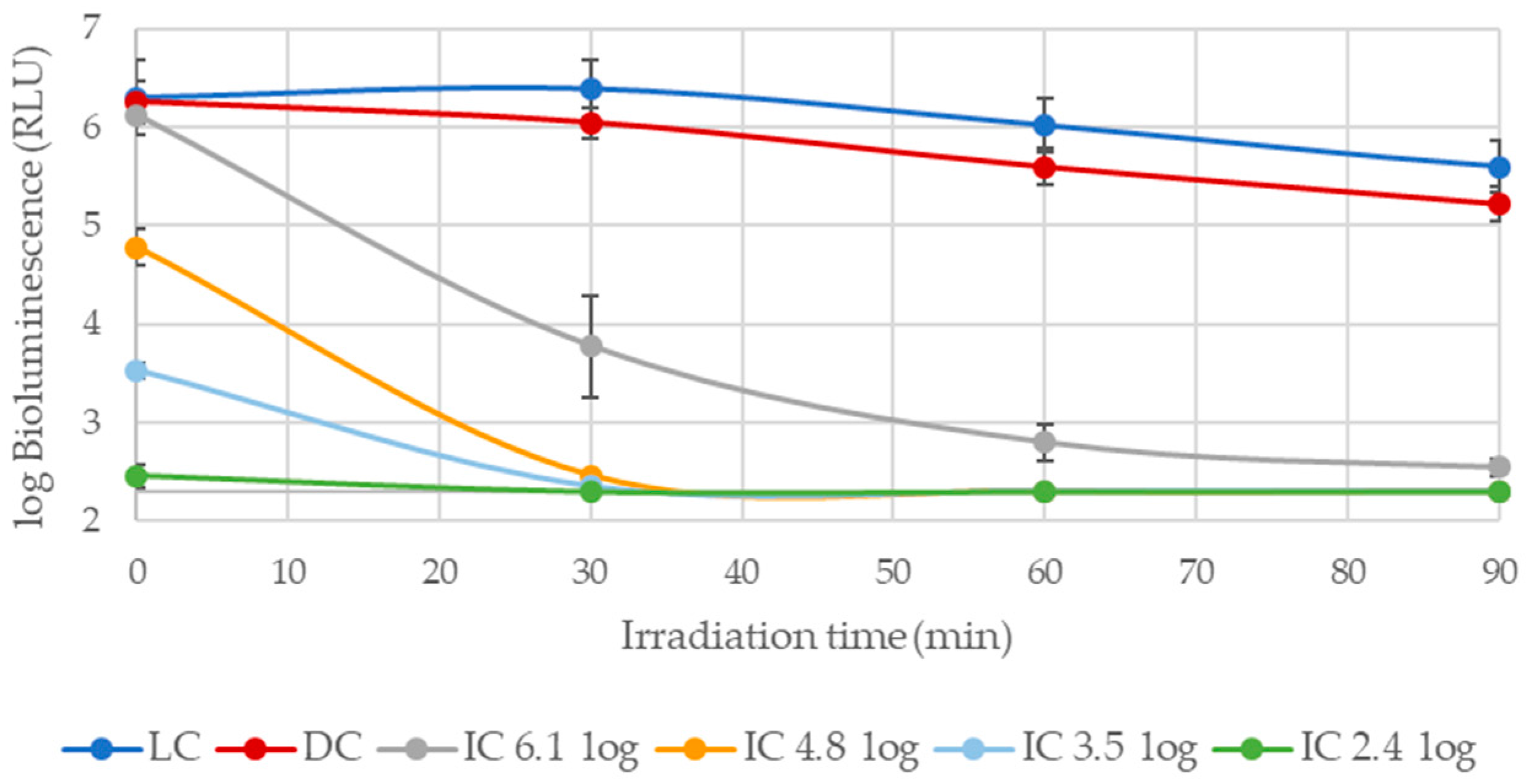
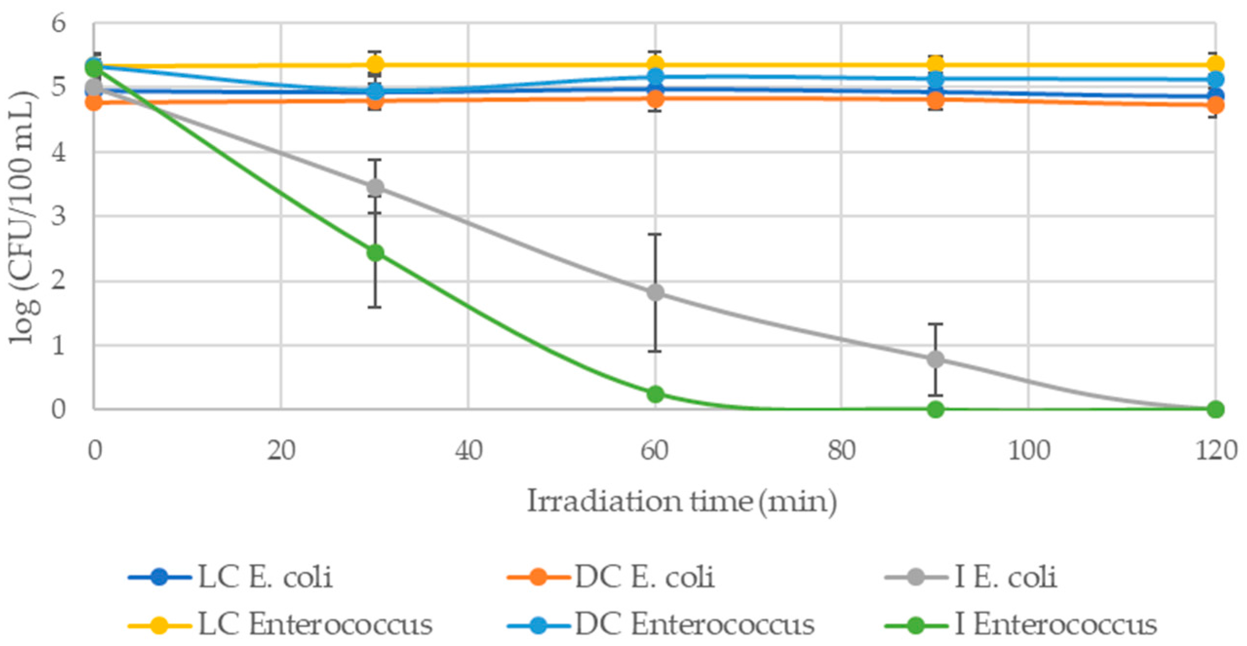
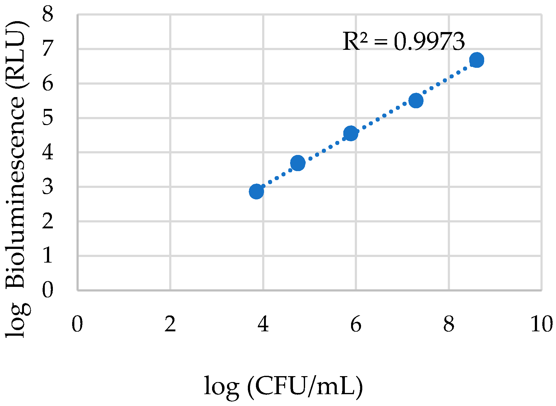
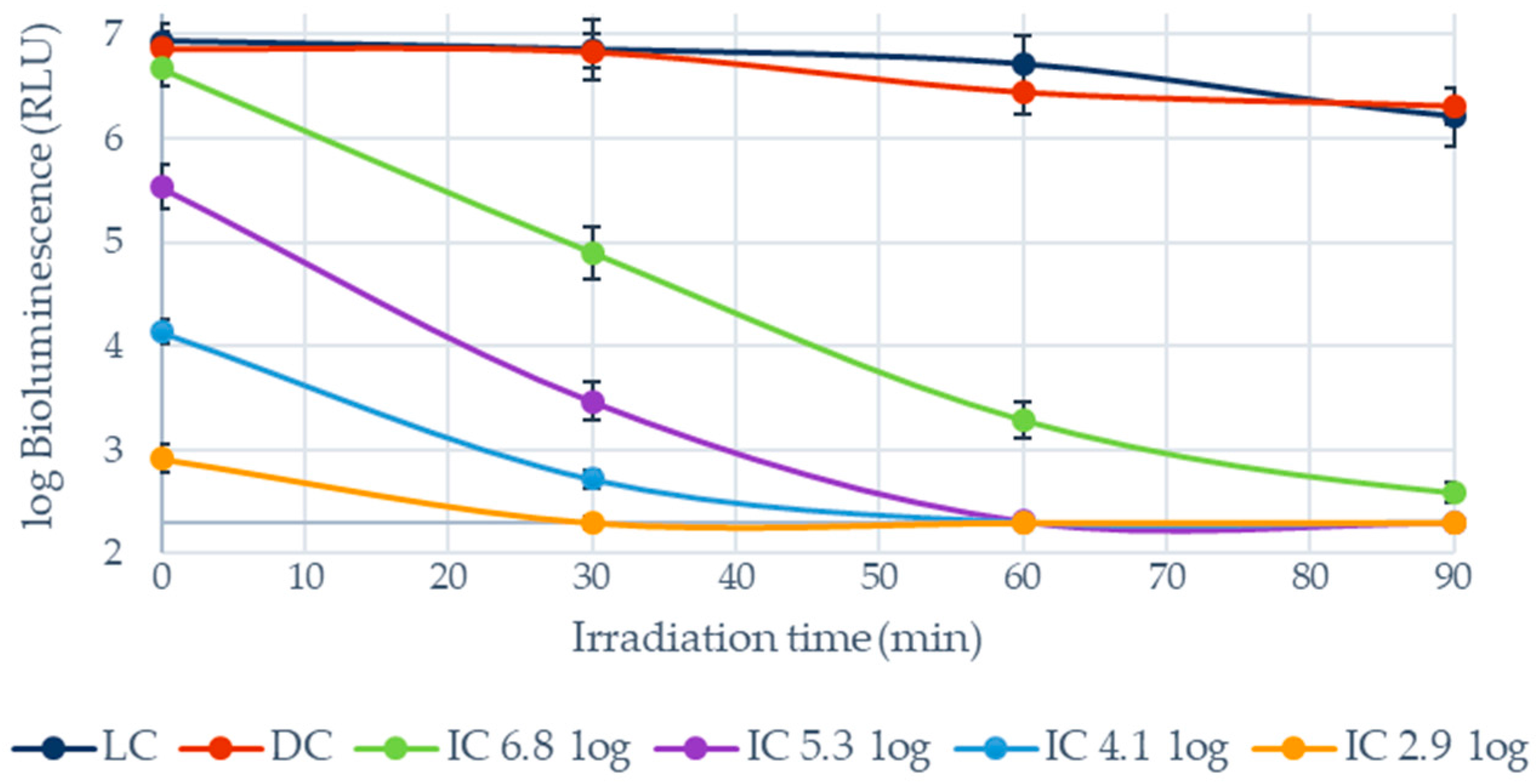
3.1.1. Photoinactivation of E. coli in Filtered WW and PBS

Cells suspensions of bioluminescent E. coli added to WW at different bacterial densities (Figure 1) were subjected to an irradiance of 40 W m−2 for 90 min in the presence of Tetra-Py+-Me at a concentration of 10 µM. Aliquots were taken at 0 min (before the treatment) and at 30, 60, and 90 min (the end of the treatment). As shown in Figure 2, for all the bacterial abundances tested, the bacteria were inactivated to the detection limit of the method, but the full inactivation was observed sooner for the samples with low bacterial abundances (p < 0.05). For the initial concentration of 6.1 log RLU, the inactivation to the detection limit was achieved after 90 min of treatment; meanwhile, for the initial bacterial concentration of 4.8 log RLU, 60 min was the required time. For the initial bacterial concentrations of 3.5 log RLU and 2.4 log RLU, the inactivation to the detection limit was attained after 30 min. At the controls (light and dark controls), the concentration of viable cells did not vary significantly (p > 0.05), indicating that the bacterial cells were not affected by the action of light alone nor by the presence of PS by itself.
For comparison, a similar study was performed but the WW was substituted by PBS. As in the previous experiments, cells suspensions of bioluminescent E. coli were added to PBS at different abundances (Figure 3). The PDI treatments were performed using the same concentration of Tetra-Py+-Me (10 µM) and the same light protocol (irradiance of 40 W m−2), and the aliquots were taken during the same period. As shown in Figure 3, the inactivation was effective for all the bacterial abundances, but the time required for the inactivation to attain the detection limit of the method was dependent, as before, on the initial bacterial abundance. The highest initial bacterial concentration of 6.8 log RLU required a treatment of 90 min, while the bacterial abundances of 5.3 and 4.1 log RLU required 60 min, and the one of 2.9 RLU, 30 min (p < 0.05). As before, the abundance of viable cells in the controls did not vary significantly, indicating that the bacterial cells were not affected by the action of light alone nor by the presence of PS by itself (p > 0.05).
In order to evaluate if the volume of WW would affect the efficiency of the PDI treatments another set of assays was considered. For these experiments, suspensions of bioluminescent E. coli at an initial bacterial abundance of 6.8 log RLU were added to two different volumes of the filtered WW: 20 mL and 500 mL. These assays were performed under the same protocol conditions as before: Tetra-Py+-Me at a concentration of 10 µM and an irradiation period of 90 min at an irradiance of 40 W m−2. The results summarized in Figure 4 show that the PDI efficacy observed using the small volume of 20 mL was maintained when it was used 25 times more volume (500 mL) of the WW suspension (p > 0.05).

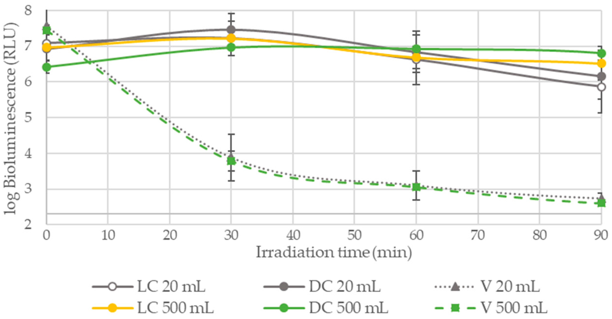
3.1.2. PDI Assays Performed in WW without the Addition of Bacteria

A suspension of WW was used directly in order to evaluate the PDI efficiency in the inactivation of native bacteria present in the WW. Tetra-Py+-Me was added to the suspension to reach the concentration of 10 µM. PDI assays at these conditions were performed for 120 min with an irradiation of 40 W m−2. Aliquots were taken at 0 min (before the treatment) and at 30, 60, 90, and 120 min (at the end of the treatment), and 10-fold serial dilutions were prepared in sterile PBS and aliquots were pour-plated, in duplicate, in m-FC agar and in m-KF agar.
As shown in Figure 5, there was a total E. coli reduction of 5.0 log CFU/100 mL and a total Enterococcus reduction of 5.3 log CFU/100 mL after respectively 120 and 60 min of irradiation. At the controls (dark and light controls), the concentration of viable cells did not vary significantly (p > 0.05), indicating that the bacterial cells were not affected by the action of light alone nor by the presence of PS by itself.

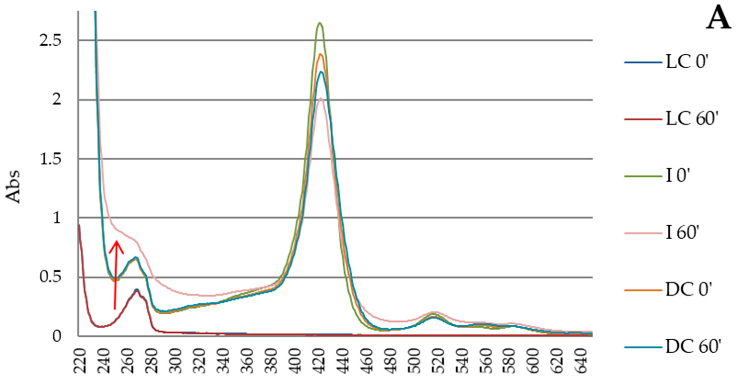
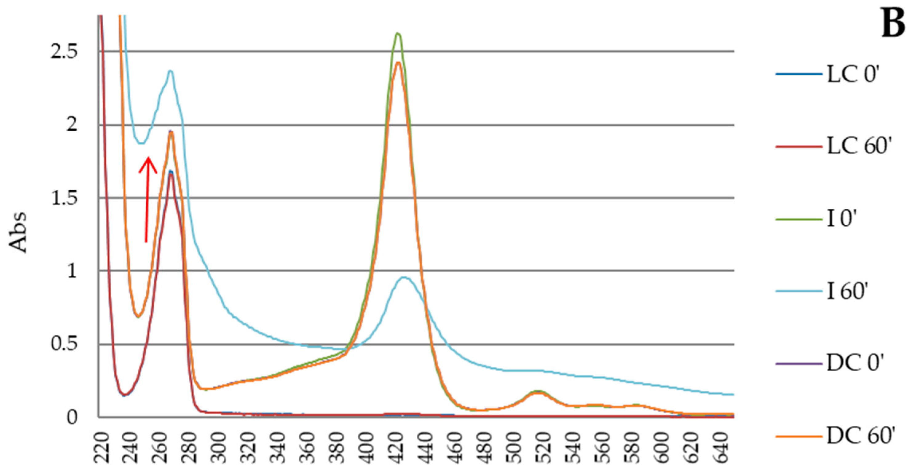
3.2. Photodegradation Tests of Phenol

3.2.1. Photodegradation Test of Phenol with Artificial Light

The photodegradation of phenol tests were performed under similar experimental conditions that were tested in PDI assays (artificial PAR white light with an irradiance of 40 W m−2 and Tetra-Py+-Me concentration of 10 µM) and a phenol concentration of 100 mg L−1, in PBS. Under these last conditions, no significant photodegradation of phenol was observed (data not shown). When the Tetra-Py+-Me concentration was increased to 25 µM in the irradiated samples, the appearance of new absorption bands at ~255 nm was observed (Figure 6). However, there was no significant photo-alteration of phenol in the control samples, since the spectrum bands appear overlain.

3.2.2. Photodegradation Test of Phenol with Solar Light

Tests of photodegradation of phenol were performed with solar light with an irradiance variance between 389 and 1206 W m−2 and a concentration of Tetra-Py+-Me of 25 µM in buffer (PBS). In this test, phenol concentrations of 20 mg L−1 and 100 mg L−1 were used. Figure 7A (phenol concentration of 20 mg L−1) shows that there was no photo-alteration of phenol by solar light alone since in the spectra of the light control sample at 0 min and 60 min, the bands appear overlain. However, the appearance of new absorption bands at ~255 nm can be detected in the samples containing the PS after the solar irradiation. A similar situation can be observed when phenol was used at a concentration of 100 mg L−1 as shown in Figure 7B; the photodegradation of phenol is only observed in the presence of Tetra-Py+-Me where new absorption bands occurs at ~255 nm as previously.

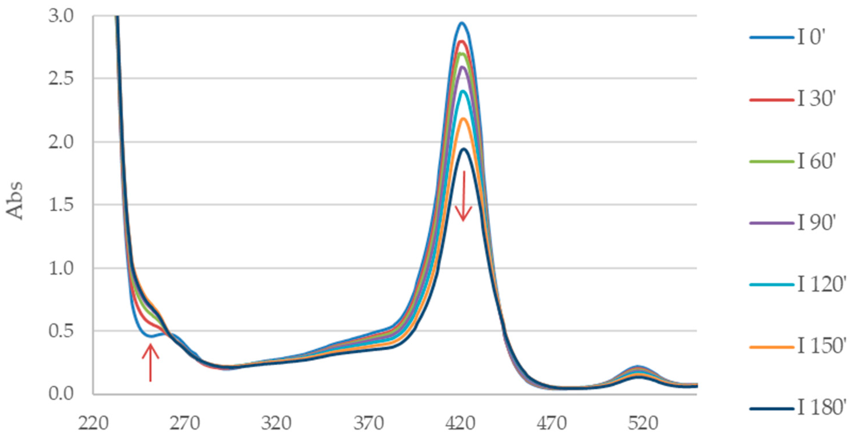
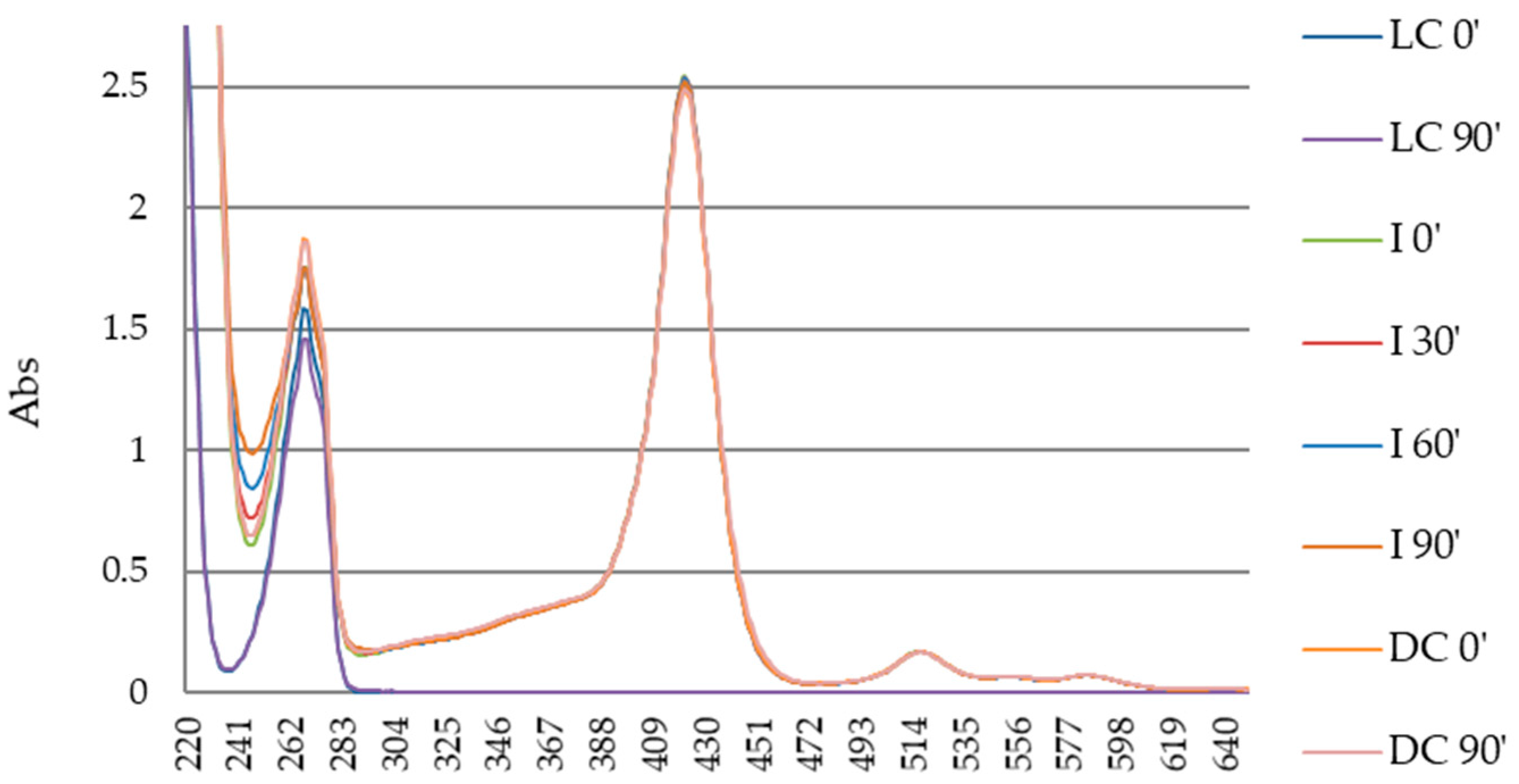
3.3. Photodegradation of Porphyrin

The assays to evaluate the porphyrin photodegradation were performed under solar light with an irradiance variance between 389 and 1206 W m−2 and at a porphyrin concentration of 25 µM in distilled water. Figure 8 shows that there was photo-alteration of the porphyrin under solar irradiation. The appearance of new absorption bands in the ~250 nm range can be detected in the samples containing the PS after the solar irradiation with the appearance of a shoulder at the ~250 nm range, in the samples containing the PS after the solar irradiation.

4. Discussion

According to literature, the PDI process using cationic porphyrins seems to be an efficient antimicrobial approach against Gram-positive and Gram-negative bacteria in clear aqueous suspensions [11,12,34]. Moreover, PDI of microorganisms in environmental waters with high concentration of suspended and dissolved matter is not as effective as in clear aqueous suspensions, but a good response can be obtained in aquaculture waters by adjusting the PS concentration and light dose [35]. However, the PDI efficiency in filtrated hospital WW was higher than in PBS [5]. This difference was interpreted by the authors as due to dissolved compounds in the hospitals WW, like pharmaceuticals, which can affect the bacteria, facilitating the photoinactivation process. As significant reductions in the bacterial number for the tested bacteria were not observed in light and dark controls, these dissolved compounds alone are not likely to affect the bacteria directly. As the effectiveness of PDI in environmental waters depends on different factors, whenever planning for PDI in field conditions, the water should be previously characterized for the establishment of environmentally efficient antimicrobial protocols, including (1) the content of the suspended solids in the medium; (2) the concentration of PS; and (3) the light parameters [36].
In this study, we performed PDI assays under different test conditions to evaluate possible variables that could affect PDI efficiency both against bacterial species and against phenol used as chemical contaminant model.
In order to evaluate if the efficiency of PDI was affected by the less clear medium of WW due to its higher levels of organic matter, different concentrations of E. coli bioluminescent (in general less susceptible to photodynamic inactivation than Gram-positive bacteria) were added to the WW suspension media filtered to eliminate the native bacteria. The results were compared with the ones obtained when similar treatments were performed in PBS, a crystalline medium. The data (Figure 2 and Figure 3) showed that, in both situations (WW and PBS), PDI was an efficient antimicrobial approach. However, the inactivation rate was higher at lower periods of treatment in the assays in the WW, reaching total bacterial inactivation at 30 min of irradiation for initial bacterial abundances of 4.8 log RLU, 3.5 log RLU, and 2.4 log RLU, but the differences were not statistically significant (p > 0.05); in the tests performed in PBS, the same total bacterial inactivation at 30 min of irradiation only occurred for the initial bacterial concentration of 2.9 log RLU. According to previous results, PDI of microorganisms in environmental waters (non-filtered water containing suspended solids) with high concentration of suspended matter and of dissolved organic matter is not as effective as in clear aqueous suspensions [35,36], but the PDI efficiency in filtrated hospital WW can be higher than that in PBS due to the presence of dissolved pharmaceuticals in the hospitals WW [5].
Although in the present study a secondarily treated urban effluent has been used, this include residues of domestic nature, resulting from the needs of individuals such as chemical contaminants (pharmaceutical and personal care products (PPCP)), including releases from kitchens, cleaning detergents, etc. [37]. Moreover, as the effluent receives also wastewater from several industrial zones, this wastewater contains also industrial chemical contaminants. The chemical contaminants can affect the quality of natural water where this WW is discharged, but can also affect the photoinactivation process of WW microorganisms. In fact, no significant reductions in the bacterial number, added E. coli and WW native bacteria, were observed in light and dark controls of WW, which means that these dissolved compounds alone are not likely to affect the bacteria directly, but can have a synergistic effect with PDI. As the WW was filtered before the PDI experiments, the effect of the suspended solids is not expected to affect the PDI results, but the potential dissolved PPCP can influence the PDI efficiency.
WW also contains other kinds of dissolved organic matter that, besides affecting the quality and biosecurity of receiving natural waters, can also affect the efficiency of PDI. The secondary treatment, utilizes bacterial biological degradation to reduce organic matter in WW, thus avoiding microbial multiplication. However, soluble microbial products (SMPs) are produced during WW treatment, as a result of the bacterial biological treatment [38,39]. These SMPs also represent a major concern for aquatic organisms. Moreover, after secondary treatment, part of the organic matter remains in the WW, leading to eutrophication of receiving waters and allowing bacterial growth. Nevertheless, once the photodynamic action occurs through the generation of ROS, the damages in the bacterial cells could occur (1) via the interaction between the excited PS and O2, resulting in 1O2 formation, which will interact with the cellular components through oxidative reactions originating their oxidative damage; or (2) via the energy transference from the excited PS to the surrounding substrates [8,24,40]. Although the occurrence of each type of interaction between PS and the surroundings highly depends on the chemical structure of the PS and according to the literature, porphyrin derivatives PS tend to generate ROS via the energy transference to O2, the higher PDI efficiency in the tests using WW as medium suspension shown in our study could be explained by the high presence of organic matter in the medium. It is well known that ROS have an extremely short lifetime due to their very unstable electronic configuration and their diffusion range is consequently small and dependent on the environment type [21]. The fact that the WW used as WW medium have a high quantity of organic matter could allow for the appearance of different microenvironments with different 1O2 diffusion rates and lifetimes [38,39]; additionally, the presence of compounds able to generate ROS [41] can affect the rate of bacterial inactivation and consequently be responsible for the differences observed in the inactivation rate between the tests performed in PBS and WW medium. Further studies using WW with different content of organic matter are needed in order to test this hypothesis.
Since the WW is a medium less crystalline letting the light penetrate at less distances through the medium, the depth of the water column to treat can be an important factor to have into account in the development of a PDI protocol to treat WW in the field. In this study, PDI assays with a different total volume of the suspension medium were performed to evaluate if it was an influential factor (Figure 4). In fact, in these tests, the results showed that, in both assay conditions, the model bacteria used was efficiently inactivated, reaching a total inactivation of 5.2 log RLU and 5.1 log RLU in the treated WW samples with a total volume of 20 mL and 500 mL, respectively. These results suggest that, since the bacterial and PS concentration are maintained, the PDI efficiency does not seem to be affected by the total volume of medium suspension in which the PDI treatment is performed. Moreover, after 30 min of irradiation, there was already a bacterial inactivation of 3.7 log RLU for both samples (V 20 mL and V 500 mL), reaching 4.8 log RLU after 90 min of irradiation.
In the assays performed without the addition of bacteria to the WW, and thus performing the PDI procedure against native bacteria already occurring in the non-filtered secondarily treated WW, the results showed that PDI was an efficient antimicrobial process against the tested bacteria, E. coli and Enterococci; the Enterococci group was totally inactivated after a period of irradiation of 60 min and the E. coli after an irradiation period of 120 min. These groups of bacteria, both recommended as indicators to water quality control, occurred in the tested WW at a concentration of ~10 000 CFU 100 mL−1, which is a value much higher than the one stipulated by the European Parliament Bathing Water Directive 2006/7/CE to the bathing water quality control [25]. This European Directive (in the case of Portugal adapted to the Portuguese Decreto Lei nº 113/2012), stipulate the limiter values of Enterococci to 660 CFU/100 mL and 350 CFU/100 mL and of E. coli to 1800 CFU/100 mL and 1200 CFU/100 mL to, respectively, interior bathing waters and coastal or transitional bathing waters [42,43]. WW from urban areas is secondarily treated and launched into rivers and seawater far from recreational waters and bivalve/fish production areas. Although the secondarily treated effluent contains high concentrations of microorganisms, water dilution makes it acceptable in terms of quality indicators. However, the emerging of MDR microorganisms involves serious risks when WW not properly treated is discharged in the environment, even in case of urban effluents, which frequently include hospital effluents. In fact, hospital WW is discharged in a municipal sewage system without prior treatment, contributing to a widespread contamination of natural waters with emerging microorganisms and hospital-specific chemical contaminants [44].
Although efficient bacterial inactivation in hospital WW by PDI has been reported [5], little is known about the feasibility of this approach on urban WW or about the degradation of chemical contaminants. As it was announced in the Official Journal of the European Union by the commission responsible by evaluating chemical risks, phenol is considered one of the contaminants, which merits priority on the development of strategies for limiting its existence [45]. In this study, an alternative to several methods already used for the phenol removal from urban and industrial effluents, such as chemical oxidation, biological treatment, ozonolysis and activated carbon adsorption, and wet oxidation, was tested. Each of these methods was shown to have some disadvantages [28]. In this study, in the tested conditions, phenol was photo-degraded once the formation of a new band was observed at ~255 nm, corresponding to p-benzoquinones [28,46,47,48]. According to the literature, benzoquinones have shown to be one of the main photochemical product of phenol [28,46,47] and have shown less toxicity than phenol itself [28]. The new bands formed at test conditions using a phenol concentration of 20 mg mL−1, irradiated with solar light at an irradiance interval between 389 and 1206 W m−2 and a Tetra-Py+-Me concentration of 25 µM, was formed at the end of 60 min of irradiation. Furthermore, the results show that the irradiation with solar light (which includes UV wavelengths) solely do not induce photo-damage in phenol molecules [28].
Parallel, we tested the photodegradation of Tetra-Py+-Me in the presence of solar light irradiation. It was observed that the formation of a shoulder at ~250 nm and the concomitant decrease of the Soret band at ~425 nm. During the photodegration tests of phenol (absorption at ~255 nm), the alterations in its UV-Vis spectrum also occurs within the range of 250–270 nm. Therefore, based on the results shown in Figure 7 and Figure 8, the modification observed at 250–270 nm range occurs due to the photodegradation of both phenol and porphyrin (even if at different extensions). Nevertheless, the photodegradation of Tetra-Py+-Me could have a positive impact once it degrades naturally in the presence of sunlight, and consequently it will not remain active in the environment.

5. Conclusions

In this study, we considered two possible applications of the cationic porphyrin Tetra-Py+-Me, as a photo-bactericidal (against E. coli and Enterococci groups) and as photo-oxidative compound (against an organic compound). In fact, this PS was shown to be effective against both bacterial groups, representing both Gram-negative and Gram-positive bacterial groups used as microbiological parameters, in testing bathing water quality and even showing the power of organic compounds photo-oxidation. Despite the fact that the traditional disinfection methods can inactivate the bacteria, there are also potential problems associated with the contamination of the receiving waters with the disinfection products used (in the case of chlorine) or the potential induction of mutagenicity on the microorganisms (in the case of UV). Additionally, when the PS is immobilized on solid matrices, it can be easily removed, recovered, and reused [8,49], making it an effective, less expensive, easily applicable, and environmentally friendly technology.
Developing this study, we understand how important it will be to build up the knowledge around the hypothesis of the integration of PDI as an asset to the wastewater disinfection process. Future studies must test (1) if PDI using solar light can be more effective in microbial inactivation comparing with the results obtained in tests performed with artificial light; (2) if the PS efficiency is affected when the PS is immobilized on solid matrices; and (3) the PDI efficiency in the photo-degradation of other chemical pollutants of extreme importance with respect to the environment, due to its maleficence, as antibiotics. In this sense, we expect to develop further studies recurring to other analytical techniques.

Acknowledgments

The authors are thankful to the University of Aveiro, to FCT/MEC for the financial support to the Centre for Environmental and Marine Studies (CESAM) unit (project Pest-C/MAR/LA0017/2013) and QOPNA research Unit (FCT UID/QUI/00062/2013), through national founds and, co-financed by the FEDER, within the PT2020 Partnership Agreement, and to the Portuguese NMR Network. M.B. is thankful to the Fundação para a Ciência e a Tecnologia (FCT) for her Ph.D. Grant (SFRH/BD/121645/2016).

Author Contributions

All authors participated in the conception and design of the experiments and analyzed the resulting data; M.B. and S.R. performed the experiments; M.B., M.G.P.M.S.N., M.A.F.F. and A.A. wrote the paper.

Conflicts of Interest

The authors declare no conflict of interest. The founding sponsors had no role in the design of the study; in the collection, analyses, or interpretation of data; in the writing of the manuscript; or in the decision to publish the results.

References

  1. Brown, K.D.; Kulis, J.; Thomson, B.; Chapman, T.H.; Mawhinney, D.H. Occurrence of antibiotics in hospital, residential, and dairy effluent, municipal wastewater, and the Rio Grande in New Mexico. Sci. Total Environ. 2006, 366, 772–783. [Google Scholar] [CrossRef] [PubMed]
  2. Costa, L.; Carvalho, C.M.B.; Faustino, M.A.F.; Neves, M.G.P.M.S.; Tomé, J.P.C.; Tomé, A.C.; Cavaleiro, J.A.S.; Cunha, Â.; Almeida, A. Sewage bacteriophage inactivation by cationic porphyrins: Influence of light parameters. Photochem. Photobiol. Sci. 2010, 9, 1126–1133. [Google Scholar] [CrossRef] [PubMed]
  3. Frigon, D.; Biswal, B.K.; Mazza, A.; Masson, L.; Gehr, R. Biological and physicochemical wastewater treatment processes reduce the prevalence of virulent Escherichia coli. Appl. Environ. Microbiol. 2013, 79, 835–844. [Google Scholar] [CrossRef] [PubMed]
  4. Oliveira, A.; Almeida, A.; Carvalho, C.M.B.; Tomé, J.P.C.; Fautino, M.A.F.; Neves, M.G.P.M.S.; Tomé, A.C.; Cavaleiro, J.A.S.; Cunha, Â. Porphyrin derivatives as photosensitizers for the inactivation of Bacillus cereus endospores. J. Appl. Microbiol. 2009, 106, 1986–1995. [Google Scholar] [CrossRef] [PubMed]
  5. Almeida, J.; Tomé, J.P.C.; Neves, M.G.P.M.S.; Tomé, A.C.; Cavaleiro, J.A.S.; Cunha, Â.; Costa, L.; Faustino, M.A.F.; Almeida, A. Photodynamic inactivation of multidrug-resistant bacteria in hospital wastewaters: Influence of residual antibiotics. Photochem. Photobiol. Sci. 2014, 13, 626–633. [Google Scholar] [CrossRef] [PubMed]
  6. Vaz-Moreira, I.; Nunes, O.C.; Manaia, C.M. Bacterial diversity and antibiotic resistance in water habitats: Searching the links with the human microbiome. FEMS Microbiol. Rev. 2014, 38, 761–778. [Google Scholar] [CrossRef] [PubMed]
  7. Darcy, C.; Lescure, I.; Payot, V.; Rouland, G. Effluents des Établissements Hospitaliers: Teneur en Microorganismes Pathogènes, Risques Sanitaires, Procédures Particulières D’épuration et de Gestion des Boues; Office International de l’eau: Limoges, France, 2002. [Google Scholar]
  8. Alves, E.; Faustino, M.A.F.; Neves, M.G.P.M.S.; Cunha, Â.; Nadais, H.; Almeida, A. Potential applications of porphyrins in photodynamic inactivation beyond the medical scope. J. Photochem. Photobiol. C Photochem. Rev. 2015, 22, 34–57. [Google Scholar] [CrossRef]
  9. Costa, L.; Alves, E.; Carvalho, C.M.B.; Tomé, J.P.C.; Faustino, M.A.F.; Neves, M.G.P.M.S.; Tomé, A.C.; Cavaleiro, J.A.S.; Cunha, Â.; Almeida, A. Sewage bacteriophage photoinactivation by cationic porphyrins: A study of charge effect. Photochem. Photobiol. Sci. 2008, 7, 415–422. [Google Scholar] [CrossRef] [PubMed]
  10. Costa, L.; Faustino, M.A.F.; Neves, M.G.P.M.S.; Cunha, Â.; Almeida, A. Photodynamic inactivation of mammalian viruses and bacteriophages. Viruses 2012, 4, 1034–1074. [Google Scholar] [CrossRef] [PubMed]
  11. Jemli, M.; Alouini, Z.; Sabbahi, S.; Gueddari, M. Destruction of fecal bacteria in wastewater by three photosensitizers. J. Environ. Monit. 2002, 4, 511–516. [Google Scholar] [CrossRef] [PubMed]
  12. Carvalho, C.M.B.; Gomes, A.T.P.C.; Fernandes, S.C.D.; Prata, A.C.B.; Almeida, A.; Cunha, A.; Tomé, J.P.C.; Faustino, M.A.F.; Neves, M.G.P.M.S.; Tomé, A.C.; et al. Photoinactivation of bacteria in wastewater by porphyrins: Bacterial β-galactosidase activity and leucine-uptake as methods to monitor the process. J. Photochem. Photobiol. 2007, 88, 112–118. [Google Scholar] [CrossRef] [PubMed]
  13. Almeida, A.; Cunha, Â.; Faustino, M.A.F.; Tomé, A.C.; Neves, M.G.P.M.S. Porphyrins as antimibrobial photosensitizing agents. In Photodynamic Inactivation of Microbial Pathogens: Medical and Environmental Applications, 11th ed.; Hamblin, M.R., Jori, G., Eds.; Royal Society of Chemistry: Cambridge, UK, 2011; pp. 83–160. [Google Scholar]
  14. Tavares, A.; Carvalho, C.M.B.; Faustino, M.A.F.; Neves, M.G.P.M.S.; Tomé, J.P.C.; Tomé, A.C.; Caveleiro, J.A.S.; Cunha, Â.; Gomes, N.C.M.; Alves, E.; et al. Antimicrobial photodynamic therapy: Study of bacterial recovery viability and potential development of resistance after treatment. Mar. Drugs 2010, 8, 91–105. [Google Scholar] [CrossRef] [PubMed]
  15. Carvalho, C.M.B.; Tomé, J.P.C.; Faustino, M.A.F.; Neves, M.G.P.M.S.; Tomé, A.C.; Cavaleiro, J.A.S.; Costa, L.; Alves, E.; Oliveira, A.; Cunha, Â.; et al. Antimicrobial photodynamic activity of porphyrin derivatives: Potential application on medical and water disinfection. J. Porphyr. Phthalocyanines 2009, 13, 574–577. [Google Scholar] [CrossRef]
  16. Carvalho, C.M.B.; Alves, E.; Costa, L.; Tomé, J.P.C.; Faustino, M.A.F.; Neves, M.G.P.M.S.; Tomé, A.C.; Cavaleiro, J.A.S.; Almeida, A. Functional cationic nanomagnet-porphyrin hybrids for the photoinactivation of microorganisms. ACS Nano 2010, 4, 7133–7140. [Google Scholar] [CrossRef] [PubMed]
  17. Bartolomeu, M.; Rocha, S.; Cunha, Â.; Neves, M.G.P.M.S.; Faustino, M.A.F.; Almeida, A. Effect of photodynamic therapy on the virulence factors of Staphylococcus aureus. Front. Microbiol. 2016, 7, 1–11. [Google Scholar] [CrossRef] [PubMed]
  18. Bonnett, R. Chemical Aspects of Photodynamic Therapy; Gordon and Breach Science Publishers: London, UK; Newark, NJ, USA, 2000. [Google Scholar]
  19. Wainwright, M.; Maisch, T.; Nonell, S.; Plaetzer, K.; Almeida, A.; Tegos, G.P.; Hamblin, M.R. Photoantimicrobials—Are we afraid of the light? Lancet Infect. Dis. 2016, 3099, 1–7. [Google Scholar] [CrossRef]
  20. St. Denis, T.G.; Dai, T.; Izikson, L.; Astrakas, C.; Anderson, R.R.; Hamblin, M.R.; Tegos, G.P. All you need is light. Virulence 2011, 2, 509–520. [Google Scholar] [CrossRef] [PubMed]
  21. Alves, E.; Faustino, M.A.F.; Neves, M.G.P.M.S.; Cunha, Â.; Tomé, J.P.C.; Almeida, A. An insight on bacterial cellular targets of photodynamic inactivation. Future Med. Chem. 2014, 6, 141–164. [Google Scholar] [CrossRef] [PubMed]
  22. Calin, M.A.; Parasca, S.V. Light sources for photodynamic inactivation of bacteria. Lasers Med. Sci. 2009, 24, 453–460. [Google Scholar] [CrossRef] [PubMed]
  23. Henderson, B.W.; Dougherty, T.J. How does photodynamic therapy work? Photochem. Photobiol. 1992, 55, 145–157. [Google Scholar] [CrossRef] [PubMed]
  24. De Oliveira, K.T.; de Souza, J.M.; Gobo, N.R.D.S.; de Assis, F.F.; Brocksom, T.J. Basic concepts and applications of porphyrins, chlorins and phthalocyanines as photosensitizers in photonic therapies. Rev. Virtual Quím. 2015, 7, 310–335. [Google Scholar] [CrossRef]
  25. European Environment Agency. Bathing Water Quality. 2015. Available online: http://www.eea.europa.eu/data-and-maps/indicators/bathing-water-quality (accessed on 27 April 2017).
  26. Jin, G.; Jeng, H.-W.; Bradford, H.; Englande, A.J. Comparison of E. coli, enterococci, and fecal coliform as indicators for brackish water quality assessment. Water Environ. Res. 2005, 76, 245–255. [Google Scholar] [CrossRef]
  27. Kujawski, W.; Warszawskp, A.; Ratajczak, W.; Porebski, T.; Capata, W.; Ostrowska, I. Removal of phenol from wastewater by different separation techniques. Desalination 2004, 163, 287–296. [Google Scholar] [CrossRef]
  28. Mahvi, A.H.; Maleki, A.; Alimohamadi, M.; Ghasri, A. Photo-oxidation of phenol in aqueous solution: Toxicity of intermediates. Korean J. Chem. Eng. 2007, 24, 79–82. [Google Scholar] [CrossRef]
  29. National Center for Biotechnology Information. Phenol. PubChem Compound Database. Available online: https://pubchem.ncbi.nlm.nih.gov/compound/996 (accessed on 27 April 2017).
  30. Santo, C.M. A Indústria de Refinação de Petróleo: Características e Tratamento das Águas Residuais. e-LP Eng. Technol. J. 2010, 1, 21–46. Available online: http://recil.grupolusofona.pt/handle/10437/2853 (accessed on 27 April 2017).
  31. Jori, G.; Fabris, C.; Soncin, M.; Ferro, S.; Coppellotti, O.; Dei, D.; Fantetti, L.; Chiti, G.; Roncucci, G. Photodynamic therapy in the treatment of microbial infections: Basic principles and perspective applications. Lasers Surg. Med. 2006, 38, 468–481. [Google Scholar] [CrossRef] [PubMed]
  32. Kharkwal, G.B.; Sharma, S.K.; Huang, Y.-Y.; Dai, T.; Hamblin, M.R. Photodynamic therapy for infections: Clinical applications. Lasers Surg. Med. 2011, 43, 755–767. [Google Scholar] [CrossRef] [PubMed]
  33. Alves, E.; Carvalho, C.M.B.; Tomé, J.P.C.; Faustino, M.A.F.; Neves, M.G.P.M.S.; Tomé, A.C.; Cavaleiro, J.A.S.; Cunha, Â.; Mendo, S.; Almeida, A. Photodynamic inactivation of recombinant bioluminescent Escherichia coli by cationic porphyrins under artificial and solar irradiation. J. Ind. Microbiol. Biotechnol. 2008, 35, 1447–1454. [Google Scholar] [CrossRef] [PubMed]
  34. Bonnett, R.; Krysteva, M.A.; Lalov, I.G.; Artarsky, S.V. Water disinfection using photosensitizers immobilized on chitosan. Water Res. 2006, 40, 1269–1275. [Google Scholar] [CrossRef] [PubMed]
  35. Arrojado, C.; Pereira, C.; Tomé, J.P.C.; Faustino, M.A.F.; Neves, M.G.P.M.S.; Tomé, A.C.; Cavaleiro, J.A.S.; Cunha, Â.; Calado, R.; Gomes, N.C.M.; et al. Applicability of photodynamic antimicrobial chemotherapy as an alternative to inactivate fish pathogenic bacteria in aquaculture systems. Photochem. Photobiol. Sci. 2011, 10, 1691–1700. [Google Scholar] [CrossRef] [PubMed]
  36. Alves, E.; Faustino, M.A.F.; Tomé, J.P.C.; Neves, M.G.P.M.S.; Tomé, A.C.; Cavaleiro, J.A.S.; Cunha, Â.; Gomes, N.C.M.; Almeida, A. Photodynamic antimicrobial chemotherapy in aquaculture: Photoinactivation studies of Vibrio fischeri. PLoS ONE 2011, 6, e20970. [Google Scholar] [CrossRef] [PubMed]
  37. Shon, H.K.; Vigneswaran, S.; Snyder, S.A. Effluent Organic Matter (EfOM) in wastewater: Constituents, effects, and treatment. Crit. Rev. Environ. Sci. Technol. 2006, 36, 327–374. [Google Scholar] [CrossRef] [Green Version]
  38. Ogilby, P.R.; Foote, C.S. Chemistry of singlet oxygen. 42. Effect of solvent, solvent isotopic substitution, and temperature on the lifetime of singlet molecular oxygen (1.DELTA.g). J. Am. Chem. Soc. 1983, 105, 3423–3430. [Google Scholar] [CrossRef]
  39. Wilkinson, F.; Helman, W.P.; Ross, A.B. Rate constants for the decay and reactions of the lowest electronically excited singlet state of molecular oxygen in solution. An expanded and revised compilation. J. Phys. Chem. 1995, 24, 663–677. [Google Scholar] [CrossRef]
  40. Wainwright, M.; Crossley, K.B. Photosensitising agents—Circumventing resistance and breaking down biofilms: A review. Int. Biodeterior. Biodegrad. 2004, 53, 119–126. [Google Scholar] [CrossRef]
  41. Filipe, O.M.S.; Mota, N.; Santos, S.A.O.; Domingues, M.R.M.; Silvestre, A.J.D.; Neves, M.G.P.M.S.; Simões, M.M.Q.; Santos, E.B.H. Identification and characterization of photodegradation products of metoprolol in the presence of natural fulvic acid by HPLC-UV-MSn. J. Hazard. Mater. 2017, 323, 250–263. [Google Scholar] [CrossRef] [PubMed]
  42. Decreto-Lei nº 113/2012. Gestão da Qualidade das Águas Balneares. Ministério da Agricultura, do Mar, do Ambiente e do Ordenamento do Território: Portugal. Available online: https://dre.pt/web/guest/pesquisa/-/search/177865/details/maximized (accessed on 22 August 2017).
  43. Sistema Nacional de Informação de Recursos Hídricos, Qualidade das Águas Balneares. 2017. Available online: http://snirh.apambiente.pt/index.php?idMain=1&idItem=2.1 (accessed on 28 April 2017).
  44. Pal, A.; Gin, K.Y.-H.; Lin, A.Y.-C.; Reinhard, M. Impacts of emerging organic contaminants on freshwater resources: Review of recent occurrences, sources, fate and effects. Sci. Total Environ. 2010, 408, 6062–6069. [Google Scholar] [CrossRef] [PubMed]
  45. Official Journal of the European Union. Commission Communication on the Results of the Risk Evaluation and the Risk Reduction Strategies for the Substances: Piperazine; Cyclohexane; Methylenediphenyl Diisocyanate; But-2yne-1,4-diol; Methyloxirane; Aniline; 2-Ethylhexylacrylate; 1,4-Dichlorobenz. Available online: http://eur-lex.europa.eu/legal-content/EN/TXT/?qid=1492559033778&uri=CELEX:52008XC0207(02) (accessed on 19 April 2017).
  46. Drozd, D.; Szczubiałka, K.; Skiba, M.; Kepczynski, M.; Nowakowska, M. Porphyrin−nanoclay photosensitizers for visible light induced oxidation of phenol in aqueous media. J. Phys. Chem. 2014, 118, 9196–9202. [Google Scholar] [CrossRef]
  47. DeRosa, M.C.; Crutchley, R.J. Photosensitized singlet oxygen and its applications. Coord. Chem. Rev. 2002, 233–234, 351–371. [Google Scholar] [CrossRef]
  48. Gerdes, R.; Wöhrle, D.; Spiller, W.; Schneider, G.; Schnurpfeil, G.; Schulz-Ekloff, G. Photo-oxidation of phenol and monochlorophenols in oxygen-saturated aqueous solutions by different photosensitizers. J. Photochem. Photobiol. A Chem. 1997, 111, 65–74. [Google Scholar] [CrossRef]
  49. Mesquita, M.Q.; Menezes, J.C.J.M.D.S.; Pires, S.M.G.; Neves, M.G.P.M.S.; Simões, M.M.Q.; Tomé, A.C.; Cavaleiro, J.A.S.; Cunha, Â.; Daniel-da-Silva, A.L.; Almeida, A.; et al. Pyrrolidine-fused chlorin photosensitizer immobilized on solid supports for the photoinactivation of Gram-negative bacteria. Dyes Pigment. 2014, 110, 123–133. [Google Scholar] [CrossRef]
Figure 1. Correlation between bacterial cell density of E. coli bioluminescent (log of cell density, log (CFU/mL)), and the emitted luminescence detected by the luminometer (log of bioluminescence (RLU)).
Water 09 00630 g001
Figure 2. E. coli inactivation in sterilized WW at different initial bacteria abundances. Tetra-Py+-Me was used at a concentration of 10 µM and the samples were irradiated with artificial PAR white light (380–700 nm) at an irradiance of 40 W m−2 for 90 min. Legend: light control (LC), dark control (DC), and different initial bacterial abundances (IC). Values represent the mean of three independent experiments with two replicates each. XX axis cross the YY axis at 2.30 log RLU, representing the detection limit of the method.
Water 09 00630 g002
Figure 3. E. coli inactivation in PBS at different initial bacteria abundance. Tetra-Py+-Me was used at a concentration of 10 µM and the samples were irradiated with artificial PAR white light (380–700 nm) with an irradiance of 40 W m−2 for 90 min. Legend: light control (LC), dark control (DC), and different initial abundances (IC). Values represent the mean of three independent experiments with two replicates each. XX axis cross the YY axis at 2.30 log RLU, representing the detection limit of the method.
Water 09 00630 g003
Figure 4. E. coli inactivation in WW with different total volume of medium suspension. Tetra-Py+-Me was used at a concentration of 10 µM, and the samples were irradiated with white light (380–700 nm) with an irradiance of 40 W m−2 for 90 min. Legend: light control (LC), dark control (DC), and samples with different volumes (V). Values represent the mean of three independent experiments with two replicates each. XX axis cross the YY axis at 2.30 log RLU, representing the detection limit of the method.
Water 09 00630 g004
Figure 5. E. coli and Enterococcus inactivation in WW without addition of bacteria. Tetra-Py+-Me was used at a concentration of 10 µM and the samples were irradiated with artificial PAR white light (380–700 nm) with an irradiance of 40 W m−2 for 120 min. Legend: light control (LC), dark control (DC), and irradiated samples (I). Values represent the mean of three independent experiments with two replicates each.
Water 09 00630 g005
Figure 6. Photodegradation of phenol at a concentration of 100 mg L−1 with artificial PAR white light (380–700 nm) with an irradiance of 40 W m−2 and a Tetra-Py+-Me concentration of 25 µM in PBS. These tests of photodegradation had an irradiation period of 90 min. Legend: light control (LC), dark control (DC), and irradiated samples (I) at different times of analysis.
Water 09 00630 g006
Figure 7. Photodegradation test of phenol at a concentration of 20 mg L−1 (A) and phenol at a concentration of 100 mg L−1 (B) in PBS with solar light with an irradiance between 389 and 1206 W m−2 and a Tetra-Py+-Me concentration of 25 µM. These tests of photodegradation had an irradiation period of 60 min. Legend: light control (LC), dark control (DC), and irradiated samples (I) at different times of analysis.
Water 09 00630 g007aWater 09 00630 g007b
Figure 8. Photodegradation of porphyrin Tetra-Py+-Me in distilled water with solar light with an irradiance between 389 and 1206 W m−2 and a Tetra-Py+-Me concentration of 25 µM. This test of photodegradation had an irradiation period of 180 min. Legend: irradiated samples (I) at different times of analysis.
Water 09 00630 g008

