Review

Alpha Particle Emitter Radiolabeled Antibody for Metastatic Cancer: What Can We Learn from Heavy Ion Beam Radiobiology?

Division of Nuclear Medicine, Russell H. Morgan Department of Radiology and Radiological Science, Johns Hopkins University School of Medicine, Baltimore, MD 21231, USA
*
Author to whom correspondence should be addressed.
Antibodies 2012, 1(2), 124-148; https://doi.org/10.3390/antib1020124
Submission received: 3 May 2012 / Revised: 17 June 2012 / Accepted: 20 June 2012 / Published: 26 June 2012
(This article belongs to the Special Issue Modes of Antibody Action for Cancer Therapy)

Abstract

:
Alpha-particle emitter labeled monoclonal antibodies are being actively developed for treatment of metastatic cancer due to the high linear energy transfer (LET) and the resulting greater biological efficacy of alpha-emitters. Our knowledge of high LET particle radiobiology derives primarily from accelerated heavy ion beam studies. In heavy ion beam therapy of loco-regional tumors, the modulation of steep transition to very high LET peak as the particle approaches the end of its track (known as the Bragg peak) enables greater delivery of biologically potent radiation to the deep seated tumors while sparing normal tissues surrounding the tumor with the relatively low LET track segment part of the heavy ion beam. Moreover, fractionation of the heavy ion beam can further enhance the peak-to-plateau relative biological effectiveness (RBE) ratio. In contrast, internally delivered alpha particle radiopharmaceutical therapy lack the control of Bragg peak energy deposition and the dose rate is determined by the administered activity, alpha-emitter half-life and biological kinetics of the radiopharmaceutical. The therapeutic ratio of tumor to normal tissue is mainly achieved by tumor specific targeting of the carrier antibody. In this brief overview, we review the radiobiology of high LET radiations learned from ion beam studies and identify the features that are also applicable for the development of alpha-emitter labeled antibodies. The molecular mechanisms underlying DNA double strand break repair response to high LET radiation are also discussed.

1. Introduction

Targeted radiopharmaceutical therapy using alpha-particle emitters is a promising treatment option for metastatic cancer. The current development of alpha-particle emitters is further warranted by the recent successful Phase III trial of the alpha particle emitter, 223Ra, in treating castration resistant prostate cancer patients with bone metastases [1]. However, due to its unique bone uptake mechanism and the lack of daughter redistribution that lead to reduced marrow toxicity, the success of 223Ra does not directly carry over to antibody-mediated delivery of alpha-particle emitters. On the other hand, the better clinical outcome of an alpha-emitter compared to other bone seeking radiopharmaceuticals emitting betas and gammas (32P, 89Sr and 153Sm) does highlight the inherent potential advantages of using alpha-emitters. A greater understanding of the radiobiology of high LET alpha particle radiation can lead to the design of safer and more efficient approaches to the delivery of alpha particles.
Compared to the relatively recent efforts to develop internal alpha-particle emitters for clinical oncology, there is a long history of using accelerator generated heavy charged particles (including helium) in clinical studies and radiobiological research. High LET heavy ions cause more severe clustered DNA damage, induce distinctive DNA damage responses compared to low LET radiation in different cell cycles, dose rate and oxygenation status. In addition, mammalian cell DNA repair machinery responds differently to high LET radiation compared to low LET radiation. There are several excellent reviews on radiobiology of heavy ions and their historical development [2,3,4]. In this brief review, we will focus on the common physical and radiobiological features in accelerator generated heavy charged particles and internal alpha-emitters and identify the unique radiobiology features that could guide the development of alpha radioimmunotherapy. We will also discuss the implication of combination therapy with antibody and radiotherapy in radioimmunotherapy when binding of the carrier antibody can disrupt DNA repair signaling as well as the applicability of it to patients with tumors that have genetic defects in DNA repair signaling pathways. It is worth pointing out that properties of the antibodies used in radioimmunotherapy, such as its biodistribution, binding affinity, internalization kinetics and tumor penetration kinetics as well as matching these properties to the radionuclide half-life and emission ranges have a significant impact on the clinical outcomes of targeted radionuclide therapy. This is not the focus of this review and there are several excellent reviews that cover this topic [5,6].

2. Alpha-Particle Radioimmunotherapy and Heavy Ion Beam Therapy of Cancer

The potential of alpha particle-emitters (224Ra) to kill tumors was recognized soon after they were discovered more than one hundred years ago [4]. The clinical development of targeted alpha particle-emitter cancer therapy, however, only became feasible in the 1980s after a series of important technological advances coupled with a greater understanding of cancer biology. These include the invention of monoclonal antibody technology, discovery of the genetic basis of cancer, elucidation of the radiobiological consequences of high LET radiation, the ongoing development of chelators, especially for alpha emitters and the establishment of a reliable supply of alpha-particle emitting radionuclides. Alpha particles are uniquely suited to eradicating small metastasized tumors because their energy is highly concentrated on the short track they travel making it possible to kill single cancer cells with very few alpha particles traversing the cell nuclei. The first human trial of alpha-particle emitter labeled antibody was reported in 1997 wherein alpha emitter 213Bi labeled anti-CD33 monoclonal antibody HuM195 was investigated in patients with myeloid leukemia [7]. Subsequently, additional human trials using other alpha emitter-labeled antibodies have been conducted, including 211At-anti-tenascin for glioblastoma [8], 225Ac-HuM195 for myeloid leukemia [9], 212Pb-Trastuzumab for ovarian cancer [10], 211At-MX35 F(ab’)2 for ovarian cancer [11] and 213Bi-substance P for glioblastoma [12].
Our radiobiological knowledge of high LET alpha particle radiation today was mostly gained through the studies using accelerated heavy ion beams. Parallel to the development of radiolabeled antibodies for cancer therapy, heavy ion beam therapy of cancer was investigated for treatment of cancer shortly after the invention of cyclotron by Ernest Lawrence at Berkeley Lab [2]. His brother, John Lawrence, also a pioneer in the field of nuclear medicine, along with Cornelius Tobias pioneered the application of proton beams and later heavy ion beams after Berkeley Lab built its Heavy Ion Linear Accelerator (HILAC) in 1957. The following year, they conducted the first human study of alpha-particle beams in patients with brain tumors [13]. After the combination of HILAC and Bevatron (Bevalac) in 1970s, a variety of heavy charged-ion beams, carbon, neon, argon etc. were investigated to treat cancer. Before the decommission of Bevalac in 1993, more than one thousand cancer patients had been treated by accelerated heavy ion beams. Several heavy ion beam facilities were subsequently built in Japan (Heavy Ion Medical Accelerator, HIMAC, 1994) and Germany (Gesellschaft Für Schwerionenforschung, GSI, 1997) that continued cancer patient treatment with heavy ion beams therapy as well as radiobiological studies with heavy ions. Modern day interest in heavy ion beam therapy has led to a number of new centers, including the Hyogo Ion Beam Medical Center (2002) and the Gunma Heavy Ion Medical Center (2010) in Japan and the Heidelberg Ion-Beam Therapy (HIT, 2009) in Germany as well as several other facilities under construction in Australia, Italy and China [14]. Most of these proton and carbon ion beams are for the treatment of locally advanced tumors or in the regions that are difficult to operate or treat by other treatment modalities, such as uveal melanoma, head and neck cancer, bone cancer, advance prostate cancer, and inoperable lung cancer [15,16]. The radiobiological basis of such treatments was first established in the 1960s mainly by Barendsen and more recently studied in light of the molecular mechanisms underlying DNA damage responses to high LET radiation.

3. High LET Alpha-Particle Radiation, Bragg Peaks and RBE

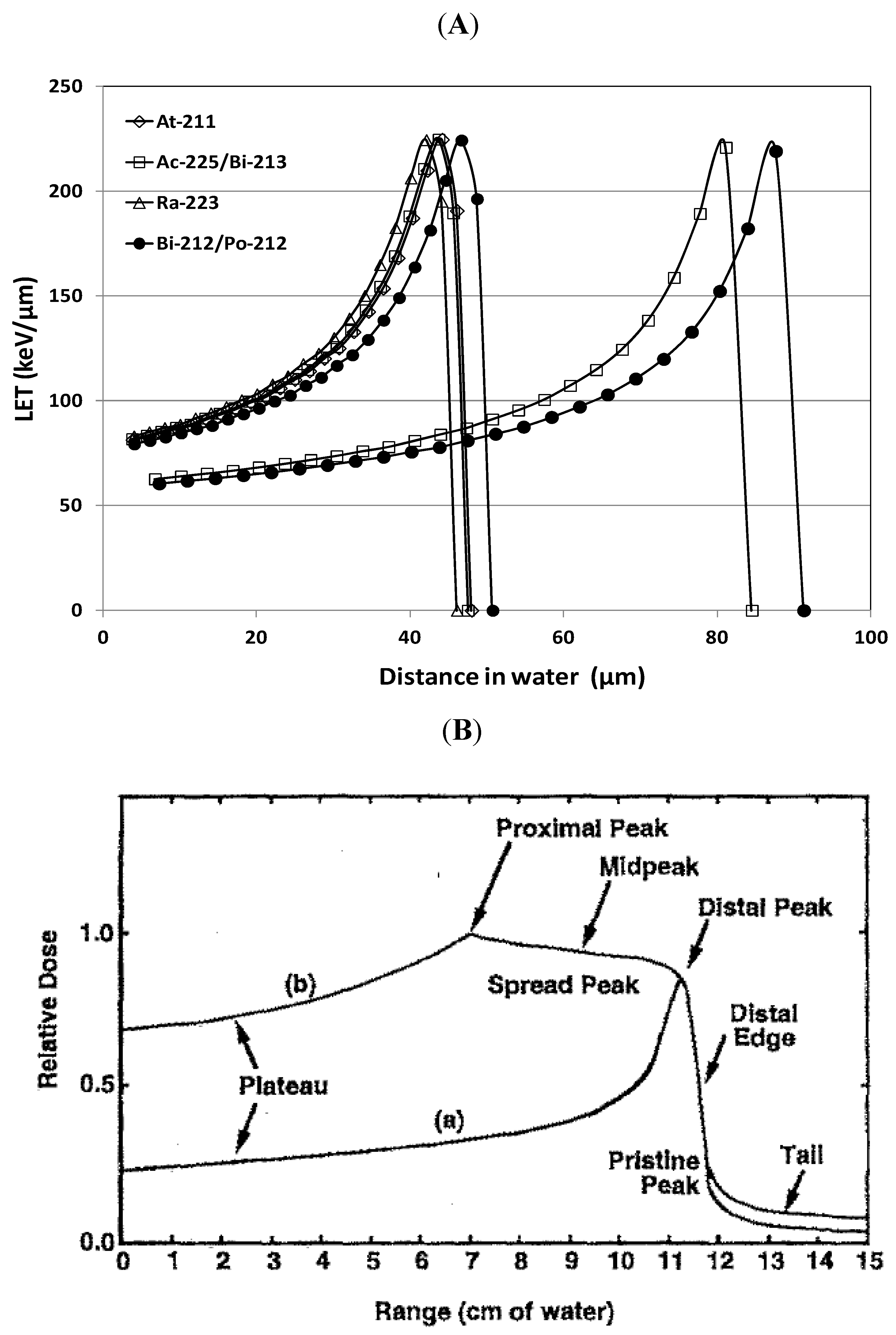
3.1. High LET Alpha-Particle Emitters
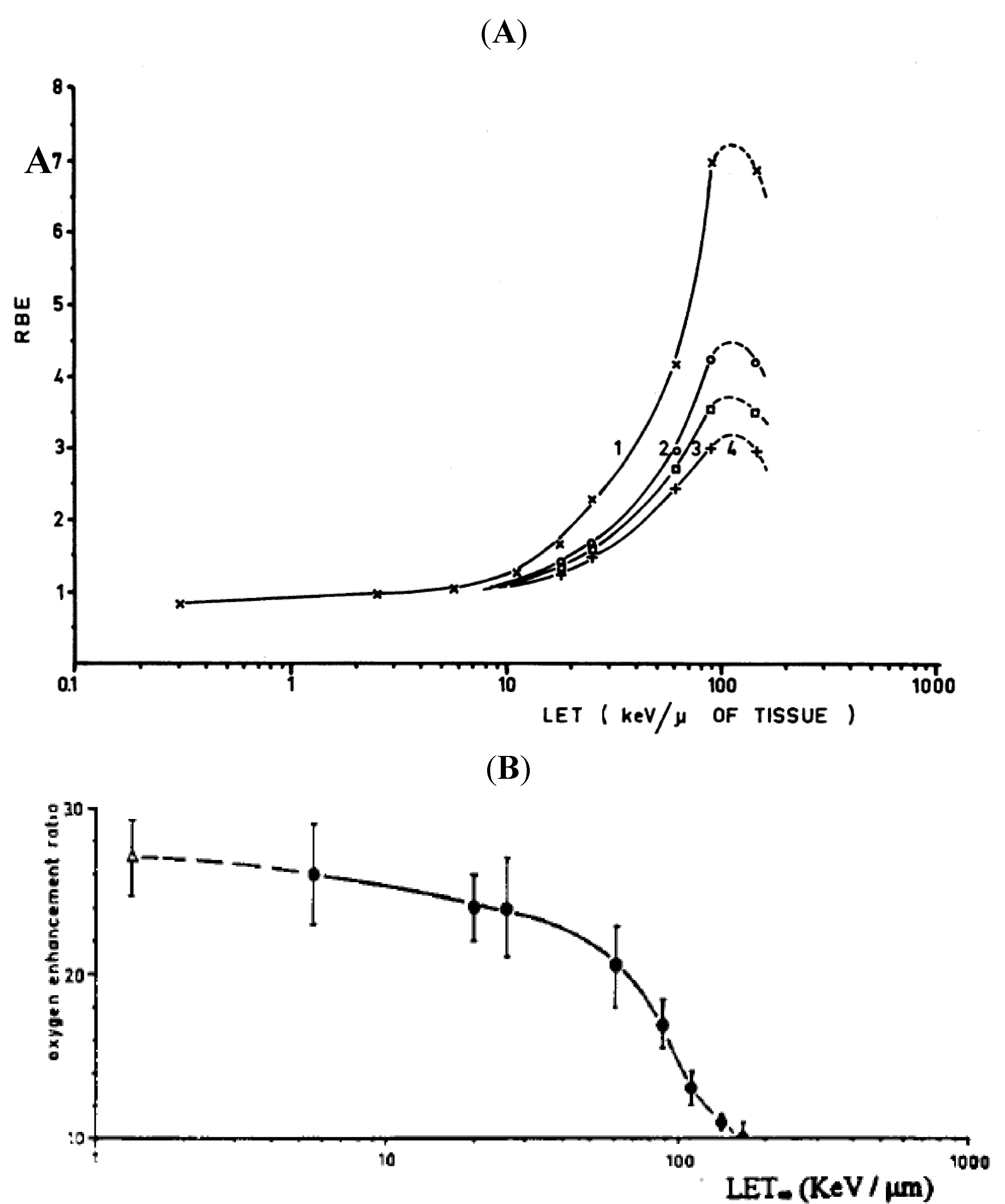
The biological effect of radiation is directly correlated with the pattern of energy transfer to biological material along its path and the amount of energy imparted per unit distance travelled is described as the linear energy transfer (LET). The LET of alpha-particles emitters under clinical development (225Ac, 211At, 213Bi, 212Pb, 223Ra, 227Th) is typically around 100 keV/µm. In comparison, the LET of 250 kVp X-rays is 2 keV/µm, 10 MeV photon is 4.7 keV/µm and the LET of beta particles from internal emitters like 90Y, 131I and 177Lu is 0.2 keV/µm. Low LET photons and electrons deposit their energy almost exponentially decreasing from the source. Heavy charged ions such as alpha particles, however, deposit energy very differently along their tracks for a much shorter range (µm vs. mm). As the alpha particles slow down due to loss of energy, the interaction cross-section increases (i.e., they have higher probability to interact with more materials) and results in higher LET at the end of their tracks known as Bragg peaks (Figure 1A). The range of the alpha particles and hence the position of the Bragg peaks are correlated to the initial energy of the alpha particles.
Figure 1. (A) LET vs. distance in water traveled by typical alpha particles emitted by radionuclides in development for alpha-particle radioimmunotherapy, 225Ac (5.829 MeV)/213Bi (8.375 MeV), 211At (5.867 MeV), 212Bi (6.08 MeV)/Po212 (8.78 MeV), 223Ra (5.716 MeV). The range of the alpha particle and the position of the Bragg peaks are correlated with the initial energy of the alpha particles. LET of alpha-particles in water was calculated using stopping-power and range tables (continuous slowing down approximation range) for electrons, protons, and helium ions from National Institute of Standards and Technology (NIST). (B) The deposition of heavy ion energy as a function of penetrating depth of (a) a pristine beam and (b) a modulated beam with widened stopping region (spread out Bragg peaks). Adapted from Chu WT et al. Review of Scientific Instrument 1993; 64, 2055–2122. Reproduced with permission of the American Institute of Physics.
Antibodies 01 00124 g001
In heavy ion beam therapy, the steep Bragg peaks can thus be modulated to deposit most of the energy in the tumor mass. Since the LET is relatively lower before the Bragg peaks and drops abruptly to zero afterwards, this physical characteristic of heavy ion particles provide a significantly better therapeutic ratio between tumors and normal tissues than the photon beams that do not exhibit Bragg peaks. In the early years of proton and alpha beam studies at Berkeley, the Bragg peak effect of alpha beams had already been utilized to treat breast cancer and glioma. Dosimetry of tumor and skin showed the sparing effect of the Bragg peaks from alpha beams delivering tumor dose of 50 to 85 Gy with less than 20 Gy to the skin [13]. Current ion beam delivery is realized with a combination of range shifter, modulator, lateral spreading and collimator to achieve uniform dose distribution in the tumors (spread out Bragg peak, SOBP) and minimize doses to the surrounding normal tissues [17] (Figure 1B).
The alpha-particles from natural decays have relatively lower energy compared to helium and carbon ions generated from the ion accelerator. Their short ranges (<100 µm, Figure 1A) enable them to deposit all their energy within the distance of about five cell diameters, sparing normal tissues surrounding them. Unlike ion beam therapy where toxicity to the normal tissues surrounding the tumors is the main concern, normal tissue toxicity in radioimmunotherapy is determined by in vivo distribution of the radiolabeled antibody and dose limiting organs are typically red marrow and, in the case of high dose myeloablative treatment, lungs, liver and kidneys. This difference between alpha-article radioimmunotherapy and ion beam therapy makes the high LET Bragg peak a much less contributing factor in determining normal tissue toxicity for internal alpha-emitters. The range of the alpha particle (Figure 1A) seems to suggest that alpha-particle emitter labeled antibody will be less effective against single cells (mammalian cells are about 20 µm in diameter) compared to multi-cellular small metastasis since the high LET Bragg peak of alpha particles emitted from cell surface will miss single cells. Measuring relative biological effectiveness (RBE) at different LET can shed some light on this possibility.

3.2. LET and RBE

The first study on RBE of heavy ion particles at different LET was performed by Barendsen et al in the 1960s using monoenergetic alpha particle beams [18,19,20,21,22,23]. Monoenergetic alpha particles with initial energy ranging from 2.5 MeV to 26 MeV were used to irradiate human kidney T1 cells with track segment (entrance plateau region of LET curve, not within Bragg peak) and compared to deuteron beam and X-rays. LET from alpha particles range from 25 keV/µm to 185 keV/µm while LET from deuterons and 250 kVp X-rays are below 20 keV/µm (Figure 2A). The data clearly showed that high LET alpha particles are more effective than low LET deuterons and X-rays. For alpha particles, the RBE increases with increasing LET until it peaks at slightly over 100 keV/µm and declines afterwards. This decline is attributed to the fact that once cells are killed applying even higher LET alpha particle is simply a waste of energy without enhancing the probability of cell kill. For internal alpha-emitters, due to the relatively low initial energy of the alpha particles, the LET is already in the range of the peak RBE (~100 keV) suggesting these antibody delivered alpha particles are optimal to kill single cell and micrometastasis.
Figure 2. (A) Relative biological effectiveness (RBE) vs. LET for human kidney T1 cells irradiated with track segments of mono-energetic heavy charged particles. Curves 1, 2 and 3 correspond to RBE measured at survival fractions of 0.8, 0.1, 0.01. Adapted from Barendsen GW et al., 1963; 18, 106-119. Reproduced with permission of the Radiation Research Society. (B) Oxygen enhancement ratio (OER) vs. LET for 250 kVp X-ray (open triangle) and mono-energetic alpha particle with different LETs. Adapted from Barendsen GW. Current Topics in Radiation Research, 1968; 293–365. Reproduced with permission of Elsevier Science.
Antibodies 01 00124 g002
Since the classic studies by Barendsen, the relationship between RBE and LET was widely accepted and a single RBE vs. LET curve was assumed to fit for different types of radiation. In the early 1990s, Belli et al., in a series of studies using low LET alpha particles generated with alpha particles accelerated to very high initial energy since the higher the energy the lower the initial LET [24,25,26,27], found that at the same low LET, protons have higher RBE than the alpha particles (1.2 MeV proton vs. 30.5 MeV alphas) with different endpoint for RBE including cell survival and mutation, but much less so for induction of DNA double strand breaks. Several other studies generated similar results [28,29,30,31]. Although LET of alpha particles from internal emitters are much higher with greater RBE than protons and other low LET radiation, these studies highlighted the importance of track structure of different radiations and the microscopic distribution of energy deposition, which is especially important for antibody delivered alpha radiation since antigen heterogeneity and slow tumor penetration of antibody lead to highly non-uniform distribution of alpha-particle emitters.
The RBEs for internal alpha-emitters have also been examined both experimentally and theoretically [4,32,33,34,35,36]. For internal alpha emitter, since cells can be irradiated with different LETs along the alpha tracks RBE is reported, instead of LET, with individual alpha emitter or by the initial energy of the alpha particle they emit. Aurlien et al. reported that alpha particle emitter 211At labeled antibody has an RBE of 3.43 for osteosarcoma cell line OHS-s1 and 1.55 for bone marrow cells using 37% cell survival as the biological endpoint and 60Co γ-rays as reference [36]. Bäck et al. reported an RBE of 4.8 for 211At labeled MX35 F(ab’)2 in an ovarian cancer NIH:OVCAR-3 tumor model [37]. In vivo measurement of RBE using mouse testes as an experimental model and testicular spermhead survival as the biological endpoint, Howell et al. found that the RBE of 212Pb with alpha particle emitting daughters 212Bi and 212Po was 4.7 using 120 kVp X-rays as reference [38]. The same model was also used to find that the RBE of two other alpha particle emitters, 148Gd and 223Ra, is 7.4 ± 2.4 and 5.4 ± 0.9, respectively [39]. In addition, in vivo studies of alpha emitter 213Bi labeled peptide found an RBE of 2-3 for control of colon cancer and surprisingly close to 1.0 for marrow toxicity using beta emitter 90Y labeled peptide as reference [40,41]. Nayak et al. found similar RBE of 3.4 for 213Bi labeled DOTATOC when treating pancreatic adenocarcinoma cells using 137Cs as reference radiation [42]. The reported RBEs of internal alpha-emitters, probably an average of LET along its tracks with different biological endpoints and reference radiations, are still in the range of those reported in the high LET ion beams studies (Figure 2A). Review of alpha-emitters for medical therapy by an expert panel on a Department of Energy workshop recommended an RBE value of 5 for 213Bi and 211At in phase I clinical trials and suggested establishment of “clinical” RBE values as trials progress [43].

3.3. Microdosimetry

In radiobiology, understanding the distribution of energy deposition in irradiated tissues is of high significance in evaluating biological effects of different types of radiation. It is widely accepted that the DNA molecules are the primary targets for radiation induced damage and that DNA DSBs are the principal cause of biological damage. Pairs of DNA damage interact with each other in the micrometer range and the probability of interaction is distance-dependent. The diameter of DNA is about 2 nanometer and the distribution of absorbed energy in the nanometer and the micrometer level can cause observed effects and their relative contributions determine the relative biological effectiveness [44].
In targeted alpha radionuclide therapy, the range of emitted particles is comparable to the size of the cells and the distance between the sites of radionuclide deposition is also small hence, random spatial distribution of disintegration has non-negligible effect on the local energy deposition. In other words, statistical variation in the energy deposition from high-LET radiation such as alpha particles is large in a small volume and that the macrodosimetic quantities such as mean absorbed dose can be a misleading index for the biological effects of high-LET radiation [4]. In such cases, microdosimetric concepts and their associated quantities such as specific energy (energy per unit mass) and lineal energy (energy per unit path length) that accounts for the stochastic nature of energy deposited in a small volume are more suited to understanding biological effects. The criterion as to when microdosimetry should be considered was defined by Kellerer and Chmelevsky [45], which states that the stochastic nature of energy deposition within the target should be taken into account when the relative deviations of the local dose from the mean in the target region exceeds 20%. The applicability of microdosimetric concepts in targeted alpha particle therapy has been extensively reviewed by Sgouros et al. [4] and also recently by Chouin and Bardies [46].
At present the use of microdosimetry in radiobiology is constrained by the lack of biological information at the microscopic level by experimental methods and not due to the lack of microdosimetric models. It is envisioned that advances in molecular techniques would shed some light in the analyses of the spatial distribution of DNA lesions that can be correlated to the spatial fluctuations of energy deposition by different ionizing radiations in the near future [47].

4. Biological Effects of High LET Radiation

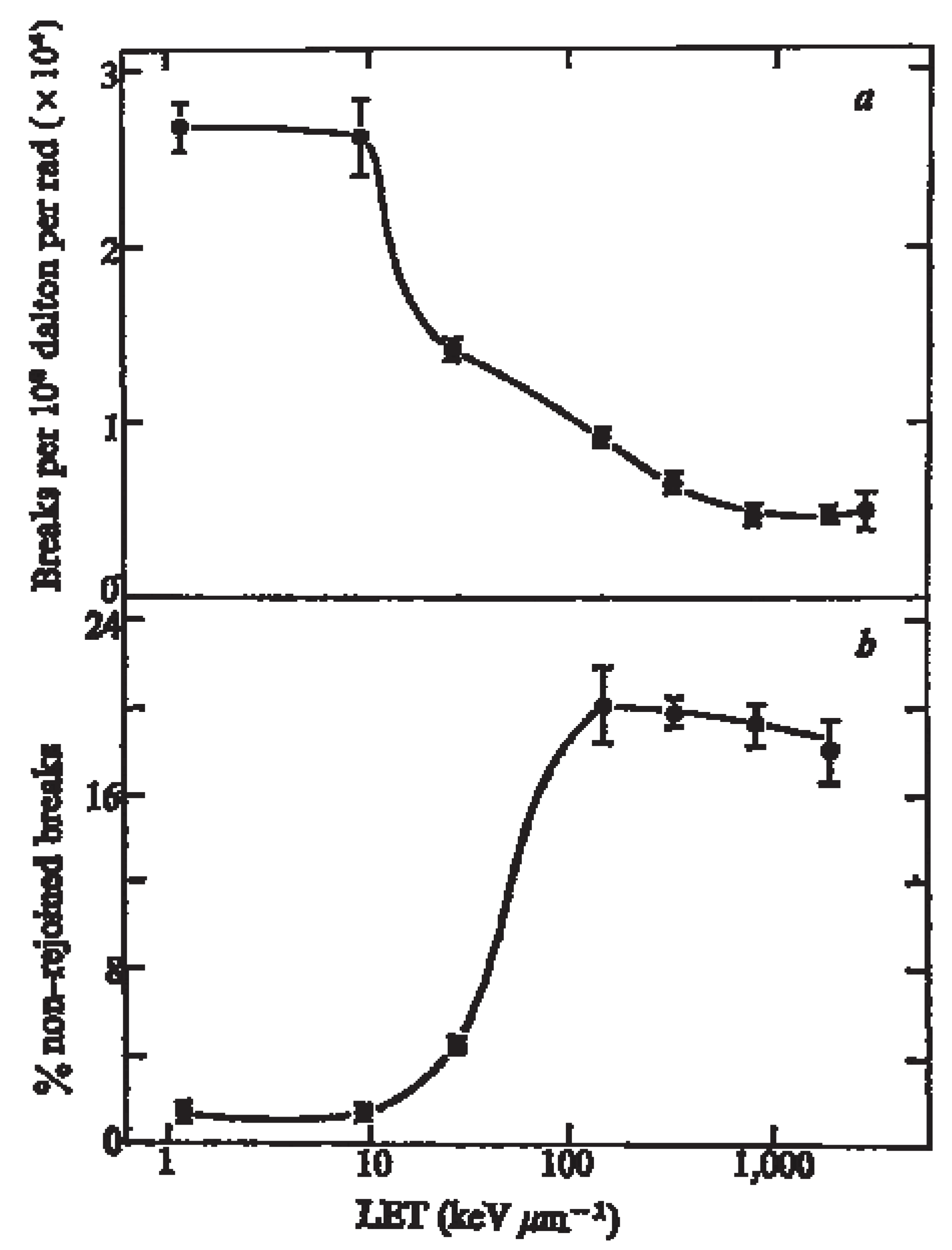
4.1. Induction of DNA Damage by High LET Radiation

Radiation kills cancer cells primarily by damaging DNA [48]. Quantity and “quality” of DNA damages are two important factors that determine the severity of ionizing radiation caused by low and high LET radiation, which is mainly due to different levels of indirect (free radicals) and direct (physical interaction between radiation and DNA) effects that will be discussed in detail in the next section. So far, evidence suggested that high LET radiation does not induce significantly higher amount of DNA breaks than low LET radiation that can explain its more severe effects on DNA damages. Both supercoiled plasmid DNA and cell based assays using pulse-field gel electrophoresis (PFGE) have shown that the yield of DNA double strand breaks (damage site/Mbp/Gy) with high LET is only slightly higher than with low LET [26,49,50,51] or in some studies decreases with increasing LET in mammalian cells [52,53]. Increasing LET meanwhile was shown to induce more DSBs per track traversing cells [54], suggesting that most DSBs induced by high LET radiation are concentrated on fewer tracks compared to low LET radiation. The induction of DNA DSBs/cell/Gy also does not correlate well with other measurement endpoints for RBE such as cell survival and mutation induction, both of which clearly show the trend of higher RBE with increasing LET. Uncertainty still exists in the measurement of DNA fragments to quantify DSBs where Monte Carlo simulation has shown that the yield of short DNA fragments (0.1–1.0 kbp) continues to increase with higher LET while yield of intermediate DNA fragments (1.0–1000 kbp) peaks at around 100 keV/µm LET raising the possibility that short DNA fragment undetected in the measurement can significantly affect the yield of DSBs and artifactually lower the calculated RBE of high LET radiations [55].
On the other hand, it has been established that high LET radiation induces more complex DNA damage where DNA lesions occurring close to each other form clustered DNA damage [56,57]. These lesions include DNA DSBs and non-DSB oxidative clustered DNA lesions (OCDL) [57,58]. For non-DSB oxidative clustered DNA lesions, both Monte Carlo simulation and experimental measurements using DNA base excision repair enzymes, such as DNA glycosylases and AP endonucleases isolated from E. Coli. [59], have confirmed that induction of non-DSB OCDL decreases with high LET radiation compared to low LET radiation [52,53,60,61,62,63]. This decrease is attributed to the possibility that high LET simply generates fewer amounts of single strand breaks (SSBs) and damaged bases relative to low LET radiation. Most importantly, these observations of DNA damage inductions by high LET radiation showed that their higher RBE in cell survival and mutation induction is not the result of higher yield of DNA DSB and OCDL lesions. Rather, most DNA lesions of higher LET radiation are concentrated in DNA damage clusters. Theoretical analysis revealed that low LET radiation can generate cluster with as many as 10 lesions while high LET radiation is able to induce significantly more, up to 25, lesions in one cluster [64]. Recently, immunofluorescent staining of DNA repair proteins, 53BP1 (DSB damage), XRCC1 (SSB damage), and hOGG1 (base damage) foci, have also shown that most clusters induced by high LET radiation have colocalization of all three DNA repair proteins suggesting the prevalence of complex DNA damage [65]. Volume of foci colocalization is also significantly higher in high LET radiation treated cells and most clustered DNA damage induced by Fe ion irradiation is irreparable [65], supporting the model that “high quality” or complexity of high LET-induced DNA damage, not the yield of DNA damage, induced by high LET radiation is the cause of its high RBE.

4.2. Effect of Dose-Rate and Fractionation

A key difference between high LET ion beam therapy and antibody delivered alpha-particle emitters is the dose rate. In the current operational heavy ion beam therapy facilities including GSI in Darmstadt Germany and the HIMAC in Japan, the typical dose rate of carbon ion beam is 1 Gy/min with maximum dose rate of 5 Gy/min [66]. In comparison, internal alpha-particle emitters are delivered at much lower dose rate. For long-lived alpha particle emitters such as 225Ac (T1/2 = 10.0 day), 223Ra (T1/2 = 11.4 day) and 227Th (T1/2 = 18.7 day), delivering 20 Gy to the tumors amounts to an initial dose rate of 0.001Gy/min, 0.0008 Gy/min and 0.0005 Gy/min, respectively. Even for short-lived alpha-emitters 213Bi (T1/2 = 45.6 min), 212Bi (T1/2 = 60.6 min) and 211At (T1/2 = 7.2 h), the initial dose rates will be approximately 0.3, 0.2, 0.03 Gy/min.
Protracting radiation dose over longer period of time lowers the RBE for low LET radiation primarily because cells are allowed more time to repair radiation induced DNA damage before it accumulates and leads to cell death. For high LET radiation, when cell survival was evaluated as the biological endpoint, alpha particle irradiation with dose rate ranging from 0.5 to 100 cGy/min did not affect RBE [67]. Interestingly, when neoplastic transformation and somatic mutation was examined, unlike low LET radiation where low dose rate causes fewer number of event, low dose rate of high LET radiation including neutrons and heavy ion beam actually lead to enhanced neoplastic transformation and somatic mutation. This effect of high LET radiation is termed inverse dose rate effects and has been shown in several cell models [68,69,70,71]. Tauchi et al. showed that the inverse dose rate effect observed with carbon ion beams could be attributed to the observations that cells in G2/M phase are hypersensitive for mutation induction by high LET radiation while low LET radiation only induces mutation in the G1 phase [71,72]. A series of other studies also found that this inverse dose rate effect is limited to the LET range of 30 to 130 keV/µm [73,74,75]. The absence of inverse dose rate effect over 130 keV/µm can be explained by fewer number of cells being hit at the same dose while lower than 30 keV/µm radiation is not sufficient to saturate DNA repair processes [70].
Similar to low dose rate studies, fractionated high LET ion beam studies have shown a sparing effect on normal tissues while maintaining cell kill on tumor cells all of which are dependent on tissue types, LET and dose rates. Barendsen et al. first investigated the effect of fractionation on cell survival. Fractionating alpha particle radiation (12 h apart) with various energies (24.6, 60.8, 85.8 keV/µm) using a human kidney cell model, they observed no significant repair which was attributed to a “single event” caused by alpha particle that is hard to repair [67]. Goldstein et al. showed and confirmed by other groups that fractionation of heavy ion beams enhanced the peak-to-plateau RBE compared to single dose radiation in mouse intestine [76,77,78] because cells irradiated with spread out Bragg peak region had less recovery after fractionation compared to cells irradiated with plateau region (Figure 3). Chang et al. reported that when an iron ion beam (146 keV/μm at the sample position) was fractionated into five daily doses, significantly lower levels of micronucleated reticulocytes in peripheral blood at 48 h were observed and lead to a sparing effect on cytotoxicity to the hematopoietic system [79]. High LET carbon ions were also used to investigate the change in surviving fraction of four human tumor cell lines after fractionated dose irradiation. Again, fractionation was found to enhance the peak-to-plateau RBE ration compared to single dose [80].
Unlike high LET ion beam therapy where effects of dose rate and fractionated dose irradiation are well established to enhance the tumor to normal tissue (peak-to-plateau) RBE ratio, controlled delivery of peak-to-plateau RBE ration is not possible for antibody delivered internal alpha-particle emitters and the dose rate depends upon the radionuclide half-life and is orders of magnitude lower than that available from heavy ion beams. Thus, the achievable therapeutic ratio between normal tissue and tumors are mostly determined by antibody targeting and, potentially, fractionation. Few studies have investigated fractionation of internal alpha particle radiation on tumor and normal tissue RBE in vivo or in vitro. Barendsen et al. observed no survival difference of the kidney cells in vitro between single and fractionated irradiation with alpha particles (3.4 MeV) from 210Po [67]. Elgqvist et al. found no advantage in therapeutic efficacy with fractionated alpha particle emitter 211At labeled MX35 F(ab’)2 compared to single administration [81]. Another important aspect of antibody delivered alpha-emitter that needs to be taken into consideration is the possible saturation and turnover rate of tumor antigens during fractionated doses of radiolabeled antibodies that could reduce fractionated doses.
Figure 3. Cell survival curves of jejuna crypt cells irradiated with single dose or fractionated doses (5 fractions or 10 fractions) of SOBP or plateau region of a 225-MeV/amu helium beam. Fractionation clearly enhanced the peak-to-plateau RBE ratio. Adapted from Goldstein LS et al. Radiation Research, 1981; 86, 542–558. Reproduced with permission of the Radiation Research Society.
Antibodies 01 00124 g003

4.3. Effect of Cell Cycle and Oxygenation

It has long been established that radiosensitivity of mammalian cells to low LET radiation is cell cycle dependent. Cells in mitosis and the G2 phase are the most sensitive and become most resistant in the S phase [48]. For high LET radiation, significant cell cycle delay was found in G2 phase in asynchronized and synchronized Chinese hamster V79 cells and increase of dose prolongs G2 arrest [82,83]. More importantly, irradiation of synchronized V79 cells with different LET radiation has found that variation of cell cycle dependent survival curves are gradually reduced with the increase of LET [84], suggesting that RBE of high LET is cell cycle independent. Claesson et al. investigated the effects of cell cycle on RBE of alpha particles from 211At labeled on Trastuzumab, non-specific to the Chinese hamster lung fibroblast cells V79-379A used in the study. It was found that RBE of high LET alpha particles from 211At is significantly higher than X-rays in both DSB induction and cell survival. Variation of cell survival between different cell cycle phases was significantly reduced for alpha radiation compared to X-rays but such reduction was not as evident for DSB induction, suggesting a weak correlation between DSB induction and cell survival [34].
Like the case for cell cycle, it has also been established for low LET radiation that oxygen has the most effect among many chemical agents to modify the biological effect of ionizing radiation. For X-rays and γ-rays, the typical oxygen enhancement ratio (OER), the ratio of doses needed under hypoxic condition to achieve the same biologic effect as aerobic condition, ranges from 2.5 to 3.5 [48]. This oxygen effect can be explained by the oxygen fixation hypothesis where DNA molecules react with free radicals, typically reversible under hypoxic conditions, become fixed with organic peroxide in the presence of oxygen and result in DNA damage. It is estimated that indirect effects of free radicals account for approximately two-thirds of the DNA damage caused by X-rays [48]. For high LET radiation, classic studies performed by Barendsen et al. and others have shown an inverse relationship between OER and LET where effect of oxygen on cell radiosensitivity becomes diminished (OER = 1.0) for LET greater than 140 keV/µm [21,85] (Figure 2B). The main hypothesis for the decrease of OER with increasing LET is that high LET radiation predominately causes direct DNA damage (estimated at about 75% for alpha particle of 150 keV/µm [86]) independent of the free radical inflicted indirect DNA damage, thereby less affected by oxygen. Clinically, poor tumor oxygenation status (hypoxia defined as O2 partial pressure less than 10 mmHg) has been repeatedly found to be a prognostic factor for disease free survival after conventional radiation therapy [87,88,89]. High LET radiation could overcome such radioresistance because of its diminished susceptibility to the OER effect. In a clinical trial of high LET carbon ion beam, Nakano et al. compared its efficacy against hypoxic and normoxic cervical tumors and showed similar disease-free survival between hypoxic (<20 mmHg) and oxygenated (>20 mmHg) tumors suggesting that high LET radiation can overcome the radioresistance caused by tumor hypoxia [90]. Modeling analysis, however, suggest that the reduction of OER with high LET radiation under the clinical tumor hypoxic environment (0.5–20 mmHg) is relatively moderate with approximately 15% benefit over photon [85]. Furthermore, the findings that patients with hypoxic tumors are associated with poor prognosis with treatments independent of oxygen status (such as surgery) suggest that hypoxic tumor could have a malignant phenotype as a result of colony selection under hypoxic condition which in turn is maintained by its fast consumption of O2. Up-regulation of key signaling pathways including angiogenesis, cell survival, glucose metabolism by hypoxia inducible factor-1 alpha (HIF-1α) further promotes the resistance and progression of this malignant phenotype [91]. More studies are needed to establish the advantage of high LET radiation in treating poorly oxygenated tumors, where difference between local control of hypoxic tumors by alpha radiation and progression free survival can indicate whether progression is due to ineffectiveness of alpha radiation against hypoxic tumors itself or against a highly malignant phenotype.
For internal alpha-particle emitters, the plateau LET is between 50 to 100 keV/µm (Figure 1A) which correspond to an OER between 1.2 to 2.0 (Figure 2B), while the LET around Bragg peaks is well above 140 keV/µm (OER = 1). As a consequence, the stochastic distribution of alpha particle radiation within or surrounding the hypoxic region of tumors will determine its overall OER. Few studies have investigated the correlation between pretreatment oxygenation status of tumors and tumor response to antibody labeled alpha-emitters while tumor response to beta emitter labeled antibody was shown to correlate with tumor pO2 [92]. It is important to note, however, since it is very difficult for antibody as a carrier to penetrate tumor hypoxic regions, the delivery of alpha-emitters is probably the dominating dose limiting factor that determines tumor control. Delivery of alpha radiation with smaller molecules such as peptide, scFv and diabody could potentially lead to a better penetration into the tumor hypoxic core [42,93].

5. Repair of DNA Damage by High LET Radiation

5.1. DNA DSB Repair After High LET Radiation

In contrast to the findings that induction of DNA DSBs by high LET heavy ion radiation does not correlate well with cell survival, studies from Tobias’ lab at Berkeley using heavy ion beams have found that the rate of DNA break rejoining becomes significantly slower as LET increases and there is a strong correlation between the efficiency of cell kill and the non-rejoined DNA strand breaks [94,95]. This impaired rejoining rate reaches maximal for LET in the range of 100 to 200 keV/µm and plateaus for higher LET (Figure 4), unlike RBE vs. LET where RBE begins to decrease for higher LET due to overkill. For LET at the maximal impaired rejoining, about 20% of the DNA breaks remain non-rejoined compared to less than 2% for low LET radiation (Figure 4). These percentages are dose independent. More recently, immunostaining of phosphorylated histone protein H2AX (γ-H2AX) had been used as a marker to quantify induction and rejoining of DNA DSB. Carboxy-terminal phosphorylation of histone H2AX is the earliest cellular response to DNA DSB that accumulates at the sites of DSB quickly (within minutes of the damage) [96]. Consistent with prior findings, similar numbers of γ-H2AX foci are formed after low LET and high LET radiation but there are more remaining γ-H2AX foci at 24 h after high LET radiation (20% of initial foci remaining after alpha particle, 120 keV/µm, compared to less than 10% after gamma ray) [54,97,98]. In addition, the repair of DNA DSB appears to consist of two kinetic components, a fast phase and a slow phase, where most of the DNA DSBs caused by high LET radiation is repaired [97]. Studies in our lab with anti-HER2 Trastuzumab labeled alpha-particle emitter 213Bi also see the same trend where higher fraction of γ-H2AX foci remained at 24 h compared to gamma irradiation (unpublished data). The “high quality” DNA DSB (clustered damage) inflicted by high LET radiation is most likely the cause of the 20% non-rejoined DSBs. The 80% rejoined DNA DSBs apparently correspond to DNA lesions that are still reparable by the mammalian cell repair machinery. Better understanding of the DNA DSB repair processes after high LET radiation might yield strategies to further enhance the RBE of high LET radiation in a tumor-specific fashion.
Figure 4. Induction of DNA strand breaks (a) and percentage of non-rejoining DNA breaks (b) at 8.5 h after X-ray or heavy ion irradiation in V79S171 cells. Higher LET radiation induces significantly more non-rejoined DNA breaks. Adapted from Ritter, M.A. Nature 1977, 266, 653–655. Reproduced with permission of Nature Publishing Group.
Antibodies 01 00124 g004
Repair of DNA DSBs is mediated mainly through homologous recombination (HR) and non-homologous end joining (NHEJ) pathways. For diploid mammalian cells, DNA DSB is repaired by the HR pathway primarily in late S and G2 phase where an intact DNA template is available, resulting in more precise repair of DNA damage. In contrast, the NHEJ pathway operates throughout the cell cycle but is the only repair mechanism available in G1 and early S phase where no sister chromatid is present. Thus, the homologous-sequence-independent NHEJ pathway is often error prone and leads to apoptosis [99]. The involvement of NHEJ and HR pathways in the repair of high LET induced DNA DSBs has been investigated using cell lines deficient with repair proteins key to each repair pathway. Irradiation of glioblastoma cells MO59J deficient in a key enzyme of the NHEJ pathway, DNA-PKcs, found that no reduction of γ-H2AX foci was detected after 21 h [54,100]. In Ku80 deficient CHO cells (also NHEJ deficient) treated with alpha particles from boron neutron capture reaction, significantly more γ-H2AX foci were present (58.4% to 69.5%) 2 h after radiation compared to normal CHO cells (36.5%–42.8%) [101]. Examination of human fibroblast 180BR with mutated DNA ligase IV, part of a complex with XRCC4 that catalyzes the final step in the NHEJ pathway, found that cell survival was further compromised and more excess chromosome fragments/cell remained after high LET radiation compared to normal fibroblast cell HFL III [102]. Zafar et al. tested the contribution of homologous recombination pathway to repair DNA DSB induced by high LET radiation using RAD51D-deficient CHO cells and found that rad51d−/− cells are more sensitive than wild type CHO cells [103]. Moreover, studies of ataxia telangiectasia-mutated (ATM) protein, functioning upstream of both NHEJ and HR repair pathways, with ATM deficient cells and ATM inhibitors have also shown that lack of ATM significantly reduced cell survival after treatment with high LET carbon ion radiation [54,104]. All cell lines with deficiencies in DNA DSB repair exhibit increased radiosensitivity to both high and low LET radiation; often the effect is more pronounced for low LET radiation, in one case, Ku80 and DNA ligase IV deficient cells exhibit similar cell survival following X-ray and carbon ion irradiation (70 keV/µm), RBE ≅ 1.0 [102]. This observation supports the idea that high-LET induced clustered damage is not easily repaired and that similar DNA repair pathways are involved in the repair of DNA DSBs induced by high and low LET radiation.
A few studies have investigated the DNA DSB repair response and involvement of repair proteins by antibody delivered alpha-particle radiation. Friesen et al. studied the efficacy of alpha emitter 213Bi labeled anti-CD45 antibody in radio- and chemo- resistant leukemia cells [105]. Using DNA ligase IV deficient cells, it was shown that alpha radiation induced slightly more apoptotic cells in lig.IV−/− cells compared to lig.IV+/+ cells while β- and γ- irradiation significantly enhances the amount of apoptotic cells in lig.IV−/− cells. Yong et al. reported that repair of DNA damage, as evaluated using a comet assay, was delayed in colon cancer cells LS-174T after treatment by 212Pb labeled Trastuzumab [10]. The activation of DNA DSB repair pathways after antibody delivered alpha radiation is not completely the same as that after high LET ion beam radiation. In part, this could be attributed to the biological effect exerted by the carrier antibody.

5.2. Impact of Antibodies that Dis-Regulates DNA Repair

One example is the anti-EGFR monoclonal antibody Cetuximab. In a clinical phase III study, Cetuximab was found to significantly enhance the loco-regional control, progression survival and overall survival (49.0 months vs. 29.3 months, P = 0.03) of advanced head and neck cancer when combined with radiotherapy compared to radiotherapy alone [106]. Dis-regulation of DNA DSB repair was proposed as one of the mechanisms underlying radiosensitization of Cetuximab. Kriegs et al. showed that inhibition of EGFR by Cetuximab down-regulates NHEJ mediated DNA DSB repair via the MAPK signaling pathway [107]. Myllynen et al. and others also found that DNA DSB repair is activated by ligand EGF and Cetuximab binding can eliminate such activation primarily via the NHEJ pathway and, to a lesser degree, also via the HR pathway independent of p53 status [108,109]. For high LET radiation, a clinical trial evaluating the efficacy of combining Cetuximab, IMRT and carbon ion beam for adenoid cystic carcinoma is underway in Germany [110]. Similarly, blocking insulin-like growth factor-I receptor (IGF-IR) by fully human anti-IGF-IR antibody A12 was found to significantly enhance the antitumor efficacy in a lung cancer xenograft model when combined with radiation compared to either modality alone [111]. γH2AX staining suggested that DNA DSB repair is partially inhibited by A12 binding and such down-regulation of DNA repair by IGF-IR inhibition is associated with impaired activation of ATM kinase [112]. Carrier antibodies whose antigen binding disrupts DNA DSB repair pathways could potentially enhance the efficacy of radioimmunotherapy.

5.3. Targeting Genetic Defects in DNA Repair by High LET Radiation

The successful (i.e., non-empiric) implementation of conventional radiotherapy largely depended on understanding the “four R’s” of radiation biology: repair of DNA damage, reoxgenation, redistribution of cell cycle and repopulation of cells, all of which are intended to maximize the differential response of tumors and normal tissues to radiation [48]. For high LET ion beam, the differential response is mainly achieved by controlling the deposition of the high LET Bragg peaks in the tumors and the relatively low LET track segment in the surrounding normal tissues. Fractionation of high LET ion beams can also enhance the peak-to-plateau (tumor-to-normal) RBE ratio compared to single dose [76]. For antibody delivered high LET alpha-particle radiation where the tumor-to-normal tissue RBE ratio is primarily achieved by high specificity of the carrier antibody, targeting tumors defective in DNA DSB repair pathways could potentially enhance this RBE ratio.
As a genetic disease, many tumor cells are defective in genes that are involved in DNA repair. For example, hereditary breast (5–10%) [113,114], ovarian (10–15%) [115,116] and pancreatic cancer (5–10%) [117] are caused by mutations in genes, BRCA1 and BRCA2, that are involved in the homologous recombination pathway of DNA DSBs repair responses. Familial form of colorectal cancer (about 3 to 4%), hereditary non-polyposis colorectal cancer (HNPCC), is associated with defective mutations in DNA mismatch repair (MMR) genes, such as MSH2 and MLH1 [118]. In contrast, the normal tissues of these patients often have heterozygous expression of the DNA repair genes that can, though the patients are predestined to higher rates of cancer incidence, still perform the DNA repair function. Nieuwenhuis et al. and others measured the rejoining of DNA breaks in fibroblast and lymphocytes cells with heterozygous BRCA1 and BRCA2 mutations after X-ray radiation with pulse PFGE and comet assay and no defect in their ability to repair DNA breaks was found [119,120,121]. Recently, using more sensitive γ-H2AX immunostaining, Beucher et al. reported that the BRCA-2 (but not BRCA-1) heterozygous carrier exhibit slightly decreased (6 to 9 more foci/cell than wild-type cells at about 10 foci/per) DNA DSB repair capacity in G2 phase (but not G1) [122]. A clinical study screening BRCA1 and BRCA2 mutations in cancer patients with severe normal tissue reactions to radiotherapy found no correlation between normal tissue radiosensitivity and BRCA1/2 mutations [123]. Likewise, heterozygous ATM genes were not linked to normal tissue hypersensitivity to radiation in cancer patients undergoing radiotherapy [124,125]. Zhou et al. found that heterozygosity in one gene, such as ATM, BRCA1 or Rad9, does not increase the transformation frequency of mouse embryo fibroblasts, after irradiation with high LET 56Fe ions, even though enhanced transformation frequency was found in cells with heterozygosity in two genes, Atmhz/Brca1hz and Atmhz/Rad9hz [126]. However, Worgul et al. reported that mice with heterozygous ATM gene are more susceptible to development of cataracts after exposure to high LET 56Fe ions compared to wild-type mice [127]. These data point to the possibility that high LET alpha-particle radiation could cause different RBE between tumors with homozygous loss of function in DNA repair proteins and normal tissues with heterozygous DNA repair genes. More studies are needed to confirm the response of tumor and normal tissues with defective DNA DSB repair genes to high LET radiation, particularly when they are delivered by monoclonal antibodies.

6. Conclusions

The radiobiology of high LET radiation was primarily established by studies using accelerated heavy ion beams. In modern-day ion beam therapy, the therapeutic ratio is achieved, in part, by targeting the high LET Bragg peak portion of the particle track in tumors while sparing normal tissues surrounding the tumors with relatively low LET track segment. Fractionation of the heavy ion beam can further enhance the peak-to-plateau RBE ratio. For internally delivered alpha particles, there is no control of the Bragg peak deposition and the dose rate is predetermined by the administered activity, pharmacokinetics of the carrier, and half-life of the alpha emitter. The therapeutic ratio of tumor to normal tissue is mainly achieved by highly tumor specific targeting of carrier antibody and, potentially, can be augmented by targeting tumor defective in DNA DSB repair and by choosing an antibody that can dis-regulate DNA repair signaling.

References

  1. Nilsson, S.; Franzen, L.; Parker, C.; Tyrrell, C.; Blom, R.; Tennvall, J.; Lennernas, B.; Petersson, U.; Johannessen, D.C.; Sokal, M.; et al. Bone-targeted radium-223 in symptomatic, hormone-refractory prostate cancer: A randomised, multicentre, placebo-controlled phase II study. Lancet Oncol. 2007, 8, 587–594. [Google Scholar] [CrossRef]
  2. Skarsgard, L.D. Radiobiology with heavy charged particles: A historical review. Phys. Med. 1998, 14 Suppl. 1, 1–19. [Google Scholar]
  3. Blakely, E.A.; Kronenberg, A. Heavy-ion radiobiology: New approaches to delineate mechanisms underlying enhanced biological effectiveness. Radiat. Res. 1998, 150, S126–S145. [Google Scholar] [CrossRef]
  4. Sgouros, G.; Roeske, J.C.; McDevitt, M.R.; Palm, S.; Allen, B.J.; Fisher, D.R.; Brill, A.B.; Song, H.; Howell, R.W.; Akabani, G.; et al. MIRD Pamphlet No. 22 (abridged): radiobiology and dosimetry of alpha-particle emitters for targeted radionuclide therapy. J. Nucl. Med. 2010, 51, 311–328. [Google Scholar] [CrossRef]
  5. Goldenberg, D.M. Targeted therapy of cancer with radiolabeled antibodies. J. Nucl. Med. 2002, 43, 693–713. [Google Scholar]
  6. Jain, M.; Venkatraman, G.; Batra, S.K. Optimization of radioimmunotherapy of solid tumors: Biological impediments and their modulation. Clin. Cancer Res. 2007, 13, 1374–1382. [Google Scholar] [CrossRef]
  7. Jurcic, J.G.; Larson, S.M.; Sgouros, G.; McDevitt, M.R.; Finn, R.D.; Divgi, C.R.; Ballangrud, A.M.; Hamacher, K.A.; Ma, D.; Humm, J.L.; et al. Targeted alpha particle immunotherapy for myeloid leukemia. Blood 2002, 100, 1233–1239. [Google Scholar]
  8. Zalutsky, M.R.; Reardon, D.A.; Akabani, G.; Coleman, R.E.; Friedman, A.H.; Friedman, H.S.; McLendon, R.E.; Wong, T.Z.; Bigner, D.D. Clinical experience with alpha-particle emitting 211At: Treatment of recurrent brain tumor patients with 211At-labeled chimeric antitenascin monoclonal antibody 81C6. J. Nucl. Med. 2008, 49, 30–38. [Google Scholar]
  9. Scheinberg, D.A.; McDevitt, M.R. Actinium-225 in targeted alpha-particle therapeutic applications. Curr. Radiopharm. 2011, 4, 306–320. [Google Scholar]
  10. Yong, K.J.; Milenic, D.E.; Baidoo, K.E.; Brechbiel, M.W. (212)Pb-radioimmunotherapy induces G(2) cell-cycle arrest and delays DNA damage repair in tumor xenografts in a model for disseminated intraperitoneal disease. Mol. Cancer Ther. 2012, 11, 639–648. [Google Scholar] [CrossRef]
  11. Andersson, H.; Cederkrantz, E.; Back, T.; Divgi, C.; Elgqvist, J.; Himmelman, J.; Horvath, G.; Jacobsson, L.; Jensen, H.; Lindegren, S.; et al. Intraperitoneal alpha-particle radioimmunotherapy of ovarian cancer patients: pharmacokinetics and dosimetry of (211)At-MX35 F(ab')2—A phase I study. J. Nucl. Med. 2009, 50, 1153–1160. [Google Scholar] [CrossRef]
  12. Cordier, D.; Forrer, F.; Bruchertseifer, F.; Morgenstern, A.; Apostolidis, C.; Good, S.; Muller-Brand, J.; Macke, H.; Reubi, J.C.; Merlo, A. Targeted alpha-radionuclide therapy of functionally critically located gliomas with 213Bi-DOTA-[Thi8,Met(O2)11]-substance P: A pilot trial. Eur. J. Nucl. Med. Mol. I 2010, 37, 1335–1344. [Google Scholar] [CrossRef]
  13. Lawrence, J.H.; Tobiascaborn, J.L.; Gottschalk, A.; Linfoot, J.A.; Kling, R.P. Alpha Particle and Proton Beams in Therapy. JAMA 1963, 186, 236–245. [Google Scholar]
  14. Muramatsu, M.; Kitagawa, A. A review of ion sources for medical accelerators (invited). Rev. Sci. Instrum. 2012, 83, 02B909. [Google Scholar] [CrossRef]
  15. Jensen, A.D.; Munter, M.W.; Debus, J. Review of clinical experience with ion beam radiotherapy. Br. J. Radiol. 2011, 84, S35–S47. [Google Scholar] [CrossRef]
  16. Schulz-Ertner, D.; Tsujii, H. Particle radiation therapy using proton and heavier ion beams. J. Clin. Oncol. 2007, 25, 953–964. [Google Scholar] [CrossRef]
  17. Chu, W.T.; Ludewigt, B.A.; Renner, T.R. Instrumentation for Treatment of Cancer Using Proton and Light-Ion Beams. Rev. Sci. Instrum. 1993, 64, 2055–2122. [Google Scholar] [CrossRef]
  18. Barendsen, G.W.; Beusker, T.L. Effects of different ionizing radiations on human cells in tissue culture. I. Irradiation techniques and dosimetry. Radiat. Res. 1960, 13, 832–840. [Google Scholar] [CrossRef]
  19. Barendsen, G.W. Impairment of the Proliferative Capacity of Human Cells in Culture by Alpha-Particles with Differing Linear-Energy Transfer. Int. J. Radiat. Biol. Relat. Stud. Phys. Chem. Med. 1964, 8, 453–466. [Google Scholar] [CrossRef]
  20. Barendsen, G.W.; Beusker, T.L.; Vergroesen, A.J.; Budke, L. Effects of different radiations on human cells in tissue culture. II. Biological experiments. Radiat. Res. 1960, 13, 841–849. [Google Scholar] [CrossRef]
  21. Barendsen, G.W.; Koot, C.J.; Van Kersen, G.R.; Bewley, D.K.; Field, S.B.; Parnell, C.J. The effect of oxygen on impairment of the proliferative capacity of human cells in culture by ionizing radiations of different LET. Int. J. Radiat. Biol. Relat. Stud. Phys. Chem. Med. 1966, 10, 317–327. [Google Scholar] [CrossRef]
  22. Barendsen, G.W.; Walter, H.M. Effects of Different Ionizing Radiations on Human Cells in Tissue Culture. Iv. Modification of Radiation Damage. Radiat. Res. 1964, 21, 314–329. [Google Scholar] [CrossRef]
  23. Barendsen, G.W.; Walter, H.M.; Fowler, J.F.; Bewley, D.K. Effects of different ionizing radiations on human cells in tissue culture. III. Experiments with cyclotron-accelerated alpha-particles and deuterons. Radiat. Res. 1963, 18, 106–119. [Google Scholar] [CrossRef]
  24. Belli, M.; Goodhead, D.T.; Ianzini, F.; Simone, G.; Tabocchini, M.A. Direct comparison of biological effectiveness of protons and alpha-particles of the same LET. II. Mutation induction at the HPRT locus in V79 cells. Int. J. Radiat. Biol. 1992, 61, 625–629. [Google Scholar] [CrossRef]
  25. Goodhead, D.T.; Belli, M.; Mill, A.J.; Bance, D.A.; Allen, L.A.; Hall, S.C.; Ianzani, F.; Simone, G.; Stevens, D.L.; Stretch, A.; et al. Direct comparison between protons and alpha-particles of the same LET: I. Irradiation methods and inactivation of asynchronous V79, HeLa and C3H 10T1/2 cells. Int. J. Radiat. Biol. 1992, 61, 611–624. [Google Scholar] [CrossRef]
  26. Jenner, T.J.; Belli, M.; Goodhead, D.T.; Ianzini, F.; Simone, G.; Tabocchini, M.A. Direct comparison of biological effectiveness of protons and alpha-particles of the same LET. III. Initial yield of DNA double-strand breaks in V79 cells. Int. J. Radiat. Biol. 1922, 61, 631–637. [Google Scholar]
  27. Belli, M.; Cherubini, R.; Finotto, S.; Moschini, G.; Sapora, O.; Simone, G.; Tabocchini, M.A. RBE-LET relationship for the survival of V79 cells irradiated with low energy protons. Int. J. Radiat. Biol. 1989, 55, 93–104. [Google Scholar] [CrossRef]
  28. Prise, K.M.; Folkard, M.; Davies, S.; Michael, B.D. The irradiation of V79 mammalian cells by protons with energies below 2 MeV. Part II. Measurement of oxygen enhancement ratios and DNA damage. Int. J. Radiat. Biol. 1990, 58, 261–277. [Google Scholar] [CrossRef]
  29. Folkard, M.; Prise, K.M.; Vojnovic, B.; Newman, H.C.; Roper, M.J.; Michael, B.D. Inactivation of V79 cells by low-energy protons, deuterons and helium-3 ions. Int. J. Radiat. Biol. 1996, 69, 729–738. [Google Scholar] [CrossRef]
  30. Furusawa, Y.; Fukutsu, K.; Aoki, M.; Itsukaichi, H.; Eguchi-Kasai, K.; Ohara, H.; Yatagai, F.; Kanai, T.; Ando, K. Inactivation of aerobic and hypoxic cells from three different cell lines by accelerated (3)He-, (12)C- and (20)Ne-ion beams. Radiat. Res. 2000, 154, 485–496. [Google Scholar] [CrossRef]
  31. Folkard, M.; Prise, K.M.; Vojnovic, B.; Davies, S.; Roper, M.J.; Michael, B.D. The irradiation of V79 mammalian cells by protons with energies below 2 MeV. Part I: Experimental arrangement and measurements of cell survival. Int. J. Radiat. Biol. 1989, 56, 221–237. [Google Scholar] [CrossRef]
  32. Charlton, D.E.; Utteridge, T.D.; Allen, B.J. Theoretical treatment of human haemopoietic stem cell survival following irradiation by alpha particles. Int. J. Radiat. Biol. 1998, 74, 111–118. [Google Scholar] [CrossRef]
  33. Charlton, D.E.; Turner, M.S. Use of chord lengths through the nucleus to simulate the survival of mammalian cells exposed to high LET alpha-radiation. Int. J. Radiat. Biol. 1996, 69, 213–217. [Google Scholar] [CrossRef]
  34. Claesson, K.; Magnander, K.; Kahu, H.; Lindegren, S.; Hultborn, R.; Elmroth, K. RBE of alpha-particles from (211)At for complex DNA damage and cell survival in relation to cell cycle position. Int. J. Radiat. Biol. 2010, 87, 372–384. [Google Scholar]
  35. Claesson, A.K.; Stenerlow, B.; Jacobsson, L.; Elmroth, K. Relative biological effectiveness of the alpha-particle emitter (211)At for double-strand break induction in human fibroblasts. Radiat. Res. 2007, 167, 312–318. [Google Scholar] [CrossRef]
  36. Aurlien, E.; Larsen, R.H.; Akabani, G.; Olsen, D.R.; Zalutsky, M.R.; Bruland, O.S. Exposure of human osteosarcoma and bone marrow cells to tumour-targeted alpha-particles and gamma-irradiation: Analysis of cell survival and microdosimetry. Int. J. Radiat. Biol. 2000, 76, 1129–1141. [Google Scholar] [CrossRef]
  37. Back, T.; Andersson, H.; Divgi, C.R.; Hultborn, R.; Jensen, H.; Lindegren, S.; Palm, S.; Jacobsson, L. 211At radioimmunotherapy of subcutaneous human ovarian cancer xenografts: evaluation of relative biologic effectiveness of an alpha-emitter in vivo. J. Nucl. Med. 2005, 46, 2061–2067. [Google Scholar]
  38. Howell, R.W.; Azure, M.T.; Narra, V.R.; Rao, D.V. Relative biological effectiveness of alpha-particle emitters in vivo at low doses. Radiat. Res. 1994, 137, 352–360. [Google Scholar] [CrossRef]
  39. Howell, R.W.; Goddu, S.M.; Narra, V.R.; Fisher, D.R.; Schenter, R.E.; Rao, D.V. Radiotoxicity of gadolinium-148 and radium-223 in mouse testes: Relative biological effectiveness of alpha-particle emitters in vivo. Radiat. Res. 1997, 147, 342–348. [Google Scholar] [CrossRef]
  40. Behr, T.M.; Behe, M.; Stabin, M.G.; Wehrmann, E.; Apostolidis, C.; Molinet, R.; Strutz, F.; Fayyazi, A.; Wieland, E.; Gratz, S.; Koch, L.; Goldenberg, D.M.; Becker, W. High-linear energy transfer (LET) alpha versus low-LET beta emitters in radioimmunotherapy of solid tumors: Therapeutic efficacy and dose-limiting toxicity of 213Bi- versus 90Y-labeled CO17–1A Fab' fragments in a human colonic cancer model. Cancer Res. 1999, 59, 2635–2643. [Google Scholar]
  41. Behr, T.M.; Behe, M.; Sgouros, G. Correlation of red marrow radiation dosimetry with myelotoxicity: Empirical factors influencing the radiation-induced myelotoxicity of radiolabeled antibodies, fragments and peptides in pre-clinical and clinical settings. Cancer Biother. Radiopharm. 2002, 17, 445–464. [Google Scholar] [CrossRef]
  42. Nayak, T.K.; Norenberg, J.P.; Anderson, T.L.; Prossnitz, E.R.; Stabin, M.G.; Atcher, R.W. Somatostatin-receptor-targeted alpha-emitting Bi-213 is therapeutically more effective than beta-emitting Lu-177 in human pancreatic adenocarcinoma cells. Nucl. Med. Biol. 2007, 34, 185–193. [Google Scholar] [CrossRef]
  43. Feinendegen, L.E.; McClure, J.J. Meeting report—Alpha-emitters for medical therapy—Workshop of the United States Department of Energy—Denver, Colorado, May 30–31, 1996. Radiat. Res. 1997, 148, 195–201. [Google Scholar] [CrossRef]
  44. Rossi, H.H. Radiation physics and radiobiology. Health Phys. 1996, 70, 828–831. [Google Scholar] [CrossRef]
  45. Kellerer, A.M.; Chmelevsky, D. Criteria for the applicability of LET. Radiat. Res. 1975, 63, 226–234. [Google Scholar] [CrossRef]
  46. Chouin, N.; Bardies, M. Alpha-particle microdosimetry. Curr. Radiopharm. 2011, 4, 266–280. [Google Scholar]
  47. Kellerer, A.M. Radiobiological challenges posed by microdosimetry. Health Phys. 1996, 70, 832–836. [Google Scholar] [CrossRef]
  48. Hall, E.J.; Giaccia, A.J. Ovid Technologies Inc. Radiobiology for the Radiologist, 6th ed.; Lippincott Williams & Wilkins: Philadelphia, PA, USA, 2006; p. 456. [Google Scholar]
  49. Franken, N.A.; ten Cate, R.; Krawczyk, P.M.; Stap, J.; Haveman, J.; Aten, J.; Barendsen, G.W. Comparison of RBE values of high-LET alpha-particles for the induction of DNA-DSBs, chromosome aberrations and cell reproductive death. Radiat. Oncol. 2011, 6, 64. [Google Scholar] [CrossRef]
  50. Belli, M.; Campa, A.; Dini, V.; Esposito, G.; Furusawa, Y.; Simone, G.; Sorrentino, E.; Tabocchini, M.A. DNA fragmentation induced in human fibroblasts by accelerated (56)fe ions of differing energies. Radiat. Res. 2006, 165, 713–720. [Google Scholar] [CrossRef]
  51. Newman, H.C.; Prise, K.M.; Folkard, M.; Michael, B.D. DNA double-strand break distributions in X-ray and alpha-particle irradiated V79 cells: Evidence for non-random breakage. Int. J. Radiat. Biol. 1997, 71, 347–363. [Google Scholar] [CrossRef]
  52. Terato, H.; Tanaka, R.; Nakaarai, Y.; Nohara, T.; Doi, Y.; Iwai, S.; Hirayama, R.; Furusawa, Y.; Ide, H. Quantitative analysis of isolated and clustered DNA damage induced by gamma-rays, carbon ion beams, and iron ion beams. J. Radiat. Res. 2008, 49, 133–146. [Google Scholar] [CrossRef]
  53. Hada, M.; Sutherland, B.M. Spectrum of complex DNA damages depends on the incident radiation. Radiat. Res. 2006, 165, 223–230. [Google Scholar] [CrossRef]
  54. Karlsson, K.H.; Stenerlow, B. Focus formation of DNA repair proteins in normal and repair-deficient cells irradiated with high-LET ions. Radiat. Res. 2004, 161, 517–527. [Google Scholar] [CrossRef]
  55. Friedland, W.; Dingfelder, M.; Jacob, P.; Paretzke, H.G. Calculated DNA double-strand break and fragmentation yields after irradiation with He ions. Radiat. Phys. Chem. 2005, 72, 279–286. [Google Scholar]
  56. Ward, J.F. The complexity of DNA damage: Relevance to biological consequences. Int. J. Radiat. Biol. 1994, 66, 427–432. [Google Scholar] [CrossRef]
  57. Hada, M.; Georgakilas, A.G. Formation of clustered DNA damage after high-LET irradiation: A review. J. Radiat. Res. 2008, 49, 203–210. [Google Scholar] [CrossRef]
  58. Gollapalle, E.; Wang, R.; Adetolu, R.; Tsao, D.; Francisco, D.; Sigounas, G.; Georgakilas, A.G. Detection of oxidative clustered DNA lesions in X-irradiated mouse skin tissues and human MCF-7 breast cancer cells. Radiat. Res. 2007, 167, 207–216. [Google Scholar] [CrossRef]
  59. Sutherland, B.M.; Bennett, P.V.; Sidorkina, O.; Laval, J. Clustered DNA damages induced in isolated DNA and in human cells by low doses of ionizing radiation. Proc. Natl. Acad. Sci. USA 2000, 97, 103–108. [Google Scholar]
  60. Tsao, D.; Kalogerinis, P.; Tabrizi, I.; Dingfelder, M.; Stewart, R.D.; Georgakilas, A.G. Induction and processing of oxidative clustered DNA lesions in Fe-56-Ion-irradiated human monocytes. Radiat. Res. 2007, 168, 87–97. [Google Scholar] [CrossRef]
  61. Milligan, J.R.; Aguilera, J.A.; Paglinawan, R.A.; Ward, J.F.; Limoli, C.L. DNA strand break yields after post-high LET irradiation incubation with endonuclease-III and evidence for hydroxyl radical clustering. Int. J. Radiat. Biol. 2001, 77, 155–164. [Google Scholar] [CrossRef]
  62. Semenenko, V.A.; Stewart, R.D. Fast Monte Carlo simulation of DNA damage formed by electrons and light ions. Phys. Med. Biol. 2006, 51, 1693–1706. [Google Scholar] [CrossRef]
  63. Nikjoo, H.; O'Neill, P.; Wilson, W.E.; Goodhead, D.T. Computational approach for determining the spectrum of DNA damage induced by ionizing radiation. Radiat. Res. 2001, 156, 577–583. [Google Scholar] [CrossRef]
  64. Semenenko, V.A.; Stewart, R.D. Monte carlo simulation of base and nucleotide excision repair of clustered DNA damage sites. II. Comparisons of model predictions to measured data. Radiat. Res. 2005, 164, 194–201. [Google Scholar] [CrossRef]
  65. Asaithamby, A.; Hu, B.; Chen, D.J. Unrepaired clustered DNA lesions induce chromosome breakage in human cells. Proc. Natl. Acad. Sci. USA 2011, 108, 8293–8298. [Google Scholar] [CrossRef]
  66. Levy, R.P.; Blakely, E.A.; Chu, W.T.; Coutrakon, G.B.; Hug, E.B.; Kraft, G.; Tsujii, H. The Current Status and Future Directions of Heavy Charged Particle Therapy in Medicine. Appl. Acc. Res. Ind. 2009, 1099, 410–425. [Google Scholar]
  67. Barendsen, G.W. Modification of Radiation Damage by Fractionation of the Dose, Anoxia, and Chemical Protectors in Relation to Let. Ann. NY Acad. Sci. 1964, 114, 96–114. [Google Scholar] [CrossRef]
  68. Hill, C.K.; Buonaguro, F.M.; Myers, C.P.; Han, A.; Elkind, M.M. Fission-Spectrum Neutrons at Reduced Dose-Rates Enhance Neoplastic Transformation. Nature 1982, 298, 67–69. [Google Scholar]
  69. Brenner, D.J.; Hall, E.J. The inverse dose-rate effect for oncogenic transformation by neutrons and charged particles: A plausible interpretation consistent with published data. Int. J. Radiat. Biol. 1990, 58, 745–758. [Google Scholar]
  70. Brenner, D.J.; Hall, E.J.; Randerspehrson, G.; Miller, R.C. Mechanistic Considerations on the Dose-Rate Let Dependence of Oncogenic Transformation by Ionizing-Radiations. Radiat. Res. 1993, 133, 365–369. [Google Scholar] [CrossRef]
  71. Tauchi, H.; Waku, H.; Matsumoto, E.; Yara, S.; Okumura, S.; Iwata, Y.; Komatsu, K.; Furusawa, Y.; Eguchi-Kasai, K.; Tachibana, A. Two major factors involved in the reverse dose-rate effect for somatic mutation induction are the cell cycle position and LET value. J. Radiat. Res. 2009, 50, 441–448. [Google Scholar] [CrossRef]
  72. Rossi, H.H.; Kellerer, A.M. The Dose-Rate Dependence of Oncogenic Transformation by Neutrons May Be Due to Variation of Response during the Cell-Cycle. Int. J. Radiat. Biol. 1986, 50, 353–361. [Google Scholar]
  73. Miller, R.C.; Randers-Pehrson, G.; Hieber, L.; Marino, S.A.; Richards, M.; Hall, E.J. The inverse dose-rate effect for oncogenic transformation by charged particles is dependent on linear energy transfer. Radiat. Res. 1993, 133, 360–364. [Google Scholar] [CrossRef]
  74. Bettega, D.; Calzolari, P.; Chiorda, G.N.; Tallonelombardi, L. Transformation of C3h 10t1/2 Cells with 4.3 Mev Alpha-Particles at Low-Doses—Effects of Single and Fractionated Doses. Radiat. Res. 1992, 131, 66–71. [Google Scholar]
  75. Hieber, L.; Ponsel, G.; Roos, H.; Fenn, S.; Fromke, E.; Kellerer, A.M. Absence of a dose-rate effect in the transformation of C3H 10T1/2 cells by alpha-particles. Int. J. Radiat. Biol. Relat. Stud. Phys. Chem. Med. 1987, 52, 859–869. [Google Scholar]
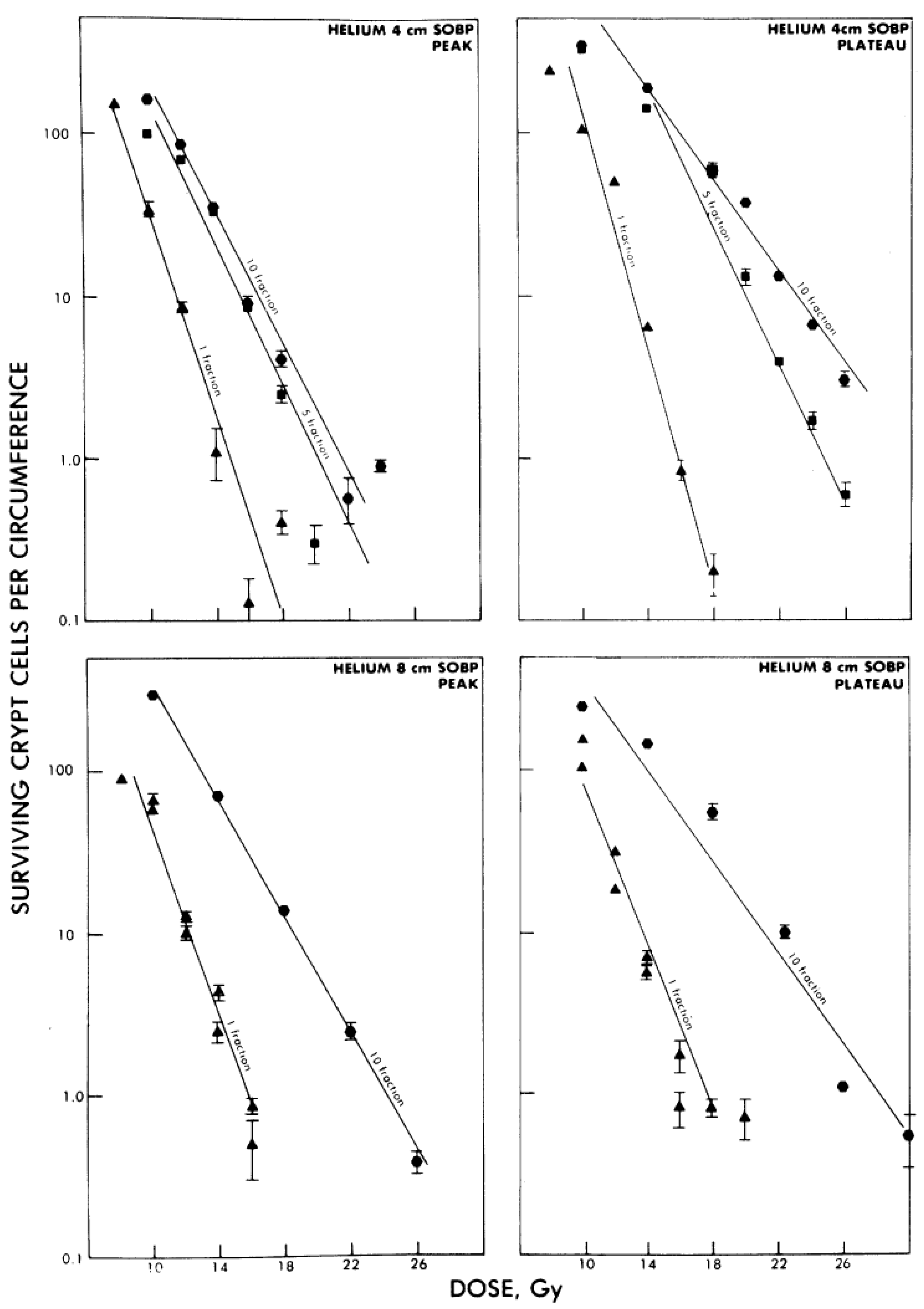
  76. Goldstein, L.S.; Philips, T.L.; Ross, G.Y. Enhancement by fractionation of biological peak-to-plateau relative biological effectiveness ratios for heavy ions. Int. J. Radiat. Oncol. Biol. Phys. 1978, 4, 1033–1037. [Google Scholar] [CrossRef]
  77. Fukutsu, K.; Kanai, T.; Furusawa, Y.; Ando, K. Response of mouse intestine after single and fractionated irradiation with accelerated carbon ions with a spread-out Bragg peak. Radiat. Res. 1997, 148, 168–174. [Google Scholar] [CrossRef]
  78. Goldstein, L.S.; Phillips, T.L.; Ross, G.Y. Biological effects of accelerated heavy ions. II. Fractionated irradiation of intestinal crypt cells. Radiat. Res. 1981, 86, 542–558. [Google Scholar] [CrossRef]
  79. Chang, P.Y.; Bakke, J.; Puey, A. Fractionated exposure of high energy iron ions has a sparing effect in vivo. Adv. Space Res. 2007, 40, 568–575. [Google Scholar] [CrossRef]
  80. Suzuki, M.; Kase, Y.; Kanai, T.; Ando, K. Change in radiosensitivity with fractionated-dose irradiation of carbon-ion beams in five different human cell lines. Int. J. Radiat. Oncol. 2000, 48, 251–258. [Google Scholar]
  81. Elgqvist, J.; Andersson, H.; Back, T.; Claesson, I.; Hultborn, R.; Jensen, H.; Lindegren, S.; Olsson, M.; Palm, S.; Warnhammar, E.; Jacobsson, L. Fractionated radioimmunotherapy of intraperitoneally growing ovarian cancer in nude mice with 211At-MX35 F(ab')2: Therapeutic efficacy and myelotoxicity. Nucl. Med. Biol. 2006, 33, 1065–1072. [Google Scholar] [CrossRef]
  82. Lucke-Huhle, C.; Blakely, E.A.; Chang, P.Y.; Tobias, C.A. Drastic G2 arrest in mammalian cells after irradiation with heavy-ion beams. Radiat. Res. 1979, 79, 97–112. [Google Scholar] [CrossRef]
  83. Luckehuhle, C. Alpha-Irradiation-Induced G2 Delay—A Period of Cell Recovery. Radiat. Res. 1982, 89, 298–308. [Google Scholar] [CrossRef]
  84. Bird, R.P.; Burki, H.J. Survival of synchronized Chinese hamster cells exposed to radiation of different linear-energy transfer. Int. J. Radiat. Biol. Relat. Stud. Phys. Chem. Med. 1975, 27, 105–120. [Google Scholar] [CrossRef]
  85. Wenzl, T.; Wilkens, J.J. Modelling of the oxygen enhancement ratio for ion beam radiation therapy. Phys. Med. Biol. 2011, 56, 3251–3268. [Google Scholar]
  86. Michalik, V. Model of DNA Damage Induced by Radiations of Various Qualities. Int. J. Radiat. Biol. 1992, 62, 9–20. [Google Scholar] [CrossRef]
  87. Fyles, A.W.; Milosevic, M.; Wong, R.; Kavanagh, M.C.; Pintilie, M.; Sun, A.; Chapman, W.; Levin, W.; Manchul, L.; Keane, T.J.; Hill, R.P. Oxygenation predicts radiation response and survival in patients with cervix cancer. Radiother. Oncol. 1998, 48, 149–156. [Google Scholar] [CrossRef]
  88. Fyles, A.; Milosevic, M.; Hedley, D.; Pintilie, M.; Levin, W.; Manchul, L.; Hill, R.P. Tumor hypoxia has independent predictor impact only in patients with node-negative cervix cancer. J. Clin. Oncol. 2002, 20, 680–687. [Google Scholar] [CrossRef]
  89. Hockel, M.; Schlenger, K.; Aral, B.; Mitze, M.; Schaffer, U.; Vaupel, P. Association between tumor hypoxia and malignant progression in advanced cancer of the uterine cervix. Cancer Res. 1996, 56, 4509–4515. [Google Scholar]
  90. Nakano, T.; Suzuki, Y.; Ohno, T.; Kato, S.; Suzuki, M.; Morita, S.; Sato, S.; Oka, K.; Tsujii, H. Carbon beam therapy overcomes the radiation resistance of uterine cervical cancer originating from hypoxia. Clin. Cancer Res. 2006, 12, 2185–2190. [Google Scholar] [CrossRef]
  91. Semenza, G.L. Targeting HIF-1 for cancer therapy. Nat. Rev. Cancer 2003, 3, 721–732. [Google Scholar] [CrossRef]
  92. O'Hara, J.A.; Blumenthal, R.D.; Grinberg, O.Y.; Demidenko, E.; Grinberg, S.; Wilmot, C.M.; Taylor, A.M.; Goldenberg, D.M.; Swartz, H.M. Response to radioimmunotherapy correlates with tumor pO2 measured by EPR oximetry in human tumor xenografts. Radiat. Res. 2001, 155, 466–473. [Google Scholar] [CrossRef]
  93. Adams, G.P.; Shaller, C.C.; Chappell, L.L.; Wu, C.; Horak, E.M.; Simmons, H.H.; Litwin, S.; Marks, J.D.; Weiner, L.M.; Brechbiel, M.W. Delivery of the alpha-emitting radioisotope bismuth-213 to solid tumors via single-chain Fv and diabody molecules. Nucl. Med. Biol. 2000, 27, 339–346. [Google Scholar] [CrossRef]
  94. Ritter, M.A.; Cleaver, J.E.; Tobias, C.A. High-Let Radiations Induce a Large Proportion of Non-Rejoining DNA Breaks. Nature 1977, 266, 653–655. [Google Scholar]
  95. Roots, R.; Yang, T.C.; Craise, L.; Blakely, E.A.; Tobias, C.A. Impaired repair capacity of DNA breaks induced in mammalian cellular DNA by accelerated heavy ions. Radiat. Res. 1979, 78, 38–49. [Google Scholar] [CrossRef]
  96. Fillingham, J.; Keogh, M.C.; Krogan, N.J. GammaH2AX and its role in DNA double-strand break repair. Biochem. Cell Biol. 2006, 84, 568–577. [Google Scholar] [CrossRef]
  97. Schmid, T.E.; Dollinger, G.; Beisker, W.; Hable, V.; Greubel, C.; Auer, S.; Mittag, A.; Tarnok, A.; Friedl, A.A.; Molls, M.; Roper, B. Differences in the kinetics of gamma-H2AX fluorescence decay after exposure to low and high LET radiation. Int. J. Radiat. Biol. 2010, 86, 682–691. [Google Scholar]
  98. Leatherbarrow, E.L.; Harper, J.V.; Cucinotta, F.A.; O'Neill, P. Induction and quantification of gamma-H2AX foci following low and high LET-irradiation. Int. J. Radiat. Biol. 2006, 82, 111–118. [Google Scholar] [CrossRef]
  99. Lieber, M.R. The mechanism of double-strand DNA break repair by the nonhomologous DNA end-joining pathway. Annu. Rev. Biochem. 2010, 79, 181–211. [Google Scholar] [CrossRef]
  100. Anderson, J.A.; Harper, J.V.; Cucinotta, F.A.; O'Neill, P. Participation of DNA-PKcs in DSB Repair after Exposure to High- and Low-LET Radiation. Radiat. Res. 2010, 174, 195–205. [Google Scholar] [CrossRef]
  101. Kinashi, Y.; Takahashi, S.; Kashino, G.; Okayasu, R.; Masunaga, S.; Suzuki, M.; Ono, K. DNA double-strand break induction in Ku80-deficient CHO cells following Boron Neutron Capture Reaction. Radiat. Oncol. 2011, 6, 106. [Google Scholar] [CrossRef]
  102. Okayasu, R.; Okada, M.; Okabe, A.; Noguchi, M.; Takakura, K.; Takahashi, S. Repair of DNA damage induced by accelerated heavy ions in mammalian cells proficient and deficient in the non-homologous end-joining pathway. Radiat. Res. 2006, 165, 59–67. [Google Scholar] [CrossRef]
  103. Zafar, F.; Seidler, S.B.; Kronenberg, A.; Schild, D.; Wiese, C. Homologous recombination contributes to the repair of DNA double-strand breaks induced by high-energy iron ions. Radiat. Res. 2010, 173, 27–39. [Google Scholar] [CrossRef]
  104. Xue, L.; Yu, D.; Furusawa, Y.; Okayasu, R.; Tong, J.; Cao, J.; Fan, S. Regulation of ATM in DNA double strand break repair accounts for the radiosensitivity in human cells exposed to high linear energy transfer ionizing radiation. Mutat. Res. 2009, 670, 15–23. [Google Scholar] [CrossRef]
  105. Friesen, C.; Glatting, G.; Koop, B.; Schwarz, K.; Morgenstern, A.; Apostolidis, C.; Debatin, K.M.; Reske, S.N. Breaking chemoresistance and radioresistance with [Bi-213]anti-CD45 antibodies in leukemia cells. Cancer Res. 2007, 67, 1950–1958. [Google Scholar]
  106. Bonner, J.A.; Harari, P.M.; Giralt, J.; Azarnia, N.; Shin, D.M.; Cohen, R.B.; Jones, C.U.; Sur, R.; Raben, D.; Jassem, J.; et al. Radiotherapy plus cetuximab for squamous-cell carcinoma of the head and neck. N. Engl. J. Med. 2006, 354, 567–578. [Google Scholar] [CrossRef]
  107. Kriegs, M.; Kasten-Pisula, U.; Rieckmann, T.; Holst, K.; Saker, J.; Dahm-Daphi, J.; Dikomey, E. The epidermal growth factor receptor modulates DNA double-strand break repair by regulating non-homologous end-joining. DNA Repair 2010, 9, 889–897. [Google Scholar] [CrossRef]
  108. Myllynen, L.; Rieckmann, T.; Dahm-Daphi, J.; Kasten-Pisula, U.; Petersen, C.; Dikomey, E.; Kriegs, M. In tumor cells regulation of DNA double strand break repair through EGF receptor involves both NHEJ and HR and is independent of p53 and K-Ras status. Radiother. Oncol. 2011, 101, 147–151. [Google Scholar] [CrossRef]
  109. Dittmann, K.; Mayer, C.; Rodemann, H.P. Inhibition of radiation-induced EGFR nuclear import by C225 (Cetuximab) suppresses DNA-PK activity. Radiother. Oncol. 2005, 76, 157–161. [Google Scholar] [CrossRef]
  110. Jensen, A.D.; Nikoghosyan, A.; Hinke, A.; Debus, J.; Munter, M.W. Combined treatment of adenoid cystic carcinoma with cetuximab and IMRT plus C12 heavy ion boost: ACCEPT [ACC, Erbitux (R) and particle therapy]. BMC Cancer 2011, 11, 70. [Google Scholar] [CrossRef]
  111. Allen, G.W.; Saba, C.; Armstrong, E.A.; Huang, S.M.; Benavente, S.; Ludwig, D.L.; Hicklin, D.J.; Harari, P.M. Insulin-like growth factor-I receptor signaling blockade combined with radiation. Cancer Res. 2007, 67, 1155–1162. [Google Scholar]
  112. Macaulay, V.M.; Salisbury, A.J.; Bohula, E.A.; Playford, M.P.; Smorodinsky, N.I.; Shiloh, Y. Downregulation of the type 1 insulin-like growth factor receptor in mouse melanoma cells is associated with enhanced radiosensitivity and impaired activation of Atm kinase. Oncogene 2001, 20, 4029–4040. [Google Scholar]
  113. Szabo, C.I.; King, M.C. Inherited breast and ovarian cancer. Hum. Mol. Genet. 1995, 4, 1811–1817. [Google Scholar]
  114. Fackenthal, J.D.; Olopade, O.I. Breast cancer risk associated with BRCA1 and BRCA2 in diverse populations. Nat. Rev. Cancer 2007, 7, 937–948. [Google Scholar] [CrossRef]
  115. Risch, H.A.; McLaughlin, J.R.; Cole, D.E.; Rosen, B.; Bradley, L.; Kwan, E.; Jack, E.; Vesprini, D.J.; Kuperstein, G.; Abrahamson, J.L.; et al. Prevalence and penetrance of germline BRCA1 and BRCA2 mutations in a population series of 649 women with ovarian cancer. Am. J. Hum. Genet. 2001, 68, 700–710. [Google Scholar] [CrossRef]
  116. Ramus, S.J.; Gayther, S.A. The contribution of BRCA1 and BRCA2 to ovarian cancer. Mol. Oncol. 2009, 3, 138–150. [Google Scholar] [CrossRef]
  117. Goggins, M.; Schutte, M.; Lu, J.; Moskaluk, C.A.; Weinstein, C.L.; Petersen, G.M.; Yeo, C.J.; Jackson, C.E.; Lynch, H.T.; Hruban, R.H.; et al. Germline BRCA2 gene mutations in patients with apparently sporadic pancreatic carcinomas. Cancer Res. 1996, 56, 5360–5364. [Google Scholar]
  118. Lynch, H.T.; de la Chapelle, A. Hereditary colorectal cancer. N. Engl. J. Med. 2003, 348, 919–932. [Google Scholar] [CrossRef]
  119. Nieuwenhuis, B.; Van Assen-Bolt, A.J.; Van Waarde-Verhagen, M.A.; Sijmons, R.H.; Van der Hout, A.H.; Bauch, T.; Streffer, C.; Kampinga, H.H. BRCA1 and BRCA2 heterozygosity and repair of X-ray-induced DNA damage. Int. J. Radiat. Biol. 2002, 78, 285–295. [Google Scholar]
  120. Trenz, K.; Schutz, P.; Speit, G. Radiosensitivity of lymphoblastoid cell lines with a heterozygous BRCA1 mutation is not detected by the comet assay and pulsed field gel electrophoresis. Mutagenesis 2005, 20, 131–137. [Google Scholar] [CrossRef]
  121. Baeyens, A.; Thierens, H.; Claes, K.; Poppe, B.; de Ridder, L.; Vral, A. Chromosomal radiosensitivity in BRCA1 and BRCA2 mutation carriers. Int. J. Radiat. Biol. 2004, 80, 745–756. [Google Scholar] [CrossRef]
  122. Beucher, A.; Deckbar, D.; Schumann, E.; Krempler, A.; Frankenberg-Schwager, M.; Lobrich, M. Elevated radiation-induced gammaH2AX foci in G2 phase heterozygous BRCA2 fibroblasts. Radiother. Oncol. 2011, 101, 46–50. [Google Scholar]
  123. Leong, T.; Whitty, J.; Keilar, M.; Mifsud, S.; Ramsay, J.; Birrell, G.; Venter, D.; Southey, M.; McKay, M. Mutation analysis of BRCA1 and BRCA2 cancer predisposition genes in radiation hypersensitive cancer patients. Int. J. Radiat. Oncol. Biol. Phys. 2000, 48, 959–965. [Google Scholar] [CrossRef]
  124. Clarke, R.A.; Goozee, G.R.; Birrell, G.; Fang, Z.M.; Hasnain, H.; Lavin, M.; Kearsley, J.H. Absence of ATM truncations in patients with severe acute radiation reactions. Int. J. Radiat. Oncol. 1998, 41, 1021–1027. [Google Scholar] [CrossRef]
  125. Shayeghl, M.; Seal, S.; Regan, J.; Collins, N.; Barfoot, R.; Rahman, N.; Ashton, A.; Moohan, M.; Wooster, R.; Owen, R.; et al. Heterozygosity for mutations in the ataxia telangiectasia gene is not a major cause of radiotherapy complications in breast cancer patients. Br. J. Cancer 1998, 78, 922–927. [Google Scholar]
  126. Zhou, G.; Smilenov, L.B.; Lieberman, H.B.; Ludwig, T.; Hall, E.J. Radiosensitivity to high energy iron ions is influenced by heterozygosity for Atm, Rad9 and Brca1. Adv. Space Res. 2010, 46, 681–686. [Google Scholar] [CrossRef]
  127. Worgul, B.V.; Smilenov, L.; Brenner, D.J.; Vazquez, M.; Hall, E.J. Mice heterozygous for the ATM gene are more sensitive to both X-ray and heavy ion exposure than are wildtypes. Adv. Space Res. 2005, 35, 254–259. [Google Scholar] [CrossRef]

