Next Article in Journal
Antibody Fragments Defining Biologically Relevant Conformations of Target Proteins
Previous Article in Journal
Reduced Culture Temperature Differentially Affects Expression and Biophysical Properties of Monoclonal Antibody Variants
 
 
Addendum published on 22 September 2015, see Antibodies 2015, 4(3), 278.
Article

Polyclonal Antibody Therapies for Clostridium difficile Infection

by
Michael R. Simon
1,*,
Stephanie M. Chervin
2 and
Stephen C. Brown
3
1
Secretory IgA, Inc., Ann Arbor, and Allergy and Clinical Immunology, William Beaumont Hospital, Royal Oak, MI 48073, USA
2
Secretory IgA, Inc., Ann Arbor, MI, USA
3
Secretory IgA, Inc., Ann Arbor, MI, USA
*
Author to whom correspondence should be addressed.
Antibodies 2014, 3(4), 272-288; https://doi.org/10.3390/antib3040272
Submission received: 26 August 2014 / Revised: 18 September 2014 / Accepted: 28 September 2014 / Published: 28 October 2014
(This article belongs to the Special Issue Antibodies Therapies against Infectious Diseases)

Abstract

:
Clostridium difficile infection has emerged as a growing worldwide health problem. The colitis of Clostridium difficile infection results from the synergistic action of C. difficile secreted toxins A and B upon the colon mucosa. A human monoclonal IgG anti-toxin has demonstrated the ability in combination therapy to reduce mortality in C. difficile challenged hamsters. This antibody is currently in a clinical trial for the treatment of human Clostridium difficile infection. More than one group of investigators has considered using polyclonal bovine colostral antibodies to toxins A and B as an oral passive immunization. A significant proportion of the healthy human population possesses polyclonal antibodies to the Clostridium difficile toxins. We have demonstrated that polyclonal IgA derived from the pooled plasma of healthy donors possesses specificity to toxins A and B and can neutralize these toxins in a cell-based assay. This suggests that secretory IgA prepared from such pooled plasma IgA may be able to be used as an oral treatment for Clostridium difficile infection.

1. Introduction

Clostridium difficile (C. difficile) infection (CDI) is a serious and growing worldwide health problem. The estimated incidence of C. difficile-infection among patients hospitalized in the U.S. is >300,000 cases annually which is about 25% of all antibiotic-associated diarrhea [1,2]. Most patients require antimicrobial treatment with metronidazole or vancomycin. Approximately 24% of patients with C. difficile colitis do not respond favorably to the standard treatment of metronidazole or vancomycin and will suffer a relapse [3]. Consequently, the development of alternative therapeutic approaches to combat this infection are urgently needed [4,5].
The colitis of Clostridium difficile-associated disease results from the synergistic action of C. difficile secreted toxins A (CdtA) and B (CdtB) (also commonly referred to as TcdA and TcdB, respectively) upon the colon mucosa [6,7,8,9]. Together, the toxins disrupt cell–cell tight junctions of the colon thereby allowing the bacterium to adhere to the underlying colon tissue and feed upon the nutrients released by the damaged epithelium [10]. Successive rounds of C. difficile colonization, replication and toxin production lead to a vigorous host inflammatory response resulting in further degradation of the gut tissue, the pseudomembranous pathology associated with the diarrhea, and the pain observed in Clostridium difficile-associated disease.
We are developing an orally available polyclonal IgA treatment for C. difficile infection. This report documents the progress that we have made toward the proof of principle for the use of semisynthetic secretory IgA produced by combining plasma-derived IgA dimer and recombinant human secretory component. IgA dimers are required because secretory component does not bind to IgA monomers. Semisynthetic secretory IgA is being developed because it withstands gastrointestinal digestion better than monomeric antibodies which do not contain secretory component.
The advantages of oral immunotherapy with IgA are that the antibodies are delivered to the intestines where the CdtA and CdtB are active, and that treatment is more easily accomplished in outpatients. We believe that such an anti-C. difficile toxin antibody administration in combination with antibiotic treatment will decrease morbidity and death in patients at increased risk.

2. Human Antibody Responses to C. difficile Toxins

IgG directed against CdtA and CdtB is present in the general population [11,12]. In general, the three major classes of circulating immunoglobulins (IgM, IgG and IgA) share the same range of antigenic specificities, i.e., they share the same idiotypic determinants [13]. Toxin-specific circulating IgA titers are similar to the titers of toxin-specific IgG [14]. Dimeric IgA is present in plasma [15,16,17]. This is important because we propose that IgA derived from the plasma of healthy donors contains IgA with specificity to the C. difficile toxins A and B.

3. Animal Studies

Animal studies demonstrate that immunity to CdtA and CdtB can be achieved and is protective. Vaccination against Clostridium difficile toxins A and B results in active immunity against C. difficile disease in hamsters [18]. Several studies have demonstrated that passive immunization delivered as an acute treatment after disease onset provides protection against Clostridium difficile-associated disease. In a hamster disease model, serum antibodies against C. difficile derived from chickens protect hamsters against C. difficile disease after oral administration [19]. Similarly, a human monoclonal IgG anti-toxin has demonstrated the ability in combination therapy to reduce mortality in C. difficile challenged hamsters from 100%–45% in a primary disease model, and from 78%–32% in a relapse disease model [20]. High potency humanized monoclonal antibodies to CdtA and CdtB have been produced and offered protection to 95% of hamsters with CDI while all of the control animals died [21]. Another group has succeeded in preparing monoclonal antibodies against CdtA and CdtB which are multivalent and effectively oligoclonal [22]. Increased valency of monoclonal antibodies may be important in engaging the host response to the antibody after being bound to toxin [23]. Polyclonal antibodies recognize multiple epitopes [24]. These studies of oligoclonal and polyvalent monoclonal antibodies support the contention that polyclonal antibody is likely to be more effective at neutralizing toxin than monoclonal antibody.
Maternal antibodies in breast milk transfer protection against C. difficile ileocecitis to infant hamsters [25]. It has been reported that bovine IgG concentrate from the colostrum of cows vaccinated with C. difficile toxoid protects hamsters against antibiotic-associated cecitis [26]. Administration of an immunoglobulin product containing specific antibodies to C. difficile results in the elimination of C. difficile toxins and also in killing the bacteria within the colon [27]. In another study, bovine anti-C. difficile cells and toxin antibody-enriched whey were administered to hamsters by gavage beginning 3 h before bacterial challenge and then for 3 days subsequently. All of the untreated animals died, while 17 of 20 treated animals survived [28]. In summary, these studies demonstrated that in the hamster model, orally administered allogeneic and xenogeic IgA (in breast milk and whey), and xenogeneic IgG (chicken and bovine) provide protection against Clostridium difficile-associated disease. This subject was reviewed by Hussack and Tanha [29].

4. Experience in Humans

4.1. Passive Immunization with Intravenous IgG

4.1.1. Polyclonal Human IgG

Passive immunization using human intravenous polyclonal IgG derived from healthy plasma donors has been reported to facilitate treatment in patients with C. difficile disease [12,30,31,32,33,34]. This is especially true for patients who lacked circulating antibodies to the C. difficile toxins [30,34]. There have also been reports that cast doubt on the efficacy of the use of intravenous immunoglobulin treatment of C. difficile infection [35,36]. This publication record has been reviewed by Hussack and Tanha [29].

4.1.2. Monoclonal IgG

A recent clinical trial demonstrated that fully human monoclonal antibodies against CdtA and CdtB delivered intravenously in combination with antibiotic therapy reduced the recurrence of C. difficile compared to patients treated with antibiotic therapy and placebo alone [5]. The clinical trial tested the intravenous administration of humanized monoclonal antibodies) (MDX-066 and MDX-1388, also known as CDA-1 and CDB-1, respectively) for the treatment of C. difficile disease. Passive immunization with this parenteral human monoclonal IgG anti-toxin A and B in combination with antibiotic therapy was demonstrated to be efficacious in reducing recurrence of C. difficile disease from 25%–7% [5]. A Phase III study is now in progress. Please see Humphreys and Wilcox [37] for a thorough review of the use of monoclonal antibodies in the treatment of C. difficile infection.

4.2. Passive Immunization with Oral Polyclonal Human Immunoglobulins A and G

An early case study of a young child with refractory antibiotic-associated diarrhea and C. difficile toxin A detected in his stools demonstrated the feasibility of a combination therapy of polyclonal plasma IgA admixed with IgG (2:1) (IgAbulin, Immuno AG, Vienna) (100 mg/mL) together with vancomycin. The child improved on this treatment [38]. A similar efficacy was noted in another case report wherein polyclonal IgG derived from pooled human plasma was administered orally to another child with refractory C. difficile diarrhea who had failed treatment with antibiotics and intravenous polyclonal IgG. This child lacked circulating antibodies to C. difficile toxin. The patient received oral polyclonal IgG at 200 mg/kg/day every 2 days for three doses together with courses of oral vancomycin and Lactobacillus. The child had recovered at follow-up evaluation 2 weeks later [39]. These reports demonstrate the efficacy of passive parenteral and oral immunization with pooled immunoglobulins derived from the general population. It appears that polyclonal monomeric circulatory immunoglobulins possess efficacy.

4.3. Passive Immunization with Oral Polyclonal Bovine Immunoglobulins A and G

Anti-C. difficile toxin bovine immunoglobulin concentrate derived from bovine colostrums was administered to 11 patients with mild to moderate C. difficile-associated diarrhea. Ten grams were administered four times daily for 10 days, while six patients received placebo. There was no difference in improvement between treatment groups [40]. In another study, bovine antibody-enriched whey was obtained from cows immunized with whole C difficile cells and toxoid [28]. IgG and secretory IgA to C. difficile cells and to toxin A in milk were demonstrated. Sixteen patients who had suffered Clostridium difficile-associated disease, nine of whom had experienced relapsed disease, were studied. They were treated with 5 g of whey given three times daily for 2 weeks following completion of antibiotic therapy. None developed relapsed disease during a median follow-up period of 333 days (range 35–365 days). Stool samples were negative for Clostridium difficile toxin at 1 week after completion of treatment with bovine antibody-enriched whey in 14 of 15 patients. The fifteenth patient’s stool became negative for toxin one week later. Recent studies have alternatively revealed response and relapse rates equivalent to that of metronidazole [41] or reduced the relapse rate by 50% [42]. A potential disadvantage of the use of bovine colostrums is that IgG comprises 86% of the antibodies in bovine colostrums [43]. This IgG is subject to digestion when swallowed by humans [44].

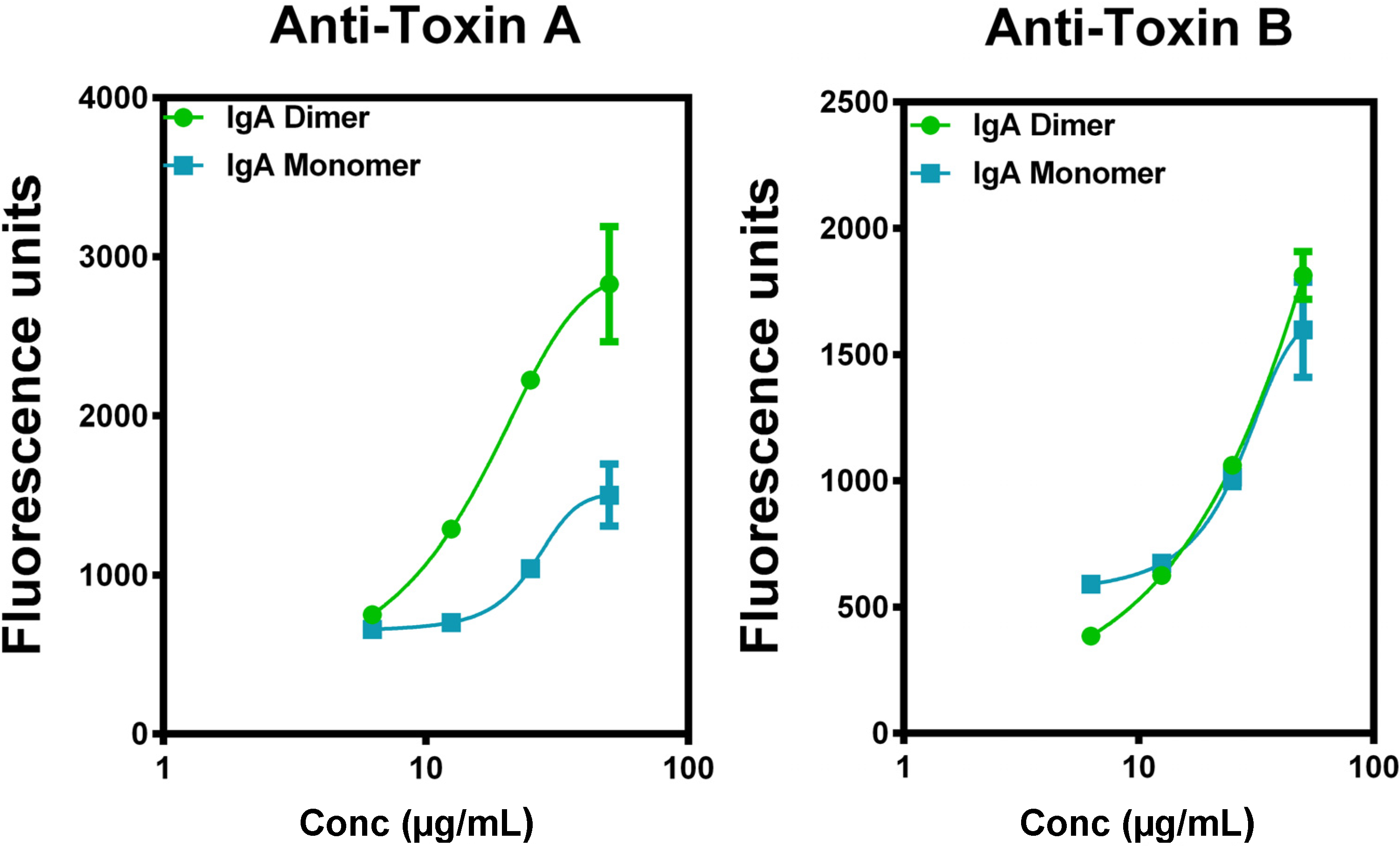
4.4. Superiority of IgA to IgG in Vitro

In vitro experiments have demonstrated that polymeric IgA is superior to monomeric IgA and IgG in preventing C. difficile toxin damage to intestinal epithelial cell monolayers [45]. Selective neutralization of C. difficile toxin by serum IgA has also been demonstrated [46]. IgA1 conjugated to secretory component is just as resistant to protease digestion as naturally occurring secretory IgA, which is predominantly of the IgA2 subclass [47]. This work showing the preparation of secretory IgA from human plasma IgA which is predominantly IgA1 has been confirmed and extended [17]. This semisynthetic secretory IgA also resists peptic digestion. Polymeric secretory IgA is likely to be more potent than IgG via the oral route, since it will not be digested.

4.5. Medical Plausibility for the Treatment of Relapsed C. difficile Infection with Secretory IgA (Table 1)

Many patients with relapsed Clostridium difficile-associated disease lack circulating antibodies to the C. difficile toxins [48,49]. Such patients appear most likely to benefit from passive immunization with intravenous IgG [30,34]. It is to be expected that increased efficacy will be achieved by oral secretory IgA as compared to oral monomeric IgA and IgG because of the propensity of monomeric immunoglobulins which are not protected by secretory component to degrade in the gastrointestinal tract [44].
Table 1. Potential Advantages of Oral Human Secretory IgA for the Treatment of C. difficile Infection.
Advantage over Parenteral Monoclonal IgG and Human Polyclonal IgG (IVIg)
Polyclonal – specificity for multiple epitopes
Delivered to the site of infection in the intestines
Advantage over Oral Bovine Colostral IgG and Oral Human Polyclonal IgG
Secretory component protects the IgA antibody from digestion
Advantage over Oral Human Colostral IgA
Raw material is available in vast quantities as an industrial byproduct
Orally-administered secretory IgA is likely to have better safety and efficacy than a parenterally-administered monoclonal IgG. This human secretory IgA will more likely be less immunogenic although xenogenic proteins are commonly eaten without a deleterious immune response. C. difficile toxins are present in the gut lumen, not the systemic circulation, and secretory IgA is much more stable in that milieu than IgG antibodies.
While demonstrating the promising role of immunoglobulin-based therapy to combat C. difficile associated disease [32,33,34,49,50,51], treatment with an IgG has the disadvantage that IgG must be administered parenterally to avoid digestive degradation. An immunoglobulin therapy administered orally has the advantage of more direct delivery of the neutralizing antibody to the gut. This therapy is possible using secretory IgA, which is resistant to digestive proteolysis [17].

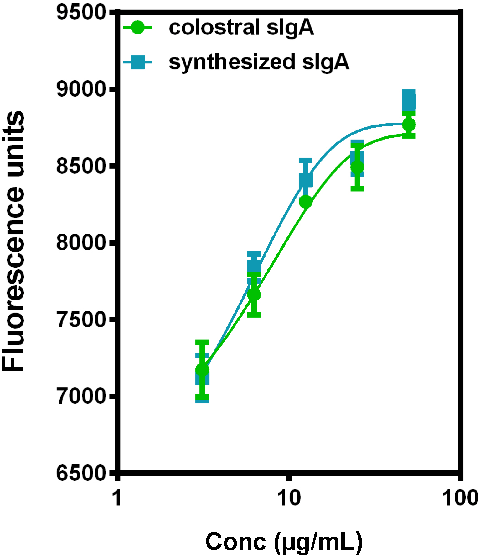
5. Current Studies of the Preparation of Anti-C. difficile Secretory IgA from Pooled Human Plasma IgA

There is a need for new non-antibiotic based therapeutic approaches to treat C. difficile infection. We are developing a novel therapeutic approach consisting of an orally administered immunotherapy consisting of polyclonal human secretory IgA (sIgA) formed by combining plasma derived dimeric IgA with recombinant human secretory component. We expect that this will provide a significant clinical advantage over passive immunization with parenterally administered polyclonal and recombinant monoclonal IgG antibodies.
We report the experiments that we have performed while working toward this goal. The starting material is Cohn fraction III precipitate which contains IgA. Cohn fraction III precipitate is a byproduct of the manufacture of intravenous immunoglobulin (IVIg). Tons of Cohn fraction III precipitate are discarded each year. IgA was purified from the Cohn fraction III precipitate by jacalin affinity chromatography [52,53]. Jacalin is a lectin extracted from the jack bean (Canavalia ensiformis). It binds the galactose side chains present on IgA. The IgA can then be eluted from jacalin affinity columns using 0.1 M galactose in phosphate buffered saline (PBS).
Figure 1 shows a diffuse band (Lane C) at the expected MW (160,000 daltons) of monomeric IgA eluted by galactose from jacalin. Figure 2 shows IgA anti-C. difficile Toxin A (CdtA) ELISAs. Figure 2A shows that CFxIII and jacalin eluate demonstrate binding to CdtA by IgA ELISA. Figure 2B shows a titration curve of a recombinant anti-CdtA-specific IgA positive control antibody (from Blaise Corthesy, Lusanne, Switzerland) and jacalin eluate in a CdtA IgA ELISA [45]. This demonstrates that the anti-CdtA binding activity isolated from CFxIII is IgA. The concentration of the monoclonal positive control is in ng/mL while the concentration of the polyclonal IgA is in µg/mL. This is because the polyclonal IgA has specificities to many antigens while the monoclonal IgA is monospecific. The monoclonal IgA not only serves as a positive control for the assay but also provides an estimate of the proportion of total plasma-derived IgA that has anti-CdtA specificity. The advantage of using polyclonal IgA is that it has specificity to all the epitopes, and especially the immunodominant epitopes, present on the CdtA and CdtB molecules. Monoclonal IgA has specificity to only one epitope. The polyclonal IgA can be enriched for the anti-CdtA and anti-CdtB antibody molecules by using CdtA and CdtB affinity column purification. The titer of anti-CdtA and anti-CdtB secretory IgA can be adjusted as needed. Figure 2C shows binding of IgA to CdtB and to CdtA in an ELISA.
Figure 1. Figure 1 shows non-reducing sodium dodecyl sulfate (SDS)-polyacrylamide gel electrophoresis (PAGE) analysis of jacalin affinity purified IgA from Cohn fraction III precipitate. Coomassie blue stain. Lane A: Molecular weight standards; Lane B: Solubilized Cohn fraction III proteins; Lane C: jacalin affinity purified IgA from Cohn fraction III proteins. A diffuse band is present in Lane C at the expected MW (160 kDa) of monomeric IgA eluted by galactose from jacalin (jackbean lectin).
Antibodies 03 00272 g001
Figure 2. Figure 2 shows IgA anti-C. difficile Toxin A ELISAs. Both the crude Cohn fraction III preparation (green line, green dots) and the jacalin eluate containing polyclonal plasma-derived IgA (blue line, blue squares) demonstrated binding to C. difficile toxin A (bound to the plate) by ELISA, while the jacalin flow-through protein fraction (red line, red triangles) did not (Figure 2A). This demonstrates that anti-C. difficile toxin A binding activity isolated from Cohn fraction III is IgA. Figure 2A shows the fluorescence units at sequentially lower dilutions beginning at a 1:10 dilution (0.1 dilution factor), 1:5 dilution (0.2 dilution factor), etc. Cohn FxIII: resolublized Cohn fraction III precipitate; Jacalin +: jacalin (jack bean lectin) IgA eluate; Jacalin -: jacalin flow-through. The figure (B) shows the anti-C. difficile Toxin A binding (CdtA is bound to the plate) of plasma derived polyclonal IgA and monoclonal anti-CdtA IgA which is used as a positive control (Blaise Corthesy, Lausanne, Switzerland). The figure (C) shows the relative anti-CdtA and CdtB binding of plasma derived polyclonal IgA (CdtA or CdtB is bound to the plate); mAB: monoclonal antibody; pAb: polyclonal antibody; anti-CdtA: anti-C. difficile Toxin A; anti- CdtB: anti-C. difficile Toxin B.
Antibodies 03 00272 g002
A typical chromatogram of jacalin purified IgA in our lab revealed that the dimer peak comprised 18% of the total IgA (Figure 3A). Figure 3C demonstrates that IgA dimer is present in plasma IgA derived from CFxIII precipitate. The plate was coated with anti-J chain antibody while the secondary antibody was specific for IgA heavy chains. This proves that the protein that bound to the plate contained J chain and IgA and is therefore IgA dimer. The colostral human secretory IgA served as the positive control. The advantage of using semisynthetic secretory IgA derived from a plasma byproduct, rather than human colostral secretory IgA, is that the plasma derived IgA is available in vast quantities (many tons annually) as the byproduct of the production of IVIg.
As shown in Figure 4, polyclonal IgA neutralizes toxicity of CdtA and CdtB toxins (CellToxGreen, Promega) in the CT26.WT mouse colon carcinoma cell culture (ATCC, Manassas, VA, USA). Figure 5 demonstrates that both dimeric and monomeric IgA bind both to CdtA and CdtB. There is more binding to CdtA by dimeric IgA than by monomeric IgA. There is similar binding to CdtB by dimers and monomers.
Figure 6 demonstrates the in vitro preparation of secretory IgA from plasma IgA which contains both monomeric and dimeric IgA. CSL Behring LTD has prepared a similar secretory IgA from their pooled human plasma and has found that it is effective in an animal model of C. difficile infection (personal communication, Blaise Corthesy). We expect that the dosing of such a medication would be lower than the dosing of monomeric IgA given to the pediatric patient described by Tjellstrom et al. [34]. We anticipate the dose to be lower because the secretory IgA is resistant to digestion. We have developed fully scalable proprietary methods for the production of the secretory IgA from Cohn fraction III precipitate. However, it is not possible to adequately understand the cost of production at this early stage of development.
Figure 3. The ELISA results shown in Figure 3 demonstrate that IgA dimer is present in plasma IgA derived from CFxIII precipitate. Figure 3A is a typical Sephacryl 300 IgA chromatogram showing the polymer, dimer and monomer peaks. The column was 85.5 cm long. The IgA chromatogram dimer peak is pictured in lane 4 of Figure 3B. The PAGE gel pictured in Figure 3B was run by Dr. George Konstantinou at the Mount Sinai School of Medicine in New York, NY, USA. Lanes 1 and 6: molecular weight ladder; lane 2: polymer peak; lanes 3 and 4: early and late portions of dimer peak; lane 5: monomer peak. Figure 3C shows the IgA dimer ELISA using IgA antibody derived from the dimer peak shown in Figure 3A. The plate was coated with anti-J chain antibody so only J chain containing dimer binds. The human colostral secretory IgA is a positive control.
Antibodies 03 00272 g003
Figure 4. Polyclonal IgA neutralizes toxicity of C. difficile toxins A and B (CellToxGreen, Promega) in the CT26.WT mouse colon carcinoma cell culture (ATCC, Manassas, VA, USA). The toxin concentration used in the cell cultures was 100 ng/mL. The amount of polyclonal plasma IgA used to neutralize the toxins was 15 mcg IgA/1 ng CdtA or CdtB. The IgA and CdtA or CdtB were incubated at 37 °C for 1 h before being added to the cell cultures. Experimental conditions from left to right: cells alone, cells plus IgA, cells plus toxin, cells plus toxin pre-incubated with IgA. Cells plus toxin A vs. cells plus toxin A pre-incubated with IgA P = 0.0067; Cells plus toxin B vs. cells plus toxin B pre-incubated with IgA P = 0.019. Crl = control; tox = toxin.
Antibodies 03 00272 g004
Figure 5. Figure 5 demonstrates that both dimeric and monomeric IgA bind both to CdtA and CdtB. There is more binding to CdtA by dimeric IgA than by monomeric IgA. There is similar binding to CdtB by dimers and monomers.
Antibodies 03 00272 g005
Figure 6. Figure 6 shows colostral sIgA (MP Biologicals, Solon, OH, USA) as a positive control compared with sIgA prepared from IgA dimer and recombinant human secretory component. Dimeric IgA associates with recombinant human secretory component (provided by Blaise Corthesy, Lausanne, Switzerland) to form secretory IgA. The ELISA plate was coated with mouse anti-secretory component antibody (antibodies-online, Atlanta, GA, USA).
Antibodies 03 00272 g006
The experiments performed in the laboratory of Secretory IgA Inc. demonstrate that human plasma derived IgA from healthy donors binds to, and neutralizes CdtA and CdtB. The published work of the CSL Behring IgA research group [17] demonstrate that plasma derived IgA dimer can be transformed into human secretory IgA that is functional. Case reports and published case series suggest that semisynthetic secretory IgA administered orally may be an effective additional treatment for C. difficile disease.

6. Experimental Section

Process for Obtaining Jacalin Affinity-Purified IgA from Cohn Fraction III Precipitate (CFxIII Precipitate). Cohn fraction III precipitate is a discarded industrial byproduct of the manufacture of intravenous immunoglobulin (IVIg). Each batch is derived from the pooled plasma of 3000–5000 healthy plasma donors. The kg aliquot used in this study was collected from the waste stream of a routinely manufactured batch in 2009. Frozen CFxIII precipitate was suspended in phosphate buffered saline (PBS), and then clarified by centrifugation and filtration (0.22 micron) to remove filter aides (diamataceous earth). IgA was isolated from solvent—detergent (1% tri (N butyl) phosphate, 1% Triton X-100) virus-inactivated CFxIII precipitate by jacalin affinity chromatography [52]. Jacalin binds to IgA via its galactose side chains. IgA is then eluted with 0.1 M galactose. Yields have approached 100% of the available IgA.
Separation of IgA Monomer and Dimer. Jacalin affinity-purified IgA in 0.1 M galactose in PBS, derived from CFxIII precipitate, was placed on a 85.5 cm long Sephacryl 300 size exclusion column on an FPLC (BioCAD Workstation for perfusion chromatography (Applied Biosystems, Foster City, CA, USA)). The monomer and dimer peaks were sufficiently separated to allow collection of pure monomer (trailing portion of the monomer peak) and pure dimer (leading portion of the dimer peak).
Human IgA anti-C. difficile Toxin A ELISA. Human IgA anti-C. difficile toxin A ELISA demonstrates IgA binding to CdtA in CFxIII and jacalin eluate, but not in the jacalin flow-through. High binding ELISA plates were coated with 1 μg/mL CdtA (TechLab, Blacksburg, VA, USA), blocked and washed with 0.1% Tween 20 in PBS. After application of positive control mouse/human chimeric IgA anti-CdtA [45] provided by Dr. Blaise Corthesy (Lausanne. Switzerland), virus-inactivated CFxIII; or jacalin eluate, or jacalin flow-through was applied. The secondary HRP-conjugated goat anti-human IgA (Zymed, Invitrogen, Carlsbad, CA, USA) was then applied. Fluorogenic Amplex Red (Invitrogen) was used for detection. Plates were read on a fluorescent plate reader (Fmax, Molecular Devices (Figure 2A); 7620, Cambridge Technologies (Figure 2B,C)). The experiments depicted in Figure 2C used plates coated with either CdtA or with CdtB. The secondary antibody remained the same. Data was plotted using a 4-parameter curve fit using Prism software (GraphPad, Inc., La Jolla, CA, USA). Dilution factors for CFxIII were normalized to the same total protein concentration as jacalin eluate and jacalin flow-through samples. Human IgA anti- CdtA concentrations were calculated using the mouse/human chimeric IgA anti- CdtA standard curve. Jacalin eluate from CFxIII at a dilution of 1:3 has the equivalent toxin A binding capacity as the standard anti- CdtA at 1 ng/mL as shown by the relative responses in this assay.
Human IgA Dimer ELISA. ELISA to quantitate J-chain-containing IgA dimers were performed and analyzed (Figure 3C) using plates coated with mouse anti-human J chain antibody (Biogenex, San Ramon, CA, USA). The secondary antibody was HRP-conjugated goat anti-human IgA (Zymed, Invitrogen). Human colostral sIgA (MP Biomedicals, Santa Ana, CA, USA) was used as the positive control. Fluorescent plate reader: 7620, Cambridge Technologies. Plasma IgA dimer was isolated by size exclusion chromatography using an 85 cm long Sephacryl column. The IgA chromatogram peak used in this ELISA is that pictured in lane 4 of Figure 3B. Figure 3B NuPAGE BisTris 4%–12% PAGE. (Invitrogen, Carlsbad, CA, USA).
In Vitro Toxin Neutralization of C. difficile Toxins A and B. IgA neutralizes C. difficile toxins A and B in CT26 cell culture (ATCC). The readout is the “CellTox Green” (Promega, Madison, WI, USA) fluorescent dye which enters dead cells and stains their DNA. The readout is obtained using an fmax (Molecular Devices) fluorescent plate reader. Higher fluorescence units means more cell death. The toxin concentration used in the cell cultures was 100 ng/mL. The amount of polyclonal plasma IgA used to neutralize the toxins was 15 mcg IgA/1 ng toxin A or B. The IgA and CdtA or CdtB were incubated at 37 °C for 1 h before being added to the cell cultures.
ELISA was used to quantitate binding of IgA dimers and monomers to toxins A and B. These ELISAs were performed using plates coated with toxin A (TechLab) or toxin B (Hanping Feng, Baltimore, MD, USA). IgA dimer and monomer were isolated by size exclusion chromatography using an 85 cm long Sephacryl 300 column. The secondary antibody was HRP-conjugated goat anti-human IgA (Zymed, Invitrogen).
ELISA to Demonstrate Laboratory Synthesis of Secretory IgA from Plasma IgA. Plate was coated with mouse anti-secretory component antibody (antibodies-online, Atlanta). Figure 6 shows colostral sIgA (MP Biologicals, Solon, OH, USA) compared with newly synthesized sIgA.
Recombinant Human Secretory Component. The secretory component used in these experiments was prepared in CHO(17) cells and was >95% pure (personal communication Blaise Corthesy). Glycosylation was primarily with galactose-β (1-4)-N-acetylglucosamine and was identical to a human colostral secretory component [54].

7. Conclusions

There is substantial evidence that antibody treatment of C. difficile infection is likely to be an important addition to antibiotic therapy. Monoclonal human IgG antibodies administered parenterally are being evaluated in a clinical trial. Semisynthetic polyclonal human secretory IgA can be prepared and is likely to be efficacious.

Acknowledgments

The authors acknowledge the contribution of George Konstantinou in the preparation of the SDS-PAGE gel pictured in Figure 3B.

Author Contributions

Dr. Simon conceived the project, wrote the manuscript and performed the laboratory work shown on Figure 2B and C, Figure 3A and C, and Figure 4 through Figure 6. Dr. Chervin assisted in the preparation of the manuscript and performed the laboratory work shown on Figure 1 and Figure 2A. Dr. Brown assisted in the preparation of the manuscript.

Conflicts of Interest

Drs. Simon, Chervin and Brown have a potential conflict of interest in that each has an equity stake in Secretory IgA, Inc. which would benefit from the comericalization of the technology described in this manuscript.

References

  1. Wilkins, T.D.; Lyerly, D.M. Clostridium difficile testing: After 20 years, still challenging. J. Clin. Microbiol. 2003, 41, 531–534. [Google Scholar]
  2. McFarland, L.V. Update on the changing epidemiology of Clostridium difficile-associated disease. Nat. Clin. Pract. Gastroenterol. Hepatol. 2008, 5, 40–48. [Google Scholar]
  3. Sunenshine, R.H.; McDonald, L.C. Clostridium difficile-associated disease: New challenges from an established pathogen. Cleve. Clin. J. Med. 2006, 73, 187–197. [Google Scholar]
  4. Shen, E.P.; Surawicz, C.M. The changing face of Clostridium difficile: What treatment options remain? Am. J. Gastroenterol. 2007, 102, 2789–2792. [Google Scholar]
  5. Lowy, I.; Molrine, D.C.; Leav, B.A.; Blair, B.M.; Baxter, R.; Gerding, D.N.; Nichol, G.; Thomas, W.D., Jr.; Leney, M.; Sloan, S.; et al. Treatment with monoclonal antibodies against Clostridium difficile toxins. N. Engl. J. Med. 2010, 362, 197–205. [Google Scholar]
  6. Lyerly, D.M.; Krivan, H.C.; Wilkins, T.D. Clostridium difficile: Its disease and toxins. Clin. Microbiol. Rev. 1988, 1, 1–18. [Google Scholar]
  7. Barroso, L.A.; Wang, S.Z.; Phelps, C.J.; Johnson, J.L.; Wilkins, T.D. Nucleotide sequence of Clostridium difficile toxin B gene. Nucleic Acids Res. 1990, 18, 4004. [Google Scholar]
  8. Dove, C.H.; Wang, S.Z.; Price, S.B.; Phelps, C.J.; Lyerly, D.M.; Wilkins, T.D.; Johnson, J.L. Molecular characterization of the Clostridium difficile toxin A gene. Infect. Immunol. 1990, 58, 480–488. [Google Scholar]
  9. Sun, X.; Savidge, T.; Feng, H. The Enterotoxicity of Clostridium difficile Toxins. Toxins 2010, 2, 1848–1880. [Google Scholar]
  10. Borriello, S.P. Pathogenesis of Clostridium difficile infection. J. Antimicrob. Chemother. 1998, 41 (Suppl. C), 13–19. [Google Scholar]
  11. Bacon, A.E., 3rd; Fekety, R. Immunoglobulin G directed against toxins A and B of Clostridium difficile in the general population and patients with antibiotic-associated diarrhea. Diagn. Microbiol. Infect. Dis. 1994, 18, 205–209. [Google Scholar]
  12. Salcedo, J.; Keates, S.; Pothoulakis, C.; Warny, M.; Castagliuolo, I.; LaMont, J.T.; Kelly, C.P. Intravenous immunoglobulin therapy for severe Clostridium difficile colitis. Gut 1997, 41, 366–370. [Google Scholar]
  13. Goodman, J.W. Immunoglobulins I: Structure & Function. In Basic & Clinical Immunology, 5th ed.; Stites, D.P., Stobo, J.D., Fudenberg, H.H., Wells, J.V., Eds.; Lange Medical Publications: Los Altos, CA, USA, 1984; Chapter 4; pp. 30–42. [Google Scholar]
  14. Johnson, S.; Gerding, D.N.; Janoff, E.N. Systemic and mucosal antibody responses to toxin A in patients infected with Clostridium difficile. J. Infect. Dis. 1992, 166, 1287–1294. [Google Scholar]
  15. Delacroix, D.L.; Hodgson, H.J.; McPherson, A.; Dive, C.; Vaerman, J.P. Selective transport of polymeric immunoglobulin A in bile. Quantitative relationships of monomeric and polymeric immunoglobulin A, immunoglobulin M, and other proteins in serum, bile, and saliva. J. Clin. Invest. 1982, 70, 230–241. [Google Scholar]
  16. Delacroix, D.L.; Elkom, K.B.; Geubel, A.P.; Hodgson, H.F.; Dive, C.; Vaerman, J.P. Changes in size, subclass, and metabolic properties of serum immunoglobulin A in liver diseases and in other diseases with high serum immunoglobulin A. J. Clin. Invest. 1983, 71, 358–367. [Google Scholar]
  17. Longet, S.; Miled, S.; Lötscher, M.; Miescher, S.M.; Zuercher, A.W.; Corthésy, B. Human plasma-derived polymeric IgA and IgM antibodies associate with secretory component to yield biologically active secretory-like antibodies. J. Biol. Chem. 2013, 288, 4085–4094. [Google Scholar]
  18. Libby, J.M.; Jortner, B.S.; Wilkins, T.D. Effects of the two toxins of Clostridium difficile in antibiotic-associated cecitis in hamsters. Infect. Immun. 1982, 36, 822–829. [Google Scholar]
  19. Kink, J.A.; Williams, J.A. Antibodies to recombinant Clostridium difficile toxins A and B are an effective treatment and prevent relapse of C. difficile-associated disease in a hamster model of infection. Infect. Immun. 1998, 66, 2018–2025. [Google Scholar]
  20. Babcock, G.J.; Broering, T.J.; Hernandez, H.J.; Mandell, R.B.; Donahue, K.; Boatright, N.; Stack, A.M.; Lowy, I.; Graziano, R.; Molrine, D.; et al. Human monoclonal antibodies directed against Toxins A and B prevent Clostridium difficile-induced mortality in hamsters. Infect. Immun. 2006, 74, 6339–6347. [Google Scholar]
  21. Marozsan, A.J.; Ma, D.; Nagashima, K.A.; Kennedy, B.J.; Kang, Y.K.; Arrigale, R.R.; Donovan, G.P.; Magargal, W.W.; Maddon, P.J.; Olson, W.C. Protection against Clostridium difficile infection with broadly neutralizing antitoxin monoclonal antibodies. J. Infect. Dis. 2012, 206, 706–713. [Google Scholar]
  22. Davies, N.L.; Compson, J.E.; Mackenzie, B.; O’Dowd, V.L.; Oxbrow, A.K.; Heads, J.T.; Turner, A.; Sarkar, K.; Dugdale, S.L.; Jairaj, M.; et al. Lightwood DJ, Humphreys DP. A mixture of functionally oligoclonal humanized monoclonal antibodies that neutralize Clostridium difficile TcdA and TcdB with high levels of in vitro potency shows in vivo protection in a hamster infection model. Clin. Vaccine Immunol. 2013, 20, 377–390. [Google Scholar]
  23. Demarest, S.J.; Hariharan, M.; Elia, M.; Salbato, J.; Jin, P.; Bird, C.; Short, J.M.; Kimmel, B.E.; Dudley, M.; Woodnutt, G.; et al. Neutralization of Clostridium difficile toxin A using antibody combinations. MAbs 2010, 2, 190–198. [Google Scholar]
  24. Shreffler, W.G.; Lencer, D.A.; Bardina, L.; Sampson, H.A. IgE and IgG4 epitope mapping by microarray immunoassay reveals the diversity of immune response to the peanut allergen, Ara h 2. J. Allergy Clin. Immunol. 2005, 116, 893–899. [Google Scholar]
  25. Kim, P.-H.; Iaconis, J.P.; Rolfe, R.D. Immunization of adult hamsters against Clostridium difficile-associated ileocecitis and transfer of protection to infant hamsters. Infect. Immun. 1987, 55, 2984–2992. [Google Scholar]
  26. Lyerly, D.M.; Bostwick, E.F.; Binion, S.B.; Wilkins, T.D. Passive immunization of hamsters against disease caused by Clostridium difficile by use of bovine immunoglobulin G concentrate. Infect. Immunol. 1991, 59, 2215–2218. [Google Scholar]
  27. Bostwick, E.F.; Hoerr, R.A. Therapeutic treatment of Clostridium difficile associated diseases. US Patent 5,773,000, 1998. [Google Scholar]
  28. Van Dissel, J.T.; de Groot, N.; Hensgens, C.M.; Numan, S.; Kuijper, E.J.; Veldkamp, P.; van’t Wout, J. Bovine antibody-enriched whey to aid in the prevention of a relapse of Clostridium difficile-associated diarrhoea: Preclinical and preliminary clinical data. J. Med. Microbiol. 2005, 54, 197–205. [Google Scholar]
  29. Hussack, G.; Tanha, J. Toxin-specific antibodies for the treatment of Clostridium difficile: Current status and future perspectives. Toxins (Basel) 2010, 2, 998–1018. [Google Scholar]
  30. Leung, D.Y.; Kelly, C.P.; Boguniewicz, M.; Pothoulakis, C.; LaMont, J.T.; Flores, A. Treatment with intravenously administered gamma globulin of chronic relapsing colitis induced by Clostridium difficile toxin. J. Pediatr. 1991, 118, 633–637. [Google Scholar]
  31. Beales, I.L. Intravenous immunoglobulin for recurrent Clostridium difficile diarrhoea. Gut 2002, 51, 456. [Google Scholar]
  32. Wilcox, M.H. Descriptive study of intravenous immunoglobulin for the treatment of recurrent Clostridium difficile diarrhoea. J. Antimicrob. Chemoth. 2004, 53, 882–884. [Google Scholar]
  33. McPherson, S.; Rees, C.J.; Ellis, R.; Soo, S.; Panter, S.J. Intravenous immunoglobulin for the treatment of severe, refractory, and recurrent Clostridium difficile diarrhea. Dis. Colon Rectum. 2006, 49, 640–645. [Google Scholar]
  34. Cone, L.A.; Lopez, C.; Tarleton, H.L.; Jodoin, D.; Taylor, M.; Gade-Andavolu, R.; Dreisbach, L.P. A durable response to relapsing Clostridium difficile colitis may require combined therapy with high-dose oral vancomycin and intravenous immune globulin. Infect. Dis. Clin. Pract. 2006, 14, 217–220. [Google Scholar]
  35. Juang, P.; Skledar, S.J.; Zgheib, N.K.; Paterson, D.L.; Vergis, E.N.; Shannon, W.D.; Ansani, N.T.; Branch, R.A. Clinical outcomes of intravenous immune globulin in severe Clostridium difficile-associated diarrhea. Am. J. Infect. Control. 2007, 35, 131–137. [Google Scholar]
  36. Abougergi, M.S.; Broor, A.; Cui, W.; Jaar, B.G. Intravenous immunoglobulin for the treatment of severe Clostridium difficile colitis: An observational study and review of the literature. J. Hosp. Med. 2010, 5, E1–E9. [Google Scholar]
  37. Humphreys, D.P.; Wilcox, M.H. Antibodies for treatment of Clostridium difficile infection. Clin. Vaccine Immunol. 2014, 21, 913–923. [Google Scholar]
  38. Tjellstrom, B.; Stenhammar, L.; Eriksson, S.; Magnusson, K.E. Oral immunoglobulin A supplement in treatment of Clostridium difficile enteritis. Lancet 1993, 341, 701–702. [Google Scholar]
  39. Saturno, E.J.; Costa, H.; Sorensen, R. Oral immunoglobulin therapy in a child with severe Clostridium Difficile diarrhea. J. Allergy Clin. Immunol. 2006, 117, S284, (abstract and poster). [Google Scholar]
  40. BB-IND 3852. In Bovine Immunoglobulin Concentrate, Clostridium difficile; Serial Number 022 Annual Report; U.S. Food and Drug Administration: Silver Spring, MD, USA, 1999.
  41. Mattila, E.; Anttila, V.J.; Broas, M.; Marttila, H.; Poukka, P.; Kuusisto, K.; Pusa, L.; Sammalkorpi, K.; Dabek, J.; Koivurova, O.P.; et al. A randomized, double-blind study comparing Clostridium difficile immune whey and metronidazole for recurrent Clostridium difficile-associated diarrhoea: Efficacy and safety data of a prematurely interrupted trial. Scand. J. Infect. Dis. 2008, 40, 702–708. [Google Scholar]
  42. Bauer, M.P.; Numan-Ruberg, S.C.; Bredewold, O.W.; Kuijper, E.J.; Mooi-Kokenberg, E.A.; Debast, S.B.; van Dissel, J.T. Recidieven van Clostridium difficile-geassocieerde diarree voorkómen door toediening van een weiconcentraat van specifiek geïmmuniseerde koeien; prospectief onderzoek [Recurrence of Clostridium difficile-associated diarrhoea prevented by the administration of a whey concentrate from specifically immunised cows; prospective study]. Ned. Tijdschr. Geneeskd 2008, 152, 1919–1926. [Google Scholar]
  43. Stelwagen, K.; Carpenter, E.; Haigh, B.; Hodgkinson, A.; Wheeler, T.T. Immune components of bovine colostrum and milk. J. Anim. Sci. 2009, 87 (Suppl. 13), 3–9. [Google Scholar]
  44. Roos, N.; Mahé, S.; Benamouzig, R.; Sick, H.; Rautureau, J.; Tomé, D. 15N-labeled immunoglobulins from bovine colostrum are partially resistant to digestion in human intestine. J. Nutr. 1995, 125, 1238–1244. [Google Scholar]
  45. Stubbe, H.; Berdoz, J.; Kraehenbuhl, J.-P.; Corthesy, B. Polymeric IgA is superior to monomeric IgA and IgG carrying the same variable domain in preventing Clostridium difficile Toxin A damaging of T84 monolayers. J. Immunol. 2000, 164, 1952–1960. [Google Scholar]
  46. Johnson, S.; Sypura, W.D.; Gerding, D.N.; Ewing, S.L.; Janoff, E.N. Selective neutralization of a bacterial enterotoxin by serum immunoglobulin A in response to mucosal disease. Infect. Immun. 1995, 63, 3166–3173. [Google Scholar]
  47. Lindh, E. Increased resistance of immunoglobulin A dimers to proteolytic degradation after binding of secretory component. J. Immunol. 1975, 114, 284–286. [Google Scholar]
  48. Kyne, L.; Warny, M.; Qamar, A.; Kelly, C.P. Asymptomatic carriage of Clostridium difficile and serum levels of IgG antibody against Toxin A. N. Engl. J. Med. 2000, 342, 390–397. [Google Scholar]
  49. Kyne, L.; Warny, M.; Qamar, A.; Kelly, C.P. Association between antibody response to to toxin A and protection against recurrent Clostridium difficile Diarrhoea. Lancet 2001, 357, 189–193. [Google Scholar]
  50. Hassoun, A.; Ibrahim, F. Use of intravenous immunoglobulin for the treatment of severe Clostridium difficile colitis. Am. J. Geriatr. Pharmacother. 2007, 5, 48–51. [Google Scholar]
  51. Jodlowski, T.Z.; Oehler, R.; Kam, L.W.; Melnychuk, I. Emerging Therapies in the Treatment of Clostridium difficile-Associated Disease. Ann. Pharmacother. 2006, 40, 2164–2169. [Google Scholar]
  52. Haun, M.; Incledon, B.; Alles, P.; Wasi, S. A rapid procedure for the purification of IgA1 and IgA2 subclasses from normal human serum using protein G and jackfruit lectin (jacalin) affinity chromatography. Immunol. Lett. 1989, 22, 273–279. [Google Scholar]
  53. Kabir, S. Jacalin: A jackfruit (Artocarpus heterophyllus) seed-derived lectin of versatile applications in immunobiological research. J. Immunol. Methods 1998, 212, 193–211. [Google Scholar]
  54. Perrier, C.; Sprenger, N.; Corthésy, B. Glycans on secretory component participate in innate protection against mucosal pathogens. J. Biol. Chem. 2006, 281, 14280–14287. [Google Scholar]

