Article

Reverse Signaling Contributes to Control of Chronic Inflammation by Anti-TNF Therapeutics

1
Institute of Medical Biology, University of Szeged, 6720, Szeged, Hungary
2
Department of Pathophysiology and Allergy Research, Center for Pathophysiology, Infectiology and Immunology, Medical University of Vienna, 1090, Vienna, Austria
*
Author to whom correspondence should be addressed.
Antibodies 2015, 4(2), 123-140; https://doi.org/10.3390/antib4020123
Submission received: 17 February 2015 / Revised: 11 May 2015 / Accepted: 29 May 2015 / Published: 9 June 2015
(This article belongs to the Special Issue TNF in the Regulation of Immune Cells)

Abstract

:
Anti-tumor necrosis factor (TNF) monoclonal antibodies and TNF receptor ectodomain fusion protein are in clinical use to neutralize circulating TNF and ameliorate symptoms of many autoimmune diseases and pathological conditions with chronic inflammation. In this paper we present data to prove that reverse signaling, elicited by agonist molecules interacting with the membrane-bound TNF of myeloid cells, significantly contributes to the therapeutic effect of these anti-TNF medicines. Interaction of agonist monoclonals with cell surface TNF significantly attenuates the expression of pro-inflammatory cytokines and induces changes in the production of extracellular and intracellular signaling molecules. This phenomenon is not dependent on the Fc portion of antibodies as Fab constructs are as efficient as full antibody molecules.

1. Introduction

Tumor necrosis factor (TNF), a master regulator of inflammation, is a cytokine that has crucial roles in regulating the intensity of immune response, mobilizing immune cells against invading pathogens, dangerous “self,” and—in pathological conditions—healthy tissues [1]. TNF is a key mediator of lethal endotoxin poisoning and septic shock. Excessive production of this cytokine leads to serious, frequently life-threatening pathological consequences, as revealed by Medzhitov [2]. TNF, as a powerful pro-inflammatory cytokine, is responsible for many of the clinical symptoms associated with autoimmune disorders. The presence of relatively low levels of TNF for extended periods of time can also lead to serious consequences, for example cachexia in malignant diseases. Some neurological disorders, like major depression or bipolar disorder [3], pulmonary diseases [4], cardiovascular diseases [5], and insulin resistance [6] are characterized as a consequence of enhanced TNF production. A drastically elevated TNF level is also demonstrated in chronic autoimmune diseases such as psoriasis [7], rheumatoid arthritis [8], Crohn’s disease [9], etc.
In chronic inflammatory disorders and autoimmune diseases, the blockade of TNF by anti-TNF antibodies (infliximab [Remicade], adalimumab [Humira], golimumab [Simponi], and certolizumab pegol [Cimzia]) or receptor (p75) ectodomain-immunoglobulin Fc fragment fusion protein (Etanercept [Enbrel]) became the therapy of choice, though their exact mechanism of action is still not totally clear. The most frequently used anti-TNF antibody is infliximab (IFX). IFX is a chimeric antibody carrying the constant region of the human immunglobulin G1 (IgG1) [10]. Infliximab was first approved by the FDA for the treatment of Crohn’s disease in 1998 [11], and now it is also used for the treatment of rheumatoid arthritis, psoriasis, psoriatic arthritis, ankylosing spondylitis, and ulcerative colitis. Unfortunately, long-term application of IFX provokes the development of anti-drug antibodies in a significant proportion (40%–45%) of the patients [12]. In these cases, change to another type of TNF-inhibitor, e.g., etanercept and certolizumab pegol (CZP), is the solution. CZP contains only the Fab fragment of the human monoclonal anti-TNF antibody, derivatized with polyethylene glycol (PEG). The presence of PEG extends the molecule’s serum half-life, allowing less frequent application [13]. Furthermore, as the constant fragment of IgG is absent from CZP, it can be applied in pregnancy. IgG is able to cross the placenta by active transport at the third trimester [14]. In contrast to antibodies, CZP penetrates the placenta only by passive diffusion, resulting in significantly lower CZP concentration in the cord blood as compared with monoclonals [15].
Though most activated immune cells (and several other cell types) are able to produce TNF, the most important source of circulating sTNF is macrophages, and most of the transmembrane TNF (mTNF) is present on activated macrophages. We have shown earlier that mTNF shows receptor-like properties [16] and several groups also reported that engagement of the transmembrane form of TNF with cognate receptor molecules or agonistic antibodies triggers a signaling event in TNF producing cells [17,18,19], defined as “reverse” signaling to distinguish it from “forward” signaling induced by activation of TNF receptors.
In this paper we report the results of our investigations, specifically how agonistic antibody-elicited reverse signaling influences the expression of genes of several cytokines and intracellular signaling molecules in different myeloid cell lines.

2. Materials and Methods

2.1. Cell Culture

THP-1, MonoMac6, and U937 human monocytic cell lines were obtained from American Type Culture Collection (ATCC; USA) and propagated in RPMI-1640 medium (Lonza), which was supplemented with 10% heat-inactivated fetal bovine serum (FBS; Euro-Clone), 2 mM L-glutamine (Lonza), and 10 µg/mL kanamycin (Sigma).
RAW264.7, DHO3, and J774 murine macrophages were grown in Dulbecco’s Modified Eagle Medium (DMEM; Lonza) in the presence of 10% heat-inactivated FBS (Euro-Clone), 2 mM L-glutamine (Lonza), and 10 µg/mL kanamycin (Sigma). Twenty-four hours before the experiments, 5 × 10⁵ cells were plated onto each well of a six-well plate (Sarstedt) in order to minimize the unwanted stress response during the experiments. Human and mouse cells were kept under standard cell culture conditions (37 °C in an atmosphere of 5% (v/v) CO2 in air).

2.2. Cell Culture Stimulation

To induce reverse signaling, monoclonal anti-TNF antibodies, namely infliximab (IFX; Schering-Plough) and Certolizumab pegol (CZP; UCB Pharma), were used in a concentration of 10 µg/mL. For mimicking inflammatory milieux, Escherichia coli 055:B5 strain derived lipopolysaccharide (LPS; Sigma) was added in a concentration of 100 ng/mL. In some experiments, immunglobulin G (IgG; Human Bioplazma Ltd) stimulation (10 µg/mL) was used as a control.

2.3. Bone Marrow-Derived Mouse Macrophages

Six-week-old SPF C57/B6 female mice were purchased from Charles River. Animals were euthanized by cervical dislocation. Femurs and tibiae were removed and kept in RPMI-1640 cell culture media (Lonza), which was supplemented with 10% heat-inactivated FBS (Euro-Clones), 2 mM L-glutamine (Lonza), and 100 µg/mL kanamycin (Sigma) for further use. Bone marrow was removed from the bones by aspirating them with RPMI-1640 medium. Bone marrow was centrifuged at 2000 rpm for 5 min. In order to remove erythrocytes, the cell pellet was resuspended in 2 mL ammonium chloride potassium lysis buffer (ACK, Lonza). An additional 8 mL of RPMI-1640 medium was added and cells were centrifuged. Bone marrow-derived cells were washed an additional two times with RPMI-1640 medium. Then, cells were resuspended in a DMEM high glucose medium (Lonza), which was supplemented with 15% heat-inactivated FBS, 20% conditioned L929 cell culture supernatant (which was used as a source of M-CSF), and 100 µg/mL kanamycin (Sigma). Five × 105 cells were plated onto six-well tissue culture plates (Sarstedt). Two days later, cells were washed with phosphate buffered saline (PBS; Lonza) and a complete DMEM cell culture medium was added. Three days later (hereafter defined as Day 1), cells were washed again and fresh DMEM was added. Of the resulting population, 93.5%–96.2% consisted of CD11b+, F4/80+ macrophages with the majority being Ly-6C, Ly-6G, and c-Fms+.

2.4. Quantitative Reverse Transcriptase Polymerase Chain Reaction (QRT-PCR)

Total RNA was extracted from cell cultures at different time points after stimulation using GeneJet RNA Purification Kit (Thermo Scientific) according to the manufacturer’s instructions. The quality and quantity of the extracted RNA were determined by photometric measurements (BioPhotometer, Eppendorf).
cDNA was synthesized from more than 500 ng of total RNA by using the Maxima First Strand cDNA Synthesis Kit for RT-qPCR (Thermo Scientific) based on the instructions of the manufacturer.
The relative abundance of selected mRNA was determined by QRT-PCR by using Bio-Rad CFX96 System (Bio-Rad). Reactions were performed using the Luminaris Color HiGreen No ROX qPCR Master Mix (Thermo Scientific). The primer sets used in QRT-PCR are listed in Table 1. As controls, reaction mixtures without cDNA were used. All of the experiments were performed in duplicate with at least three biological replicates. The ratio of each mRNA is relative to glycerinaldehide-3-phosphate-dehydrogenase (GAPDH) and calculated using the ∆∆Ct method.
Table 1. Specific exon–exon spanning primer sets used in QRT-PCR experiments. (A) human; (B) mouse. (Expression of a number of other genes was also investigated; here primers of only those genes that are discussed in this paper are presented).
A
Target nameForward (5'→3')Reverse (3'→5')
GAPDHCAGTCAGCCGCATCTTCTTTTGCGCCCAATACGACCAAATCC
TNFGCCTCTTCTCCTTCCTGATCGGCTTGAGGGTTTGCTACAACAT
CKIP-1ACCTGCAACCGACGATTCTTCATTCCATGAAGTCAGCGATATGT
IL-1βCCACCTCCAGGGACAGGATATTTGGGATCTACACTCTCCAGC
IL-6GGATTCAATGAGGAGACTTGCCCTGGCATTTGTGGTTGGGTC
IL-8ACTCCAAACCTTTCCACCCCTTCTCAGCCCTCTTCAAAAACTTC
IL-10GGCACCCAGTCTGAGAACAGGGCAACCCAGGTAACCCTTAAA
CXCL9AGTGCAAGGAACCCCAGTAGTCACATCTGCTGAATCTGGGTT
ICAM1TGTGACCAGCCCAAGTTGTTTGGAGTCCAGTACACGGTGA
TGF-βACCAACTATTGCTTCAGCTCCACTTGCTGTACTGCGTGTCCA
TNFAIP3GCTGAAAACGAACGGTGACGGAGAGACTCCAGTTGCCAGCGG
B
GAPDHCTGGAAAGCTGTGGCGTGATATACTTGGCAGGTTTCTCCAGG
TNFTAGCCCACGTCGTAGCAAACGCAGCCTTGTCCCTTGAAGA
CKIP-1GGATGAGGTCACCGTTGAGGCGGAAGCTCTCAGCACAAGA
CXCL10TCATCCTGCTGGGTCTGAGTTCATCGTGGCAATGATCTCAACA
IL-1βAACCTTTGACCTGGGCTGTCCAGAGGATGGGCTCTTCTTCAA

2.5. Data Representation and Statistical Analysis

Bars show the average ± standard error of the mean (SEM). The significance of differences between sets of data was determined by Student’s paired t-test or One-Way ANOVA following the Neuman–Keuls post hoc test using GraphPad Prism for Windows. A probability value (p) of less than 0.05 was considered significant.

3. Results

3.1. Infliximab Stimulation Attenuates the LPS-Induced Gene Expression of Pro-Inflammatory Molecules in THP-1 Cells

In previous reports, we and others [16] have shown that reverse signaling influenced the transcription activity of different genes, and Mitoma and colleagues reported induction of apoptosis and cell cycle arrest in transmembrane TNF-expressing Jurkat T cells [20]. To investigate this phenomenon at relative gene expression level, THP-1 human monocytes were treated with IFX and/or LPS according to the model described at Figure 1A. In brief, cells were exposed to 10 µg/mL IFX and 30 min later IFX was removed and the cells were challenged with LPS in a concentration of 100 ng/mL or left untreated. After four hours the cells were washed with PBS and resuspended in fresh cell culture medium. Another four hours later, total RNA was extracted and QRT-PCR experiments were performed as described in the Materials and Methods section.
As was reviewed by Akira and his colleagues, the activation of the TLR4 by bacterial LPS results in the phosphorylation of the members of the MAP kinase cascade, which leads to the activation of the nuclear factor kappa B (NF-κB) [21]; thus, it was not surprising that LPS stimulation alone resulted in remarkable upregulation of relative gene expression in the case of pro-inflammatory cytokines and chemokines (Figure 1B). While IFX treatment alone had practically no impact on the relative gene expression pattern of the investigated molecules, IFX pretreatment of the cells significantly attenuated the effect of LPS challenge. This finding suggests that reverse signaling elicited by IFX treatment has a regulatory role only under inflammatory conditions. Neutralization of soluble and mTNF by anti-TNF antibodies inhibits TNF receptor signaling, which influence the expression of genes of inflammatory cytokines in activated myeloid cells. Previous experiments with NF-κB-luc transformed THP-1 cells (Fejer G. and Kusz E. unpublished data) [22] showed that LPS challenge induced a dramatic increase in NF-κB activity that returned to pre-treatment levels within 3 h. In the present experiments we followed the changes in gene activity at least 4 h after LPS treatment, when TNF receptor signaling already faded away.
Although IL-10 is mostly characterized as an anti-inflammatory cytokine that interferes with pro-inflammatory cytokine production and inhibits the antigen-presenting capacity of macrophages and dendritic cells [23], our data demonstrate remarkable similarity between the expression patterns of IL-10 and pro-inflammatory cytokines. A more complex role of IL-10 is also emerging from recent publications [24]. Synovial fluid-derived macrophages isolated from rheumatoid arthritis (RA) patients produced higher levels of IL-10 than peripheral blood mononuclear cells of the same patients or healthy individuals [25,26].
Casein kinase 2-interacting protein 1 (CKIP-1) was previously identified by yeast-two hybrid experiments as a protein interacting with the cytoplasmic domain of TNF. We have shown that during reverse signaling it translocates with the processed N-terminal fragment of TNF from the plasma membrane onto the cytoplasm [18]. In the present experiments the gene of CKIP-1 was the only one that was induced both by IFX stimulation and LPS challenge.

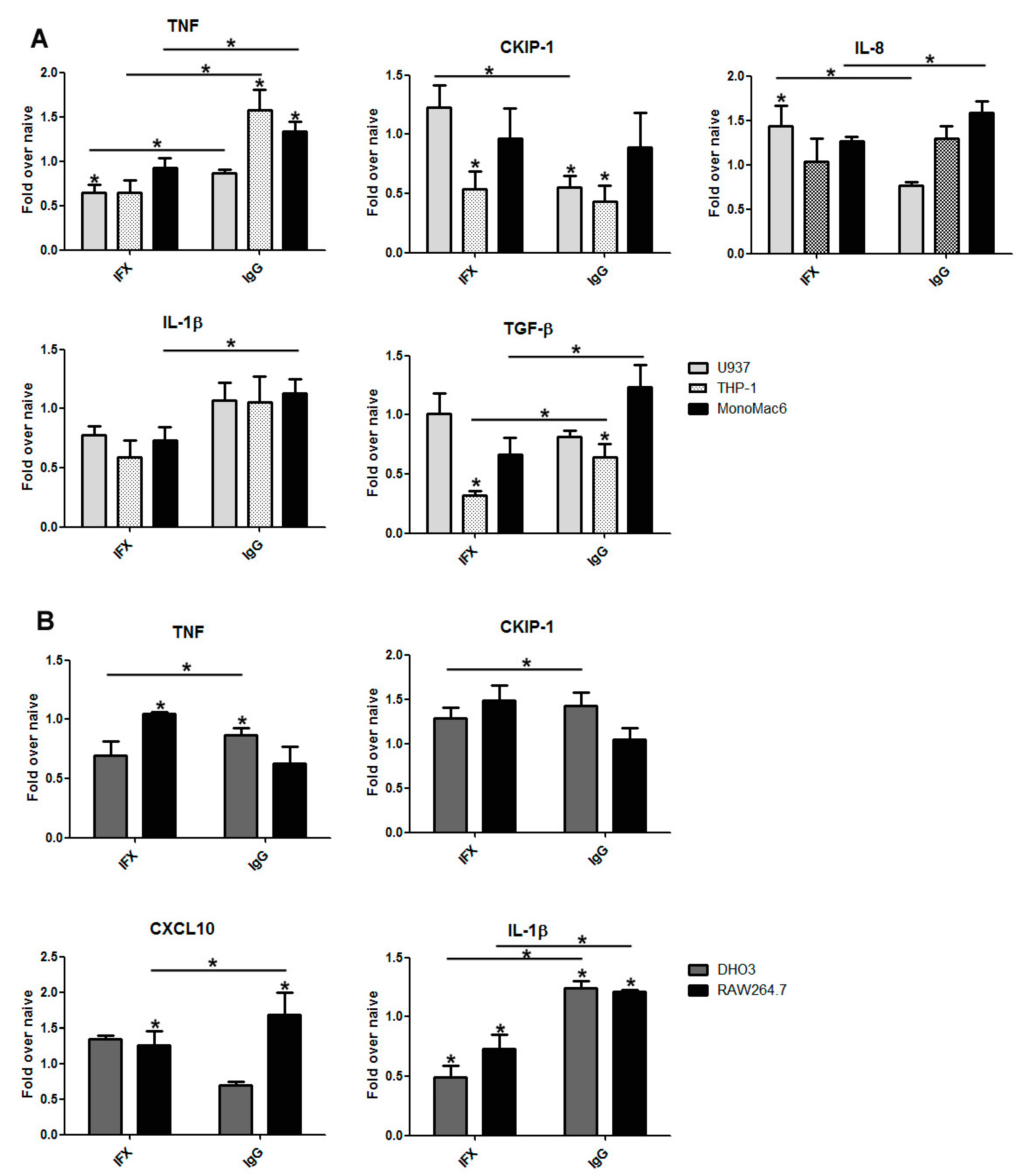
3.2. The Effect of Infliximab Does Not Depend on Its Fc Fragment

The primary therapeutic role of IFX is neutralization of the circulating TNF. There are publications describing its cytotoxic- [20] and reverse signal-inducing effects on mTNF-producing cells [27]. The chimeric IFX contains the constant fragment of human IgG1 (Fc) that is required for complement fixation and antibody-dependent cell-mediated cytotoxicity (ADCC). On the other hand, reverse signaling is elicited only by the interaction of mTNF with the agonist Fab—at least theoretically.
Figure 1. The induction of reverse signaling with IFX on THP-1 cells alters the expression pattern of inflammation-related effector molecules. (A) THP-1 human monocytes were treated with IFX and/or LPS. Total RNA and cell culture supernatants were collected 8 h post-IFX treatment. (B) IFX treatment alone had no significant impact on relative gene expression pattern of the genes, with the notable exception of CKIP-1. The CKIP-1 protein is involved in inflammation and in reverse signaling. Bars show means ± SEM from at least three independent experiments. * Significantly different from controls (One-Way ANOVA, following the Neuman–Keuls post hoc test).
Antibodies 04 00123 g001
In order to test the role of the constant fragment of IFX on human (Figure 2A) and mouse monocyte and macrophage cell lines (Figure 2B), they were exposed to IFX or IgG1 for a day or left untreated. RNA isolation was performed 24 h later. In all human cell lines, IFX stimulation decreased the relative expression of TNF and IL-1β genes as compared with naïve and IgG treated samples. In the case of other genes, different cells responded in different ways to both IFX and IgG1, probably reflecting differences in the density and ratio of Fc receptors and TNF molecules on the cell surface.
Figure 2. The effect of IFX is not dependent on the presence of the constant region (Fc) of IgG. Although IFX is a chimeric antibody that contains the Fc fragment of human IgG1, and myeloid cells carry Fc receptors, distinct relative gene expression patterns were demonstrated after IFX or IgG challenge in all investigated cell lines. To verify that the effect of reverse signaling is different from that of FcR signaling, the relative gene expression pattern of inflammation-related effector molecules were investigated in human (A) and mouse (B) monocytic cell lines 24 h after IFX or IgG stimulation. All of the investigated molecules show distinct relative gene expression pattern after IFX stimulation as compared with IgG treatment. Data show average ± SEM from at least three independent experiments. * Significantly different from controls (One-Way ANOVA following Neuman–Keuls post hoc test).
Antibodies 04 00123 g002
In mouse macrophage cells moderate downregulation of relative gene expression of IL-1β was measured 24 h after IFX treatment. In the cases of TNF, CKIP-1, and CXCL10, as in human cell lines, no gene- or treatment-specific tendencies were observed. However, in practically all cases the effect of IFX on a certain cell line was dissimilar from the effect of IgG1 on the same cells. We might conclude that the effect of infliximab could be influenced by, but does not depend on, its Fc fragment.

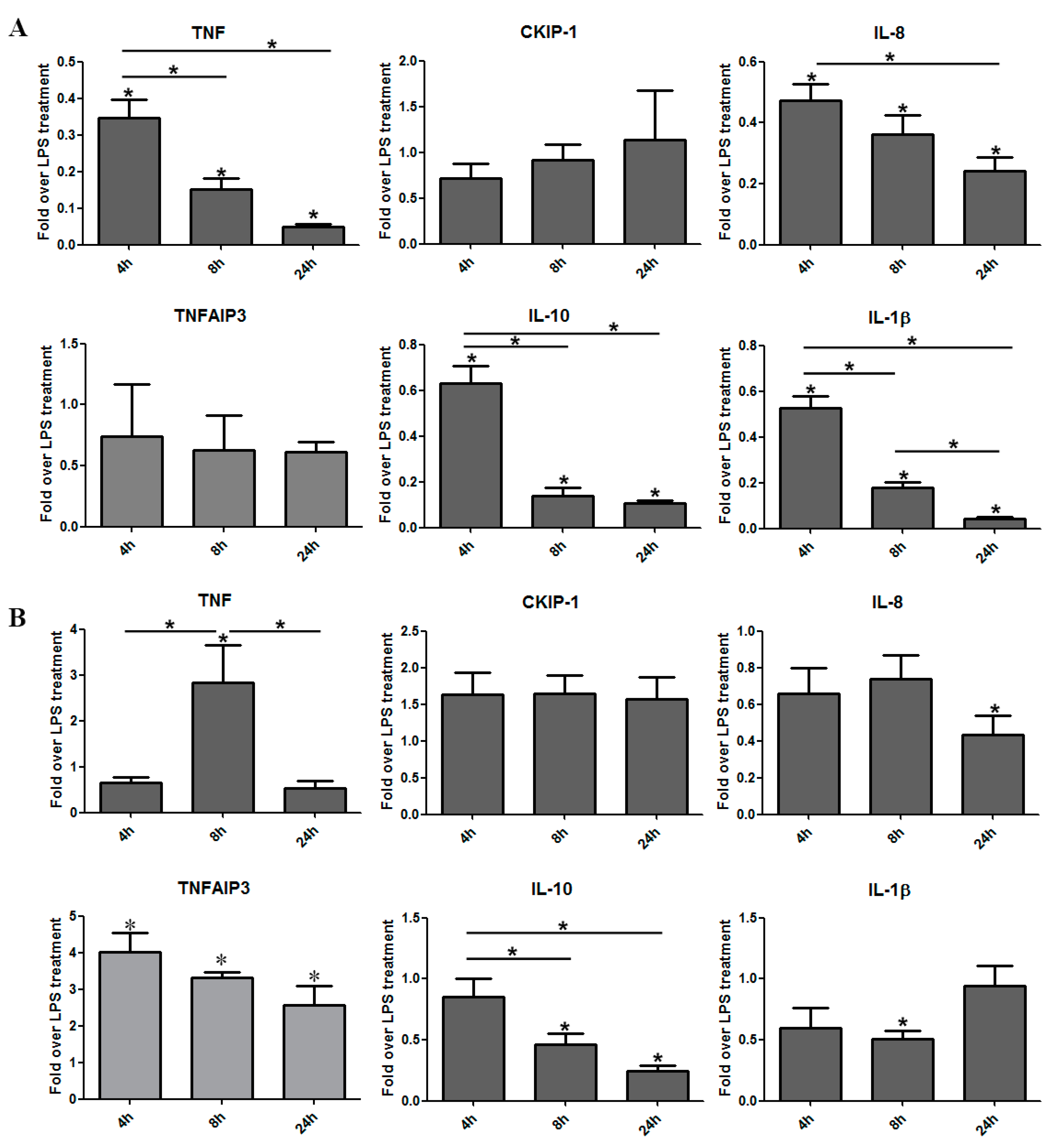
3.3. The Induction of Reverse Signaling by Monoclonal Antibodies Modulates the Expression Pattern of Cytokines and Chemokines in a Time-Dependent Manner

As monoclonal anti-TNF antibodies are widely used in autoimmune inflammatory diseases, we next aimed to test their impact on THP-1 human monocytes under inflammatory conditions. THP-1 cells were challenged with bacterial LPS for 2 h before anti-TNF treatment. In this study, we also tested the effect of certolizumab pegol (CZP) on the gene expression of cytokines and chemokines.
Measurement of relative gene expression was carried out 4 and 8 h after anti-TNF treatment. In order to test the dependence of IFX on the presence of the Fc fragment, in parallel with anti-TNF stimulation, IgG challenge was performed.
IgG treatment increased the expression of IL-8 but had no significant alteration on the gene expression patterns of investigated effector molecules as compared with controls 4 h post-treatment. The relative gene expression of all investigated genes was downregulated by 8 h after IgG treatment as compared with 4 h samples; however, the statistical significance was demonstrated only in the case of TNF (Figure 3A) and IL-1β (Figure 3D).
The application of different anti-TNF antibodies under inflammatory conditions resulted in significant decrease of relative gene expression as compared with either IgG stimulated samples or controls. The regulatory effect of the monoclonals is a time-dependent phenomenon, as 8 h after stimulation more robust relative gene expression decrease was detected compared with earlier time points. In accordance with our previous findings, the regulatory effect of IFX seems to be very different than that of IgG1, as IFX treatment resulted in significant decrease of the relative expression of TNF (A), IL-8 (C), and IL-1β (D) genes as compared with IgG-treated samples or controls at all investigated time points.
IFX slightly decreased the expression of the CKIP-1 gene, as did IgG1 at 8 h and CZP at 4 h. However, in the investigated cell lines neither IgG nor the monoclonal treatment caused significant changes in the expression of this gene.

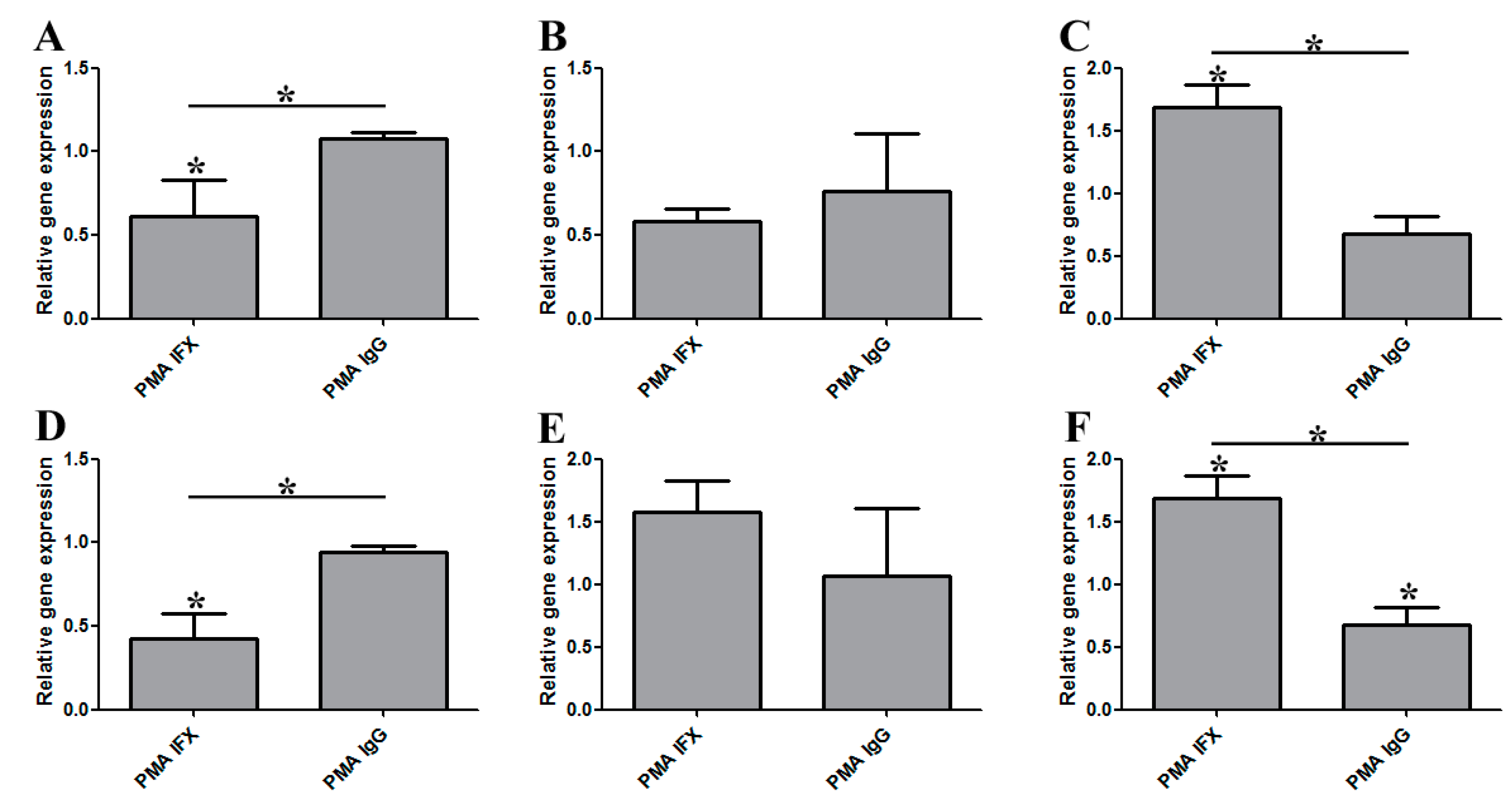
3.4. Certolizumab Pegol Decreases the Relative Gene Expression of Inflammation-Related Effector Molecules in a Time-Dependent Manner under Inflammatory Conditions

In order to investigate the transcriptional effect of CZP-elicited reverse signaling in an inflammatory setting, THP-1 (Figure 4A) and U937 (Figure 4B) human monocytes were pretreated with LPS for two hours, then cells were exposed to 10 µg/mL CZP to induce reverse signaling. Total RNA was harvested 4, 8, and 24 h after CZP treatment.
Figure 3. Induction of reverse signaling by distinct anti-TNF monoclonals modulates gene expression patterns in a time-dependent manner. Relative gene expression pattern of TNF (A), CKIP-1 (B), IL-8 (C), and IL-1β (D) were investigated by QRT-PCR on THP-1 human monocytes. To induce inflammatory response, LPS treatment was performed. Two hours post-LPS challenge, samples were stimulated with IgG1, IFX, or CZP, respectively. RNA was isolated from the cells four and eight hours after stimulation. IFX- and CZP-treated samples showed comparable effect on the gene expression of inflammatory cytokines, although CZP does not carry any Fc fragment. Data show average ± SEM from at least three independent experiments. * Significantly different from unstimulated controls (One-Way ANOVA following Neuman–Keuls post hoc test).
Antibodies 04 00123 g003
In THP-1 cells (Figure 4A), CZP treatment significantly decreased the relative gene expression of TNF, IL-8, IL-10, and IL-1β compared with untreated samples 4 h after anti-TNF treatment. An even more pronounced relative gene expression decline was detected at later time points, reaching the minimum level at 24 h after stimulation. In contrast to the above cytokines—and in accordance with our previous findings—the gene of CKIP-1 demonstrated an opposite pattern. Although at first a slight decrease was detected in relative gene expression (4 h after CZP treatment), at subsequent time points a minor upregulation of relative gene expression was demonstrated, as compared with the 4 h samples or untreated control, though the increase was not significant.
THP-1 and U937 cells have developed into myeloid leukemia cell lines as a result of mutations causing different differentiation abnormalities. These cell lines represent different myeloid lineages and frequently respond to different stimuli in different ways. CZP-induced reverse signaling changed the gene expression pattern of IL-8 and L-10 in U937 cells (Figure 4B); like in THP-1 cells, the relative expression of these genes showed a decline in a time-dependent manner.
Figure 4. The effect of CZP-induced reverse signaling on LPS-induced gene expression in THP-1 and U937 human monocytes. THP-1 (A) and U937 (B) human monocytes were pretreated with LPS to induce inflammatory response. Two hours later, CZP was added. Total RNA was harvested 4, 8, and 24 h after the addition of CZP. The regulatory effect of CZP treatment was time-dependent. In the case of both THP-1 and U937 cells, mild relative expression upregulation was detected in the CKIP-1 gene, suggesting the induction of reverse signaling. Data show the average ± SEM from at least three independent experiments. * Significantly different from unstimulated controls (One-Way ANOVA following Neuman–Keuls post hoc test).
Antibodies 04 00123 g004
On the other hand, the expression pattern of the two principal pro-inflammatory cytokines, TNF and IL-1β, showed fairly interesting time courses. The relative gene expression of TNF was moderately downregulated 4 h after CZP challenge, turning into significant upregulation after another 4 h. Finally, the long term effect of reverse signaling was a dramatic decrease in the relative expression of the TNF gene. In the case of IL-1β, a decrease of relative gene expression was detected 4 h post-CZP challenge, which dropped further at 8 h after CZP treatment (with statistical significance as compared with untreated samples). Here again, the long-term effect was the opposite: 24 h after treatment, we repeatedly detected a moderate relative gene expression upregulation.
The different myeloid cell lines used in the experiments represent macrophages with different differentiation patterns, expressing different markers. Summarizing the above experiment, we can say that in all these cell lines anti-TNF monoclonal-induced reverse signaling resulted in attenuation of expression of pro-inflammatory genes and IL-10. This can significantly contribute to the beneficial therapeutic effect of anti-TNF antibodies in autoimmune and chronic inflammatory diseases.

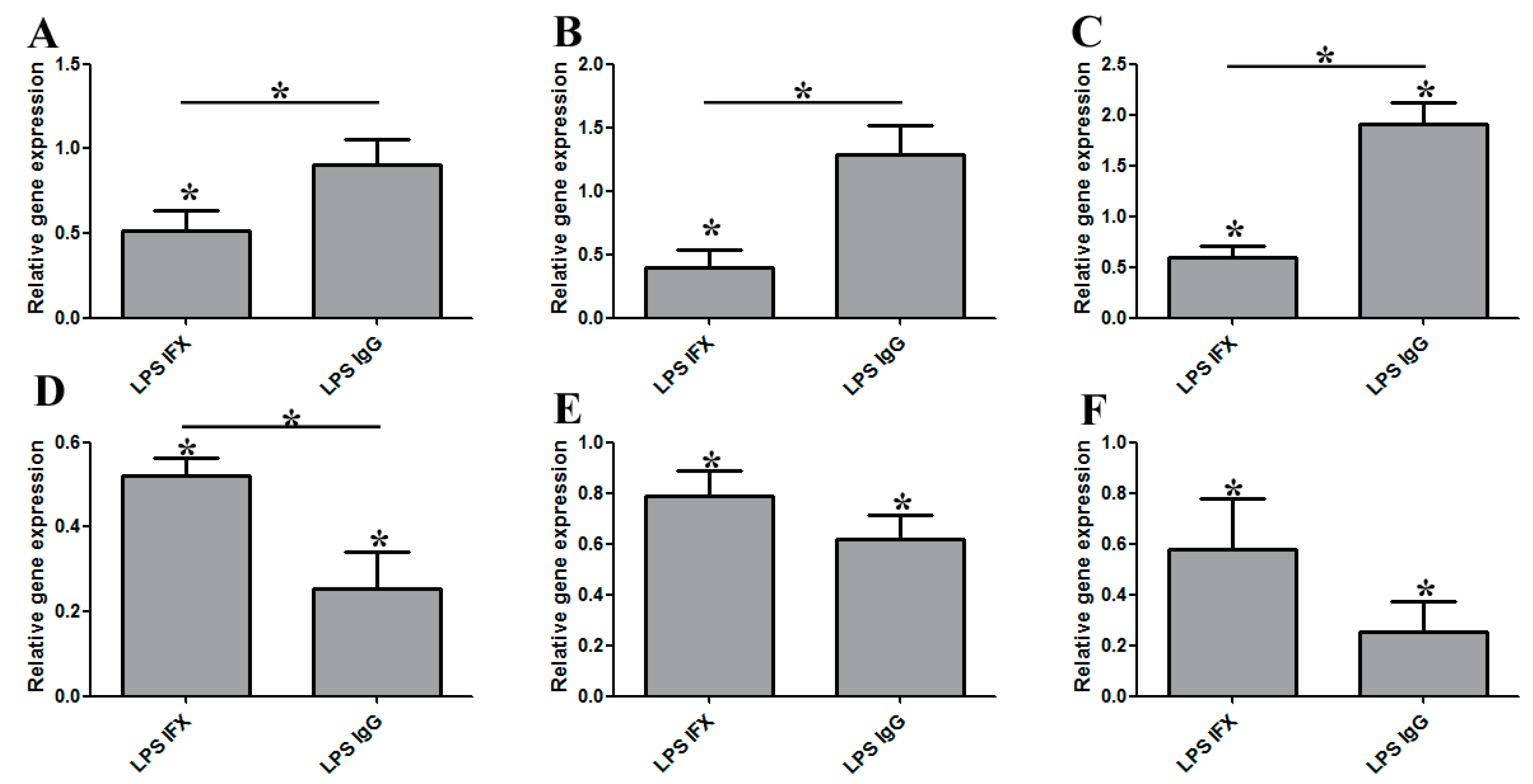
3.5. The Regulatory Effect of IFX Treatment on Activated Bone Marrow-Derived Mouse Macrophages

Primary bone marrow-derived macrophages were differentiated from C57/B6 SPF mice by standard protocol. Cells were treated with phorbol 12-myristate 13-acetate (PMA, Sigma, 100 nM final concentration). On Day 2, cells were washed twice with PBS and fresh cell culture medium was added. Reverse signaling was induced by IFX treatment. Relative gene expression studies were carried out 24 h after IFX (or IgG) stimulation.
IFX-induced reverse signaling remarkably reduced the relative gene expression of TNF and CXCL9 (Figure 5A,D, respectively) as compared with PMA-stimulated control cells. On the other hand, in the case of CXCL10 or PDCD1, and ICAM-1 (Figure 5C,E,F, respectively), moderate or significant upregulation was detected. IFX and IgG stimulation clearly had different effects on gene expression of different cytokines, especially in the cases of PDCD1 (Figure 5C) and ICAM-1 (Figure 5F).
Figure 5. IFX (and IgG) treatment of PMA-induced primary mouse macrophages. Primary mouse macrophages were activated by PMA and IFX or IgG stimulation was performed. PMA-stimulated cells were used as control. The relative gene expression pattern of TNF (A), IL-1β (B), PDCD1 (C), CXCL9 (D), CXCL10 (E), and ICAM1 (F) were determined by qRT-PCR 24 h after the induction of reverse signaling. Bars show means ± SEM from at least three independent experiments. * Significantly different (One-Way ANOVA, following Neuman–Keuls post hoc test).
Antibodies 04 00123 g005
Next, we examined how IFX induced reverse signaling acts on BMDMs under inflammatory conditions. For this, 24 h before IFX or IgG treatment, cells were stimulated with E.coli-derived LPS, an inflammatory ligand of TLR-4 [21,28]. Compared to control cells, IFX treatment downregulated relative the gene expression of all investigated effectors (Figure 6A,F). On the other hand, IgG treatment resulted in relative gene expression increase in case of PDCD1 (C) and expression of CXCL9 (D), CXCL10 (E), and ICAM-1 (F) was also lower as compared with control samples. These data suggest that IFX-induced reverse signaling has a modulatory role under inflammatory conditions (mostly on M1-like macrophages), which may explain the beneficial effect of anti-TNF antibodies in autoimmune inflammatory diseases.
Figure 6. The effect of IFX on LPS-challenged primary mouse macrophages. Macrophages were activated by the presence of LPS before IFX treatment. Relative gene expression of TNF (A), IL-6 (B), PDCD1 (C), CXCL9 (D), CXCL10 (E), and ICAM-1 (F) was determined by qRT-PCR. LPS- and IgG-treated, as well as only LPS-stimulated, samples were used as controls. Bars show average ± SEM from at least three independent experiments. * Significantly different (One-Way ANOVA, Neuman–Keuls post hoc test).
Antibodies 04 00123 g006
In other experiments (Figure 7), the response of bone marrow-derived primary mouse macrophages was investigated in the absence of inflammatory stimuli (under naïve conditions). When the cells were not exposed to LPS one day earlier, IFX-induced reverse signaling caused only minor regulatory effects on the relative gene expression pattern of TNF (Figure 7A), CXCL9 (D), CXCL10 (E), and ICAM-1 (F). It is important to note here that this expression pattern shows strong correlations with our results obtained from THP-1 human monocytes (Figure 1). In the case of IL-1β (Figure 7B), IFX treatment resulted in significant relative gene expression downregulation as compared with naïve cells, while it strongly increased the expression of PDCD1 (C). IgG exposure increased the expression of TNF (A), ICAM-1 (F), and especially CXCL-10 (D). IFX and IgG treatments had opposing effects on the expression of TNF (A), PDCD1 (C), and ICAM-1 (F).
We can conclude that in the case of most effector molecules, the gene regulatory function of reverse signaling is restricted to inflammatory conditions. Its PDCD1-inducing effect on naïve BMDMs is interesting and might play a role in polarization of macrophages.
Figure 7. IFX-elicited reverse signaling had a marginal effect in the absence of inflammatory conditions. Naïve bone marrow-derived mouse macrophages were treated with IFX, IgG, or left unstimulated. Relative gene expression of TNF (A), IL-1β (B), PDCD1 (C), CXCL9 (D), CXCL10 (E), and ICAM-1 (F) was determined 48 h after IFX or IgG stimulation. Bars show the average ± SEM from at least three independent experiments. * Significantly different (One-Way ANOVA, following Neuman–Keuls post hoc test).
Antibodies 04 00123 g007

4. Discussion

LPS tolerance evolved to prevent the tissue-damaging effects of excessive inflammation.
Reverse signaling of the TNF superfamily (recently reviewed by Juhász et al. [29] is also a regulatory mechanism, exerting—in different settings—both activating and inhibitory effects. The 26 kDa membrane-bound TNF (mTNF) is processed by TACE (TNF converting enzyme, ADAM17), liberating the soluble cytokine. sTNF can interact with the 55kDa TNF receptor TNFRI and the 75 kDa TNFR-II [30]. While TNFR-I is present on the surface of all nucleated cells, expression of TNFR-II is restricted to neurons, endothelial cells, and professional immune cells. TNFR-I possesses a death domain (DD) that can initiate apoptosis but can also activate the NF-kB and the p38 pathways to grant survival or induce proliferation, respectively. TNF-elicited responses (changes induced by receptor activation by cognate ligand) are regarded as “forward” signaling.
However, when mTNF-producing cells bind to cell surface receptors, soluble receptors, or agonistic antibodies, the interaction also induces signaling in ligand-expressing cells (hereafter defined as reverse signaling, outside-to-inside signaling, or bidirectional signaling) [17] (Anti-TNF antibodies are regarded as antagonistic as far as inflammation and TNF receptor signaling is concerned. However, when induction of reverse signaling is concerned, the active ones are regarded as agonistic.) The cytoplasmic N terminus of mTNF contains a nuclear localization signal and during reverse signaling translocates into the nucleus and modifies—among others—the expression pattern of inflammation-related genes [16]. Although this fine tuning of the immune response is believed to be one of the most important roles of TNF reverse signaling, it is also necessary for the appropriate axonal development during embryogenesis [31] and it has a pivotal role in nociceptor development [32]. Until now, 19 ligands and 29 receptors of the TNF family members have been identified [33], and reverse signaling was demonstrated in all cases investigated (Fas Ligand [34], BAFF [35], CD137 Ligand [36], and GITRL [37]).
Neutralization of sTNF by the monoclonals was demonstrated in the case of IFX, CZP, etanercept, and adalimumab [38]. The impressive effects of anti-TNF agents suggest that other mechanisms may also contribute to efficacy. Antibody-dependent cell-mediated cytotoxicity (ADCC) and complement activation (CDC) may also play a role in this phenomenon [20].
TNF reverse signaling might also contribute to the therapeutic effect as it is shown in this paper to be able to downregulate expression of pro-inflammatory genes. The effects we observed are not consequences of inhibition of autocrine TNF receptor signaling on macrophages by TNF neutralizing agents. LPS-induced TNF production is restricted for short periods of 1–2 h, unless the cells are also exposed to adenoviruses [39]. In our in vitro experiments, we did not observe reverse signal-induced apoptosis of myeloid cells, only changes in the expression of genes and the production of cytokines. Our data strongly suggest that IFX stimulation before or after LPS challenge drastically downregulates the relative gene expression of inflammation-related effector molecules in THP-1 human monocytes. These data show strong correlations with the results of Domonkos (IL-1beta) [16], Eissner (IL-1, IL-6, and IL-10) [40], and Mitoma (IL-1beta) [20]. It is also known that IFX treatment robustly downregulates the relative expression of the IL-6 and CCL1 genes in Staphylococcus aureus-derived peptidoglycan stimulated primary human dendritic cells [41]. The interesting, opposing effects of both IFX and IgG on PDCD1 expression of PMA- and LPS-stimulated cells should be further investigated, as this receptor seems to play an important role in the polarization of macrophages [42] and immune suppression.
Our research group has previously demonstrated that CKIP-1 interacts with AP-1/c-Jun [40], and CKIP-1 can also interact with a wide variety of proteins, including Akt, ATM, IFP35/Nmi, and Smurf1; thus, it can induce a wide variety of physiological processes, such as changes in cell motility, proliferation, differentiation, or cell death [18]. Both forced CKIP-1 overexpression and TNF reverse signaling increase the apoptotic tendency of myeloid cells when induced separately, but interfere with each other to promote survival if applied simultaneously. This and the fairly unpredictable expression of CKIP-1 in macrophages need to be further investigated [18].
The bone marrow-derived mouse macrophages and human and mouse myeloid cell lines studied in this report represented cells with different routes of differentiation and (in the case of leukemic cells) different defects of development. According to their origin and activation state, reverse signaling evoked different, sometimes opposing effects on the expression of certain genes in these cells. However, as far as the therapeutic importance of anti-TNF biologicals is concerned, the anti-inflammatory effect of reverse signaling seems to be the most important in activated inflammatory macrophages. The primary function, neutralization of soluble TNF, is only one factor to achieve the therapeutic effects of anti-TNF monoclonals. Further mechanisms, including CDC, ADCC, and TNF reverse signaling-induced attenuated expression of inflammatory cytokines and increased apoptosis of specially activated macrophages (e.g., pathological synovial macrophages but not normal macrophages), are also contributing to the efficacy of these agents [24].

5. Conclusions

Activated macrophages and TNF play decisive roles in the development of pathological symptoms of autoimmune and chronic inflammatory diseases. Anti-TNF biologicals are used in the clinic to attenuate these symptoms by neutralizing soluble (and cell-associated) TNF. It is an unexpected development that anti-TNF monoclonals do more than that. By binding to the transmembrane form of TNF on activated macrophages, they activate reverse signaling, an intracellular signaling pathway that leads to the modification of gene activity pattern. As shown in the reported experiments, reverse signaling interferes with the production of inflammatory mediator molecules, contributing to the long-term therapeutic effects of anti-TNF antibodies.

Acknowledgements

This study was supported by an NKTH-A*STAR Hungarian-Singaporean bilateral Grant to Erno Duda. The authors acknowledge the help of Annamária Marton and Erzsébet Kúsz in some of the experiments. The authors are grateful to Prof. Gyula Szabó (Department of Pathophysiology, University of Szeged) for giving us the opportunity to perform some of the real-time PCR experiments.

Author Contributions

Kata Filkor contributed to the conception of the study, designed the experimental procedures, performed the experiments, and participated in data analysis and writing of the manuscript; Erno Duda conceived the study and took part in the writing of the paper. Orsolya Sipos, Annamária Török, and Tanja Kalic performed the experiments and participated in data analysis.

Conflicts of Interest

The authors declare that they have no conflict of interests.

References

  1. Ramadori, G.; Van Damme, J.; Rieder, H. Meyer zum Buschenfelde, K.H. Interleukin 6, the third mediator of acute-phase reaction, modulates hepatic protein synthesis in human and mouse. Comparison with interleukin 1 beta and tumor necrosis factor-alpha. Eur. J. Immunol. 1988, 18, 1259–1264. [Google Scholar] [CrossRef] [PubMed]
  2. Medzhitov, R. Origin and physiological roles of inflammation. Nature 2008, 454, 428–435. [Google Scholar] [CrossRef] [PubMed]
  3. Dowlati, Y.; Herrmann, N.; Swardfager, W.; Liu, H.; Sham, L.; Reim, E.K.; Lanctot, K.L. A meta-analysis of cytokines in major depression. Biol. Psychiatry 2010, 67, 446–457. [Google Scholar] [CrossRef] [PubMed]
  4. Matera, M.G.; Calzetta, L.; Cazzola, M. TNF-alpha inhibitors in asthma and COPD: We must not throw the baby out with the bath water. Pulm. Pharmacol. Ther. 2010, 23, 121–128. [Google Scholar] [CrossRef] [PubMed]
  5. Levine, B.; Kalman, J.; Mayer, L.; Fillit, H.M.; Packer, M. Elevated circulating levels of tumor necrosis factor in severe chronic heart failure. N. Engl. J. Med. 1990, 323, 236–241. [Google Scholar] [CrossRef] [PubMed]
  6. Hotamisligil, G.S.; Shargill, N.S.; Spiegelman, B.M. Adipose expression of tumor necrosis factor-alpha: direct role in obesity-linked insulin resistance. Science 1993, 259, 87–91. [Google Scholar] [CrossRef] [PubMed]
  7. Nickoloff, B.J.; Xin, H.; Nestle, F.O.; Qin, J.Z. The cytokine and chemokine network in psoriasis. Clin. Dermatol. 2007, 25, 568–573. [Google Scholar] [CrossRef] [PubMed]
  8. Feldmann, M.; Maini, R.N. Anti-TNF alpha therapy of rheumatoid arthritis: What have we learned? Annu. Rev. Immunol. 2001, 19, 163–196. [Google Scholar] [CrossRef] [PubMed]
  9. Chey, W.Y. Infliximab for patients with refractory ulcerative colitis. Inflamm. Bowel Dis. 2001, 7 (Suppl. 1), S30–S33. [Google Scholar] [CrossRef] [PubMed]
  10. Knight, D.M.; Trinh, H.; Le, J.; Siegel, S.; Shealy, D.; McDonough, M.; Scallon, B.; Moore, M.A.; Vilcek, J.; Daddona, P.; et al. Construction and initial characterization of a mouse-human chimeric anti-TNF antibody. Mol. Immunol. 1993, 30, 1443–1453. [Google Scholar] [CrossRef]
  11. Nightingale, S.L. From the Food and Drug Administration. JAMA 1998, 280, 1128. [Google Scholar] [CrossRef] [PubMed]
  12. Hsu, L.; Armstrong, A.W. Anti-drug antibodies in psoriasis: A critical evaluation of clinical significance and impact on treatment response. Expert Rev. Clin. Immunol. 2013, 9, 949–958. [Google Scholar] [CrossRef] [PubMed]
  13. Horiuchi, T.; Mitoma, H.; Harashima, S.; Tsukamoto, H.; Shimoda, T. Transmembrane TNF-alpha: Structure, function and interaction with anti-TNF agents. Rheumatology (Oxford) 2010, 49, 1215–1228. [Google Scholar] [CrossRef] [PubMed]
  14. Kane, S.V.; Acquah, L.A. Placental transport of immunoglobulins: a clinical review for gastroenterologists who prescribe therapeutic monoclonal antibodies to women during conception and pregnancy. Am. J. Gastroenterol. 2009, 104, 228–233. [Google Scholar] [CrossRef] [PubMed]
  15. Mahadevan, U.; Wolf, D.C.; Dubinsky, M.; Cortot, A.; Lee, S.D.; Siegel, C.A.; Ullman, T.; Glover, S.; Valentine, J.F.; Rubin, D.T.; et al. Placental transfer of anti-tumor necrosis factor agents in pregnant patients with inflammatory bowel disease. Clin. Gastroenterol. Hepatol. 2013, 11, 286–292, quiz e224. [Google Scholar] [CrossRef] [PubMed]
  16. Domonkos, A.; Udvardy, A.; Laszlo, L.; Nagy, T.; Duda, E. Receptor-like properties of the 26 kDa transmembrane form of TNF. Eur. Cytokine Netw. 2001, 12, 411–419. [Google Scholar] [PubMed]
  17. Eissner, G.; Kirchner, S.; Lindner, H.; Kolch, W.; Janosch, P.; Grell, M.; Scheurich, P.; Andreesen, R.; Holler, E. Reverse signaling through transmembrane TNF confers resistance to lipopolysaccharide in human monocytes and macrophages. J. Immunol. 2000, 164, 6193–6198. [Google Scholar] [CrossRef] [PubMed]
  18. Juhasz, K.; Zvara, A.; Lipp, A.M.; Nimmervoll, B.; Sonnleitner, A.; Balogi, Z.; Duda, E. Casein kinase 2-interacting protein-1, an inflammatory signaling molecule interferes with TNF reverse signaling in human model cells. Immunol. Lett. 2013, 152, 55–64. [Google Scholar] [CrossRef] [PubMed]
  19. Harashima, S.; Horiuchi, T.; Hatta, N.; Morita, C.; Higuchi, M.; Sawabe, T.; Tsukamoto, H.; Tahira, T.; Hayashi, K.; Fujita, S.; et al. Outside-to-inside signal through the membrane TNF-alpha induces E-selectin (CD62E) expression on activated human CD4+ T cells. J. Immunol. 2001, 166, 130–136. [Google Scholar] [CrossRef] [PubMed]
  20. Mitoma, H.; Horiuchi, T.; Hatta, N.; Tsukamoto, H.; Harashima, S.; Kikuchi, Y.; Otsuka, J.; Okamura, S.; Fujita, S.; Harada, M. Infliximab induces potent anti-inflammatory responses by outside-to-inside signals through transmembrane TNF-alpha. Gastroenterology 2005, 128, 376–392. [Google Scholar] [CrossRef] [PubMed]
  21. Akira, S.; Uematsu, S.; Takeuchi, O. Pathogen recognition and innate immunity. Cell 2006, 124, 783–801. [Google Scholar] [CrossRef] [PubMed]
  22. Fejer, G.; Kusz, E.; Biological Research Centre of the Hungarian Academy of Sciences, Szeged, Hungary. Unpublished work. 2006.
  23. de Waal Malefyt, R.; Abrams, J.; Bennett, B.; Figdor, C.G.; de Vries, J.E. Interleukin 10(IL-10) inhibits cytokine synthesis by human monocytes: an autoregulatory role of IL-10 produced by monocytes. J. Exp. Med. 1991, 174, 1209–1220. [Google Scholar] [CrossRef] [PubMed]
  24. Fiorentino, D.F.; Bond, M.W.; Mosmann, T.R. Two types of mouse T helper cell. IV. Th2 clones secrete a factor that inhibits cytokine production by Th1 clones. J. Exp. Med. 1989, 170, 2081–2095. [Google Scholar] [CrossRef] [PubMed]
  25. Alanara, T.; Karstila, K.; Moilanen, T.; Silvennoinen, O.; Isomaki, P. Expression of IL-10 family cytokines in rheumatoid arthritis: Elevated levels of IL-19 in the joints. Scand. J. Rheumatol. 2010, 39, 118–126. [Google Scholar] [CrossRef] [PubMed]
  26. Iyer, S.S.; Cheng, G. Role of interleukin 10 transcriptional regulation in inflammation and autoimmune disease. Crit. Rev. Immunol. 2012, 32, 23–63. [Google Scholar] [CrossRef] [PubMed]
  27. Kirchner, S.; Holler, E.; Haffner, S.; Andreesen, R.; Eissner, G. Effect of different tumor necrosis factor (TNF) reactive agents on reverse signaling of membrane integrated TNF in monocytes. Cytokine 2004, 28, 67–74. [Google Scholar] [CrossRef] [PubMed]
  28. Akira, S.; Takeda, K. Toll-like receptor signalling. Nat. Rev. Immunol. 2004, 4, 499–511. [Google Scholar] [CrossRef] [PubMed]
  29. Cheng, C.; Huang, C.; Ma, T.T.; Bian, E.B.; He, Y.; Zhang, L.; Li, J. SOCS1 hypermethylation mediated by DNMT1 is associated with lipopolysaccharide-induced inflammatory cytokines in macrophages. Toxicol. Lett. 2014, 225, 488–497. [Google Scholar] [CrossRef] [PubMed]
  30. Chan, F.K.; Chun, H.J.; Zheng, L.; Siegel, R.M.; Bui, K.L.; Lenardo, M.J. A domain in TNF receptors that mediates ligand-independent receptor assembly and signaling. Science 2000, 288, 2351–2354. [Google Scholar] [CrossRef] [PubMed]
  31. Kisiswa, L.; Osorio, C.; Erice, C.; Vizard, T.; Wyatt, S.; Davies, A.M. TNFalpha reverse signaling promotes sympathetic axon growth and target innervation. Nat. Neurosci. 2013, 16, 865–873. [Google Scholar] [CrossRef] [PubMed]
  32. Wheeler, M.A.; Heffner, D.L.; Kim, S.; Espy, S.M.; Spano, A.J.; Cleland, C.L.; Deppmann, C.D. TNF-alpha/TNFR1 signaling is required for the development and function of primary nociceptors. Neuron 2014, 82, 587–602. [Google Scholar] [CrossRef] [PubMed]
  33. Aggarwal, B.B.; Gupta, S.C.; Kim, J.H. Historical perspectives on tumor necrosis factor and its superfamily: 25 years later, a golden journey. Blood 2012, 119, 651–665. [Google Scholar] [CrossRef] [PubMed]
  34. Lee, S.M.; Kim, E.J.; Suk, K.; Lee, W.H. Stimulation of FasL induces production of proinflammatory mediators through activation of mitogen-activated protein kinases and nuclear factor-kappaB in THP-1 cells. Inflammation 2012, 35, 1–10. [Google Scholar] [CrossRef] [PubMed]
  35. Jeon, S.T.; Kim, W.J.; Lee, S.M.; Lee, M.Y.; Park, S.B.; Lee, S.H.; Kim, I.S.; Suk, K.; Choi, B.K.; Choi, E.M.; et al. Reverse signaling through BAFF differentially regulates the expression of inflammatory mediators and cytoskeletal movements in THP-1 cells. Immunol. Cell Biol. 2010, 88, 148–156. [Google Scholar] [CrossRef] [PubMed]
  36. Tang, Q.; Koh, L.K.; Jiang, D.; Schwarz, H. CD137 ligand reverse signaling skews hematopoiesis towards myelopoiesis during aging. Aging (Albany NY) 2013, 5, 643–652. [Google Scholar] [PubMed]
  37. Bae, E.M.; Kim, W.J.; Suk, K.; Kang, Y.M.; Park, J.E.; Kim, W.Y.; Choi, E.M.; Choi, B.K.; Kwon, B.S.; Lee, W.H. Reverse signaling initiated from GITRL induces NF-kappaB activation through ERK in the inflammatory activation of macrophages. Mol. Immunol. 2008, 45, 523–533. [Google Scholar] [CrossRef] [PubMed]
  38. Ramos-Casals, M.; Brito-Zeron, P.; Munoz, S.; Soto, M.J. A systematic review of the off-label use of biological therapies in systemic autoimmune diseases. Medicine (Baltimore) 2008, 87, 345–364. [Google Scholar] [CrossRef] [PubMed]
  39. Fejer, G.; Szalay, K.; Gyory, I.; Fejes, M.; Kusz, E.; Nedieanu, S.; Pali, T.; Schmidt, T.; Siklodi, B.; Lazar, G., Jr.; et al. Adenovirus infection dramatically augments lipopolysaccharide-induced TNF production and sensitizes to lethal shock. J. Immunol. 2005, 175, 1498–1506. [Google Scholar] [CrossRef]
  40. Kirchner, S.; Boldt, S.; Kolch, W.; Haffner, S.; Kazak, S.; Janosch, P.; Holler, E.; Andreesen, R.; Eissner, G. LPS resistance in monocytic cells caused by reverse signaling through transmembrane TNF (mTNF) is mediated by the MAPK/ERK pathway. J. Leukoc. Biol. 2004, 75, 324–331. [Google Scholar] [CrossRef] [PubMed]
  41. Filkor, K.; Hegedus, Z.; Szasz, A.; Tubak, V.; Kemeny, L.; Kondorosi, E.; Nagy, I. Genome wide transcriptome analysis of dendritic cells identifies genes with altered expression in psoriasis. PLoS One 2013, 8, e7343. [Google Scholar] [CrossRef] [PubMed] [Green Version]
  42. Cho, H.Y.; Choi, E.K.; Lee, S.W.; Jung, K.O.; Seo, S.K.; Choi, I.W.; Park, S.G.; Choi, I. Programmed death-1 receptor negatively regulates LPS-mediated IL-12 production and differentiation of murine macrophage RAW264.7 cells. Immunol. Lett. 2009, 127, 39–47. [Google Scholar] [CrossRef] [PubMed]

