Ammonium-Carbamate-Rich Organogels for the Preparation of Amorphous Calcium Carbonates


Department of Materials and Environmental Chemistry, Stockholm University, SE 106 91 Stockholm, Sweden
*
Author to whom correspondence should be addressed.
Minerals 2017, 7(7), 110; https://doi.org/10.3390/min7070110
Submission received: 31 May 2017
/
Revised: 17 June 2017
/
Accepted: 18 June 2017
/
Published: 27 June 2017
(This article belongs to the Special Issue Nucleation of Minerals: Precursors, Intermediates and Their Use in Materials Chemistry)
Abstract
:Amine-CO2 chemistry is important for a range of different chemical processes, including carbon dioxide capture. Here, we studied how aspects of this chemistry could be used to prepare calcium carbonates. Chemically crosslinked organogels were first prepared by reacting hyperbranched polyethylene imine (PEI) dissolved in DMSO with carbon dioxide. The crosslinks of the organogel consisted of ammonium-carbamate ion pairs as was shown by IR spectroscopy. These carbamate-rich organogels were subsequently subjected to aqueous solutions of calcium acetate, and amorphous calcium carbonate (ACC) precipitated. The ACC did not crystalize during the mixing for up to 20 h, as was shown by a combination of IR spectroscopy, X-ray diffraction, scanning electron microscopy, and thermal analysis. Some PEI had been included or adsorbed on the ACC particles. Traces of calcite were observed in one sample that had been subjected to water in a work-up procedure.
1. Introduction
The understanding of the details of precipitation and crystallization of calcium carbonates has changed. Now it commonly involves aspects of non-traditional mechanisms of crystallization where clusters and amorphous calcium carbonates (ACCs) are important objects [1]. An enhanced understanding of the formation of calcium carbonates is important as they contribute to the carbon balance globally, and there are, for example, indications that ocean acidification is already affecting the growth of coral reefs negatively [2]. In addition, calcium carbonates are important in various technical applications. For example, calcite can be added to a certain degree in Portland cement [3] and be used as a filler in papers [4]. Calcium carbonates are very important for many organisms in their biomineralized tissues, where the calcium carbonate contributes with a functional toughening [5,6]. ACCs occur in certain biomineralized tissues [5,7,8,9] but also in synthetic calcium carbonates [10,11,12] where they have been shown to display both structural and chemical differences [13,14,15].
Polymers have been shown to moderate the precipitation and crystallization of calcium carbonates by affecting the time scales of aggregation and crystallization, as well as the polymorphism and the morphologies of the particles [16,17,18,19]. Specifically, amines and polyamines have been shown to influence the precipitation and crystallization of calcium carbonates by various interactions with carbonate ions, affecting the buffering capacity, and their tendencies to adsorb to carbonate-rich interfaces [20,21,22].
Amines and polyamines have been studied to a large extent when it comes to their abilities to capture carbon dioxide from flue gas or air [23,24,25,26]. The CO2-amine chemistry is surprisingly rich and involves coupled reaction networks with bicarbonate, carbamates, and carbamic acid moieties [27,28,29]. In many solvents, ammonium-carbamate ion pairs are the dominant species and form rapidly [30]. From a technological point of view, carbamates are typically preferred over bicarbonates in carbon-capture systems as they form and decompose relatively rapidly [27,31].
For polyamines in selected solvents, organogels have been shown to form when being contacted with CO2(g). For example, Carretti et al. have derived and studied such organogels that had been prepared from polyallylamine and CO2(g), and they related the gelation to ammonium-carbamate-based crosslinks [32]. Carretti et al. also extended these studies to include organogels of polyethyleneimine (PEI) induced by reactions with CO2, and studied these ammonium-carbamate crosslinked gels in the context of art preservation [33,34].
We got inspired by the option to prepare ammonium-carbamate crosslinked organogels of PEI and CO2 and wanted to investigate if such could be used to prepare calcium carbonates. The case of using such gels as sources of CO2 was strengthened by the study of Prah et al. [35], who had shown that molecular ammonium carbamates could be used in the crystallization of calcium carbonates using solutions of calcium acetate.
The novelty of this study is that we use polymeric carbamates that were prepared from reactive CO2 absorption in PEI solutions. It could be relevant to combined CO2 capture and mineralization. The organogels were prepared from hyperbranched and low molecular weight PEI and CO2, and we studied aspects of the precipitation of ACC when the organogels were mixed with aqueous solutions of calcium acetate.
2. Materials and Methods
A solution of hyperbranched PEI (Sigma-Aldrich, Saint Louis, MO, USA, CAS number: 25987-06-8, average Mw = 800) in dimethyl sulfoxide (DMSO) was prepared, by adding and mixing 0.3677 g (2.76 mmol) of PEI in 2 mL DMSO. The organogel was prepared by bubbling CO2 through the PEI solution with a flow rate of 2.3 mL/min for 1 h. Subsequently, the gel was centrifuged to reduce the amount of DMSO. A solution of calcium acetate hydrate (Sigma-Aldrich, CAS number: 114460-21-8) was prepared in water; by adding and mixing 5 g of calcium acetate hydrate in 50 mL of deionized water. 4.6 mL of the calcium acetate solution was added at a flow rate of 0.75 mL/min to the CO2-induced organogel of PEI using a syringe pump. Such reactive mixtures were stirred (500 rpm) for 1 h (samples SW and SE) or 20 h (samples LW and LE) at room temperature. The formed solids were filtered off and washed carefully with 5 mL of deionized water and 2 × 5 mL of ethanol (samples SW and LW) or with 3 × 5 mL of ethanol (samples SE and LE). The white solids were dried at room temperature. The sample nomenclature used is: S for short mixing and L for long mixing; W for washed with water and E for washed with ethanol.
The samples were studied with infrared (IR) spectroscopy using a Varian 670-IR spectrometer (Varian, Mulgrave, Australia) equipped with a single reflection ATR device (Specac, Orpington, UK) with a diamond ATR element. X-ray diffraction was used to study the crystallinity of the solids formed using a XPERT-PRO PANalytical powder diffractometer (PANalytical B.V., Almelo, The Netherlands) (reflection mode with an X’Celerator detector (Cu Kα1 radiation, λ = 1.5418 Å) from 5.0° to 85.0° (2θ). The step size was set to 0.0131°, and the step time was 0.0104 °/min, which resulted in a total accumulation time of 126 min. Scanning electron microscopy (SEM) was used to study the morphologies of the solids using a JEOL JSM-7401F scanning electron microscope (JEOL USA Inc., Peabody, MA, USA) that operated at a voltage of 1 kV. The amount of organics in the solids was estimated by thermal analysis using a TA Instruments gravimetric thermal analyzer. The samples were subjected to an air flow 20 mL/min, the temperature range was 25–900 °C, and the temperature rate was 5 °C/min.
3. Results and Discussion
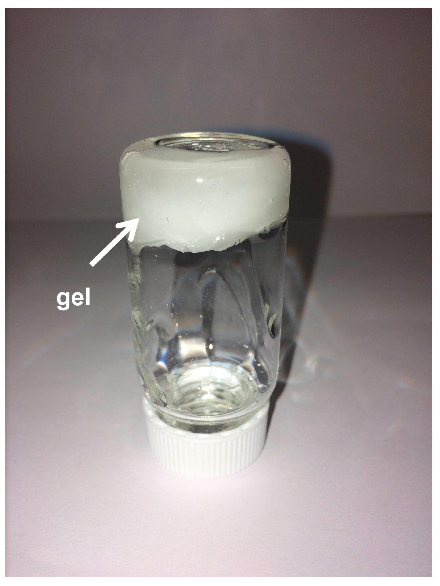
Gels formed rapidly when CO2 was bubbled through solutions of PEI in DMSO, and Figure 1 displays a corresponding image. For the gelation, we presumed that all the amine groups had reacted with CO2 forming ammonium-carbamate ion pairs following the stoichiometry of amine: CO2 = 2:1. In analogy to the study of Carretti et al. [32], we hypothesized that chemical crosslinks had formed among the PEI chains when the amino groups had reacted with CO2. This hypothesis of chemical crosslinks with alkylammonium-alkylcarbamate ion pairs was supported by IR analyzes.
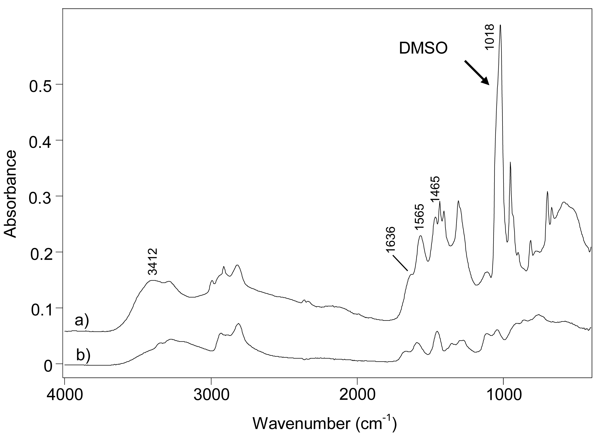
Figure 2 shows the IR spectra of the organogel and the corresponding PEI. Alkylammonium-alkylcarbamate ion pairs were detected by analyzing the IR spectrum of the organogel. In this spectrum, presented in Figure 2a, the broad band at frequencies between 3350 and 2000 cm−1 and the bands at 1636 cm−1 and 1465 cm−1 are typical for ammonium groups, and the band at a frequency of 1565 cm−1 is a clear signature of the C=O group of carbamates [36]. Bands were also observed for DMSO, of which the one with the highest intensity appeared at a frequency of 1018 cm−1.
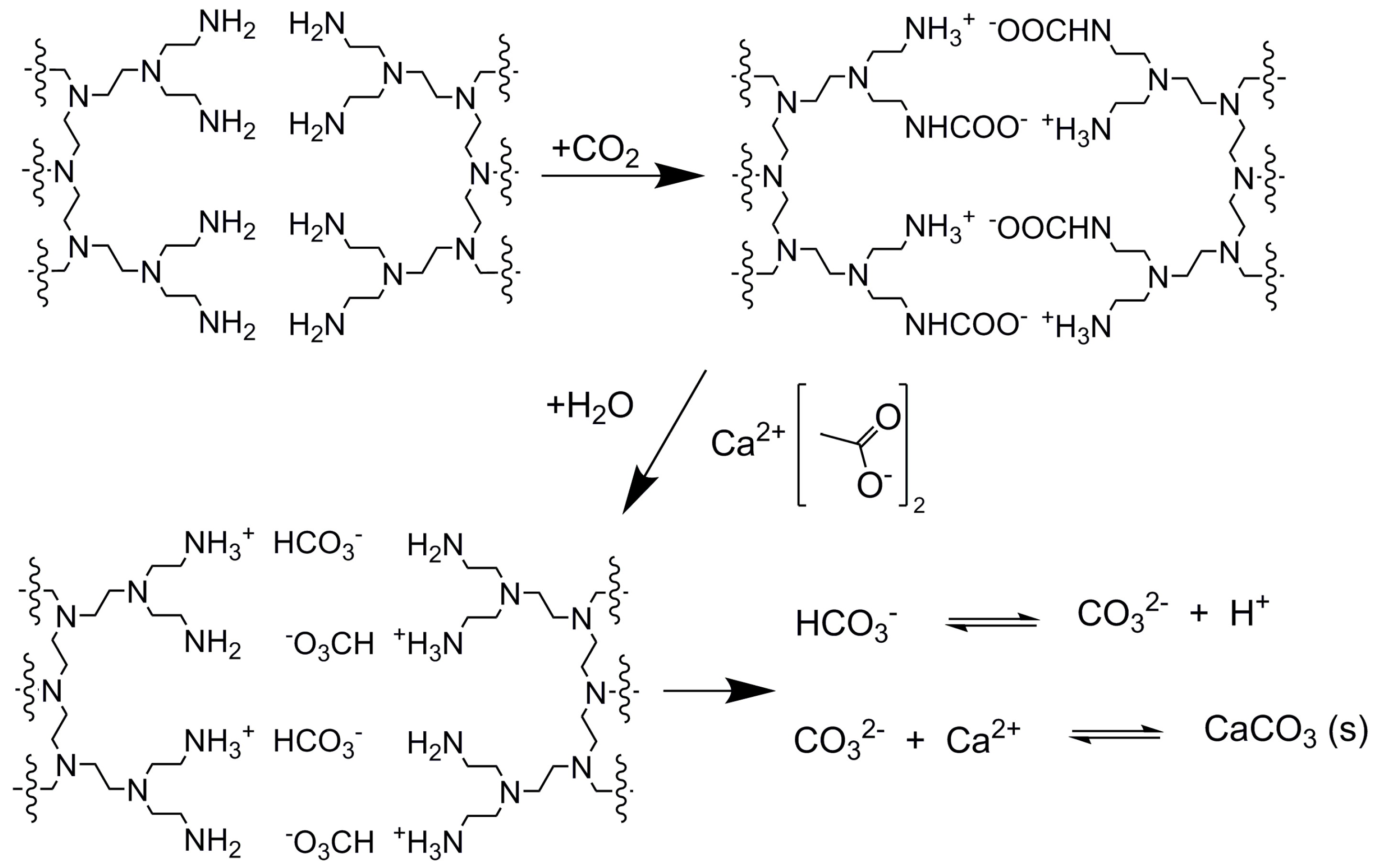
The organogels were centrifuged to reduce the amount of DMSO. Subsequently, aqueous solutions of calcium acetate were added and white solid precipitates formed, see the reactions in Scheme 1. We added a stoichiometric amount of Ca2+ ions, corresponding to a 1:1 ratio of Ca2+:carbamate. For this addition, we assumed that all of the amine groups of the PEI had formed ammonium-carbamates giving an N–carbamate ratio of 2:1. Two different groups of samples were prepared and subjected to short (samples SE and SW) and long (samples LE and LW) mixing after the calcium acetate solutions had been added.
In a control experiment when only water had been added to the organogel, the gel disappeared, which was expected by the instability of ammonium-carbamate ion pairs in aqueous solutions. Bicarbonate moieties tend to form on addition of water, and the PEI is expected to protonated and soluble in water.
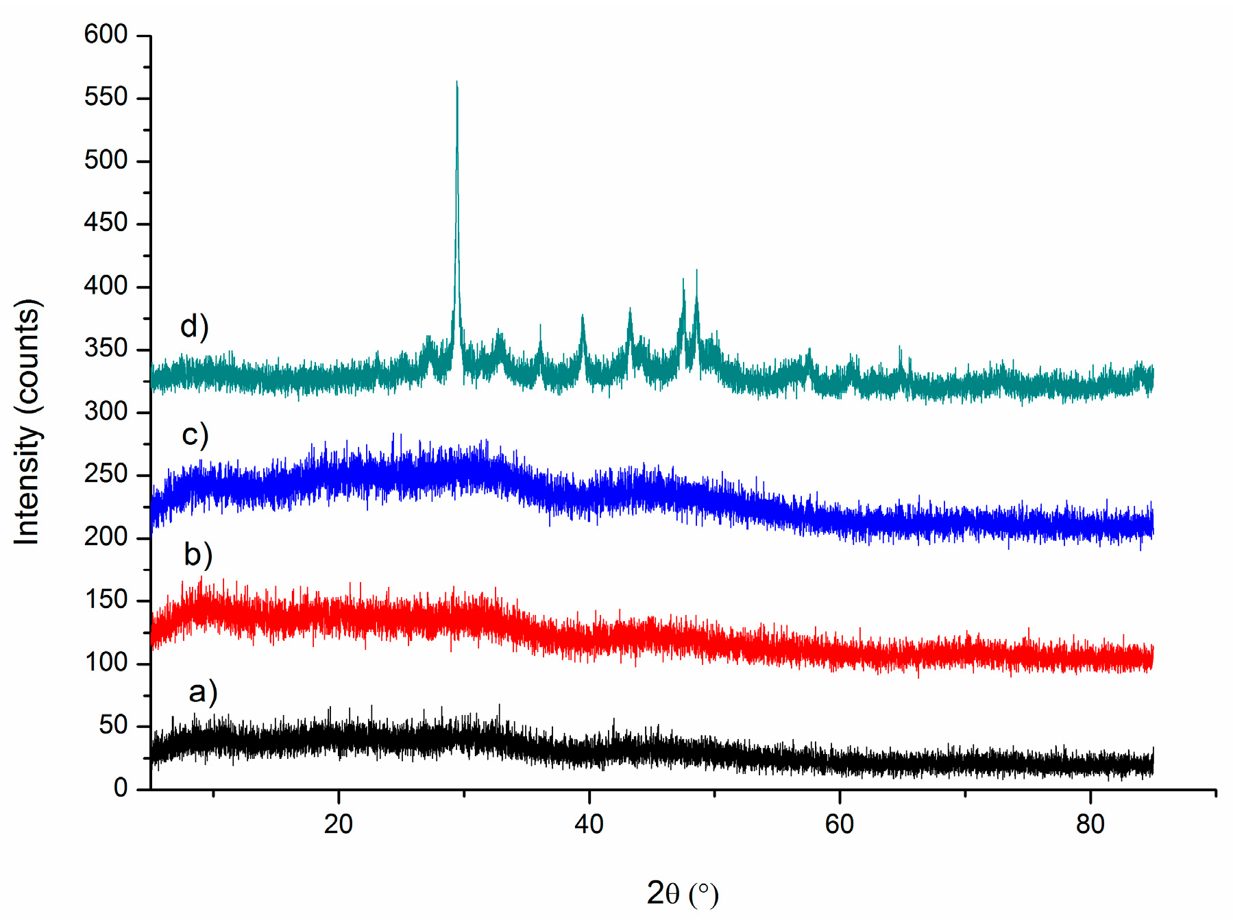
The solid precipitates were either washed in ethanol (samples SE and LE) or washed in water and ethanol (samples SW and LW). These experiments were performed to assess eventual contributions of water washing on the crystallization of calcium carbonate. The samples were studied with several methods. The XRD patterns revealed that the samples SE, SW, and LE were amorphous, and that the sample LW (20 h of mixing and water washing) contained a minor fraction of calcite with broad diffraction lines. Figure 3 shows the diffractograms of the samples, and the low signal-to-noise levels related to the amorphous nature of the samples.
The XRD diffraction patterns in Figure 3 indicated the absence of crystallinity but could not easily be used to detect ACCs, although the wide-angle scattering in the patterns is typical for ACC. IR spectroscopy, however, can be very useful for studying ACCs [14,37]. The IR spectra of samples SE, SW, LE, and LW are shown in Figure 4. The spectra clearly showed that the calcium carbonates in samples SE, SW, and LE consisted of fully of ACC, and mainly of ACC in sample LW. The bands observed at a frequency of 1380 cm−1, with a shoulder at 1465 cm−1, belong to the asymmetric stretching of carbonates. The band at a frequency of 1071 cm−1 corresponds to the symmetric stretching, and the band at 861 cm−1 to the out-of-plane bending modes. These frequency values were typical for ACCs. The band for the in-plane bending mode of the carbonate ions were observed at frequencies of around 700 cm−1. This band is the most useful to distinguish ACCs and various polymorphs of crystalline calcium carbonates. For the ACCs of this study, a broad doublet was observed at a frequency of ~700 cm−1. In the spectrum of sample LW, a sharp band also appeared at 711 cm−1, which is typical for calcite and corroborated the findings from XRD of a small fraction of calcite in this sample. IR bands of the PEI were observed in the C-H stretching region at frequencies of 2800–3000 cm−1.
The morphologies of the particles were studied by SEM, and the features observed in the images supported the amorphous nature of samples. Figure 5 shows SEM images of the ACC-rich samples of SE and LE and note that spherical particles are typically observed for ACC [14,38,39,40].
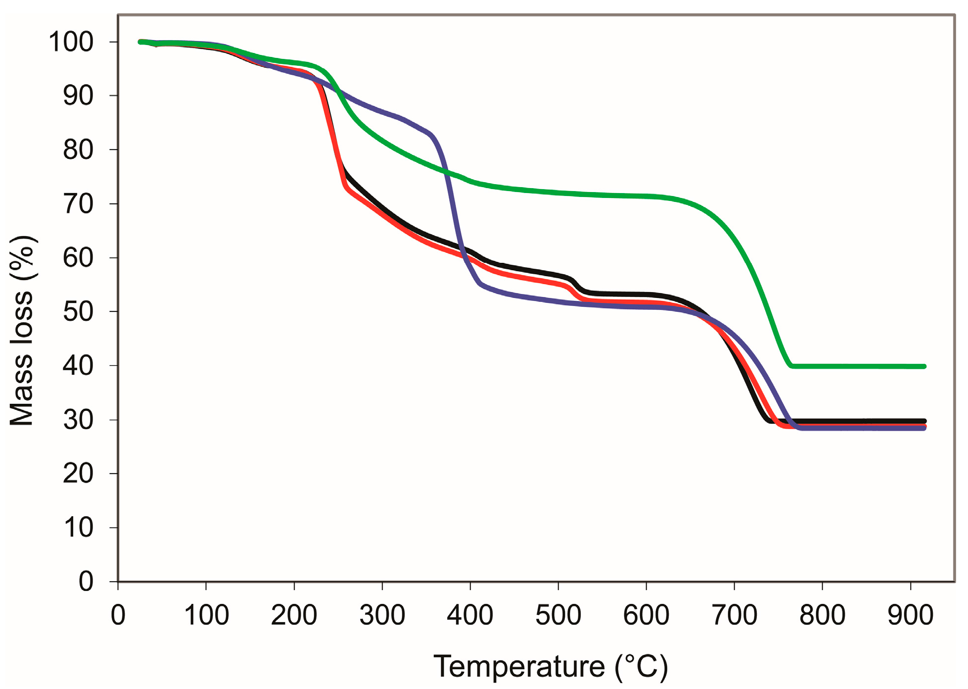
The amounts of amines in samples SE, SW, LE, and LW, were determined by thermogravimetric (TG) experiments. Figure 6 shows the TG curves, and they were surprisingly different. All curves had mass losses in three major temperature regions. The mass loss in the temperature range between room temperature and 200 °C was attributed to water loss. The second region of mass losses occurred at temperatures between 200 and 450 °C. These losses were attributed to the combustion of PEI. Sample SE (red), SW (black), and LW (green) had similar trends in this region, but sample LE (blue) had a significantly different tendency with a very significant step of mass loss related to combustion at a temperature of 370 °C. In the third regions, there were defined mass loss steps related to the decomposition of the carbonate groups (calcination) in all samples. From the mass losses observed in this region, the amounts of calcium carbonate were estimated. The amount of calcium carbonate in the samples SE, SW, and LE were similar and about 50 wt %, and sample LW consisted of ~70 wt % calcium carbonate. We speculate that the higher amount of CaCO3 in sample LW can be explained by the calcite formation and consequently a more effective washing of the sample. Considering that the amount of water appeared to be around 5 wt % in each sample, the amount of polymer and solvent in the samples was between 25 and 45 wt %. In this context, note that the molecular formula for ACC is typically observed to be CaCO3·xH2O; however, the significant amount of organics in the samples made it very difficult to robustly establish the composition of the ACC in the studied samples.
4. Conclusions
Ammonium-carbamate-rich organogels were successfully used to prepare ACC. It is our understanding that the ammonium-carbamate ion pairs are coupled chemically to bicarbonates in the aqueous calcium acetate solution used for the precipitation of ACC. The details of this chemistry were not studied. A natural extension of this study could be to study this chemistry in some detail. ACC was stabilized at the two mixing times used in this study (1 h and 20 h), the extent of this stabilization is relevant for further studies as well the interfacial details of the PEI–ACC interactions.
Acknowledgments
Swedish Research Council (2016-03568) and the Carl Trygger Foundation are thanked.
Author Contributions
Z.B. and N.H. conceived and designed the experiments, analyzed data, and wrote the paper; P.Z. performed most of the experiments and analyzed data. Z.B. performed the IR experiments.
Conflicts of Interest
The authors declare no conflict of interest.
References
- Gebauer, D.; Kellermeier, M.; Gale, J.D.; Bergström, L.; Cölfen, H. Pre-nucleation clusters as solute precursors in crystallisation. Chem. Soc. Rev. 2014, 43, 2348–2371. [Google Scholar] [CrossRef] [PubMed]
- Albright, R.; Caldeira, L.; Hosfelt, J.; Kwiatkowski, L.; Maclaren, J.K.; Mason, B.M.; Nebuchina, Y.; Ninokawa, A.; Pongratz, J.; Ricke, K.L.; et al. Reversal of ocean acidification enhances net coral reef calcification. Nature 2016, 531, 362–365. [Google Scholar] [CrossRef] [PubMed]
- Matschei, T.; Lothenbach, B.; Glasser, F.P. The role of calcium carbonate in cement hydration. Cem. Concr. Res. 2007, 37, 551–558. [Google Scholar] [CrossRef]
- Tegethoff, F.W. Calcium Carbonate—From the Cretaceous Period into the 21st Century, 1st ed.; Springer: Basel, Switzerland, 2001. [Google Scholar]
- Politi, Y.; Arad, T.; Klein, E.; Weiner, S.; Addadi, L. Sea Urchin Spine Calcite Forms via a Transient Amorphous Calcium Carbonate Phase. Science 2004, 306, 1161–1164. [Google Scholar] [CrossRef] [PubMed]
- Rodriguez-Navarro, C.; Burgos Cara, A.; Elert, K.; Putnis, C.V.; Ruiz-Agudo, E. Direct Nanoscale Imaging Reveals the Growth of Calcite Crystals via Amorphous Nanoparticles. Cryst. Growth Des. 2016, 16, 1850–1860. [Google Scholar] [CrossRef]
- Gayathri, S.; Lakshminarayanan, R.; Weaver, J.C.; Morse, D.E.; Kini, R.M.; Valiyaveettil, S. In Vitro Study of Magnesium-Calcite Biomineralization in the Skeletal Materials of the Seastar Pisaster giganteus. Chem. Eur. J. 2007, 13, 3262–3268. [Google Scholar] [CrossRef] [PubMed]
- Wilt, F.H. Developmental biology meets materials science: Morphogenesis of biomineralized structures. Dev. Biol. 2005, 280, 15–25. [Google Scholar] [CrossRef] [PubMed]
- Lowenstam, H.A. Minerals formed by organisms. Science 1981, 211, 1126–1131. [Google Scholar] [CrossRef] [PubMed]
- Brečević, L.; Nielsen, A.E. Solubility of amorphous calcium carbonate. J. Cryst. Growth 1989, 98, 504–510. [Google Scholar] [CrossRef]
- Reddy, M.M.; Nancollas, G.H. The crystallization of calcium carbonate. J. Cryst. Growth 1976, 35, 33–38. [Google Scholar] [CrossRef]
- Nakahara, Y.; Tazawa, T.; Miyata, K. Properties of Calcium Carbonate Prepared by Interfacial Reaction Method. Nippon Kagaku Kaishi 1976, 5, 732–736. [Google Scholar] [CrossRef]
- Sun, S.; Chevrier, D.M.; Zhang, P.; Gebauer, D.; Cölfen, H. Distinct Short-Range Order Is Inherent to Small Amorphous Calcium Carbonate Clusters (<2 nm). Angew. Chem. Int. Ed. 2016, 55, 12206–12209. [Google Scholar] [CrossRef] [PubMed]
- Gebauer, D.; Gunawidjaja, P.N.; Ko, J.Y.P.; Bacsik, Z.; Aziz, B.; Liu, L.; Hu, Y.; Bergström, L.; Tai, C.-W.; Sham, T.-K.; et al. Proto-Calcite and Proto-Vaterite in Amorphous Calcium Carbonates. Angew. Chem. Int. Ed. 2010, 49, 8889–8891. [Google Scholar] [CrossRef] [PubMed]
- Farhadi-Khouzani, M.; Chevrier, D.M.; Zhang, P.; Hedin, N.; Gebauer, D. Water as the Key to Proto-Aragonite Amorphous CaCO3. Angew. Chem. Int. Ed. 2016, 55, 8117–8120. [Google Scholar] [CrossRef] [PubMed]
- Gower, L.B.; Odom, D.J. Deposition of calcium carbonate films by a polymer-induced liquid-precursor (PILP) process. J. Cryst. Growth 2000, 210, 719–734. [Google Scholar] [CrossRef]
- Xu, A.-W.; Antonietti, M.; Yu, S.-H.; Cölfen, H. Polymer-Mediated Mineralization and Self-Similar Mesoscale-Organized Calcium Carbonate with Unusual Superstructures. Adv. Mater. 2008, 20, 1333–1338. [Google Scholar] [CrossRef]
- Kim, Y.-Y.; Schenk, A.S.; Ihli, J.; Kulak, A.N.; Hetherington, N.B.J.; Tang, C.C.; Schmahl, W.W.; Griesshaber, E.; Hyett, G.; Meldrum, F.C. A critical analysis of calcium carbonate mesocrystals. Nat. Commun. 2014, 5, 4341. [Google Scholar] [CrossRef] [PubMed]
- Abebe, M.; Hedin, N.; Bacsik, Z. Spherical and Porous Particles of Calcium Carbonate Synthesized with Food Friendly Polymer Additives. Cryst. Growth Des. 2015, 15, 3609–3616. [Google Scholar] [CrossRef]
- Schenk, A.S.; Cantaert, B.; Kim, Y.-Y.; Li, Y.; Read, E.S.; Semsarilar, M.; Armes, S.P.; Meldrum, F.C. Systematic Study of the Effects of Polyamines on Calcium Carbonate Precipitation. Chem. Mater. 2014, 26, 2703–2711. [Google Scholar] [CrossRef]
- Yasumoto, K.; Yasumoto-Hirose, M.; Yasumoto, J.; Murata, R.; Sato, S.; Baba, M.; Mori-Yasumoto, K.; Jimbo, M.; Oshima, Y.; Kusumi, T.; et al. Biogenic Polyamines Capture CO2 and Accelerate Extracellular Bacterial CaCO3 Formation. Mar. Biotechnol. 2014, 16, 465–474. [Google Scholar] [CrossRef] [PubMed]
- Vinoba, M.; Bhagiyalakshmi, M.; Grace, A.N.; Chu, D.H.; Nam, S.C.; Yoon, Y.; Yoon, S.H.; Jeong, S.K. CO2 Absorption and Sequestration as Various Polymorphs of CaCO3 Using Sterically Hindered Amine. Langmuir 2013, 29, 15655–15663. [Google Scholar] [CrossRef] [PubMed]
- Rochelle, G.T. Amine Scrubbing for CO2 Capture. Science 2009, 325, 1652–1654. [Google Scholar] [CrossRef] [PubMed]
- Xu, X.C.; Song, C.S.; Andresen, J.M.; Miller, B.G.; Scaroni, A.W. Novel polyethylenimine-modified mesoporous molecular sieve of MCM-41 type as high-capacity adsorbent for CO2 capture. Energy Fuels 2002, 16, 1463–1469. [Google Scholar] [CrossRef]
- Wang, X.; Ma, X.; Schwartz, V.; Clark, J.C.; Overbury, S.H.; Zhao, S.; Xu, X.; Song, C. A solid molecular basket sorbent for CO2 capture from gas streams with low CO2 concentration under ambient conditions. Phys. Chem. Chem. Phys. 2012, 14, 1485–1492. [Google Scholar] [CrossRef] [PubMed]
- Bacsik, Z.; Atluri, R.; Garcia-Bennett, A.E.; Hedin, N. Temperature-Induced Uptake of CO2 and Formation of Carbamates in Mesocaged Silica Modified with n-Propylamines. Langmuir 2010, 26, 10013–10024. [Google Scholar] [CrossRef] [PubMed]
- Bacsik, Z.; Ahlsten, N.; Ziadi, A.; Zhao, G.; Garcia-Bennett, A.E.; Martín-Matute, B.; Hedin, N. Mechanisms and kinetics for sorption of CO2 on bicontinuous mesoporous silica modified with n-propylamine. Langmuir 2011, 27, 11118–11128. [Google Scholar] [CrossRef] [PubMed]
- Danon, A.; Stair, P.C.; Weitz, E. FTIR Study of CO2 Adsorption on Amine-Grafted SBA-15: Elucidation of Adsorbed Species. J. Phys. Chem. C 2011, 115, 11540–11549. [Google Scholar] [CrossRef]
- Mafra, L.; Čendak, T.; Schneider, S.; Wiper, P.V.; Pires, J.; Gomes, J.R.B.; Pinto, M.L. Structure of Chemisorbed CO2 Species in Amine-Functionalized Mesoporous Silicas Studied by Solid-State NMR and Computer Modeling. J. Am. Chem. Soc. 2017, 139, 389–408. [Google Scholar] [CrossRef] [PubMed]
- Masuda, K.; Ito, Y.; Horiguchi, M.; Fujita, H. Studies on the solvent dependence of the carbamic acid formation from ω-(1-naphthyl)alkylamines and carbon dioxide. Tetrahedron 2005, 61, 213–229. [Google Scholar] [CrossRef]
- Choi, S.; Drese, J.H.; Jones, C.W. Adsorbent Materials for Carbon Dioxide Capture from Large Anthropogenic Point Sources. ChemSusChem 2009, 2, 796–854. [Google Scholar] [CrossRef] [PubMed]
- Carretti, E.; Dei, L.; Baglioni, P.; Weiss, R.G. Synthesis and Characterization of Gels from Polyallylamine and Carbon Dioxide as Gellant. J. Am. Chem. Soc. 2003, 125, 5121–5129. [Google Scholar] [CrossRef] [PubMed]
- Carretti, E.; Dei, L.; Weiss, R.G.; Baglioni, P. A new class of gels for the conservation of painted surfaces. J. Cult. Herit. 2008, 9, 386–393. [Google Scholar] [CrossRef]
- Carretti, E.; Bonini, M.; Dei, L.; Berrie, B.H.; Angelova, L.V.; Baglioni, P.; Weiss, R.G. New Frontiers in Materials Science for Art Conservation: Responsive Gels and Beyond. Acc. Chem. Res. 2010, 43, 751–760. [Google Scholar] [CrossRef] [PubMed]
- Prah, J.; Maček, J.; Dražič, G. Precipitation of calcium carbonate from a calcium acetate and ammonium carbamate batch system. J. Cryst. Growth 2011, 324, 229–234. [Google Scholar] [CrossRef]
- Bacsik, Z.; Hedin, N. Effects of carbon dioxide captured from ambient air on the infrared spectra of supported amines. Vib. Spectrosc. 2016, 87, 215–221. [Google Scholar] [CrossRef]
- Andersen, F.A.; Brečević, L. Infrared Spectra of Amorphous and Crystalline Calcium Carbonate. Acta Chem. Scand. 1991, 45, 1018–1024. [Google Scholar] [CrossRef]
- Zou, Z.; Bertinetti, L.; Politi, Y.; Jensen, A.C.S.; Weiner, S.; Addadi, L.; Fratzl, P.; Habraken, W.J.E.M. Opposite Particle Size Effect on Amorphous Calcium Carbonate Crystallization in Water and during Heating in Air. Chem. Mater. 2015, 27, 4237–4246. [Google Scholar] [CrossRef]
- Gorna, K.; Hund, M.; Vučak, M.; Gröhn, F.; Wegner, G. Amorphous calcium carbonate in form of spherical nanosized particles and its application as fillers for polymers. Mater. Sci. Eng. A 2008, 477, 217–225. [Google Scholar] [CrossRef]
- Foran, E.; Weiner, S.; Fine, M. Biogenic Fish-gut Calcium Carbonate is a Stable Amorphous Phase in the Gilt-head Seabream, Sparus aurata. Sci. Rep. 2013, 3, 1700. [Google Scholar] [CrossRef] [PubMed]
Figure 1.
Photograph of the organogel formed by subjecting a solution of polyethylene imine in DMSO to CO2.
Figure 1.
Photograph of the organogel formed by subjecting a solution of polyethylene imine in DMSO to CO2.

Figure 2.
IR spectra of the (a) polyethylene imine (PEI)/ammonium-carbamate/DMSO organogel and (b) PEI.
Figure 2.
IR spectra of the (a) polyethylene imine (PEI)/ammonium-carbamate/DMSO organogel and (b) PEI.

Scheme 1.
Chemical reactions in the synthesis of amorphous calcium carbonate by bubbling CO2 into solution of PEI and subjecting the formed organogel (PEI–ammonium-carbamate) to aqueous solutions of calcium acetate.
Scheme 1.
Chemical reactions in the synthesis of amorphous calcium carbonate by bubbling CO2 into solution of PEI and subjecting the formed organogel (PEI–ammonium-carbamate) to aqueous solutions of calcium acetate.

Figure 3.
X-ray diffraction patterns of the amorphous calcium-carbonate-rich samples (a) SE, (b) SW, (c) LE, and (d) LW that had been precipitated when subjecting organogels (PEI–ammonium-carbamate) to aqueous solutions of calcium acetate. S and L stand for mixing for 1 h and 20 h; E and W stand for ethanol and water-and-ethanol washing.

Figure 4.
IR spectra of the amorphous-calcium-carbonate-rich samples (a) SE, (b) SW, (c) LE, and (d) LW. These samples had precipitated when subjecting the organogels (PEI–ammonium-carbamate) to aqueous solutions of calcium acetates. S and L stand for mixing for 1 h and 20 h; E and W stand for ethanol and water-and-ethanol washing.
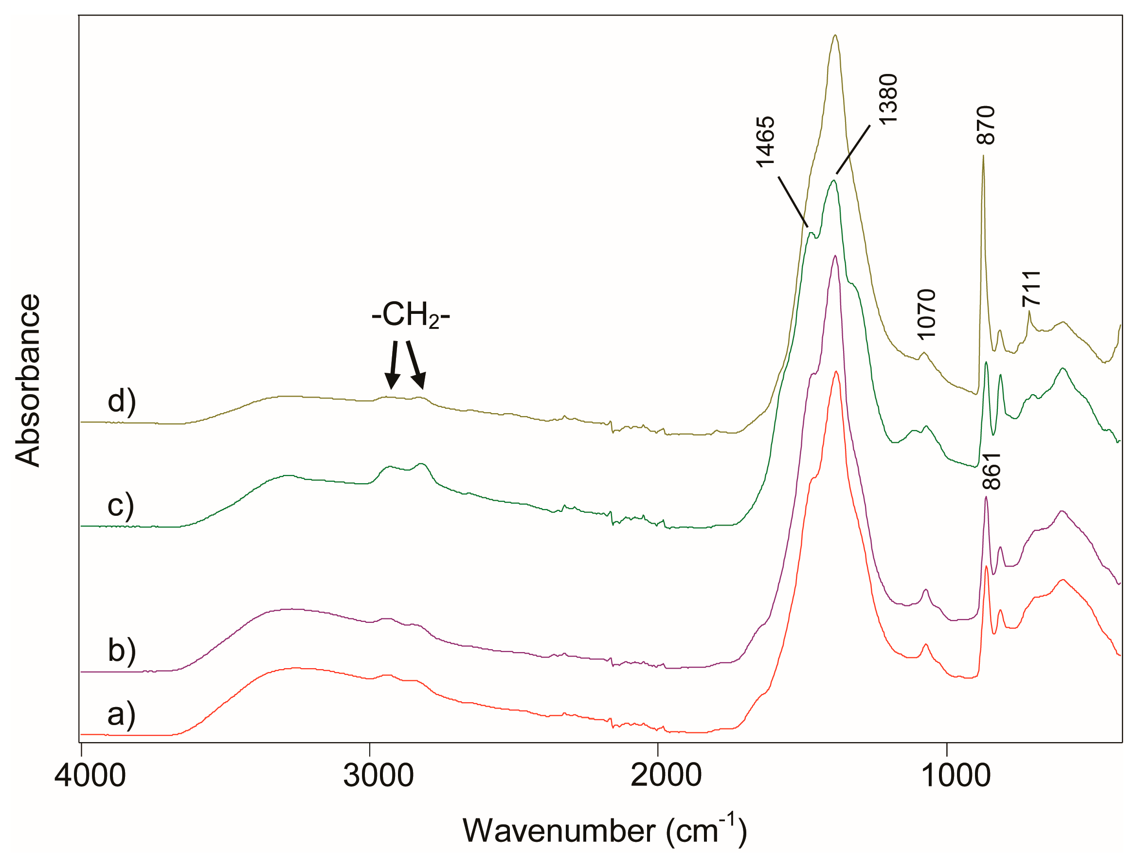
Figure 5.
SEM image of the amorphous-calcium-carbonate-rich samples (a) SE and (b) LE. They had been precipitated when subjecting an organogel (PEI–ammonium-carbamate) to an aqueous solution of calcium acetate.
Figure 5.
SEM image of the amorphous-calcium-carbonate-rich samples (a) SE and (b) LE. They had been precipitated when subjecting an organogel (PEI–ammonium-carbamate) to an aqueous solution of calcium acetate.
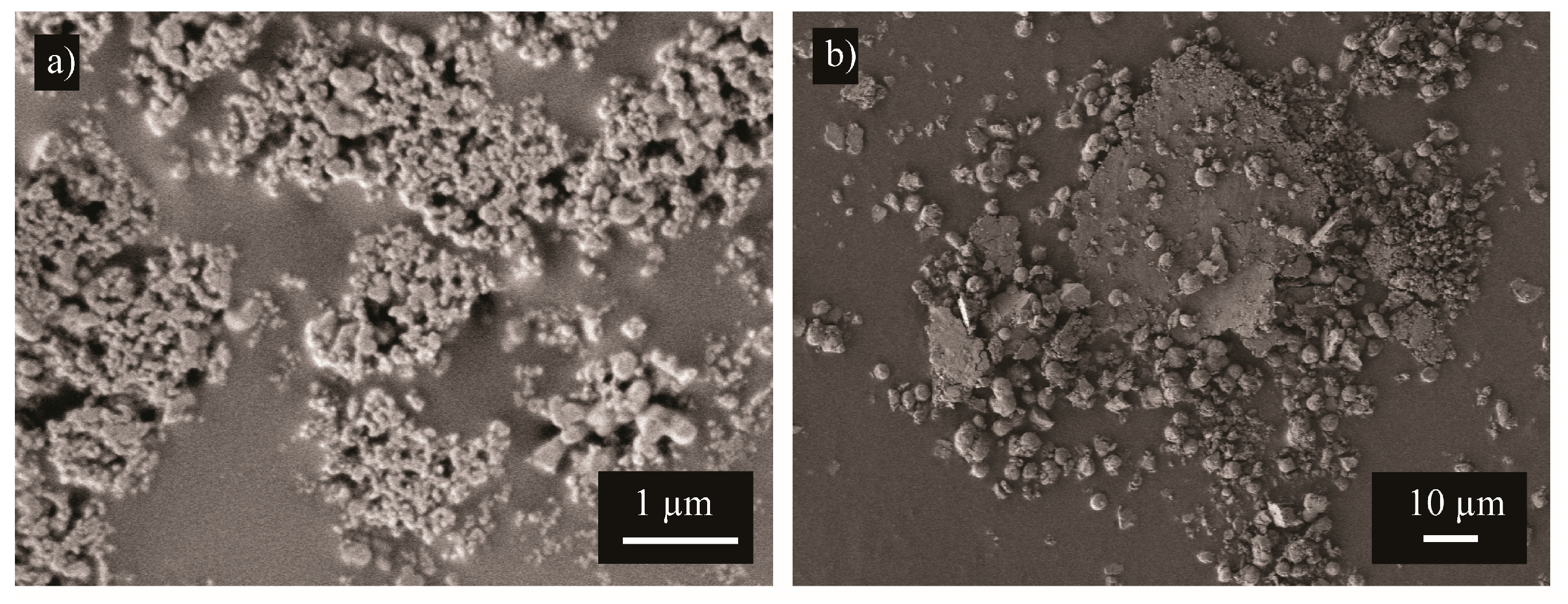
Figure 6.
Thermogravimetric curves of the amorphous-calcium-carbonate-rich samples SE (red), SW (black), LE (blue), and LW (green). These samples had precipitated when subjecting organogels (PEI–ammonium-carbamate) to aqueous solutions of calcium acetates. S and L stand for mixing for 1 h and 20 h; E and W stand for ethanol and water-and-ethanol washing.
© 2017 by the authors. Licensee MDPI, Basel, Switzerland. This article is an open access article distributed under the terms and conditions of the Creative Commons Attribution (CC BY) license (http://creativecommons.org/licenses/by/4.0/).
Share and Cite
MDPI and ACS Style
Bacsik, Z.; Zhang, P.; Hedin, N. Ammonium-Carbamate-Rich Organogels for the Preparation of Amorphous Calcium Carbonates. Minerals 2017, 7, 110. https://doi.org/10.3390/min7070110
AMA Style
Bacsik Z, Zhang P, Hedin N. Ammonium-Carbamate-Rich Organogels for the Preparation of Amorphous Calcium Carbonates. Minerals. 2017; 7(7):110. https://doi.org/10.3390/min7070110
Chicago/Turabian StyleBacsik, Zoltán, Peng Zhang, and Niklas Hedin. 2017. "Ammonium-Carbamate-Rich Organogels for the Preparation of Amorphous Calcium Carbonates" Minerals 7, no. 7: 110. https://doi.org/10.3390/min7070110
Note that from the first issue of 2016, this journal uses article numbers instead of page numbers. See further details here.