Article

Mineralogical and Elemental Composition of Pectinatella magnifica and Its Statoblasts

1
Kanazawa University, Kakuma, Kanazawa, Ishikawa 920-1192, Japan
2
Headquarters for Innovative Society Academia Cooperation, University of Fukui, 3-9-1 Bunkyo, Fukui 910-8507, Japan
3
Department of Occupational Therapy, Osaka Kawasaki Rehabilitation University, 158 Mizuma, Kaizuka, Osaka Prefecture 597-0104, Japan
4
Department of Earth and Planetary Sciences, Kanazawa University, Kakuma, Kanazawa, Ishikawa 920-1192, Japan
5
Kahokugata Lake Institute, Na 9-9, Kitachujo, Tsubata, Kahokugun, Ishikawa 929-0342, Japan
6
Medical Research Institute, Kanazawa Medical University, 1-1 Daigaku, Uchinada, Kahokugun, Ishikawa 929-0342, Japan
7
Yamato Environment Analysis Co., Ltd, 273 Santanda, Kawakita, Nomi-gun, Ishikawa 923-1253, Japan
8
Graduate School of Natural Science and Technology, Kanazawa University, Kakuma, Kanazawa, Ishikawa 920-1192, Japan
*
Author to whom correspondence should be addressed.
Minerals 2018, 8(6), 242; https://doi.org/10.3390/min8060242
Submission received: 19 March 2018 / Revised: 28 May 2018 / Accepted: 31 May 2018 / Published: 7 June 2018

Abstract

:
Several massive colonies of Pectinatella magnifica have been observed during the summer almost every year since 1974 in agricultural reservoir ponds and lakes with dirty freshwater environments in Ishikawa, Japan, which has posed serious environmental problems on the shores of Hokuriku District. We collected Pectinatella magnifica during the summer at Kahokugata Lake and Makiyama agricultural reservoir pond in June and July 2016. However, scientific data for the statoblasts of Pectinatella magnifica are limited. Our results for scanning electron microscopy equipped with energy-dispersive spectroscopy (SEM-EDS), inductively coupled plasma-mass spectrometry (ICP-MS), and X-ray powder diffraction (XRD) analyses of Pectinatella magnifica indicated immobilization of the chemical elements that were involved in the mass during the summer. We also reported the characterization of an invasive species of bryozoan (Pectinatella magnifica) in lakes and ponds in Ishikawa, Japan, based on field observations in 2016. We studied the microstructure, mineralogy, chemical composition, and radioactivity associated with these organisms, using a combination of micro-techniques, SEM-EDS, associated with ICP-MS, and XRD. This study aims to illustrate the capability of Pectinatella magnifica to produce minerals within statoblasts and gelatinous material. Obtained results may indicate forming quartz, palygorskite, dolomite, bischofite, pyrolusite, and pyrite, associated with native sulfur and copper in the statoblast. The mass of gelatinous material contains talc and vermiculite as well as non-crystalline phase. The mechanism of biomineral formation has important implications for water–mineral–organism or microorganism interactions both in lower drainage basin systems, such as Kahokugata Lake, and upper water areas, such as Makiyama agricultural reservoir pond. Many types with variety of sizes and shapes of bryozoan (Pectinatella magnifica) were found in lakes and ponds in Japan. The biomineralization systems will be made available for use not only in researching bryozoans (Pectinatella magnifica), but also for environmental change systems from upstream to downstream of the lake. To date, there have been no reports on related electron microscopy observations, including the real-life occurrence of “bioremediation”. These observations could lead to simple methods of removing statoblasts of the invasive alien species Pectinatella magnifica from agricultural and reservoir environments, because there was limited microbial immobilization of the ions during the winter.

1. Introduction

The distribution of Pectinatella magnifica has been restricted to eastern North America and central Europe. Pectinatella magnifica is also abundant on the Korean peninsula, northeastern China, and Japan. Several gelatinous colonial masses of P. magnifica appeared in the water near the shore of Lake Kawaguchi, at the foot of Mt. Fuji, Yamanashi Prefecture, Japan, in the autumn of 1972 [1]. Shuzitu Oda found giant colonial masses occurred in Lake Shoji, Yamanashi Prefecture, Japan, in 1973 [2]. Several colonial masses of Pectinatella magnifica appeared in Lake Kiba near Lake Shibayama, Ishikawa Prefecture, Japan, in 1974 and 1975 [2,3].
Since 1973, several massive colonies of Pectinatella magnifica have appeared every year in Lake Shoji, one of the five lakes at the foot of Mt. Fuji, in central Japan. In connection with a study on the life cycle of Pectinatella magnifica, Dr. Oda was able to obtain a cluster of floating statoblasts from this lake in early summer, and he distinguished six species of freshwater bryozoans among these statoblasts [4]. He reported the biological significance of these statoblasts and the distribution of Pectinatella magnifica in Japan. Every year in spring, a large number of hibernating statoblasts of Pectinatella magnifica float in clusters on the surface of Lake Shoji, though they are frozen in winter. By chance, he collected a small cluster of hibernating statoblasts from this lake on 29 June 1976. At that time, the water temperature was 21.6 °C. The majority of the statoblasts of Pectinatella magnifica, which was the main species in this cluster, had germinated. However, the valves of these statoblasts had opened and the germinal materials were lost, and only a few polypides appeared between the valves. The statoblasts of other species had not yet germinated [5]. Other species, such as Plumatella repens, Plumatella emarginata, and Stephanella hina, are also common in Japan.
Bryozoans comprise a phylum of sessile, colonial animals that inhabit marine and freshwater environments. Only around 90 species from fresh habitats have been described, most belonging to the class Phylactolaemata. Phylactolaemata can propagate asexually by forming encapsulated dormant bodies known as statoblasts, which have characteristics that are highly useful in their taxonomy and identification. To date, 23 species of Phylactolaemata in 10 genera have been reported in Japan; however, no taxonomical reviews on these have been published. Furthermore, the current taxonomy of Phylactolaemata depends heavily on the observation of statoblast microstructures using scanning electron microscopy, which poses difficulties associated with simple identification [6,7]. Pectinatella magnifica has absolutely distinctive statoblast and colony morphology. Therefore, statoblasts are useful for identification of plumatellid bryozoans.
On the other hand, the minerals and their chemical composition in bryozoans are very important for understanding the conditions of the bio-mineralization process. Therefore, there are many previous works on biomineralization of bryozoans [8,9]. Taylor et al. [9] reviewed biomineralization in bryozoans. In particular, they noted that the Mg/Ca ratio in Mg-bearing calcite, calcite/aragonite ratio, and temperature are important for understanding the location of mineral formation. To investigate these characters, they used the X-ray diffraction method. However, they did not review other minerals in bryozoans.
Various types of minerals were present in the statoblasts and gelatinous material. The chemical data of Pectinatella “gelatinous parts” (not really agar) were described with no reference to the single previous analysis of this gelatinous material [10]. There have been no reports of the mineralogical and elemental composition of Pectinatella magnifica and its statoblasts on the surface and inside gelatinous material of Pectinatella magnifica to date.
In this study, the mineralogy and chemistry of colonies and statoblasts of the freshwater species Pectinatella magnifica in Kanazawa, Ishikawa, Japan, are investigated by inductively coupled plasma-mass spectrometry (ICP-MS) and X-ray powder diffraction (XRD) techniques. Furthermore, the characteristics of colony and statoblast micro morphology and chemistry were also determined for Pectinatella magnifica using electron microscopy with an energy-dispersive microanalyzer (SEM-EDS).

2. Materials and Methods

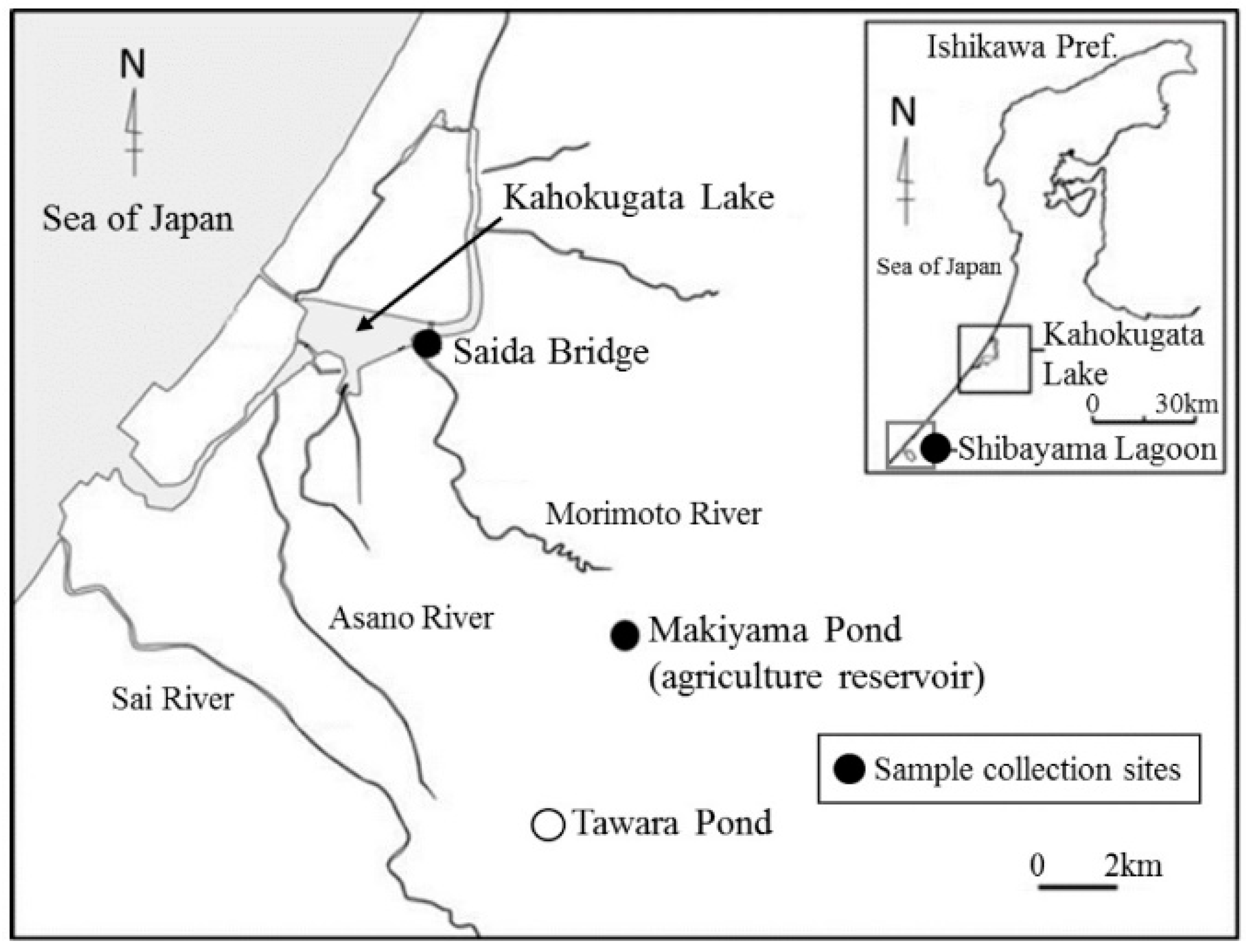
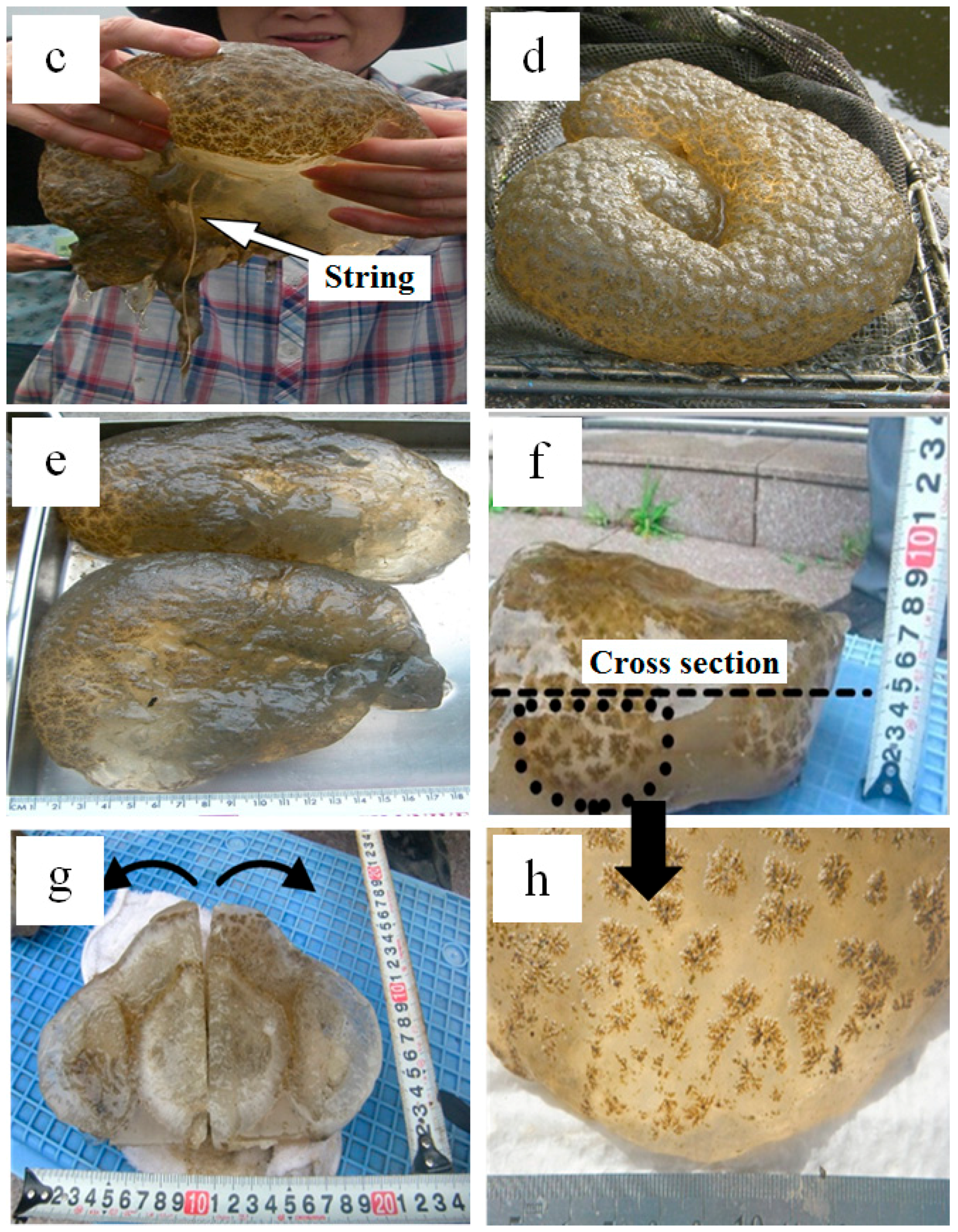
2.1. Exploring Pectinatella magnifica Field and Investigated Specimens

The freshwater Magnificent Bryozoan, Pectinatella magnifica, a confirmed native invertebrate of North America, was found in Ishikawa prefecture, Japan. This was visually confirmed on the surface of the water at four spots in ponds and lakes in Ishikawa prefecture, and samples were obtained from four locations (Figure 1), Shibayama Gata Lagoon (Figure 2a,b), Kahokugata Lake (Figure 2c,d), Makiyama agricultural reservoir pond (Figure 2e–h), and Tawara Pond [11] in Ishikawa prefecture, Japan, in July 2015 and 2016; as a result, the distribution of Pectinatella magnifica was confirmed and attracted attention. However, until now, there have been no reports of its chemical, physical, biological, and mineralogical composition.
Water characteristics such as pH, redox potential, electrical conductivity, dissolved oxygen concentration, and water temperature were measured at Makiyama agricultural reservoir pond and Kahokugata Lake during the summer of 2016 (Table 1). Furthermore, the radioactivity of water samples collected from Kahokugata Lake and Makiyama agricultural reservoir pond was recorded by measuring β(γ) rays for dried statoblasts by an Aloka GM survey meter TGS-136 (Mitaka, Japan) with a Geiger–Müller (GM) tube (Table 1).
The samples were washed in distilled and deionized (DD) water and dried under the sunshine for a few days during the summer.
The pH of the water was 6.8–7.5, the water temperature was 26–27 °C, the humidity was 48–56%, and the air temperature was 33–36 °C at noon on 18 June 2016 at Makiyama agricultural reservoir pond in Kanazawa. The dissolved oxygen (DO) was quite low at 3–4 mg/L compared with normal values (7–8 mg/L). We collected several Pectinatella magnifica samples from Makiyama agricultural pond (Figure 2e–h) and Kahokugata Lake (Figure 2c,d) to determine the major characteristics of the colonies and statoblasts. We observed their size and form and identified them as the species Pectinatella magnifica, characterizing their structure and properties based on the methods of Hirose [6,7,12] and Miyashita et al. [13]. Views of a cross section of frozen Pectinatella magnifica in Makiyama agricultural reservoir pond show two colonies 10 cm in diameter, with star patterns on their surfaces (Figure 2f–h with an arrow mark).
The frozen Pectinatella magnifica collected from Makiyama agricultural reservoir pond (f), a cross section of two colonies (g), and a statoblast pattern on the surface are shown (h) in Figure 2.
The gelatinous material varies widely in weight from 0.2 to 14 kg. Their radioactivity was 110–130 cpm (Back ground 80–100 cpm) in Kahokugata Lake during summer 2017. The water quality characteristics were pH 7.6–7.7, transparency 30–42 cm, water temperature 27 °C, electrical conductivity (EC) 12 μS/cm, and DO 6.8–7.7 mg/L for yellow brownish water on 7 and 14 July 2016.
We observed statoblast samples from Makiyama agricultural reservoir pond and Kahokugata Lake with an optical microscope (Nikon NTF2A, Tokyo, Japan) for the identification of Pectinatella magnifia.

2.2. ICP-MS Analyses

Semi-quantitative analyses of statoblast samples of Pectinatella magnifica collected from Kahokugata Lake were conducted using a Thermo Fisher Scientific (Bremen, Germany) inductively coupled argon plasma emission spectrometer (iCAP), ICP-MS Elementar Analysensysteme GmbH Vario MAX type instrument (ICP-MS) (Langenselbold, Germany).

2.3. Scanning Electron Microscopy (SEM) with Energy-Dispersive Spectroscopy (EDS)

A scanning electron microscope (S-3400N, Hitachi, Tokyo, Japan) equipped with an energy-dispersive X-ray analyzer (Horiba EMAX, Kyoto, Japan) was used to study the Pectinatella magnifica and Asajirellar gelatinosa samples collected from Makiyama agricultural reservoir pond and Kahokugata Lake. We attempted to analyze the appearance, distribution, tissue structure, and inorganic and organic constituents of the statoblasts, evaluating the microscale morphology, associated with elemental distribution maps. The operating conditions used were as follows: 15 kV accelerating voltage, 70–80 μA current, an analytical time of 1000 s, and an area of 10 mm × 10 mm on carbon double-sided tape with a Pt coating.
We also observed statoblast samples collected from Makiyama agricultural reservoir pond and performed observation and analysis at an acceleration voltage of 10 kV, in a low vacuum mode using a JEOL 6010 PLUS/LA (Tokyo, Japan) scanning electron microscope and an energy analysis of variance tools.

2.4. X-Ray Powder Diffraction Measurements

In this study, we focused on the mineralogical characterization of statoblasts and the gelatinous part of Pectinatella magnifica. Therefore, to identify minerals and analyze the structure of crystals and the crystallinity of the statoblasts and gelatinous material structure in statoblasts collected from Makiyama agricultural reservoir pond, we carried out X-ray powder diffraction measurements (XRD) under ambient conditions (Shimazu, Kyoto, Japan, XRD-6100; Cu-Kα radiation, 40 kV, 40 mA, scan speed of 2°(2θ)/min, scan range 2θ = 5–60°). Three statoblast powder samples were prepared by placing them on a glass filter with a Zn plate and two samples of the gelatinous part of Pectinatella magnifica painted directly on the sample holder were used for measurements.

2.5. X-Ray Fluorescence Analyses (XRF) for Deposited Minerals on Makiyama Pond

For comparing XRD results, the chemical compositions of the sediment samples near the Makiyama agricultural reservoir pond were also analyzed by the X-ray fluorescence analysis (XRF) technique using a XRF analyzer (Rigaku Promus II, Tokyo, Japan), which operated at an acceleration voltage of 60 kV, current of 150 mA under vacuum conditions. The sediment samples were heated to 600 °C for 30 min using an electric furnace and were pressed for XRF measurements.

3. Results

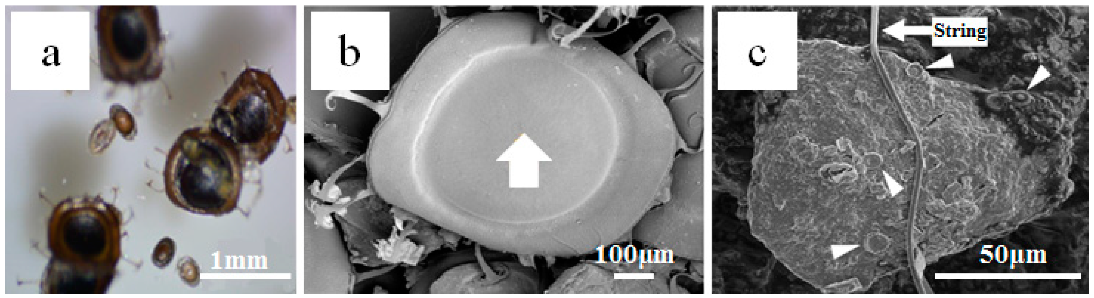
3.1. Optical Microscopy Observation and Identification of Species

We observed and analyzed statoblast samples collected from Makiyama agricultural reservoir pond using an optical microscope and SEM (Figure 3) and identified Pectinatella magnifica according to the number of statoblasts in the collected cluster [11]. Most of the statoblasts collected from Makiyama agricultural reservoir pond could be identified as Pectinatella magnifica according to their statoblasts, which have 11–22 anchors around them. Some statoblasts have 11–15 anchors. On the other hand, a few oval statoblasts with a diameter of 0.5 mm can be seen. A few reddish Cristatella mucedo were also found in Makiyama agricultural reservoir pond.

3.2. ICP-MS Analyses of Pectinatella magnifica
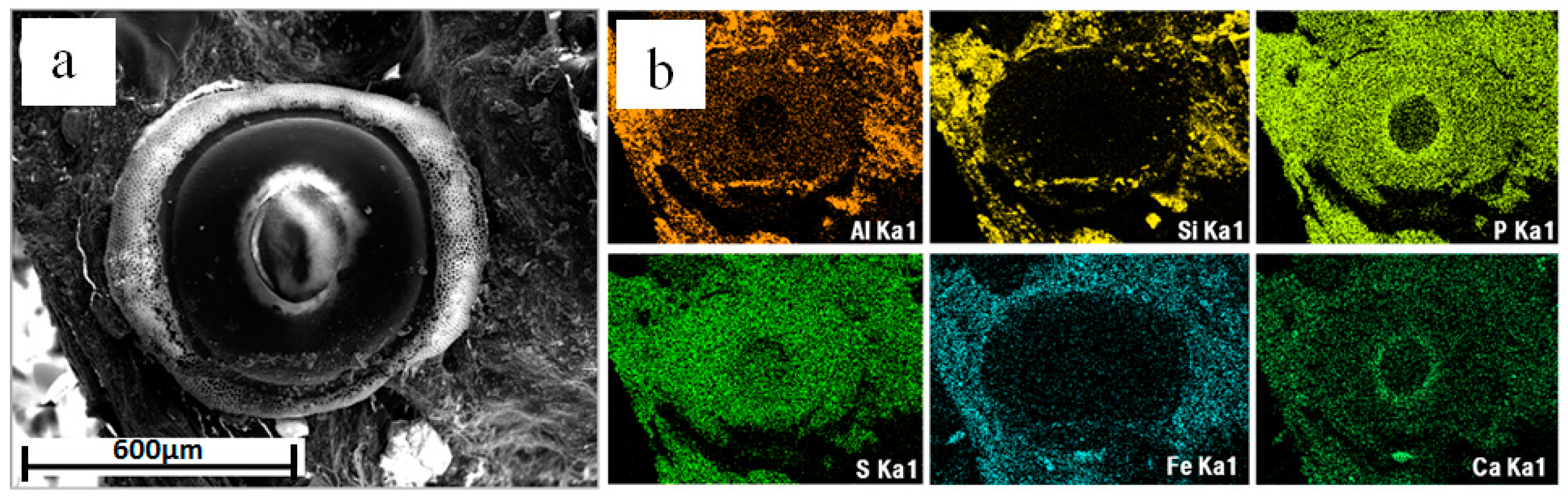
Semi-quantitative ICP-MS results for dried colonies of Pectinatella magnifica collected from Kahokugata Lake are shown in Table 2. Pectinatella magnifica contained many kinds of elements; the main constituents were Fe (290 mg/L) and Al (204 mg/L), associated with S (53.7 mg/L), Mg (46.5 mg/L), Ca (45.6 mg/L), P (26.2 mg/L), K (22.4 mg/L), and Na (13.1 mg/L) with traces of Ti (5.87 mg/L), Zn (1.58 mg/L), and Cu (0.15 mg/L) (Table 2). Traces of Sr (0.11 mg/L) can also be seen. Si cannot be measured by this method.

3.3. SEM Micrograph

SEM micrograph of a statoblast sample collected from Makiyama agricultural reservoir pond shows in Figure 3b. Pectinatella magnifica from Makiyama can be seen with a 1 mm diameter and more than 10 anchors around the rim.

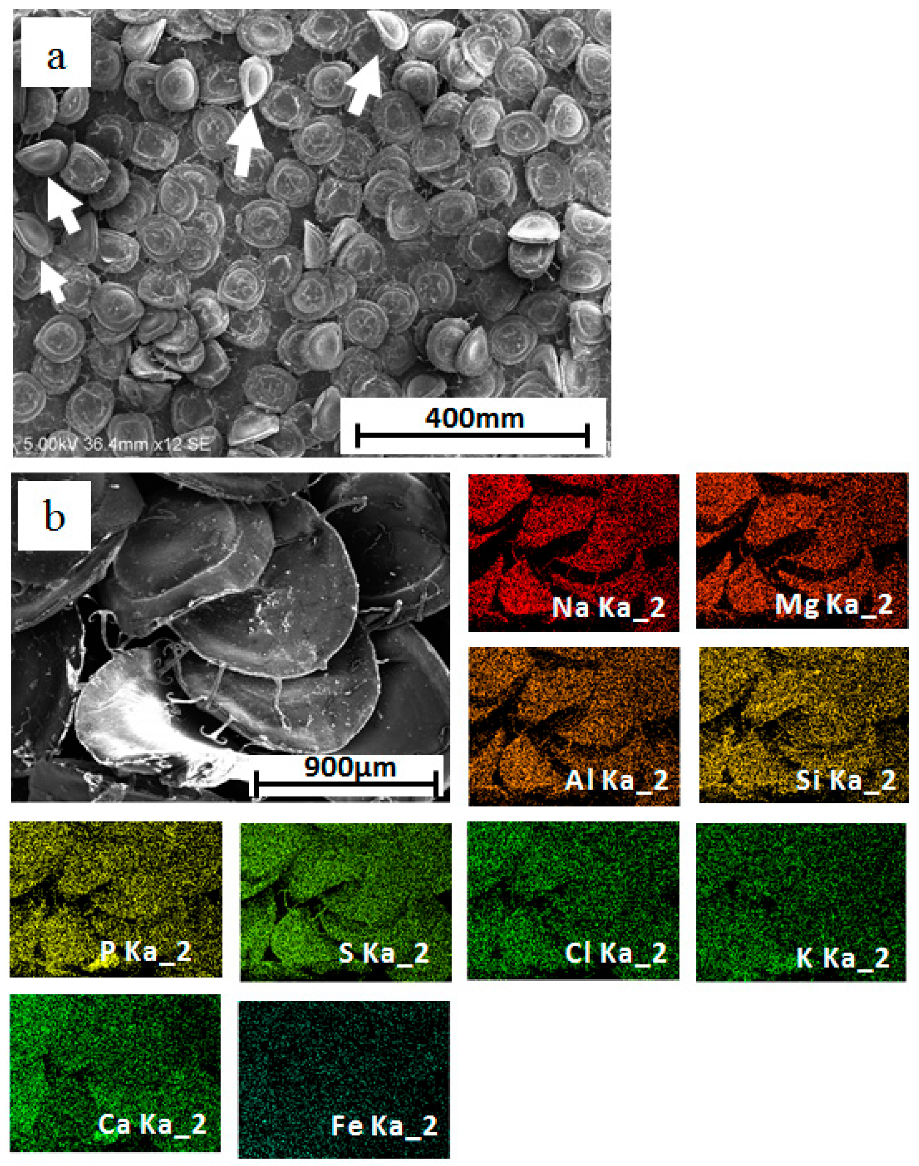
3.4. SEM Observation and Energy-Dispersive Spectroscopic (EDS) Analyses

Figure 4 shows clusters and statoblasts from Makiyama agricultural reservoir pond. Clusters with various shapes, sizes (5–10–500 μm in diameter), and numbers of statoblasts were collected from Kahokugata Lake, whereas uniform sizes (900 μm in diameter) and simple round shapes with anchors were collected from Makiyama. The samples were observed by SEM-EDS. Various types of bryozoan statoblasts lived in Kahokugata Lake, whereas only one type of extraneous with anchor type of Pectinatella magnifica lived in Makiyama agricultural reservoir pond.
Semi-quantitative analysis data of whole gelatinous material or statoblasts collected from Kahokugata Lake and Makiyama agricultural reservoir pond are shown in Table 3 and Table 4. Values are represented as mass %. The whole gelatinous material and statoblasts from Kahokugata Lake contained high Si (46–56%), associated with Al (7–15%) and Cu (7–14%) (Table 3). On the other hand, high concentrations of S (37–54%), Ca (12–15%), Si (6–10%), and traces of Sr (0.6%) can be seen in the Makiyama agricultural reservoir pond (Table 4). Statoblasts of Pectinatella magnifica from Makiyama agricultural reservoir pond showed richness in Si, S, and Ca, associated with traces of Sr, which indicates that only one anchor type of Pectinatella magnifica is present in Makiyama agricultural reservoir pond (Table 4 Points 1, 2, 3).

3.5. SEM Elemental Distribution Maps

SEM elemental distribution maps of statoblasts of Asajirella gelatinosa or Cristatella mucedo from Kahokugata Lake showed a constant distribution of P and S, whereas Al, Si, Fe, and Ca were partially concentrated at the rim or the central parts of statoblasts (Figure 5).
On the other hand, the elemental distribution maps of the statoblasts of Pectinatella magnifica from Makiyama agricultural reservoir pond showed an even distribution of almost all elements (Na, Mg, Al, Si, P, S, Cl, K, Ca, and Fe) over all the statoblasts (Figure 4b).

3.6. X-Ray Powder Diffraction (XRD) for Mineralogy of Pectinatella magnifica in Makiyama Agriculture Reservoir Pond

Some samples show almost equal XRD patterns. Therefore, we show typical profiles of each sample in Figure 6 and also show the diffraction peak position of minerals on Table 5. The mineralogy of the statoblasts of Pectinatella magnifica in Makiyama agricultural reservoir pond (Figure 6a), the gelatinous material (Figure 6b), and the zinc plate under the sample together with the glass filter (Figure 6c) was determined using XRD. Samples were placed on a Zn board with a thickness of 2 mm. The XRD profile of Figure 6a suggests that statoblasts contained mainly quartz (4.255 Å (16), 3.344 Å (100), 1.818 Å (13)) and small amounts of native sulfur (3.85 Å (100), 3.21 Å (60)) and palygorskite (Mg, Al)2[(OH, O) Si4O10, 3.17 Å (19)]. The diffractions of these minerals may contribute to the strong peak at around 2θ = 27°. This profile did not show the other strong refraction peaks of palygorskite. Therefore, the presence of palygorskite in the sample is controversial. The peaks of dolomite MgCa(CO3)2 (2.89 Å), bischofite MgCl2·6H2O (4.10 Å), and pyrolusite β-MnO2 (3.14 Å) gave wider and gradual reflections (Figure 6a), whereas Cu (2.088 Å, 1.808 Å) and pyrite FeS2 (2.4 Å) gave weak reflections. As peaks for Cu are very weak, the presence of Cu is also controversial. This XRD results are consistent with the SEM-EDS and ICP-MS results. The zinc board and glass filter used under the samples indicated reflections for a strong peak for zinc board (2.488 Å, 2.321 Å, 2.102 Å, and 1.6S93 Å), associated with a relatively strong halo pattern of gelatinous non-crystalline material, which contained talc Mg3Si4O10(OH)2 (4.55 Å) with low crystallinity and a small amount of vermiculite Mg3(Si3, Al) O10(OH)2·Mg0.5 4H2O (14–15 Å) (Figure 6b). However, talc shows only a broad halo without a sharp diffraction peak. Therefore, in order to confirm talc, more precise analyses by XRD and TEM techniques will be needed in a future study.
The statoblast crystallizes as water evaporates or biogenetic minerals, forming palygorskite, dolomite, bischofite, pyrolusite, pyrite, sulfur, and copper. The mass of gelatinous material contains talc and vermiculite as well as gelatinous non-crystalline materials. XRD and SEM-EDS electron micrographs (elemental distribution maps) may indicate Pectinatella magnifica, confirming the role of microorganisms in mediating the transfer of metal solutes from the hydrosphere to sediments.

3.7. X-Ray Fluorescence Analyses (XRF) for Deposited Minerals in Makiyama Agricultural Reservoir Pond

The chemical compositions of sediment samples near the Makiyama agricultural reservoir pond obtained by XRF analyses are shown in Table 6.

4. Discussion

In this paper, mineralogical and elemental composition of Pectinatella magnifica and its statoblasts in freshwater are summarized (Figure 7). The distribution of elements and minerals inside of the bryozoan colony of Pectinatella magnifica indicates the role of bryozoans in mediating the transfer of metal solutes from hydrosphere to sediments. Abundant intracellular and cell wall Sr sorption has been identified in phytoplankton biomass and Pectinatella magnifica in Tawara Pond, Kanazawa, Ishikawa, Japan, at pH 7–8.5 and 20–30 °C, with C (42.9 wt %), H (5.8 wt %) and N (7.6 wt %) in summer 2015 [10]. Isolates of the phytoplankton community and Pectinatella magnifica were found to contain 0.65 mg/L of Sr by dry weight using the ICP-MS method. Concentration factors for Rb1–1.55 mass % and Sr 5–7.5 mass % were found to be 5–7.5 mass % from point analyses, using the SEM-EDS method, confirming the role of microorganisms in mediating the transfer of metal solutes from the hydrosphere to sediments.
We think that the adsorption of part of some elements such as Al, Si, and Fe may be possible. On the other hand, the results of powder X-ray diffraction patterns (Figure 6) and rather high quantity of Si (Table 3 and Table 4) show the existence of some silicate minerals such as vermiculite. Therefore, a high proportion of these elements exist as biominerals in Pectinatella magnifica.

4.1. Characteristics of Statoblasts

A key to species based on colony and statoblast gross micro morphology has already been provided [14] to aid in the identification of freshwater bryozoans in Makiyama agricultural reservoir pond and Kahokugata Lake using SEM-EDS, which provides useful characteristics related to the taxonomy of freshwater species described in this paper. Elemental distribution maps of statoblasts (Figure 4) have never previously been studied, and these showed high concentrations of P and S throughout the species, whereas Al, Si, and Fe were concentrated around the rim, showing the presence of minerals such as highly crystallized sulfur and palygorskite in statoblasts, whereas talc and vermiculite were present in the gelatinous material, as shown by XRD. Therefore, sulfur may be included in the statoblasts as native S and pyrite under redox conditions. On the other hand, phosphorous may be included as non-crystalline matter. Because the XRD pattern shows a large background (halo) centered around 30°(2θ), analysis of non-crystalline matter is also important to understand Pectinatella magnifica totally. However, this kind of study requires more work. Therefore, we would like to deal with this material in a future work. On the other hand, the presence of Si is consistent with the quartz crystals identified in the XRD analysis. There is a possibility of quartz crystal due to the contamination of quartz crystal from sediments in Makiyama agricultural reservoir pond (Table 6). If the sediments contaminate the Pectinatella magnifica associated with gelatinous material, the XRD pattern of gelatinous materials (Figure 6b) may also show diffraction peaks of quartz. However, the XRD pattern of gelatinous materials did not show diffraction peaks of quartz. From these analyses, minerals in statoblasts detected by XRD measurements may be water-evaporated minerals and biominerals.
Otherwise, statoblasts of Pectinatella magnifica associated with gelatinous material will revive the following summer. The statoblasts of Pectinatella magnifica located upstream then drain downstream and are deposited in a lake, such as Kahokugata. It was particularly notable that quantities of Pectinatella magnifica have been found in Kahokugata Lake over a long period of time, since 1959. Furthermore, it is recognized that the factors affecting the size and quantity of Pectinatella magnifica are complex. Colonies accumulate in many lakes almost every summer in Japan [15,16,17,18,19,20,21].

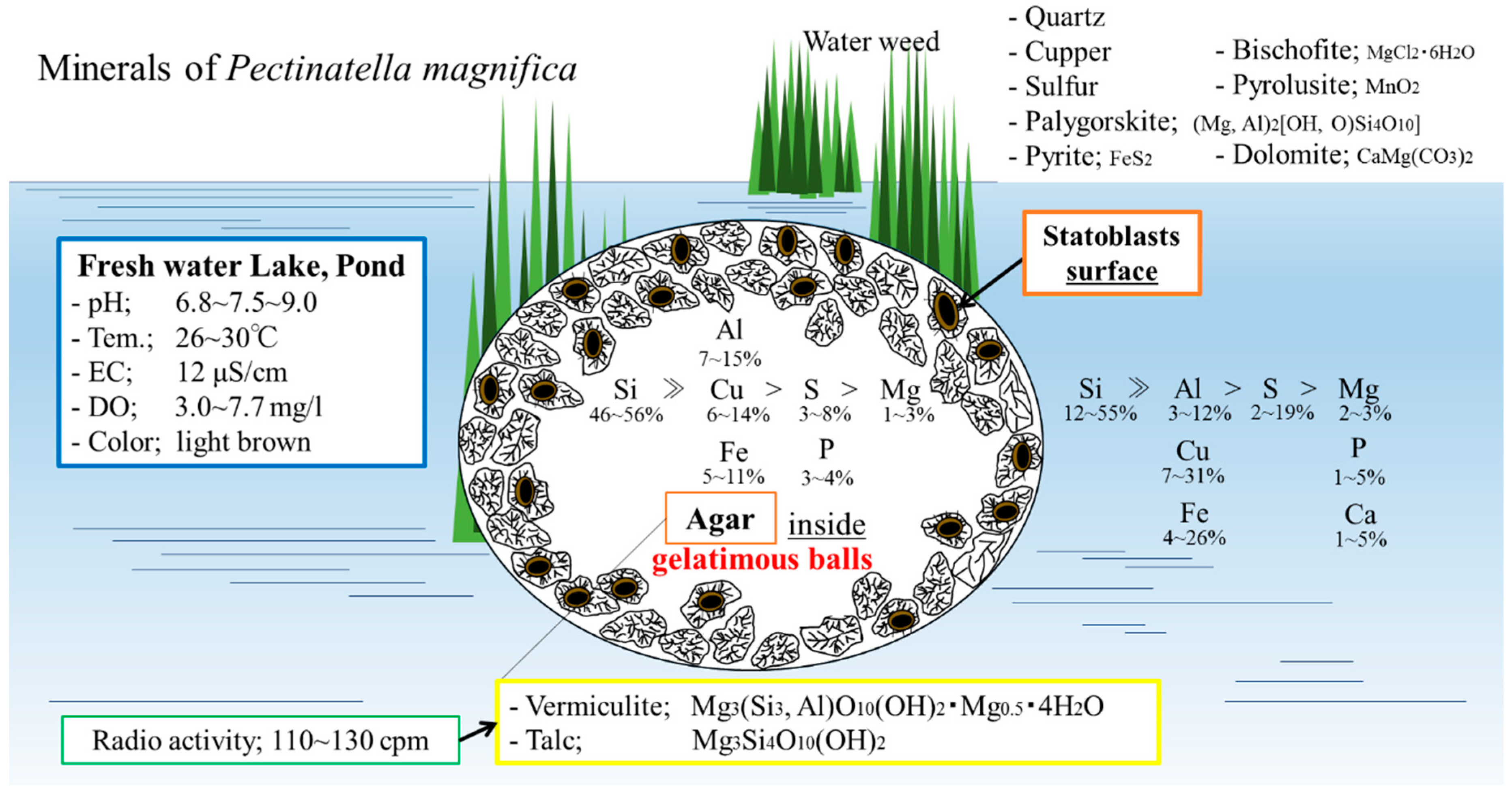
4.2. Mineralogical and Elemental Composition of Pectinatella magnifica and Its Statoblasts

In this paper, we report not only the characterization of Pectinatella magnifica in freshwater systems, but also the formation of minerals inside gelatinous material and statoblasts on the surface of freshwater ponds and lakes in Kanazawa, Ishikawa, Japan, based on the data for chemicals and minerals in the field. Therefore, we report many data on the elements and minerals including in Pectinatella magnifica obtained using SEM-EDS, ICP-MS, and XRD techniques. These results, which are summarized in Figure 7, can provide insights into the identification of mineral species and characteristic structures. We analyzed the microchemistry of Pectinatella magnifica using a combination of micro-techniques. Previously, many mineralogical studies in bryozoans were limited to carbonate minerals such as calcite, Mg-rich calcite, and aragonite. However, in this work, we identified many other minerals as follows. The gelatinous material portions of Pectinatella magnifica may contain mainly of vermiculite and talc as well as organic non-crystalline materials, whereas highly crystalline quartz was associated with native sulfur, palygolskite, bischofite, pyrolusite, dolomite, pyrite, and copper in statoblasts of Pectinatella magnifica (Figure 6a). However, the presence of minerals such as palygolskite and copper is controversial. Therefore, more precise XRD analyses may be necessary for analyzing the mineralogy in detail.
The existence of clay minerals such as vermiculite and talc in gelatinous materials may be partially caused by the contamination of lake and pond sediments. It may be possible that materials including clay minerals flow into lakes and ponds based on the data on the chemical composition of the sediment. XRF data of the sediment for Makiyama agricultural reservoir pond (Table 6) are consistent with this interpretation. The existence of dolomite is consistent with similar quantities of Ca and Mg (Table 2 and Table 3). Other minerals found in this study such as pyrite and native sulfur show reducing environment. Particular elements, such as abundant Si, Al, Cu, and Fe, were associated with S and Mg in the gelatinous material. Also, Si, Al, Cu, and Fe accumulated at the statoblast surfaces of Pectinatella magnifica, as shown in Figure 7, which illustrates Pectinatella magnifica based on chemical and mineralogical data. On the other hand, phosphorus may be included as a non-crystalline material, as mentioned above.

4.3. Distribution of Elements and Minerals inside the Bryozoan Colony

Elements often occur in close proximity in separate mineral phases, or substitute for one another as, for example, in silicate minerals, whose crystal chemical characteristics are now well known. Iron is also an essential element in the metabolic pathways of oxidation–reduction in all species from microbes to man. From the simple aqueous chemistry of Si, Al, and Fe, it is clear that particular chemical species and clay minerals that occur in surficial environments are strongly dependent on environmental conditions [22,23,24,25,26,27,28].
This paper presents an overview of mineral species and essential elements, and the possible mechanisms involved in their precipitation. Depending on the specific environmental conditions, newly-formed Fe minerals are predominantly oxides, oxyhydroxides, sulfides, sulfates, carbonates, and phosphates. The main Fe mineral formation processes are discussed in many papers, e.g., by Schwertman and Fitzpatrick [29,30].
In Actinopoda, Zoomastigina, and Mollusca, different minerals are formed at different developmental stages. In the case of the oyster Crassostrea virginica, for example, the larval “shell gland” cells form aragonite, whereas in the adult, the mantle cells form calcite. In the holothurian species Molpadiidae, the juveniles initially form spicules of calcite. An example of a calcite spicule formed by a juvenile is reported in the literature [11,29,30,31,32,33,34,35,36].
It is also important to understand the possible mechanisms of metal uptake, whether biologically mediated, inorganically controlled, or working in concert, during the precipitation of minerals in Makiyama agricultural reservoir pond upstream and Kahokugata Lake downstream of the Morimoto River. Studying these mechanisms would add to our understanding of mineral formation and growth in surface water environments [37,38,39,40].

5. Conclusions

We studied the microstructure, mineralogy, chemical composition, and radioactivity associated with Pectinatella magnifica using a combination of micro techniques, scanning electron microscopy equipped with energy-dispersive spectroscopy (SEM-EDS), associated with inductively coupled plasma-mass spectrometry (ICP-MS), and X-ray powder diffraction analyses (XRD). This study aims to illustrate the capacity of Pectinatella magnifica to produce minerals within statoblasts, forming quartz, palygorskite, dolomite, bischofite, pyrolusite, pyrite, native sulfur, and native copper. The mass of gelatinous material may principally contain vermiculite and talc as well as non-crystalline organic materials. However, the presence of palygoskite, native copper, and talc is controversial. The mechanism of mineral formation has important implications for water–mineral–microorganism interactions in both lower drainage basin systems such as Kahokugata Lake, and upper water areas such as Makiyama agricultural reservoir pond. A SEM-EDS electron micrograph showing Pectinatella magnifica confirms the role of microorganisms in mediating the transfer of metal solutes (elemental distribution maps) from the hydrosphere to sediments. Many types of bryozoans (Pectinatella magnifica) were found in the lake and the pond in Japan. To date, there have been no reports describing electron microscopy observations, elementary content maps, and mineral formation. Therefore, this work is important for understanding the minerals include in bryozoans.

Author Contributions

K.T., Y.H., and S.H. conceived and designed the fieldwork and sampling; K.N. and K.T. performed the XRD and ICP-MS experiments; K.T. and T.T. operated the SEM-EDS; K.T., A.F. and M.O. interpreted the data and wrote the manuscript; K.T., A.F., T.K., M.O. and F.T. designed the figures.

Acknowledgments

We are grateful for the support from Wakura Co. (Yusuke Katayama). The authors also wish to thank Sachiko Yamamoto, Hirono Tazaki, Hisashi Takahashi, and Nanae Kawahara for their kind support in this research. The authors would also like to thank Enago (www.enago.jp) for the English language review. We express our gratitude to Referees 1 and 2 for their helpful comments and advice on our work.

Conflicts of Interest

The authors declare no conflict of interest.

References

  1. Mawatari, S. New occurrence of Pectinatella magnifica (Leidy) in Japanese Lake. Proc. Jpn. Soc. Syst. Zool. 1973, 9, 41–43. [Google Scholar]
  2. Oda, S. Pectinatella magnifica occurring in Lake Shoji, Japan. Proc. Jpn. Soc. Syst. Zool. 1974, 10, 31–39. [Google Scholar]
  3. Oda, S. Notes on Pectinatella magnifica occurring in Japan. Collect. Breed. Tokyo 1975, 37, 40–53. (In Japanese) [Google Scholar]
  4. Oda, S. A life of Pectinatella magnifica a freshwater bryozoan. Ibid 1976, 38, 434–443. (In Japanese) [Google Scholar]
  5. Oda, S. A note on bryozoan in Lake Shoji, Japan. Proc. Jpn. Soc. Syst. Zool. 1978, 15, 19–23. [Google Scholar]
  6. Hirose, M. Taxonomy as a basis and a goal of zoology, with some examples of bryozoology in museums. TAXA Proc. Jpn. Soc. Syst. Zool. 2013, 34, 25–31. [Google Scholar]
  7. Hirose, M. Taxonomy of Lophophorata; Description of diversity of morphology and biology of bryozoans. TAXA Proc. Jpn. Soc. Syst. Zool. 2014, 37, 1–13. [Google Scholar]
  8. Smith, A.M.; Key, M.M., Jr.; Gordon, D.P. Skeletal mineralogy of bryozoans: Taxonomic and temporal patterns. Earth Sci. Rev. 2010, 78, 271–277. [Google Scholar] [CrossRef]
  9. Taylor, P.D.; Lombardi, C.; Cocito, S. Biomineralization in bryozoans: Present, past, future. Biol. Rev. 2015, 90, 1118–1150. [Google Scholar] [CrossRef] [PubMed]
  10. Withrow, M. The chemical constitution of Pectinatella. Science 1930, 71, 265. [Google Scholar]
  11. Tazaki, K.; Brandon, K.; Yamamoto, S.; Sato, M.; Hayashi, T.; Takehara, T. Strontium sorption in Phytoplankton biomass and Pectinatell magnifica; Implications for bioremediation. Radioisotopes 2016, 65, 383–392. [Google Scholar] [CrossRef]
  12. Hirose, M. Taxonomy and identification of freshwater bryozoans in Japan. TAXA Proc. Jpn. Soc. Syst. Zool. 2012, 33, 17–32. [Google Scholar]
  13. Miyashita, M.; Harada, M.; Noguch, M. First Report of Magnificent Bryozoan Pectinatella magnifica (LEIDY) in Wakayama Prefecture and Its Distribution at Kinokawa and Iwada Cities; Memoirs Institute of Advanced Technology, Kinki University: Osaka, Japan, 2014; Volume 19, pp. 1–12. [Google Scholar]
  14. Hashida, Y.; Hashida, S.; Tazaki, K.; Nakamura, I.; Takehara, H.; Nakamura, K. Environmental activity and the ecotourism at Makiyama, Kanazawa, Ishikawa in Japan. Kahokugata Lake Sci. 2017, 20, 19–31. [Google Scholar]
  15. Mukai, H. Germination of the statoblasts of freshwater bryozoan, Pectinatella gelatinosa. J. Exp. Zool. 1974, 183, 27–40. [Google Scholar] [CrossRef]
  16. Oda, S. Freshwater bryozoan in Japan. Biol. Jpn. 1990, 4, 50–57. [Google Scholar]
  17. Mawatari, S. USA originated Pectinatella magnifica (LEIOY) found in Japan. Anim. Taxon. 1973, 9, 41–44. [Google Scholar]
  18. Kaneko, Y.; Iwatsuki, K.; Noda, Y. Pectinatella magnifica in Kagawa Prefecture. J. Biol. Kagawa 1994, 21, 63–66. [Google Scholar]
  19. Hirose, M.; Ikezawa, H.; Kanek, N.; Mawatari, S. Distribution of freshwater bryozoans in Ibaragi Prefecture. Ibaragi Nat. Mus. Rep. 2006, 9, 31–46. [Google Scholar]
  20. Iwasaki, K. Introduced sessile animals and invasive alien species act in Japan. Sess. Org. 2006, 23, 13–24. [Google Scholar]
  21. Oda, S. Germination of the statoblasts in freshwater Gryozoa. Sci. Rep. Tokyo Kyoiku Daigaku B 1959, 9, 90–132. [Google Scholar]
  22. Robin, N.; Bermard, S.; Miot, I.; Blanc-Valleron, M.M.; Charbonnier, S.; Petit, G. Calcification and diagenesis of bacterial colonies. Minerals 2015, 5, 488–506. [Google Scholar] [CrossRef]
  23. Bindschedler, S.; Cailleau, G.; Verrecchia, E. Role of Fungi in the biomineralization of calcite. Minerals 2016, 6, 41. [Google Scholar] [CrossRef]
  24. Simkiss, K.; Wilbur, K.M. Biomineralization: Cell Biology and Mineral Deposition; Academic Press Inc.: Waltham, MA, USA, 1989; p. 337. [Google Scholar]
  25. Krumbein, W.E. Microbial Geochemistry; Blackwell Scientific Publications: Oxford, UK, 1983; p. 330. [Google Scholar]
  26. Tazaki, K.; Okuno, M.; Furumoto, M.; Watanabe, H. Biomineralization of pisoliths in hot springs. Mater. Sci. Eng. C 2006, 26, 617–623. [Google Scholar] [CrossRef]
  27. Tazaki, K.; Morii, I. Microbial immobilization of Si, Mn, Fe and Sr ions in the nacreous layer of Sinohyliopsis schlegeli and environmental factors. Earth Sci. Front. 2008, 15, 55–65. [Google Scholar] [CrossRef]
  28. Tazaki, K. Microbial Mats; Their Biomineralization and Bioremediation; Takakuwa Art Printing Office: Ishikawa, Japan, 2009; 228p. [Google Scholar]
  29. Lowenstam, H.A.; Weiner, S. On Biomineralization; Oxford University Press: Oxford, UK, 1989; pp. 45–46. [Google Scholar]
  30. Mann, H.; Tazaki, K.; Fyfe, W.S.; Kerrich, R. Microbial Accumulation of Iron and Manganese in Different Aquatic Environments: An Electron Optical Study; CATENA SUPPLEMENT 21; CATENA: Cremlingen, Germany, 1992; pp. 115–131. [Google Scholar]
  31. Tazaki, K.; Takehara, T.; Ishigaki, Y.; Nakagawa, H. The distribution of Sr, Cs, Np and Pu in the radioactive paddy rice field in Fukushima. J. Foss. Res. 2013, 46, 35–57. [Google Scholar]
  32. Tazaki, K.; Simojima, Y.; Takehara, T.; Nakano, M. Formation of microbial mats and salt in radioactive paddy soils in Fukushima, Japan. Minerals 2015, 5, 849–862. [Google Scholar] [CrossRef]
  33. Seckbach, J.; Oren, A. Microbial Mats: Modern and Ancient Microorganisms in Stratified Systems; Springer: New York, NY, USA, 2010; p. 606. [Google Scholar]
  34. Morikawa, H.; Takahashi, M.; Kawamura, Y. New progress in environmental biotechnology using phytoremediation. J. Environ. Biotechnol. 2001, 1, 1–14. [Google Scholar]
  35. Ehrlich, H.L.; Newman, D.K.; Kappler, A. Ehrlich’s Geomicrobiology, 6th ed.; CRC Press, Taylor & Francis Group: Boca Raton, FL, USA, 2016; 635p. [Google Scholar]
  36. Sekine, M.; Shimizu, F. Chemical Structure of Gel Material of Pectinatella magnifica. Abstract of Japanese Agricultural Chemical Society. 2013. Available online: http://www.jsbba.or.jp/MeetingofJSBBA/2013/MeetingofJSBBA2013.pdf (accessed on 24 September 2017).
  37. Oono, K.; Shibasaki, T. The lagoon and bay sediments of the Hokuriku district. Earth Sci. 1967, 21, 25–34. [Google Scholar]
  38. Tsukawaki, S.; Asano, I. Sedimentation in Kahoku-gata Lagoon in Ishikawa Pref. Central Japan; Bulletin of the Japan Sea Research Institute, Kanazawa University: Kanazawa, Japan, 1999; Volume 30, pp. 69–97. [Google Scholar]
  39. Tazaki, K. Architecture of biomats reveals history of geo-, aqua-, and bio-systems. Episodes 1999, 22, 21–25. [Google Scholar]
  40. Tazaki, K.; Tazaki, F.; Okuno, M.; Takehara, T.; Ishigaki, Y.; Nakagawa, H. Formation of pisolite at hot springs in Saturnia, Toscana, Italy. J. Geol. Soc. Jpn. 2016, 122, 45–60. [Google Scholar] [CrossRef]
Figure 1. Location map of the study area where samples were collected in Ishikawa Prefecture, Japan.
Minerals 08 00242 g001
Figure 2. Colonies of Pectinatella magnifica appearing during the summer season in polluted freshwater environments at Shibayama Lagoon (a,b); Kahokugata Lake (c,d); and Makiyama agricultural reservoir pond (e) in Ishikawa Prefecture, Japan. The frozen Pectinatella magnifica collected from Makiyama agricultural reservoirs (f); a normal pattern of zooid clusters (g); and statoblast pattern on the surface (h).
Minerals 08 00242 g002aMinerals 08 00242 g002b
Figure 3. Optical micrographs of the statoblasts of Pectinatella magnifica (a) collected from Makiyama agricultural reservoir pond. Scanning electron microscope (SEM) micrograph of the statoblasts of P. magnifica (b) collected from Makiyama agricultural reservoir pond. SEM micrographs of massive colonies of Pectinatella magnifica, showing a long adherent string ((c), an arrow) and four spherical Pectinatella magnifica on the surface of gelatinous material ((c), four arrows).
Minerals 08 00242 g003
Figure 4. SEM micrographs of gathered statoblasts of Pectinatella magnifica (a) and the enlarged elemental distribution maps (b). The samples were collected from Makiyama agricultural reservoir Pond.
Minerals 08 00242 g004
Figure 5. SEM-EDS elemental distribution maps of Asajirella gelatinosa collected from Kahokugata Lake. Elemental content maps indicated in this area associated with Asajirella gelatinosa. SEM image (a) Elemental content maps (b).
Minerals 08 00242 g005
Figure 6. XRD results for the statoblasts gathered on the surface of Pectinatella magnifica (a); the gelatinous material (agar parts) of this species (b); and the sample holder of zinc board and glass filter without the samples (c), showing the existence of various types of minerals.
Minerals 08 00242 g006
Figure 7. The distribution of elements and minerals inside the bryozoan colony, determined based on chemical data obtained from ICP-MS and SEM-EDS and mineralogical data from XRD. Characteristics of water, such as pH, temperature, EC, DO, and color are excerpted from Hashida et al. [14].
Minerals 08 00242 g007
Table 1. Characteristics of water in Kahokugata Lake and Makiyama Pond in 2016. EC: electrical conductivity; DO: dissolved oxygen; BG: Back ground.
Kahokugata LakeMakiyama Agriculture Reservoirs Pond
Date27 June, 14 July in 201618 June, 1 July in 2016
pH6.8–7.7–9.06.8–7.5
Water Temp.26–27 °C33–36 °C
EC12 μS/cm12 μS/cm
DO6.8–7.7 mg/L3.0–4.0 mg/L
Water Colorlight brownlight brown
Radio Activity110–130 cpm (BG 80–100cpm)110–120 cpm (BG 80–100 cpm)
Size of 30–40–60 cm20–50 cm
Pectinatella magnifica200 g, 800 g, 14 kg500 g, 1 kg, 5 kg
Table 2. ICP-MS semi-quantitative analytical results for dried colonies of Pectinatella magnifica collected from Kahokugata Lake. (The unit mg/L means the weight of the element in one liter of wet sample.)
Atomic NumberElementsmg/L
5B0.046
11Na13.100
12Mg46.500
13Al204.000
14Si-
15P26.200
16Si53.700
17Cl1.400
19K22.400
20Ca45.600
22Ti5.870
23V0.630
24Cr0.320
25Mn3.980
26Fe290.000
27Co0.100
28Ni0.170
29Cu0.150
30Zn1.580
33As0.100
37Rb0.048
38Sr0.110
39Y0.084
40ZrND
48Cd0.004
55Ce0.540
56Ba0.470
80HgND
82Pb0.150
-: not measured; ND: not detected.
Table 3. Results of SEM-EDS semi-quantitative analyses of dried colonies of Pectinatella magnifica collected from Kahokugata Lake in July and September 2016. The unit is mass %.
Elements1234
Mg1.601.222.093.45
Al10.497.4712.6715.34
Si46.0556.4848.6747.03
P3.523.183.543.39
S7.987.927.063.16
Cl0.430.310.680.18
K1.140.261.241.31
Ca1.701.512.041.30
Ti0.24NDNDND
Mn0.04NDNDND
Fe6.504.864.7310.90
Cu13.6911.0913.716.55
Total93.3894.3096.4392.61
ND: not detected.
Table 4. Results of SEM-EDS semi-quantitative analyses of carbon-coated dried statoblasts and anchor-shaped thorns collected from Makiyama agricultural reservoir pond in November 2016, coated with carbon, indicating richness in Si, S, and Ca associated with traces of Sr. Values are represented as mass %.
Makiyama Pond: Carbon Coated Statobolasts (mass %, ±3δ)
Elements123
Na4.706.846.64
Mg5.685.344.32
Al2.261.971.20
Si9.435.7210.09
P3.050.530.47
S45.8337.0153.75
Cl3.812.33 3.51
K3.55ND3.02
Ca14.7812.7812.43
Ti0.42ND0.59
MnNDNDND
Fe4.01ND1.89
Ni0.630.59ND
CuND15.95ND
ZnND8.22ND
SrND0.590.64
Total98.1597.8798.55
StatoblastsStatoblastsAnchor shape thorn
ND: not detected.
Table 5. Peak positions and d values of XRD patterns.
(a) Statoblasts
2θ (°)d-Value (Å)Relative Intensity
20.0504.42810
21.4004.14925
21.5604.11825
22.5713.93620
22.7373.90820
24.3863.65010
27.2003.276100
27.3403.25985
27.4803.24350
28.3763.14320
30.8772.89610
35.3542.53715
36.2962.47315
39.9602.25420
40.1602.24420
43.1582.09615
50.6401.80120
50.8201.79520
(b) Agar Part (Gelatinous Part)
2θ (°)d-Value (Å)Relative Intensity
8.05610.974- *
12.2377.233- **
19.7304.488- *
35.9392.49740 ***
38.6442.32819 ***
42.8812.107100 ***
54.0071.69725 ***
* broad peak, ** weak peak, *** Zn plate of holder.
Table 6. Results of XRF analyses of sediments in Makiyama agricultural reservoir pond, Kanazawa, Japan (mass %).
Components12345
SiO260.300058.100055.800056.800057.9000
Al2O320.100019.500023.100014.900015.8000
CO26.55009.14009.310015.600013.0000
Fe2O36.30004.56005.20004.65004.9900
K2O2.41002.25002.17002.38002.4100
MgO1.98001.22001.14001.75001.8100
TiO20.80500.45900.44400.57300.6170
Na2O0.73101.66001.05001.25001.2500
CaO0.49802.32001.11001.25001.1900
P2O50.09470.40300.35400.40400.4620
SO30.06030.15400.13600.18500.1900
MnO0.52900.04340.05880.10700.1060
Total100.359099.810099.928099.859099.7250

