Next Issue
Volume 7, December
Previous Issue
Volume 7, June


Diagnostics, Volume 7, Issue 3 (September 2017) – 16 articles

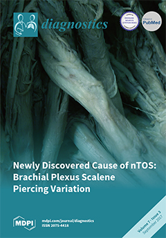
Cover Story (view full-size image): We identified a common anatomical variation in the brachial plexus of a cadaveric sample in which nerve fibers course through the anterior scalene muscle, potentially becoming impinged, which could lead to a predisposition for neurogenic thoracic outlet syndrome (nTOS). Using ultrasonography, we identified this variant in living subjects and found that individuals with nTOS symptoms were significantly more likely to have this variation. The implications for the diagnosis and treatment of nTOS are discussed in detail. View this paper
  • Issues are regarded as officially published after their release is announced to the table of contents alert mailing list.
  • You may sign up for e-mail alerts to receive table of contents of newly released issues.
  • PDF is the official format for papers published in both, html and pdf forms. To view the papers in pdf format, click on the "PDF Full-text" link, and use the free Adobe Reader to open them.
Order results
Result details
Section
Select all
Export citation of selected articles as:
250 KiB  
Review
Recent Progress in the Development of Diagnostic Tests for Malaria
by Francis D. Krampa, Yaw Aniweh, Gordon A. Awandare and Prosper Kanyong
Diagnostics 2017, 7(3), 54; https://doi.org/10.3390/diagnostics7030054 - 19 Sep 2017
Cited by 52 | Viewed by 11055
Abstract
The impact of malaria on global health has continually prompted the need to develop effective diagnostic strategies. In malaria endemic regions, routine diagnosis is hampered by technical and infrastructural challenges to laboratories. These laboratories lack standard facilities, expertise or diagnostic supplies; thus, therapy [...] Read more.
The impact of malaria on global health has continually prompted the need to develop effective diagnostic strategies. In malaria endemic regions, routine diagnosis is hampered by technical and infrastructural challenges to laboratories. These laboratories lack standard facilities, expertise or diagnostic supplies; thus, therapy is administered based on clinical or self-diagnosis. There is the need for accurate diagnosis of malaria due to the continuous increase in the cost of medication, and the emergence and spread of drug resistant strains. However, the widely utilized Giemsa-stained microscopy and immunochromatographic tests for malaria are liable to several drawbacks, including inadequate sensitivity and false-positive outcomes. Alternative methods that offer improvements in performance are either expensive, have longer turnaround time or require a level of expertise that makes them unsuitable for point-of-care (POC) applications. These gaps necessitate exploration of more efficient detection techniques with the potential of POC applications, especially in resource-limited settings. This minireview discusses some of the recent trends and new approaches that are seeking to improve the clinical diagnosis of malaria. Full article
14119 KiB  
Review
The Usefulness of Contrast-Enhanced Ultrasound in the Assessment of Early Kidney Transplant Function and Complications
by Sara Álvarez Rodríguez, Vital Hevia Palacios, Enrique Sanz Mayayo, Victoria Gómez Dos Santos, Víctor Díez Nicolás, María Dolores Sánchez Gallego, Javier Lorca Álvaro and Francisco Javier Burgos Revilla
Diagnostics 2017, 7(3), 53; https://doi.org/10.3390/diagnostics7030053 - 15 Sep 2017
Cited by 27 | Viewed by 8440
Abstract
Objectives: The routine diagnostic method for assessment of renal graft dysfunction is Doppler ultrasound. However, contrast-enhanced ultrasound (CEUS) may provide more information about parenchymal flow and vascular status of kidney allografts. The aim of the study was to assess the effectiveness of CEUS [...] Read more.
Objectives: The routine diagnostic method for assessment of renal graft dysfunction is Doppler ultrasound. However, contrast-enhanced ultrasound (CEUS) may provide more information about parenchymal flow and vascular status of kidney allografts. The aim of the study was to assess the effectiveness of CEUS in the immediate post-transplant period, focusing on acute vascular complications. A brief review of available literature and a report of our initial experience is made. Material and methods: 15 kidney transplant (KT) cases with clinical suspicion of acute surgical complication were assessed with CEUS and conventional Doppler ultrasound (US). In addition, bibliographic review was conducted through PubMed, Embase, and ClinicalKey databases. Results: 10% of KT underwent CEUS, useful for detecting vascular complication or cortical necrosis in 4 (26%) and exclude them in 74%. Grafts with acute vascular complications have a delayed contrast-enhancement with peak intensity lower than normal kidneys. Perfusion defects can be clearly observed and the imaging of cortical necrosis is pathognomonic. Conclusions: CEUS is a useful tool in the characterization of renal graft dysfunction with special interest on acute vascular complications after renal transplant. It is a feasible technique for quantitative analysis of kidney perfusion, which provides information on renal tissue microcirculation and regional parenchymal flow. Exploration could be done by a urologist at the patient’s bedside while avoiding iodinated contrast. Full article
(This article belongs to the Special Issue Functional and Molecular Imaging of Kidney and Urogenital Disease)
Show Figures

Graphical abstract

971 KiB  
Review
Development of Multiplexed Infectious Disease Lateral Flow Assays: Challenges and Opportunities
by Khayriyyah Mohd Hanafiah, Norsyahida Arifin, Yazmin Bustami, Rahmah Noordin, Mary Garcia and David Anderson
Diagnostics 2017, 7(3), 51; https://doi.org/10.3390/diagnostics7030051 - 07 Sep 2017
Cited by 45 | Viewed by 11207
Abstract
Lateral flow assays (LFAs) are the mainstay of rapid point-of-care diagnostics, with the potential to enable early case management and transform the epidemiology of infectious disease. However, most LFAs only detect single biomarkers. Recognizing the complex nature of human disease, overlapping symptoms and [...] Read more.
Lateral flow assays (LFAs) are the mainstay of rapid point-of-care diagnostics, with the potential to enable early case management and transform the epidemiology of infectious disease. However, most LFAs only detect single biomarkers. Recognizing the complex nature of human disease, overlapping symptoms and states of co-infections, there is increasing demand for multiplexed systems that can detect multiple biomarkers simultaneously. Due to innate limitations in the design of traditional membrane-based LFAs, multiplexing is arguably limited to a small number of biomarkers. Here, we summarize the need for multiplexed LFA, key technical and operational challenges for multiplexing, inherent in the design and production of multiplexed LFAs, as well as emerging enabling technologies that may be able to address these challenges. We further identify important areas for research in efforts towards developing multiplexed LFAs for more impactful diagnosis of infectious diseases. Full article
(This article belongs to the Special Issue Novel Point-of-Care Technologies in Diagnostics)
Show Figures

Graphical abstract

990 KiB  
Brief Report
Using Naïve Bayesian Analysis to Determine Imaging Characteristics of KRAS Mutations in Metastatic Colon Cancer
by Yash Pershad, Siddharth Govindan, Amy K. Hara, Mitesh J. Borad, Tanios Bekaii-Saab, Alex Wallace, Hassan Albadawi and Rahmi Oklu
Diagnostics 2017, 7(3), 50; https://doi.org/10.3390/diagnostics7030050 - 02 Sep 2017
Cited by 11 | Viewed by 8014
Abstract
Genotype, particularly Ras status, greatly affects prognosis and treatment of liver metastasis in colon cancer patients. This pilot aimed to apply word frequency analysis and a naive Bayes classifier on radiology reports to extract distinguishing imaging descriptors of wild-type colon cancer patients and [...] Read more.
Genotype, particularly Ras status, greatly affects prognosis and treatment of liver metastasis in colon cancer patients. This pilot aimed to apply word frequency analysis and a naive Bayes classifier on radiology reports to extract distinguishing imaging descriptors of wild-type colon cancer patients and those with v-Ki-ras2 Kirsten rat sarcoma viral oncogene homolog (KRAS) mutations. In this institutional-review-board-approved study, we compiled a SNaPshot mutation analysis dataset from 457 colon adenocarcinoma patients. From this cohort of patients, we analyzed radiology reports of 299 patients (> 32,000 reports) who either were wild-type (147 patients) or had a KRAS (152 patients) mutation. Our algorithm determined word frequency within the wild-type and mutant radiology reports and used a naive Bayes classifier to determine the probability of a given word belonging to either group. The classifier determined that words with a greater than 50% chance of being in the KRAS mutation group and which had the highest absolute probability difference compared to the wild-type group included: “several”, “innumerable”, “confluent”, and “numerous” (p < 0.01). In contrast, words with a greater than 50% chance of being in the wild type group and with the highest absolute probability difference included: “few”, “discrete”, and “[no] recurrent” (p = 0.03). Words used in radiology reports, which have direct implications on disease course, tumor burden, and therapy, appear with differing frequency in patients with KRAS mutations versus wild-type colon adenocarcinoma. Moreover, likely characteristic imaging traits of mutant tumors make probabilistic word analysis useful in identifying unique characteristics and disease course, with applications ranging from radiology and pathology reports to clinical notes. Full article
Show Figures

Figure 1

738 KiB  
Interesting Images
18F-FDG PET/CT Findings in a Patient with Chikungunya Virus Infection
by Michala Vaaben Rose, Anna Sophie L. Kjaer, Elena Markova and Jesper Graff
Diagnostics 2017, 7(3), 49; https://doi.org/10.3390/diagnostics7030049 - 25 Aug 2017
Cited by 3 | Viewed by 6399
Abstract
We present a case demonstrating the diagnostic work-up and follow-up of a patient with Chikungunya infection. An 18F-FDG PET/CT performed four weeks after debut of symptoms revealed pathological 18F-FDG uptake in enlarged lymph nodes on both side of the diaphragm, and [...] Read more.
We present a case demonstrating the diagnostic work-up and follow-up of a patient with Chikungunya infection. An 18F-FDG PET/CT performed four weeks after debut of symptoms revealed pathological 18F-FDG uptake in enlarged lymph nodes on both side of the diaphragm, and inflammation of both shoulder and hip joints. Lymphoma and infection were the main differential diagnoses. Follow-up 18F-FDG PET/CT scan in the patient performed 14 weeks after the abnormal scan, revealed almost complete resolution of the metabolically active disease. This case is to our knowledge the first to demonstrate sequential 18F-FDG PET/CT scan results in a patient with Chikungunya virus infection. Full article
(This article belongs to the Collection Hybrid Imaging in Medicine)
Show Figures

Figure 1

5410 KiB  
Article
Incorporating Oxygen-Enhanced MRI into Multi-Parametric Assessment of Human Prostate Cancer
by Heling Zhou, Rami R. Hallac, Qing Yuan, Yao Ding, Zhongwei Zhang, Xian-Jin Xie, Franto Francis, Claus G. Roehrborn, R. Douglas Sims, Daniel N. Costa, Ganesh V. Raj and Ralph P. Mason
Diagnostics 2017, 7(3), 48; https://doi.org/10.3390/diagnostics7030048 - 24 Aug 2017
Cited by 27 | Viewed by 8270
Abstract
Hypoxia is associated with prostate tumor aggressiveness, local recurrence, and biochemical failure. Magnetic resonance imaging (MRI) offers insight into tumor pathophysiology and recent reports have related transverse relaxation rate (R2*) and longitudinal relaxation rate (R1) measurements to tumor hypoxia. [...] Read more.
Hypoxia is associated with prostate tumor aggressiveness, local recurrence, and biochemical failure. Magnetic resonance imaging (MRI) offers insight into tumor pathophysiology and recent reports have related transverse relaxation rate (R2*) and longitudinal relaxation rate (R1) measurements to tumor hypoxia. We have investigated the inclusion of oxygen-enhanced MRI for multi-parametric evaluation of tumor malignancy. Multi-parametric MRI sequences at 3 Tesla were evaluated in 10 patients to investigate hypoxia in prostate cancer prior to radical prostatectomy. Blood oxygen level dependent (BOLD), tissue oxygen level dependent (TOLD), dynamic contrast enhanced (DCE), and diffusion weighted imaging MRI were intercorrelated and compared with the Gleason score. The apparent diffusion coefficient (ADC) was significantly lower in tumor than normal prostate. Baseline R2* (BOLD-contrast) was significantly higher in tumor than normal prostate. Upon the oxygen breathing challenge, R2* decreased significantly in the tumor tissue, suggesting improved vascular oxygenation, however changes in R1 were minimal. R2* of contralateral normal prostate decreased in most cases upon oxygen challenge, although the differences were not significant. Moderate correlation was found between ADC and Gleason score. ADC and R2* were correlated and trends were found between Gleason score and R2*, as well as maximum-intensity-projection and area-under-the-curve calculated from DCE. Tumor ADC and R2* have been associated with tumor hypoxia, and thus the correlations are of particular interest. A multi-parametric approach including oxygen-enhanced MRI is feasible and promises further insights into the pathophysiological information of tumor microenvironment. Full article
(This article belongs to the Special Issue Functional and Molecular Imaging of Kidney and Urogenital Disease)
Show Figures

Graphical abstract

1792 KiB  
Article
Feasibility Study of an Optical Caustic Plasmonic Light Scattering Sensor for Human Serum Anti-Dengue Protein E Antibody Detection
by Antonio A. García, Lina S. Franco, Miguel A. Pirez-Gomez, José L. Pech-Pacheco, Jorge F. Mendez-Galvan, Carlos Machain-Williams, Lourdes Talavera-Aguilar, José H. Espinosa-Carrillo, Miriam M. Duarte-Villaseñor, Christian Be-Ortiz, Luz E. Espinosa-de los Monteros, Ariel Castillo-Pacheco and Julian E. Garcia-Rejon
Diagnostics 2017, 7(3), 47; https://doi.org/10.3390/diagnostics7030047 - 17 Aug 2017
Cited by 2 | Viewed by 6030
Abstract
Antibody detection and accurate diagnosis of tropical diseases is essential to help prevent the spread of disease. However, most detection methods lack cost-effectiveness and field portability, which are essential features for achieving diagnosis in a timely manner. To address this, 3D-printed oblate spheroid [...] Read more.
Antibody detection and accurate diagnosis of tropical diseases is essential to help prevent the spread of disease. However, most detection methods lack cost-effectiveness and field portability, which are essential features for achieving diagnosis in a timely manner. To address this, 3D-printed oblate spheroid sample chambers were fabricated to measure green light scattering of gold nanoparticles using an optical caustic focus to detect antibodies. Scattering signals of 20–200 nm gold nanoparticles using a green laser were compared to green light emitting diode (LED) light source signals and to Mie theory. The change in signal from 60 to 120 nm decreased in the order of Mie Theory > optical caustic scattering > 90° scattering. These results suggested that conjugating 60 nm gold nanoparticles and using an optical caustic system to detect plasmonic light scattering, would result in a sensitive test for detecting human antibodies in serum. Therefore, we studied the light scattering response of conjugated gold nanoparticles exposed to different concentrations of anti-protein E antibody, and a feasibility study of 10 human serum samples using dot blot and a handheld optical caustic-based sensor device. The overall agreement between detection methods suggests that the new sensor concept shows promise to detect gold nanoparticle aggregation in a homogeneous assay. Further testing and protocol optimization is needed to draw conclusions on the positive and negative predictive values for this new testing system. Full article
Show Figures

Graphical abstract

2555 KiB  
Review
Pectoralis Minor Syndrome: Subclavicular Brachial Plexus Compression
by Richard J. Sanders and Stephen J. Annest
Diagnostics 2017, 7(3), 46; https://doi.org/10.3390/diagnostics7030046 - 28 Jul 2017
Cited by 42 | Viewed by 35066
Abstract
The diagnosis of brachial plexus compression—either neurogenic thoracic outlet syndrome (NTOS) or neurogenic pectoralis minor syndrome (NPMS)—is based on old fashioned history and physical examination. Tests, such as scalene muscle and pectoralis minor muscle blocks are employed to confirm a diagnosis suspected on [...] Read more.
The diagnosis of brachial plexus compression—either neurogenic thoracic outlet syndrome (NTOS) or neurogenic pectoralis minor syndrome (NPMS)—is based on old fashioned history and physical examination. Tests, such as scalene muscle and pectoralis minor muscle blocks are employed to confirm a diagnosis suspected on clinical findings. Electrodiagnostic studies can confirm a diagnosis of nerve compression, but cannot establish it. This is not a diagnosis of exclusion; the differential and associated diagnoses of upper extremity pain are always considered. Also discussed is conservative and surgical treatment options. Full article
(This article belongs to the Special Issue Diagnosis and Treatment of Thoracic Outlet Syndrome)
Show Figures

Figure 1

4711 KiB  
Review
Graphene Field Effect Transistors for Biomedical Applications: Current Status and Future Prospects
by Rhiannan Forsyth, Anitha Devadoss and Owen J. Guy
Diagnostics 2017, 7(3), 45; https://doi.org/10.3390/diagnostics7030045 - 26 Jul 2017
Cited by 61 | Viewed by 14928
Abstract
Since the discovery of the two-dimensional (2D) carbon material, graphene, just over a decade ago, the development of graphene-based field effect transistors (G-FETs) has become a widely researched area, particularly for use in point-of-care biomedical applications. G-FETs are particularly attractive as next generation [...] Read more.
Since the discovery of the two-dimensional (2D) carbon material, graphene, just over a decade ago, the development of graphene-based field effect transistors (G-FETs) has become a widely researched area, particularly for use in point-of-care biomedical applications. G-FETs are particularly attractive as next generation bioelectronics due to their mass-scalability and low cost of the technology’s manufacture. Furthermore, G-FETs offer the potential to complete label-free, rapid, and highly sensitive analysis coupled with a high sample throughput. These properties, coupled with the potential for integration into portable instrumentation, contribute to G-FETs’ suitability for point-of-care diagnostics. This review focuses on elucidating the recent developments in the field of G-FET sensors that act on a bioaffinity basis, whereby a binding event between a bioreceptor and the target analyte is transduced into an electrical signal at the G-FET surface. Recognizing and quantifying these target analytes accurately and reliably is essential in diagnosing many diseases, therefore it is vital to design the G-FET with care. Taking into account some limitations of the sensor platform, such as Debye–Hükel screening and device surface area, is fundamental in developing improved bioelectronics for applications in the clinical setting. This review highlights some efforts undertaken in facing these limitations in order to bring G-FET development for biomedical applications forward. Full article
(This article belongs to the Special Issue Carbon Biosensors in Diagnostics)
Show Figures

Graphical abstract

199 KiB  
Review
New Biomarkers and Diagnostic Tools for the Management of Fever in Low- and Middle-Income Countries: An Overview of the Challenges
by Camille Escadafal, Christian Nsanzabana, Julie Archer, Violet Chihota, William Rodriguez and Sabine Dittrich
Diagnostics 2017, 7(3), 44; https://doi.org/10.3390/diagnostics7030044 - 21 Jul 2017
Cited by 16 | Viewed by 8705
Abstract
A lack of simple, inexpensive, and rapid diagnostic tests for febrile illnesses other than malaria leads to overtreatment with antibiotics for those who test negative for malaria, and contributes to the global rise in antimicrobial resistance. New tests for the detection of host [...] Read more.
A lack of simple, inexpensive, and rapid diagnostic tests for febrile illnesses other than malaria leads to overtreatment with antibiotics for those who test negative for malaria, and contributes to the global rise in antimicrobial resistance. New tests for the detection of host biomarkers provide promising tools to differentiate bacterial from non-bacterial infections in febrile patients. However, most available biomarker tests are not currently used in resource-limited settings, and very few evaluations have been performed in low- and middle-income country populations with non-severe febrile illness. As a result, our knowledge of the performance of these tests in settings with high prevalence of infectious and poverty-related diseases such as malaria, HIV, malnutrition and intestinal parasites is poor. This paper describes challenges faced during the process of getting to an approved test, including difficulties in selecting the most appropriate fever biomarkers; suitable study designs and sites for test evaluations; lack of available reference tests to evaluate the performance of new tests; and lack of clear regulatory pathways to introduce such tests. As many new biomarker assays are in development, understanding these challenges will better enable those working in this area to address them during product development. Full article
(This article belongs to the Special Issue Novel Point-of-Care Technologies in Diagnostics)
Show Figures

Graphical abstract

1973 KiB  
Review
The Use of Imaging in the Prediction and Assessment of Cancer Treatment Toxicity
by Hossein Jadvar
Diagnostics 2017, 7(3), 43; https://doi.org/10.3390/diagnostics7030043 - 20 Jul 2017
Cited by 3 | Viewed by 6785
Abstract
Multimodal imaging is commonly used in the management of patients with cancer. Imaging plays pivotal roles in the diagnosis, initial staging, treatment response assessment, restaging after treatment and the prognosis of many cancers. Indeed, it is difficult to imagine modern precision cancer care [...] Read more.
Multimodal imaging is commonly used in the management of patients with cancer. Imaging plays pivotal roles in the diagnosis, initial staging, treatment response assessment, restaging after treatment and the prognosis of many cancers. Indeed, it is difficult to imagine modern precision cancer care without the use of multimodal molecular imaging, which is advancing at a rapid pace with innovative developments in imaging sciences and an improved understanding of the complex biology of cancer. Cancer therapy often leads to undesirable toxicity, which can range from an asymptomatic subclinical state to severe end organ damage and even death. Imaging is helpful in the portrayal of the unwanted effects of cancer therapy and may assist with optimal clinical decision-making, clinical management, and overall improvements in the outcomes and quality of life for patients. Full article
(This article belongs to the Special Issue Imaging of Early Response in Cancer Management)
Show Figures

Figure 1

837 KiB  
Review
Cerebrospinal Fluid Biomarkers in Alzheimer’s Disease—From Brain Starch to Bench and Bedside
by Matthias Pawlowski, Sven G. Meuth and Thomas Duning
Diagnostics 2017, 7(3), 42; https://doi.org/10.3390/diagnostics7030042 - 13 Jul 2017
Cited by 17 | Viewed by 10535
Abstract
Alzheimer’s disease is the most common cause of dementia. Over the last three decades, research has advanced dramatically and provided a detailed understanding of the molecular events underlying the pathogenesis of Alzheimer’s disease. In parallel, assays for the detection of biomarkers that reflect [...] Read more.
Alzheimer’s disease is the most common cause of dementia. Over the last three decades, research has advanced dramatically and provided a detailed understanding of the molecular events underlying the pathogenesis of Alzheimer’s disease. In parallel, assays for the detection of biomarkers that reflect the typical Alzheimer’s disease-associated pathology have been developed and validated in myriads of clinical studies. Such biomarkers complement clinical diagnosis and improve diagnostic accuracy. The use of biomarkers will become even more important with the advent of disease-modifying therapies. Such therapies will likely be most beneficial when administered early in the disease course. Here, we summarise the development of the core Alzheimer’s disease cerebrospinal fluid biomarkers: amyloid-β and tau. We provide an overview of their role in cellular physiology and Alzheimer’s disease pathology, and embed their development as cerebrospinal fluid biomarkers into the historical context of Alzheimer’s disease research. Finally, we summarise recommendations for their use in clinical practice, and outline perspectives for novel cerebrospinal fluid candidate biomarkers. Full article
(This article belongs to the Special Issue Alzheimer's Disease Imaging Biomarkers)
Show Figures

Figure 1

975 KiB  
Article
Dual-energy X-ray Absorptiometry of Both Hips Helps Appropriate Diagnosis of Low Bone Mineral Density and Osteoporosis
by Pia Afzelius, Mette-Marie Garding and Stig Molsted
Diagnostics 2017, 7(3), 41; https://doi.org/10.3390/diagnostics7030041 - 09 Jul 2017
Cited by 7 | Viewed by 11625
Abstract
Controversy still remains regarding the use of bilateral hip scanning when bone mineral density (BMD) is measured, and bilateral hip scanning is not mandatory in international guidelines for screening of osteoporosis. BMD of both hips and the lumbar spine was analyzed in 133 [...] Read more.
Controversy still remains regarding the use of bilateral hip scanning when bone mineral density (BMD) is measured, and bilateral hip scanning is not mandatory in international guidelines for screening of osteoporosis. BMD of both hips and the lumbar spine was analyzed in 133 consecutive individuals. There were discrepancies between the lowest T-scores of both hips. Fourteen of the 133 participants (11%) were diagnosed with a poorer BMD status when the BMD of the hip of the dominant leg was analyzed. The total hip BMD of the dominant hip was lower than in the non-dominant hip, (p = 0.035), whereas there were no differences in the femoral neck area of the dominant and the non-dominant leg (p = 0.754). When classified by Z- or T-scores, there was consistency in 60 cases (45%) and inconsistency in 59 cases (44%). In 14 cases (11%), T- or Z-scores were the same, and it did not matter whether the non-dominant hip or the dominant hip had been chosen. A diagnostic discordance of 11% between the left and the right hip was observed when the lumbar spine was evaluated. The lowest Z- and T-scores of the hips were, in 44% of the cases, found in the hip of the assumed dominant leg. BMD measurements of both hips are recommended as clinical practice. Full article
Show Figures

Figure 1

8995 KiB  
Article
Ultrasonographic Diagnosis of Thoracic Outlet Syndrome Secondary to Brachial Plexus Piercing Variation
by Vanessa Leonhard, Gregory Caldwell, Mei Goh, Sean Reeder and Heather F. Smith
Diagnostics 2017, 7(3), 40; https://doi.org/10.3390/diagnostics7030040 - 04 Jul 2017
Cited by 21 | Viewed by 10778
Abstract
Structural variations of the thoracic outlet create a unique risk for neurogenic thoracic outlet syndrome (nTOS) that is difficult to diagnose clinically. Common anatomical variations in brachial plexus (BP) branching were recently discovered in which portions of the proximal plexus pierce the anterior [...] Read more.
Structural variations of the thoracic outlet create a unique risk for neurogenic thoracic outlet syndrome (nTOS) that is difficult to diagnose clinically. Common anatomical variations in brachial plexus (BP) branching were recently discovered in which portions of the proximal plexus pierce the anterior scalene. This results in possible impingement of BP nerves within the muscle belly and, therefore, predisposition for nTOS. We hypothesized that some cases of disputed nTOS result from these BP branching variants. We tested the association between BP piercing and nTOS symptoms, and evaluated the capability of ultrasonographic identification of patients with clinically relevant variations. Eighty-two cadaveric necks were first dissected to assess BP variation frequency. In 62.1%, C5, superior trunk, or superior + middle trunks pierced the anterior scalene. Subsequently, 22 student subjects underwent screening with detailed questionnaires, provocative tests, and BP ultrasonography. Twenty-one percent demonstrated atypical BP branching anatomy on ultrasound; of these, 50% reported symptoms consistent with nTOS, significantly higher than subjects with classic BP anatomy (14%). This group, categorized as a typical TOS, would be missed by provocative testing alone. The addition of ultrasonography to nTOS diagnosis, especially for patients with BP branching variation, would allow clinicians to visualize and identify atypical patient anatomy. Full article
(This article belongs to the Special Issue Diagnosis and Treatment of Thoracic Outlet Syndrome)
Show Figures

Graphical abstract

843 KiB  
Review
Advancing Point-of-Care (PoC) Testing Using Human Saliva as Liquid Biopsy
by Rabia Sannam Khan, Zohaib Khurshid and Faris Yahya Ibrahim Asiri
Diagnostics 2017, 7(3), 39; https://doi.org/10.3390/diagnostics7030039 - 04 Jul 2017
Cited by 87 | Viewed by 14460
Abstract
Salivary diagnostics is an emerging field for the encroachment of point of care technology (PoCT). The necessity of the development of point-of-care (PoC) technology, the potential of saliva, identification and validation of biomarkers through salivary diagnostic toolboxes, and a broad overview of emerging [...] Read more.
Salivary diagnostics is an emerging field for the encroachment of point of care technology (PoCT). The necessity of the development of point-of-care (PoC) technology, the potential of saliva, identification and validation of biomarkers through salivary diagnostic toolboxes, and a broad overview of emerging technologies is discussed in this review. Furthermore, novel advanced techniques incorporated in devices for the early detection and diagnosis of several oral and systemic diseases in a non-invasive, easily-monitored, less time consuming, and in a personalised way is explicated. The latest technology detection systems and clinical utilities of saliva as a liquid biopsy, electric field-induced release and measurement (EFIRM), biosensors, smartphone technology, microfluidics, paper-based technology, and how their futuristic perspectives can improve salivary diagnostics and reduce hospital stays by replacing it with chairside screening is also highlighted. Full article
(This article belongs to the Special Issue Novel Point-of-Care Technologies in Diagnostics)
Show Figures

Graphical abstract

1528 KiB  
Article
Salivary Immune and Metabolic Marker Analysis (SIMMA): A Diagnostic Test to Predict Caries Risk
by Alex Mira, Alejandro Artacho, Anny Camelo-Castillo, Sandra Garcia-Esteban and Aurea Simon-Soro
Diagnostics 2017, 7(3), 38; https://doi.org/10.3390/diagnostics7030038 - 27 Jun 2017
Cited by 12 | Viewed by 8691
Abstract
By using ELISA and colorimetric tests, we have measured 25 compounds in individuals with and without dental caries at different time points of dental biofilm formation and time of the day. We find that some compounds appear to be affected by circadian rhythms, [...] Read more.
By using ELISA and colorimetric tests, we have measured 25 compounds in individuals with and without dental caries at different time points of dental biofilm formation and time of the day. We find that some compounds appear to be affected by circadian rhythms, others by dental plaque maturity, and others show constant values during a 24 h period. Using univariate analysis and cross-validation techniques, we have selected six components measured at specific time points that maximize the diagnostic separation of health and disease conditions. Two out of the six selected compounds are related to immune competence, another two to the adhesion capacity of micro-organisms, and another two to acid production or pH buffering. We conclude that, in order to design a robust caries risk test, the time of saliva sampling must be standardized and biomarkers from different categories must be included. The preliminary data shown in this paper provide a proof of principle of a caries risk test based on risk-associated categories. Thus, the test will provide not only a general caries risk assessment, but also the likely biological origin of that risk, namely: immune imbalance, and/or a tendency to adhesion of cariogenic organisms, and/or a lack of acid buffering. When tested longitudinally and validated in larger cohorts, this could open the possibility to develop preventive and personalized treatments. Full article
(This article belongs to the Special Issue Oral Fluid-Based Molecular Diagnostics)
Show Figures

Graphical abstract

Previous Issue
Next Issue
Back to TopTop