Review

Collagen-Nanoparticles Composites for Wound Healing and Infection Control

by
Mădălina Elena Grigore
1,
Alexandru Mihai Grumezescu
2,3,
Alina Maria Holban
3,4,
George Dan Mogoşanu
5,* and
Ecaterina Andronescu
2
1
Faculty of Engineering in Foreign Languages, University Politehnica of Bucharest, 313 Independenţei Avenue, Sector 6, 060042 Bucharest, Romania
2
Department of Science and Engineering of Oxide Materials and Nanomaterials, Faculty of Applied Chemistry and Materials Science, University Politehnica of Bucharest, 1–7 Gheorghe Polizu Street, Sector 1, 011061 Bucharest, Romania
3
Academy of Romanian Scientists (AOSR), 54 Independenţei Avenue, Sector 5, 050085 Bucharest, Romania
4
Division of Earth, Environmental and Life Sciences, Faculty of Biology, Research Institute of the University of Bucharest (ICUB), 1–3 Portocalelor Lane, Sector 6, 060101 Bucharest, Romania
5
Department of Pharmacognosy & Phytotherapy, Faculty of Pharmacy, University of Medicine and Pharmacy of Craiova, 2 Petru Rareş Street, 200349 Craiova, Romania
*
Author to whom correspondence should be addressed.
Metals 2017, 7(12), 516; https://doi.org/10.3390/met7120516
Submission received: 7 October 2017 / Revised: 9 November 2017 / Accepted: 15 November 2017 / Published: 23 November 2017
(This article belongs to the Special Issue Metallic and Metal Oxide Nanoparticles: Novel Approaches)

Abstract

:
Nowadays, the world is facing a serious crisis represented by the rapid emergence of resistant bacteria, which jeopardizes the efficacy of antibiotics. This crisis has been attributed to the overuse and misuse of antibiotics, as well as the cessation of new drug production by the pharmaceutical industry. Therefore, bacterial strains with resistance to multiple antibiotic classes have appeared, such as Staphylococcus aureus, Acinetobacter spp. and Pseudomonas aeruginosa. This review aims to provide an updated summary of the current approach to the treatment of infections due to resistant microorganisms, with a focus on the application of the antimicrobial effects of inorganic nanoparticles in combination with collagen to promote wound healing. In addition, the paper describes the current approaches in the field of functionalized collagen hydrogels capable of wound healing and inhibiting microbial biofilm production.

1. Introduction

One of the most significant problems faced by the world in recent years is the appearance of antibiotic resistance and the adaptation of microorganisms to conventional therapies as a result of improper use [1,2].
Bacteria have evolved in order to survive antimicrobial treatments by effectively developing resistance mechanisms. These resistance mechanisms include: (i) the modification of the antibiotic by the synthesis of degradative enzymes that inactivate the molecule; (ii) the alteration of bacterial proteins that act as therapeutic targets; (iii) changes in membrane permeability; (iv) preventing the chemical agent from entering the cell and the active efflux of the antibiotic. Because of these resistance mechanisms, many Gram-positive bacteria, including pneumococci and staphylococci, are already showing a high level of resistance to existing treatments [3].
One particularly important example of bacterial adaptation is the ability to grow as part of a sessile community, commonly referred to as biofilm formation, which represents a survival strategy for bacteria and fungi to adapt to their living environment. From the moment that cells are under the protection of biofilm, they become resistant to antibiotics and to immune responses, which increase the difficulties for the clinical treatment of biofilm infections. It has been reported that bacteria in biofilms have a greater resistance to antimicrobial stress than their planktonic counterparts and the antimicrobial concentrations necessary to inhibit bacterial biofilms can be up to 10–1000 times higher than those needed to inhibit the same bacteria grown planktonically [4,5,6]. Strategies for biofilm-associated infections include: removal of the foreign bodies, prevention of initial contamination, minimization of microbial cell attachment, use of antimicrobials to penetrate the biofilm matrix and inactivate the embedded microorganisms [5]. One of the significant advantages of a rapid bactericidal action may be that it permits wound healing to proceed without bacterial interference and reduces the likelihood for resistance to develop [7]. New composites, based on collagen and nanoparticles, are currently under research to be used for the wound healing [8,9,10].

2. Challenges in Antimicrobial Therapy

The rapid emergence of resistant bacteria is occurring worldwide, causing a serious problem in our society. This problem has been attributed to the overuse and misuse of these medications and to the lack of the development of new drugs by the pharmaceutical industry [11]. Strains with resistance to multiple antibiotic classes have emerged among major Gram-positive and Gram-negative species, including Staphylococcus aureus, Pseudomonas aeruginosa, Enterobacteriaceae or Acinetobacter spp. [12]. For example, in the USA, at least two million people become infected with bacteria that are resistant to antibiotics and each year at least 23,000 people die as a direct result of these infections. In 2013, the Centers for Disease Control (CDC) and Prevention published a report of the top 18 drug-resistant threats to the USA and these threats were categorized into three groups based on the severity of the threat they pose. The first two categories require more monitoring and prevention activities and are presented below (Table 1) [13].
Escherichia coli is a widespread bacterium in nature, which is normally found in the human and animal intestines. Although this species is a member of normal microbiota, numerous E. coli strains could be the cause of many common bacterial infections, including wounds. In a study performed on 696 abdominal operations, it was observed that E. coli dominated in cultures from infected wounds after contaminated surgery and patients with wound infection were also prone to develop other postoperative infections [14].
Multidrug-resistant (MDR) P. aeruginosa is a nosocomial pathogen, which is resistant to two or more classes of antibiotics. Multidrug resistance is responsible for 10% of all hospital-acquired infections and this pathogen can cause severe side effects due to its limited susceptibility to antimicrobial agents and the high frequency of the emergence of antibiotic resistance during therapy [15,16]. This opportunistic pathogen causes severe respiratory infections in critical care patients, being highly difficult to treat in patients with burns and chronic wound injuries. The inefficiency of the current anti-pseudomonadal therapy relies on the high resistance rates of this microorganism, but also in its ability to colonize and develop difficult to eradicate biofilms on various surfaces. It was reported that treatment options for infections caused by MDR pathogens are limited, and it will take a long time before more experimental agents become available for clinical use [15,17].
The world now faces a public health issue, in that the high rate of associated resistance prevents the use of conventional antibiotics. In the case of a person infected with an antibiotic-resistant bacterium, the treatment becomes more difficult and the modified microorganism may spread to other people, transmitting its resistance features. With the emergence of the antibiotic resistance crisis, there has been a push to discover new antibiotics and some new drugs with activity against the urgent and serious threats are currently found in advanced stages of development [12,18,19]. Clinicians’ opinion on wound healing is that common pathogens such as S. aureus, P. aeruginosa, and beta-hemolytic streptococci are the primary causes of delayed healing and infection in both acute and chronic wounds [20]. For example, Tzaneva et al. performed a study on 110 patients with chronic vascular wounds that had been hospitalized for a year. They reported that most frequent diagnosed chronic wound was peripheral arteriopathy and 30% of patients presented multiple microbial etiology, the species most frequently isolated from chronic wounds being S. aureus, E. coli, Enterococcus faecalis, P. aeruginosa and Proteus mirabilis, with a significant predominance of the Gram-negative species (55.1%). Many of the isolated microorganisms were resistant to one or more of the five most commonly utilized and major classes of antibiotics: Beta-Lactams (Penicillins and Cephalosporins), Macrolides, Fluoroquinolones, Tetracyclines and Aminoglycosides. In addition, it was reported that an efficient antibiotic treatment of chronic wounds should be based on the optimal selection, dosage and duration of administration of antimicrobial agents, with an aim to minimize both the toxicity to the patients and the impact on subsequent resistance selection [21]. Another similar study was performed on 676 surgery patients with signs and symptoms indicative for wound infections. At the end of the study, it was reported that a single etiologic agent was identified in 271 patients, multiple agents were found in 343, and no bacterial agent was identified in 62 cases. It was observed that the most common etiologies were S. aureus (191 patients), P. aeruginosa (170 patients), E. coli (53 cases), Staphylococcus epidermidis (48 cases), and E. faecalis (38 patients) [22].

3. Wound Management and Healing

Current therapies in the case of wound healing are limited because of the cellular and molecular mechanisms underpinning tissue repair. Poor wound healing affects millions of people worldwide each year due to the poorly regulated elements of the healthy tissue repair response (inflammation, angiogenesis, matrix deposition, cell recruitment, and infection). After skin injury, the wound healing process requires extensive communication between the different cellular constituents of the diverse compartments of the skin and its extracellular matrix (ECM). In the next phase, there may be two cases:
  • If the wound healing occurs in normal physiological conditions, restoration of a functional epidermal barrier is highly efficient, while postnatal repair of the deeper dermal layer is less present, resulting in a scar with a substantial loss of original tissue structure and function;
  • If the normal repair process goes wrong, producing an ulcerative skin defect or an excessive formation of scar tissue (which may be a hypertrophic scar or keloid) [23].
One of the big problems regarding wound healing is the great heterogeneity observed among diverse organisms: some organisms perfectly regenerate injured tissues and organs, whereas others replace the damaged tissue by scar tissue [24]. In the human body, the tissues that maintain the highest regenerative capacity are gut epithelium, postnatal human epidermis, and the hematopoietic system. In several studies, fingertip regeneration in young children has been reported [22]. In soft tissues and organs that contain connective tissue, the parenchymal tissue can be replaced by the deposition of excessive extracellular matrix, leading initially to tissue fibrosis and, ultimately, to loss of organ function [25]. In addition, delayed healing in wounds occurs in the presence of a high bacterial load, in a sustained influx of pro-inflammatory cells and increased inflammation. Wounds infected with biofilm-embedded bacteria are more difficult to heal, causing a number of inconveniences such as rising health care costs, patient inconvenience and prolonged hospitalization [26]. Current wound management refers to the utilization of various antiseptic solutions applied on the affected site, along with systemic antibiotic use and the local application of different dressings to avoid microbial colonization and subsequent biofilm formation [27]. Due to the alarming statistics regarding antimicrobial resistance rates in wound-related pathogens, alternative preventive and therapeutic approaches for wound management are currently being investigated. For example, the development of tailored wound dressings made by natural materials and functional nanoparticles able to promote healing while avoiding microbial contamination and biofilm formation is a much-studied approach.

4. Collagen: Structure and Properties in Wound Management

Collagen is the most abundant fibril protein in the human and animal constitution, representing approximately 30% of total protein mass. Collagen members are included in a family of several genetically different types (isotypes), serving both a structural role as the basic protein of connective tissues such as skin and bone, as well as a functional role by being involved in complex mechanisms of tissue growth and repair [28,29].
Currently, at least 28 different types of collagen (Table 2) composed of at least 46 distinct polypeptide chains have been identified in vertebrates, showing remarkable diversity in their molecular and supramolecular organization [30,31,32]. This protein is a natural polymer formed by the polymerization of 20 amino acids, arranged in sequences characteristic for the specific type of collagen molecule, which has a unique triple helix conformational structure. In the composition of collagen, glycine (Gly) amino acid represents about 33%, and proline (Pro) and hydroxyproline (hPro) amino acids about 22% [33].
Collagens exhibit properties associated with both their gelling behavior (thickening and water-binding capacity) and their surface behavior (formation and stabilization of emulsions) [34]. Besides these properties, collagen presents excellent tissue compatibility, facile biodegradation and its degradation products are absorbed easily without inflammation [35].
The stages of wound healing proceed in an organized way and follow four processes: hemostasis, inflammation, proliferation and maturation. Although the stages of wound healing are linear, wounds can progress backward or forward depending on the internal and external patient conditions (age, stress, diabetes, medications, obesity, alcohol consumption, smoking, nutrition, etc.) [36]. In the case of the wound healing process, collagen plays an important role in each of these phases of wound healing, due to a series of functions and properties, such as: chemotactic properties, the large surface of collagen fibers can attract fibrotic cells that can help the wound healing; guidance function, the collagen fibers help with the guidance of fibroblasts leading to a good vascularization; nucleation, another role of exogenous collagen in wound healing, based on the observation that neutral salt molecules favor the production of fibrillation [33]. Thus, a fibrillated collagen network in the form of sponge serves as a guide support for the orientation of new collagen deposition for capillary growth and repair of damaged tissue [33]. For example, to study the role of the basement membrane protein collagen VII (COL7A1) in wound healing, Nyström et al. used two different mouse models of genetic skin fragility. It was reported that COL7A1 is a critical player in physiological wound healing in humans and helps with the skin wound closure by two interconnected mechanisms. In the first stage, COL7A1 helps with re-epithelialization through organization of laminin-332 at the dermal-epidermal junction, and, in the second stage, COL7A1 regulates cytokine production in the granulation tissue [37].

5. Collagen-Inorganic Nanoparticles Composites

In the last decade, nanoparticles (NPs) have been explored as an alternative to antibiotics in the treatment of the bacterial infections because of their cost-effectiveness and powerful outcomes. Various types of inorganic NPs (such as silver, gold, copper-titanium and zinc oxide) have been shown to have potential therapeutic effects on wound healing, suppressing local skin inflammation and preventing pathogens from entering the skin [38,39,40]. Compared to organic nanostructures, the disadvantages of inorganic NPs are represented by their limited chemical and mechanic stability and swelling [41].
Because NPs present greater chemical reactivity than micro-sized materials, the cytotoxicity of these NPs has raised many questions. In the last several years, many authors have examined the toxicity of inorganic NPs in cultured cell lines and in animal models [42,43,44]. Several factors must be tracked in the case of inorganic NPs cytotoxicity: the first step is represented by in vitro studies that are essential for screening the toxicological effects of NPs on cultured cells. However, in vitro studies lack the system complexity found in vivo because of the immune response or biological interactions with proteins, which may lead to make false interpretations on the NPs toxicity. In addition, another important factor is represented by the changes in physicochemical properties of inorganic NPs in biological fluids (particle size, agglomerates, surface charge). In particular, for inorganic NPs, such as silver NPs, several factors play important roles in mediating cellular responses: i.e., size, shape, surface chemistry, dose, and exposure time and cell types [45,46].
There are several known mechanisms of the action of NPs against bacteria, including oxidative stress induction, metal ion release and non-oxidative mechanisms.
Inorganic NPs are used in several applications in infection control and wound management: i.e., antibiotic delivery systems to treat disease, antibacterial coatings for implantable devices and improved materials to prevent infection and promote wound healing, antibacterial vaccines to control bacterial infections, and bacterial detection systems for microbial diagnostics [47].
Several studies have investigated the antimicrobial activity of inorganic NPs, such as silver (Ag), copper oxide (CuO) and zinc oxide (ZnO), against bacteria over time, especially with regard to minimum bactericidal concentrations (MBCs) of inorganic NPs (Table 3). All of these tested NPs demonstrated antimicrobial activity against a range of Gram-positive and Gram-negative bacteria, including antibiotic resistant strains (MBC value for Ag NPs was 100 μg/mL against all strains tested, MBC values for CuO NPs ranged from 100 μg/mL to 5000 μg/mL and, for ZnO NPs, MBC values ranged from 2500 μg/mL to 5000 μg/mL) [48].

5.1. Collagen-Silver (Ag) NPs Composites

For centuries, silver, in ionic or nanocrystalline form has been used as an antimicrobial agent for the treatment of severe bacterial infections, including antibiotic-resistant bacteria [49]. Currently, Ag NPs play important roles in many diverse applications such as medicine or biosensing, and can be obtained with smaller size and larger surface area due their strong chemical, magnetic, and plasmonic properties. These NPs can be synthesized by various methods like chemical reduction, sonochemical, hydrothermal and green synthesis. Chemical reduction is the most commonly used method due to its large-scale production of nano-suspension free from aggregation (dimension and morphology variable) with low preparation costs. In addition, it was reported that the reduction of Ag NPs using hydrazine hydrate (HH) helps the large scale production of Ag NPs that could be more interesting in biomedical applications [50,51].
Sondi and Salopek-Sondi reported that the E. coli cells treated with Ag NPs were damaged, showing the formation of “pits” in the cell wall of the bacteria, while the Ag NPs were found to accumulate in the bacterial membrane. A membrane presenting this morphology exhibits a significant increase in permeability, resulting in cell death. These NPs, which can be prepared in a simple and cost-effective manner, are suitable for the formulation of new types of bactericidal materials [51,52].
A recent study tested the antimicrobial activity of Ag NPs biosynthesized by Penicillium spp. against E. coli, Salmonella spp., Pseudomonas spp. and Bacillus spp. It was reported that, when Ag NPs were used in combination with Streptomycin, the maximum antimicrobial activity was observed against Salmonella spp., Pseudomonas spp. and E. coli, followed by Bacillus spp. In addition, the maximum antimicrobial activity of Ag NPs in combination with Tetracycline was observed against Bacillus spp. and Pseudomonas spp., followed by E. coli and Salmonella spp. [53]. In another study, an acidophilic Actinobacteria strain was used as a novel reducing agent for a single-step synthesis of Ag NPs and a Streptacidiphilus durhamensis HGG16n isolate was used for the efficient synthesis of bioactive Ag NPs. It was reported that Ag NPs with crystalline structure, mostly spherical shape, and size ranged from 8 to 48 nm were obtained. The antimicrobial activity of these NPs was tested against pathogenic bacteria, with the highest antimicrobial activity observed against P. aeruginosa, S. aureus and Proteus mirabilis, followed by E. coli, Klebsiella pneumoniae and B. subtilis [54].
Kim et al. reported that yeasts and E. coli were inhibited at low concentrations of Ag NPs (>6.6 nM and >3.3 nM, respectively), whereas the growth inhibitory effects on S. aureus were mild (>33 nM) [54]. In addition, Hsueh et al. reported that a concentration of 10 ppm of Ag NPs exerts significant toxicity against B. subtilis, causing chromosomal DNA (deoxyribonucleic acid) degradation [55].
Ag NPs are amongst the most promising candidates in the case of the safety and efficacy against MDR bacteria, due to their wide spectrum of antimicrobial action. The recent development of dressing impregnated with silver has widened its use for many other wound types that are either colonized or infected [56,57]. Several studies were performed to incorporate Ag NPs into collagen hydrogels for tissue engineering applications. It was reported that this hybrid material retained the mechanical properties and biocompatibility for primary human skin fibroblasts and keratinocytes of collagen hydrogels. In addition, it was observed that these materials present remarkable anti-infective properties against S. aureus, E. coli, S. epidermidis and P. aeruginosa at considerably lower concentrations than silver nitrate [57,58].
Cardoso et al. synthesized Ag NPs stabilized with type I collagen to build a nanomaterial with antimicrobial activity. It was reported that this material showed antimicrobial activity against both S. aureus and E. coli with no observed cellular toxicity at the tested concentrations (1:1, 1:6 and 1:15) [58]. In another study, an antiseptic collagen sponge was obtained by chemical reduction of Ag+ in the presence of glucose and by plasma sputtering of Ag NPs onto the collagen sponge. The collagen sponge was obtained by cross-linking of the collagen gel with glutaraldehyde. It was observed by in vitro assays that the antiseptic activity against E. coli was present even at a low content of Ag NPs (10 ppm) [59]. In another study, a composite material from TiO2-Ag was obtained by precipitation and then it was encapsulated in the collagen matrix. It was observed that the presence of collagen did not change the antimicrobial activity, and increased both biocompatibility and skin regeneration. By in vitro microbiology tests, the nanocomposites displayed a good resistance towards S. aureus, and it was reported that the antibacterial activity was induced by the presence of Ag NPs [60].
You et al. used an Ag NPs-collagen/chitosan hybrid scaffold (NAg-CCS) to investigate the potential effects on wound healing. The scaffold was applied in full-thickness skin defects in Sprague-Dawley (SD) rats to evaluate the therapeutic effects of treatment. By an in vivo test, an increased level of pro-inflammatory and scar-related factors was observed, and the markers for macrophage activation were upregulated. After 60 days post-transplantation, it was reported that the regenerated skin by NAg-CCS had a similar structure to normal skin [61]. In addition, Rath et al. incorporated Ag NPs in collagen nanofibers to study the wound healing potential. The rate of wound healing of the Ag NPs composite nanofiber was more accelerated compared with plain collagen nanofibers and the composite nanofiber seemed to provide an aseptic environment at the wound site. In addition, in the case of Ag NPs composite collagen nanofibers, an accelerated re-epithelialization, collagen production, and better wound contraction was reported [10].

5.2. Collagen-Copper Oxide (CuO) NPs Composites

Although the specific mechanism of the antimicrobial effect related to the use of CuO NPs is not fully known, it has been reported that the generation of reactive oxygen species (ROS) within bacterial cells is enhanced when using CuO-water suspensions [62,63].
Ren et al. synthesized CuO NPs by thermal plasma technology, with a size between 20 and 95 nm, to test their antimicrobial activity. It was reported that CuO NPs in suspension showed activity against a range of bacterial pathogens such as MRSA and E. coli, with MBCs ranging from 100 to 5000 μg/mL [47]. In another study, it was reported that CuO NPs exhibit significant toxicity, at pH 5, against three different S. aureus strains (Newman, SA113, and ATCC6538). The toxicity of CuO NPs at pH 5 is caused by Cu2+ release, and it was observed that these NPs dramatically decreased the reductase activity in the strains tested [64]. In another study, CuO NPs were synthesized from copper sulphatepentahydrate by the simple precipitation method; CuO NPs were loaded into a collagen matrix and then different herbs (Andrographis paniculata, Senna auriculata and Mimosa pudica) were incorporated into the collagen-CuO matrix. It was reported that these composites presented excellent activity against S. aureus and E. coli and the highest antimicrobial activity was obtained in the case of the composite with Andrographis herb [64].
Comparing the bactericidal effects of Ag NPs and CuO NPs, it was observed that this effect depends on the microbial species. For example, disk diffusion studies with E. coli and S. aureus revealed a greater effectiveness of the Ag NPs compared with CuO NPs. B. subtilis presented the highest sensitivity to NPs compared to the other strains and it was more affected by the CuO NPs than by Ag NPs [65,66]. In addition, the highest susceptibility (Z = 0.0734 mL/μg) was reported in the case of CuO NPs (100 nm) reaction with B. subtilis, whereas Ag NPs (40 nm) reaction with E. coli showed the lowest one (Z = 0.0236 mL/μg) [66,67].
It seems that CuO NPs incorporated into polymers produce a release of Cu2+ ions, which may be required for optimum killing [50]. At this time, this issue is not very well developed; there are only a few studies on the incorporation of the copper NPs at the surface of polymers. For example, Cu2+ implantation by plasma immersion was used to create an antimicrobial surface on polyethylenes against E. coli and S. aureus [68].

5.3. Functionalized Collagen Hydrogels for Wound Healing Applications

Collagen hydrogels have been used for various applications including wound healing, as a scaffold for cartilage tissue engineering and for the investigation of adherence of bone marrow stromal cells [69] (Figure 1). Several studies have been developed for wound healing and inhibition of microbial biofilm production for chronic wounds. For example, functionalized Ag NPs were synthesized with dimensions between 10 and 50 nm; these NPs were cross-linked with succinylated collagen and then lyophilized. It was reported that the functionalized scaffold was more stable in comparison to the control (collagen scaffold), displaying an improved tensile strength, that which is essential for wound healing purposes. In addition, the functionalized scaffold shows lower minimum inhibitory concentration (MIC) with both in vitro and in vivo studies, suggesting that it can be used as a wound dressing material for clinical applications [69].
Albu et al. developed new antimicrobial collagen/zinc titanate (ZnTiO3) biomaterials using a sol-gel cryogenic drying technology, keeping the native collagen activity. The antimicrobial activity of these composites was observed for S. epidermidis, B. cereus, Candida lusitaniae, E. coli, S. enterica and P. putida. It was reported that the optimal balance between antimicrobial activity and cytotoxicity could be achieved by a variation of ZnTiO3 concentration. It was demonstrated that the properties of these composites (antifungal and broad-spectrum antibacterial activity) make them a promising anti-infection biomaterial [70]. In another study, Păunica-Panea et al. developed new composites to be used in wound healing. These composites were based on collagen hydrogels with various concentrations of dextran (used as natural polymers) and zinc oxide (used as antimicrobial agent). It was reported that the results were strongly influenced by the nature and concentration of composite components. From the obtained results (performances of the hydrogels, stationary rheometry, porous structure, morphology, and biological behavior), it was observed that the composite based on collagen and dextran with 50% ZnO was the most promising for future applications in wound dressing and as a biomaterial with a high potential for skin regeneration [8].
Akturk et al. prepared collagen scaffolds incorporating gold NPs for wound healing applications. Initially, it was observed that a dose of Au NPs (<20 ppm) was not cytotoxic on HaCat keratinocytes and 3T3 fibroblasts. Subsequently, collagen scaffolds (Cs) incorporating Au NPs were cross-linked with glutaraldehyde (Cs-Au-GTA). By incorporation of Au NPs into cross-linked scaffolds, both an improved stability against enzymatic degradation and increase in tensile strength was observed. The in vitro biocompatibility of the scaffold was examined via histopathological and biomechanical tests after 14 days of operation. It was reported that the inflammatory reaction against the Cs-Au-GTA was milder than the control and the neovascularization was higher in the case of Cs-Au-GTA. In addition, the wound closure was better than control, indicating a faster course of dermal healing [9]. In another study, nanocomposites were developed consisting of DNA-polyethyleneimine-mesoporous silica nanoparticles (MSNs) complexes co-encapsulated with fibroblasts within collagen hydrogels. It was reported that particle encapsulation limits DNA and silica dissemination outside the collagen hydrogels. In addition, it was demonstrated that these composites are promising materials for the development of novel gene delivery systems to promote tissue repair [71].

6. The Role of Vascular Endothelial Growth Factor in Wound Healing

Vascular endothelial growth factor (VEGF) is a homodimeric glycoprotein that shares almost 20% amino acid homology with platelet-derived growth factor and functions as a chemotactic agent, endothelial cell mitogen and inducer of vascular permeability. In addition, in the case of wound healing, VEGF presents unique activities such as angiogenesis, and, more recently shown, epithelialization and collagen deposition [72,73]. Another advantage of a VEGF receptor in wound healing is represented by the fact that this receptor can be expressed by a variety of other cell types, e.g., macrophages and keratinocytes, which both carry out important functions during wound healing [74]. In a recent study, it was reported that keratinocytes in the wound are a major source of VEGF [74].
Kim et al. loaded VEGF on MSNs, which was then incorporated into a type I collagen sponge. The obtained composite released VEGF sustainably over the test period of 28 days, and it was observed that the release of VEGF improved cell proliferation and induced significantly increased number of blood vessel complexes, when compared with the VEGF-free scaffold. The composite presented good biocompatibility, as examined in rat subcutaneous tissue. These results demonstrate that the scaffold can be potentially used to accelerate the wound healing process for tissue regeneration [72]. In another research, Tian et al. used Ag NPs to study the wound healing in diabetic mice. It was reported that Ag NPs accelerated wound healing in comparison to the control group. In addition, VEGF expression patterns were investigated using quantitative real-time polymerase chain reaction (RT-PCR), and it was found that both transforming growth factor-beta (TGF-β) had increased, while significantly higher VEGF mRNA (messenger ribonucleic acid) levels were also maintained in the early stage of wound healing [75].

7. Conclusions

In the last several years, wound healing has been the subject of intense research because of antibiotic resistant bacteria, which may colonize wounds and delay the healing process. New research in wound healing involves many different fields, such as gene therapy to materials science. However, this challenge remains unsolved. Current trends are moving towards the development of innovative wound care treatments employing collagen, which is the most abundant fibril protein in the animal constitution. This protein displays very good properties such as biocompatibility, biodegradability, thickening and water binding capacity. Another advantage of the collagen is the cost-effective production of materials that utilize non-mammalian sources of collagen or extracellular matrix components. Therefore, this protein is capable of providing an optimal structure for cellular ingrowth to facilitate healing [76].
The latest research in the field demonstrated that antibiotic-loaded NPs-collagen composite hydrogels are suitable for wound healing and inhibition of microbial biofilm development in chronic wounds. The future directions are heading towards providing time-released delivery of bioactive molecules or drugs based on the degradation rate of the scaffold or specific signals from the wound microenvironment [77,78].

Acknowledgments

This paper was supported by the UEFISCDI through PN-II-PT-PCCA-2013-4-0891 Project: “Innovative dental products with multiple applications No. 229/2014” and PN-III-P2-2.1-PTE-2016-0177, project number 52PTE⁄06/10/2016.

Author Contributions

All authors collaborated to write the review paper and agreed the final manuscript.

Conflicts of Interest

The authors declare no conflict of interest.

References

  1. Leekha, S.; Terrell, C.L.; Edson, R.S. General principles of antimicrobial therapy. Mayo Clin. Proc. 2011, 86, 156–167. [Google Scholar] [CrossRef] [PubMed]
  2. Martens, E.; Demain, A.L. The antibiotic resistance crisis, with a focus on the United States. J. Antibiot. (Tokyo) 2017, 70, 520–526. [Google Scholar] [CrossRef] [PubMed]
  3. Suay-García, B.; Pérez-Gracia, M.T. The antimicrobial therapy of the future: Combating resistances. J. Infect. Dis. Ther. 2014, 2, 146. [Google Scholar] [CrossRef]
  4. Wu, H.; Moser, C.; Wang, H.Z.; Høiby, N.; Song, Z.J. Strategies for combating bacterial biofilm infections. Int. J. Oral Sci. 2015, 7, 1–7. [Google Scholar] [CrossRef] [PubMed]
  5. Simões, M. Antimicrobial strategies effective against infectious bacterial biofilms. Curr. Med. Chem. 2011, 18, 2129–2145. [Google Scholar] [CrossRef] [PubMed]
  6. Mureşan, A.; Sârbu, I.; Pelinescu, D.; Ionescu, R.; Csutak, O.; Stoica, I.; Rusu, E.; Vassu-Dimov, T. Virulence profiles of pathogenic bacterial strains isolated from different sources. Biointerface Res. Appl. Chem. 2016, 6, 1631–1636. [Google Scholar]
  7. Ip, M.; Lui, S.L.; Poon, V.K.; Lung, I.; Burd, A. Antimicrobial activities of silver dressings: An in vitro comparison. J. Med. Microbiol. 2006, 55, 59–63. [Google Scholar] [CrossRef] [PubMed]
  8. Păunica-Panea, G.; Ficai, A.; Marin, M.M.; Marin, Ş.; Albu, M.G.; Constantin, V.D.; Dinu-Pîrvu, C.; Vuluga, Z.; Corobea, M.C.; Ghica, M.V. New collagen-dextran-zinc oxide composites for wound dressing. J. Nanomater. 2016. [Google Scholar] [CrossRef]
  9. Akturk, O.; Kismet, K.; Yasti, A.C.; Kuru, S.; Duymus, M.E.; Kaya, F.; Caydere, M.; Hucumenoglu, S.; Keskin, D. Collagen/gold nanoparticle nanocomposites: A potential skin wound healing biomaterial. J. Biomater. Appl. 2016, 31, 283–301. [Google Scholar] [CrossRef] [PubMed]
  10. Rath, G.; Hussain, T.; Chauhan, G.; Garg, T.; Goyal, A.K. Collagen nanofiber containing silver nanoparticles for improved wound-healing applications. J. Drug Target. 2016, 24, 520–529. [Google Scholar] [CrossRef] [PubMed]
  11. Ventola, C.L. The antibiotic resistance crisis: Part 1: Causes and threats. Pharm. Ther. 2015, 40, 277–283. [Google Scholar]
  12. Rossolini, G.M.; Arena, F.; Pecile, P.; Pollini, S. Update on the antibiotic resistance crisis. Curr. Opin. Pharmacol. 2014, 18, 56–60. [Google Scholar] [CrossRef] [PubMed]
  13. Centers for Disease Control and Prevention (CDC). Antibiotic/Antimicrobial Resistance. Available online: https://www.cdc.gov/drugresistance/biggest_threats.html (accessed on 21 May 2017).
  14. Renvall, S.; Niinikoski, J.; Aho, A.J. Wound infections in abdominal surgery. A prospective study on 696 operations. Acta Chir. Scand. 1980, 146, 25–30. [Google Scholar] [PubMed]
  15. Kanj, S.S.; Kanafani, Z.A. Current concepts in antimicrobial therapy against resistant Gram-negative organisms: Extended-spectrum β-lactamase-producing Enterobacteriaceae, carbapenem-resistant Enterobacteriaceae, and multidrug-resistant Pseudomonas aeruginosa. Mayo Clin. Proc. 2011, 86, 250–259. [Google Scholar] [CrossRef] [PubMed]
  16. Aloush, V.; Navon-Venezia, S.; Seigman-Igra, Y.; Cabili, S.; Carmeli, Y. Multidrug-resistant Pseudomonas aeruginosa: Risk factors and clinical impact. Antimicrob. Agents Chemother. 2006, 50, 43–48. [Google Scholar] [CrossRef] [PubMed]
  17. Merezeanu, N.; Gheorghe, I.; Popa, M.; Chifiriuc, M.C.; Lazăr, V.; Pântea, O.; Banu, O.; Bolocan, A.; Grigore, R.; Berteşteanu, Ş.V. Virulence and resistance features of Pseudomonas aeruginosa strains isolated from patients with cardiovascular diseases. Biointerface Res. Appl. Chem. 2016, 6, 1117–1121. [Google Scholar]
  18. Ionescu, B.; Ionescu, D.; Gheorghe, I.; Mihaescu, G.; Bleotu, C.; Sakizlian, M.; Banu, O. Staphylococcus aureus virulence phenotypes among Romanian population. Biointerface Res. Appl. Chem. 2015, 5, 945–948. [Google Scholar]
  19. Ionescu, D.; Chifiriuc, M.C.; Ionescu, B.; Cristea, V.; Sacagiu, B.; Duta, M.; Neacsu, G.; Gilca, R.; Gheorghe, I.; Curutiu, C.; et al. Study of virulence gene profiles in β-hemolytic Streptococcus sp. strains. Biointerface Res. Appl. Chem. 2016, 6, 1607–1611. [Google Scholar]
  20. Bowler, P.G.; Duerden, B.I.; Armstrong, D.G. Wound microbiology and associated approaches to wound management. Clin. Microbiol. Rev. 2001, 14, 244–269. [Google Scholar] [CrossRef] [PubMed]
  21. Tzaneva, V.; Mladenova, I.; Todorova, G.; Petkov, D. Antibiotic treatment and resistance in chronic wounds of vascular origin. Clujul Med. 2016, 89, 365–370. [Google Scholar] [CrossRef] [PubMed]
  22. Eming, S.A.; Martin, P.; Tomic-Canic, M. Wound repair and regeneration: Mechanisms, signaling, and translation. Sci. Transl. Med. 2014, 6. [Google Scholar] [CrossRef] [PubMed]
  23. Lorenz, H.P.; Longaker, M.T.; Perkocha, L.A.; Jennings, R.W.; Harrison, M.R.; Adzick, N.S. Scarless wound repair: A human fetal skin model. Development 1992, 114, 253–259. [Google Scholar] [PubMed]
  24. Vidal, P.; Dickson, M.G. Regeneration of the distal phalanx: A case report. J. Hand Surg. Br. 1993, 18, 230–233. [Google Scholar] [CrossRef]
  25. Mihai, M.M.; Holban, A.M.; Giurcăneanu, C.; Popa, L.G.; Buzea, M.; Filipov, M.; Lazăr, V.; Chifiriuc, M.C.; Popa, M.I. Identification and phenotypic characterization of the most frequent bacterial etiologies in chronic skin ulcers. Rom. J. Morphol. Embryol. 2014, 55, 1401–1408. [Google Scholar] [PubMed]
  26. Mihai, M.M.; Holban, A.M.; Giurcaneanu, C.; Popa, L.G.; Oanea, R.M.; Lazar, V.; Chifiriuc, M.C.; Popa, M.; Popa, M.I. Microbial biofilms: Impact on the pathogenesis of periodontitis, cystic fibrosis, chronic wounds and medical device-related infections. Curr. Top. Med. Chem. 2015, 15, 1552–1576. [Google Scholar] [CrossRef] [PubMed]
  27. Giacometti, A.; Cirioni, O.; Schimizzi, A.M.; Del Prete, M.S.; Barchiesi, F.; D’Errico, M.M.; Petrelli, E.; Scalise, G. Epidemiology and microbiology of surgical wound infections. J. Clin. Microbiol. 2000, 38, 918–922. [Google Scholar] [PubMed]
  28. Almeida, T.; Valverde, T.; Martins-Júnior, P.; Ribeiro, H.; Kitten, G.; Carvalhaes, L. Morphological and quantitative study of collagen fibers in healthy and diseased human gingival tissues. Rom. J. Morphol. Embryol. 2015, 56, 33–40. [Google Scholar] [PubMed]
  29. Neagu, T.P.; Ţigliş, M.; Cocoloş, I.; Jecan, C.R. The relationship between periosteum and fracture healing. Rom. J. Morphol. Embryol. 2016, 57, 1215–1220. [Google Scholar] [PubMed]
  30. Shoulders, M.D.; Raines, R.T. Collagen structure and stability. Annu. Rev. Biochem. 2009, 78, 929–958. [Google Scholar] [CrossRef] [PubMed]
  31. Ricard-Blum, S. The collagen family. Cold Spring Harb. Perspect. Biol. 2011, 3. [Google Scholar] [CrossRef] [PubMed]
  32. Luz, G.M.; Mano, J.F. Mineralized structures in nature: Examples and inspirations for the design of new composite materials and biomaterials. Compos. Sci. Technol. 2010, 70, 1777–1788. [Google Scholar] [CrossRef]
  33. Trandafir, V.; Popescu, G.; Albu, M.G.; Iovu, H.; Georgescu, M. Bioproducts Based on Collagen; Ars Docendi Publishing House: Bucharest, Romania, 2007; pp. 17–20. ISBN 978-973-558-291-3. [Google Scholar]
  34. Gómez-Guillén, M.C.; Giménez, B.; López-Caballero, M.E.; Montero, M.P. Functional and bioactive properties of collagen and gelatin from alternative sources: A review. Food Hydrocoll. 2011, 25, 1813–1827. [Google Scholar] [CrossRef] [Green Version]
  35. Grigore, M.E. Hydrogels for cardiac tissue repair and regeneration. J. Cardiovasc. Med. Cardiol. 2017, 4. [Google Scholar] [CrossRef]
  36. Guo, S.; DiPietro, L.A. Factors affecting wound healing. J. Dent. Res. 2010, 89, 219–229. [Google Scholar] [CrossRef] [PubMed]
  37. Nyström, A.; Velati, D.; Mittapalli, V.R.; Fritsch, A.; Kern, J.S.; Bruckner-Tuderman, L. Collagen VII plays a dual role in wound healing. J. Clin. Investig. 2013, 123, 3498–3509. [Google Scholar] [CrossRef] [PubMed]
  38. Hamblin, M.R.; Avci, P.; Prow, T.W. Preface. In Nanoscience in Dermatology, 1st ed.; Hamblin, M.R., Avci, P., Prow, T.W., Eds.; Academic Press-Elsevier: London, UK, 2016; pp. 287–306. ISBN 978-0-12-802926-8. [Google Scholar]
  39. Oyarzun-Ampuero, F.; Vidal, A.; Concha, M.; Morales, J.; Orellana, S.; Moreno-Villoslada, I. Nanoparticles for the treatment of wounds. Curr. Pharm. Des. 2015, 21, 4329–4341. [Google Scholar] [CrossRef] [PubMed]
  40. Fufă, O.; Andronescu, E.; Grumezescu, V.; Holban, A.M.; Mogoantă, L.; Mogoşanu, G.D.; Socol, G.; Iordache, F.; Chifiriuc, M.C.; Grumezescu, A.M. Silver nanostructurated surfaces prepared by MAPLE for biofilm prevention. Biointerface Res. Appl. Chem. 2015, 5, 1011–1017. [Google Scholar]
  41. De la Fuente, J.M.; Grazu, V. (Eds.) Nanobiotechnology: Inorganic Nanoparticles vs. Organic Nanoparticles, 1st ed.; Book Series: Frontiers of Nanoscience; Elsevier: Oxford, UK, 2012; Volume 4, pp. 36–38. ISBN 978-0-12-415769-9. [Google Scholar]
  42. Vasanth, S.B.; Kurian, G.A. Toxicity evaluation of silver nanoparticles synthesized by chemical and green route in different experimental models. Artif. Cells Nanomed. Biotechnol. 2017, 45, 1721–1727. [Google Scholar] [CrossRef] [PubMed]
  43. Soenen, S.J.; Rivera-Gil, P.; Montenegro, J.M.; Parak, W.J.; De Smedt, S.C.; Braeckmans, K. Cellular toxicity of inorganic nanoparticles: Common aspects and guidelines for improved nanotoxicity evaluation. Nano Today 2011, 6, 446–465. [Google Scholar] [CrossRef]
  44. Takamiya, A.S.; Monteiro, D.R.; Bernabé, D.G.; Gorup, L.F.; Camargo, E.R.; Gomes-Filho, J.E.; Oliveira, S.H.; Barbosa, D.B. In vitro and in vivo toxicity evaluation of colloidal silver nanoparticles used in endodontic treatments. J. Endod. 2016, 42, 953–960. [Google Scholar] [CrossRef] [PubMed]
  45. Zhang, T.; Wang, L.; Chen, Q.; Chen, C. Cytotoxic potential of silver nanoparticles. Yonsei Med. J. 2014, 55, 283–291. [Google Scholar] [CrossRef] [PubMed]
  46. Wang, L.; Hu, C.; Shao, L. The antimicrobial activity of nanoparticles: Present situation and prospects for the future. Int. J. Nanomed. 2017, 12, 1227–1249. [Google Scholar] [CrossRef] [PubMed]
  47. Ren, G.; Hu, D.; Cheng, E.W.; Vargas-Reus, M.A.; Reip, P.; Allaker, R.P. Characterisation of copper oxide nanoparticles for antimicrobial applications. Int. J. Antimicrob. Agents 2009, 33, 587–590. [Google Scholar] [CrossRef] [PubMed]
  48. Rai, M.; Yadav, A.; Gade, A. Silver nanoparticles as a new generation of antimicrobials. Biotechnol. Adv. 2009, 27, 76–83. [Google Scholar] [CrossRef] [PubMed]
  49. Gurusamy, V.; Krishnamoorthy, R.; Gopal, B.; Veeraravagan, V.; Neelamegam, P. Systematic investigation on hydrazine hydrate assisted reduction of silver nanoparticles and its antibacterial properties. Inorg. Nano-Metal Chem. 2017, 47, 761–767. [Google Scholar] [CrossRef]
  50. Lu, H.; Liu, Y.; Guo, J.; Wu, H.; Wang, J.; Wu, G. Biomaterials with antibacterial and osteoinductive properties to repair infected bone defects. Int. J. Mol. Sci. 2016, 17, 334. [Google Scholar] [CrossRef] [PubMed]
  51. Sondi, I.; Salopek-Sondi, B. Silver nanoparticles as antimicrobial agent: A case study on E. coli as a model for Gram-negative bacteria. J. Colloid Interface Sci. 2004, 275, 177–182. [Google Scholar] [CrossRef] [PubMed]
  52. Verma, S.; Abirami, S.; Mahalakshmi, V. Anticancer and antibacterial activity of silver nanoparticles biosynthesized by Penicillium spp. and its synergistic effect with antibiotics. J. Microbiol. Biotechnol. Res. 2013, 3, 54–71. [Google Scholar]
  53. Buszewski, B.; Railean-Plugaru, V.; Pomastowski, P.; Rafińska, K.; Szultka-Mlynska, M.; Golinska, P.; Wypij, M.; Laskowski, D.; Dahm, H. Antimicrobial activity of biosilver nanoparticles produced by a novel Streptacidiphilus durhamensis strain. J. Microbiol. Immunol. Infect. 2016. [Google Scholar] [CrossRef] [PubMed]
  54. Kim, J.S.; Kuk, E.; Yu, K.N.; Kim, J.H.; Park, S.J.; Lee, H.J.; Kim, S.H.; Park, Y.K.; Park, Y.H.; Hwang, C.Y.; et al. Antimicrobial effects of silver nanoparticles. Nanomedicine 2007, 3, 95–101. [Google Scholar] [CrossRef] [PubMed]
  55. Hsueh, Y.H.; Lin, K.S.; Ke, W.J.; Hsieh, C.T.; Chiang, C.L.; Tzou, D.Y.; Liu, S.T. The antimicrobial properties of silver nanoparticles in Bacillus subtilis are mediated by released Ag+ ions. PLoS ONE 2015, 10, e0144306. [Google Scholar] [CrossRef] [PubMed]
  56. Jones, V.; Grey, J.E.; Harding, K.G. Wound dressings. BMJ 2006, 332, 777–780. [Google Scholar] [CrossRef] [PubMed]
  57. Alarcon, E.I.; Udekwu, K.I.; Noel, C.W.; Gagnon, L.B.; Taylor, P.K.; Vulesevic, B.; Simpson, M.J.; Gkotzis, S.; Islam, M.M.; Lee, C.J.; et al. Safety and efficacy of composite collagen-silver nanoparticle hydrogels as tissue engineering scaffolds. Nanoscale 2015, 7, 18789–18798. [Google Scholar] [CrossRef] [PubMed]
  58. Cardoso, V.S.; Quelemes, P.V.; Amorin, A.; Primo, F.L.; Gobo, G.G.; Tedesco, A.C.; Mafud, A.C.; Mascarenhas, Y.P.; Corrêa, J.R.; Kuckelhaus, S.A.; et al. Collagen-based silver nanoparticles for biological applications: Synthesis and characterization. J. Nanobiotechnol. 2014, 12, 36. [Google Scholar] [CrossRef] [PubMed] [Green Version]
  59. Patrascu, J.M.; Nedelcu, I.A.; Sonmez, M.; Ficai, D.; Ficai, A.; Vasile, B.S.; Ungureanu, C.; Albu, M.G.; Andor, B.; Andronescu, E.; et al. Composite scaffolds based on silver nanoparticles for biomedical applications. J. Nanomater. 2015, 2015, 587989. [Google Scholar] [CrossRef]
  60. Spoiala, A.; Voicu, G.; Ficai, D.; Ungureanu, C.; Albu, M.G.; Vasile, B.S.; Ficai, A.; Andronescu, E. Collagen/TiO2-Ag composite nanomaterials for antimicrobial applications. UPB Sci. Bull. Ser. B 2015, 77, 275–290. [Google Scholar]
  61. You, C.; Li, Q.; Wang, X.; Wu, P.; Ho, J.K.; Jin, R.; Zhang, L.; Shao, H.; Han, C. Silver nanoparticle loaded collagen/chitosan scaffolds promote wound healing via regulating fibroblast migration and macrophage activation. Sci. Rep. 2017, 7, 10489. [Google Scholar] [CrossRef] [PubMed]
  62. Grigore, M.E.; Biscu, E.R.; Holban, A.M.; Gestal, M.C.; Grumezescu, A.M. Methods of synthesis, properties and biomedical applications of CuO nanoparticles. Pharmaceuticals (Basel) 2016, 9, 75. [Google Scholar] [CrossRef] [PubMed]
  63. Hsueh, Y.H.; Tsai, P.H.; Lin, K.S. pH-dependent antimicrobial properties of copper oxide nanoparticles in Staphylococcus aureus. Int. J. Mol. Sci. 2017, 18, 793. [Google Scholar] [CrossRef] [PubMed]
  64. Premlatha, T.S.; Kothai, S. Synthesis and characterization of collagen copper oxide nanocomposites incorporated with herb and their antibacterial applications. Int. J. Innov. Res. Sci. Eng. 2016, 4, 130–135. [Google Scholar]
  65. Ruparelia, J.P.; Chatterjee, A.K.; Duttagupta, S.P.; Mukherji, S. Strain specificity in antimicrobial activity of silver and copper nanoparticles. Acta Biomater. 2008, 4, 707–716. [Google Scholar] [CrossRef] [PubMed]
  66. Yoon, K.Y.; HoonByeon, J.; Park, J.H.; Hwang, J. Susceptibility constants of Escherichia coli and Bacillus subtilis to silver and copper nanoparticles. Sci. Total Environ. 2007, 373, 572–575. [Google Scholar] [CrossRef] [PubMed]
  67. Muñoz-Bonilla, A.; Fernández-García, M. Polymeric materials with antimicrobial activity. Prog. Polym. Sci. 2012, 37, 281–339. [Google Scholar] [CrossRef]
  68. Baniasadi, M.; Minary-Jolandan, M. Alginate-collagen fibril composite hydrogel. Materials (Basel) 2015, 8, 799–814. [Google Scholar] [CrossRef] [PubMed]
  69. Mandal, A.; Sekar, S.; Chandrasekaran, N.; Mukherjee, A.; Sastry, T.P. Synthesis, characterization and evaluation of collagen scaffolds crosslinked with aminosilane functionalized silver nanoparticles: In vitro and in vivo studies. J. Mater. Chem. B Mater. Biol. Med. 2015, 3, 3032–3043. [Google Scholar] [CrossRef]
  70. Albu, M.G.; Vladkova, T.G.; Ivanova, I.A.; Shalaby, A.S.; Moskova-Doumanova, V.S.; Staneva, A.D.; Dimitriev, Y.B.; Kostadinova, A.S.; Topouzova-Hristova, T.I. Preparation and biological activity of new collagen composites, Part I: Collagen/zinc titanate nanocomposites. Appl. Biochem. Biotechnol. 2016, 180, 177–193. [Google Scholar] [CrossRef] [PubMed]
  71. Wang, X.; Hélary, C.; Coradin, T. Local and sustained gene delivery in silica-collagen nanocomposites. ACS Appl. Mater. Interfaces 2015, 7, 2503–2511. [Google Scholar] [CrossRef] [PubMed]
  72. Kim, J.H.; Kim, T.H.; Kang, M.S.; Kim, H.W. Angiogenic effects of collagen/mesoporous nanoparticle composite scaffold delivering VEGF165. BioMed Res. Int. 2016, 2016, 9676934. [Google Scholar] [CrossRef] [PubMed]
  73. Bao, P.; Kodra, A.; Tomic-Canic, M.; Golinko, M.S.; Ehrlich, H.P.; Brem, H. The role of vascular endothelial growth factor in wound healing. J. Surg. Res. 2009, 153, 347–358. [Google Scholar] [CrossRef] [PubMed]
  74. Johnson, K.E.; Wilgus, T.A. Vascular endothelial growth factor and angiogenesis in the regulation of cutaneous wound repair. Adv. Wound Care 2014, 3, 647–661. [Google Scholar] [CrossRef] [PubMed]
  75. Tian, J.; Wong, K.K.; Ho, C.M.; Lok, C.N.; Yu, W.Y.; Che, C.M.; Chiu, J.F.; Tam, P.K. Topical delivery of silver nanoparticles promotes wound healing. ChemMedChem 2007, 2, 129–136. [Google Scholar] [CrossRef] [PubMed]
  76. Bereznicki, L. Factors affecting wound healing. Aust. Pharm. 2012, 31, 484–487. [Google Scholar]
  77. Gould, L.J. Topical collagen-based biomaterials for chronic wounds: Rationale and clinical application. Adv. Wound Care 2016, 5, 19–31. [Google Scholar] [CrossRef] [PubMed]
  78. Han, G.; Ceilley, R. Chronic wound healing: A review of current management and treatments. Adv. Ther. 2017, 34, 599–610. [Google Scholar] [CrossRef] [PubMed]
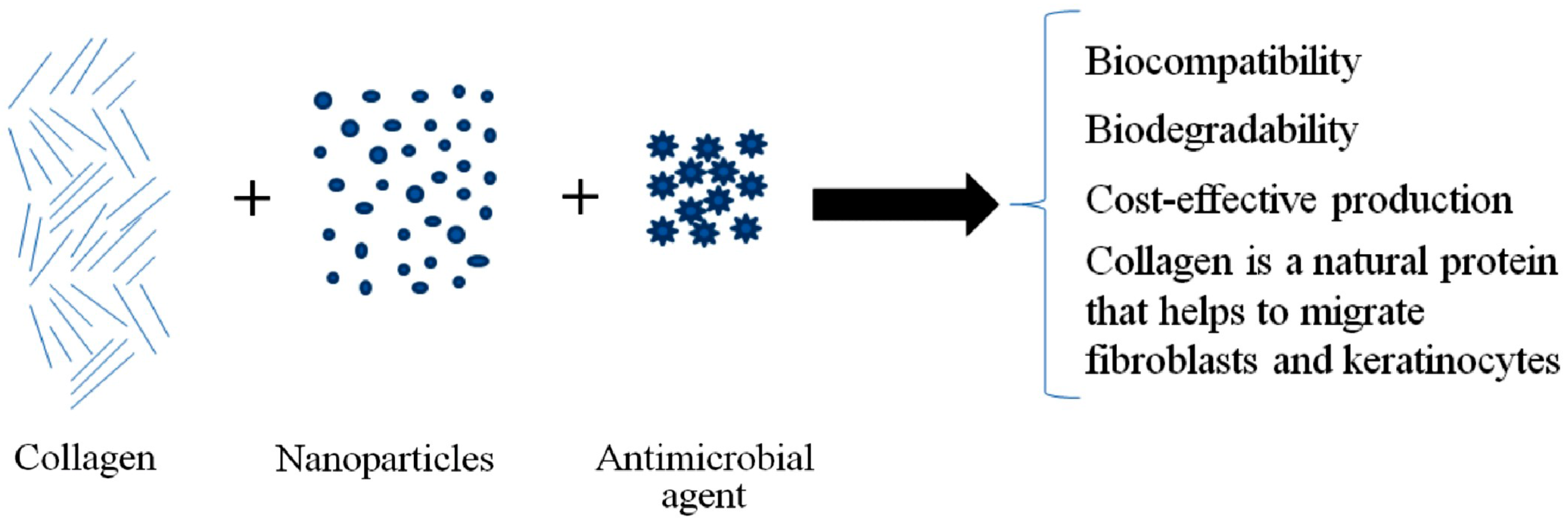
Figure 1. The main advantages of the functionalized collagen hydrogels.
Metals 07 00516 g001
Table 1. The antibiotic resistance threats published by the CDC (Centers for Disease Control) [13].
ThreatsDrug-Resistant Microbes
Urgent threatsClostridium difficile (C-diff)
Carbapenem-resistant Enterobacteriaceae (CRE)
Neisseria gonorrhoeae
Serious threatsMultidrug-resistant Acinetobacter
Drug-resistant Campylobacter
Fluconazole-resistant Candida
Extended-spectrum β-lactamase (ESBL)-producing Enterobacteriaceae
Vancomycin-resistant Enterococcus (VRE)
Multidrug-resistant Pseudomonas aeruginosa
Drug-resistant non-typhoidal Salmonella
Drug-resistant Salmonella enterica serotype typhi
Drug-resistant Shigella
Methicillin-resistant Staphylococcus aureus (MRSA)
Drug-resistant Streptococcus pneumoniae
Drug-resistant tuberculosis
Concerning threatsVancomycin-resistant S. aureus (VRSA)
Erythromycin-resistant Group A Streptococcus
Clindamycin-resistant Group B Streptococcus
Table 2. The distribution of collagen types in connective tissues [33].
TypeClassDistribution
IFibrillarDermis, bone, tendon
IIFibrillarCartilage, vitreous
IIIFibrillarBlood vessels
IVNetworkBasement membranes
VFibrillarDermis, bone, tendon
VIFilaments, 100 nmDermis, bone, tendon
VIIFibers with antiparallel dimersDermis, bladder
VIIIHexagonal matrixMembrane
IXFibril-associated collagens with interrupted triple helicesCartilage, vitreous
XHexagonal matrixCartilage
XIFibrillarCartilage
XIIFibril-associated collagens with interrupted triple helicesTendon
Table 3. MBCs (minimum bactericidal concentrations) of some inorganic NPs (nanoparticles) [48].
Bacterial StrainMBC (μg/mL)
AgCuOZnO
Staphylococcus aureus (Golden)10025002500
S. aureus (Oxford)1001005000
Escherichia coli NCTC (National Collection of Type Cultures) 9001100250>5000
Pseudomonas aeruginosa PAO11005000>5000
Proteus spp.1005000>5000
S. epidermidis SE-410025002500
Meticillin-resistant S. aureus 2521001000>5000
Epidemic meticillin-resistant S. aureus 151002505000
Epidemic meticillin-resistant S. aureus 1610010005000