The Origins of Human Modernity

International Federation of Rock Art Organizations (IFRAO), P.O. Box 216, Caulfield South, VIC 3162, Australia
Humanities 2012, 1(1), 1-53; https://doi.org/10.3390/h1010001
Submission received: 18 July 2011
/
Revised: 17 August 2011
/
Accepted: 25 August 2011
/
Published: 2 September 2011
Abstract
:This paper addresses the development of the human species during a relatively short period in its evolutionary history, the last forty millennia of the Pleistocene. The hitherto dominant hypotheses of “modern” human origins, the replacement and various other “out of Africa” models, have recently been refuted by the findings of several disciplines, and by a more comprehensive review of the archaeological evidence. The complexity of the subject is reconsidered in the light of several relevant frames of reference, such as those provided by niche construction and gene-culture co-evolutionary theories, and particularly by the domestication hypothesis. The current cultural, genetic and paleoanthropological evidence is reviewed, as well as other germane factors, such as the role of neurodegenerative pathologies, the neotenization of humans in their most recent evolutionary history, and the question of cultural selection-based self-domestication. This comprehensive reassessment leads to a paradigmatic shift in the way recent human evolution needs to be viewed. This article explains fully how humans became what they are today.
1. Introduction
In the history of the exploration of human evolution, no subject has caused more controversy than the question of when and how indications of modernity first appeared. There is an implicit acceptance that modernity in behavior emerged together with modernity in physical appearance, and yet there is no evidence for this, or even any logical reason as to why it should be expected to be so. Somatic modernity is widely regarded as being manifested in what is called “modern human anatomy”, although even this concept is fraught with difficulties [1]. On the other hand, there is no significant disagreement about what might constitute modern behavior, but here the problem is that Pleistocene archaeology has substituted perceived technological or purported cultural variables for human behavior. The discipline has little capacity to determine both cultural and behavioral dimensions, because it has become focused on those of technology to the extent of confusing cultural and technological variables. Stone tools, after all, or their relative combinations within an assemblage, do not define cultures any more than we have knife or can-opener cultures today. Cultures, obviously, are determined by cultural factors, including language, art, constructs of reality, and socially transmitted behavior patterns. Tools and technologies, on the other hand, are often used across different cultures, or members of a single culture might use different combinations of tools. It is for this reason that we may safely assume that most of the “cultures” invented by Pleistocene archaeology are etic taxonomic devices, or “institutional facts” sensu Searle [2] that have no real existence in the past. The same applies to the hypothetical peoples endowed with these cultures, which of course never existed as distinctive nations, tribes, ethnic groups or language groups.
These circumstances illustrate a rather unsatisfactory state: Pleistocene archaeology has neglected cultural factors and instead created cultural taxonomies based essentially on combinations of tool types, especially of stone tools. By the same token, the hominin bearers of these invented cultures have been placed in pigeonhole taxonomies of paleoanthropologists who, as recently as during the last few years, have been unable to determine credibly whether a small human from the cave Liang Bua in Flores is an ape or a modern human, or anything between. At the rate at which new hominin or hominid species are being created, we will have many hundreds within a couple of centuries, perhaps more than the 300 species of grizzly bears we once had (when in fact the grizzly is not even a separate species). So in a classical case of circular reasoning the separation of Upper from Middle Paleolithic “cultures” has been explained with the replacement of robust Homo sapiens people (such as those called “Neanderthals” and claimed by many to have been a separate species) with gracile ones. To the believers the replacement is then demonstrated with the purported sudden change in technology.
This model, which has almost totally dominated all relevant discourse in recent decades, neatly defines the core of the problem. Its dogma excludes the attribution of human modernity to any Robusts, linking modern behavior and developed culture exclusively with what it defines as “anatomically modern humans”. It perceives their appearance in southern Africa around 150,000 to 200,000 years (200 ka) ago, in the Levant by 100 ka ago, in Australia by 60 ka, and in Europe a mere 35 ka ago. In this it overlooks some obvious initial contradictions: the Pleistocene Australians had an exclusively Middle Paleolithic (or Mode 3) technology, and there is no indication of an Upper Paleolithic (Mode 4) technology across northern Africa until around 20 ka ago. Therefore the conflation of Moderns or Graciles with Upper Paleolithic technocomplexes is as false as that of the Robusts with Middle Paleolithic traditions.
In considering the beginnings of human modernity, these issues need to be resolved beforehand. The replacement hypothesis demands that Moderns (however defined) developed through natural selection and genetic drift, and within the confines of those models there is no realistic alternative. In this paper it will be shown that a powerful alternative model does exist, and that it is much better reconciled with the empirical evidence. This will be demonstrated by recourse to the current human genome, but the involvement of introgression, niche construction, and selective breeding or domestication will also be examined at length; and gene-culture co-evolution will be identified as the prime mover of recent human development. All of these explanations of evolutionary processes contrasting with plain natural selection are at significant variance with the currently still dominant paleoanthropological and archaeological model of recent human evolution, the replacement hypothesis (“African Eve”) and the various other Out-of-Africa (OoA) models. How was it possible that their highly unlikely demographic scenario, completely lacking any archaeological, paleoanthropological or even credible genetic evidence, was ever capable of gaining such prominence? Although “absence of evidence seldom slows the spread of fashionable ideas” [3], this is an issue that does cry out for detailed historical examination. The hypotheses were initially derived from the claims of an academic charlatan, Professor Reiner Protsch “von Zieten” (the aristocratic title was as bogus as his second doctorate; [4,5]), who invented the notion of a recent rise of “anatomically modern humans” exclusively in Africa, from where they conquered the world [6,7,8,9,10]. By the late 1980s, the academic memes created by this idea, of mitochondria mutations and OoA, were taking over world archaeology [11], despite the voices cautioning against this notion even then:
This does not mean that there was a single female from whom we are all descended, but rather that out of a population numbering perhaps several thousand, by chance, only one set of mitochondrial genes was passed on (This finding, perhaps the most surprising to us, is the least disputed by population geneticists and others familiar with genetic drift and other manifestations of the laws of probability.).[12]
Before any aspect of recent human evolution can be considered meaningfully, it is essential that paleoanthropology be entirely purged of these refuted hypotheses, a process that may still take many more years. Competition between scientific paradigms is not the sort of battle that can be resolved by proofs [13]. As Max Planck has pointed out, “[a] new scientific truth does not triumph by convincing its opponents and making them see the light, but rather because its opponents eventually die, and a new generation grows up that is familiar with it”. In this paper, only a short explanation can be given for the failure of the replacement hypothesis, before moving on to more important matters.
2. The African Hoax
The OoA or short-range model of recent human evolution (not to be confused with the totally different issue of the much earlier African origins of the human clade) perceives a sudden cultural change occurring in Europe about 35 ka ago, which it attributes to an incursion of African immigrants of superior cognition and technology, often linking it to the introduction of language [14,15,16]. It rejects all evidence of symbolism and many other markers of human modernity prior to the advent of these gifted Africans, at some point between 100 ka and 200 ka ago, and regards any cultural complexity in the rest of the world as having been introduced by these colonizers. Sometimes called the “discontinuist” model [17], its first version, by Protsch, was emulated by the “Afro-European sapiens” model [18], followed by the “African Eve” complete replacement scenario [11,19], the “wave theory” [20] and the “assimilation theory” [21]. Of these, the mitochondrial Eve model is the most extreme, in the sense that it demands a complete lack of interbreeding between its purported African species and any other humans. It therefore has no choice but to postulate that these Africans, which it calls “anatomically modern humans” or simply “Moderns”, are a species different from the robust recent humans they either displaced or exterminated. The more moderate varieties of the short-range model accept the occurrence of mixing between robust and gracile forms and therefore are merely variations of the multiregional theory [22,23,24], simply claiming a strong inflow of African genes.
These various hypotheses are of considerable relevance here: somehow a local population in sub-Saharan Africa must have become so isolated from the rest of the continent’s human genome that rapid genetic change became possible, creating a new species that was no longer interfertile with other Africans. This would explain the postulated bottleneck, culminating in the notion of the single female all Holocene humans are related to. But when protagonists of the replacement hypothesis cite possible genetic bottlenecks to contrive explanations for inherent weaknesses of their model, they overlook that such bottlenecks tend to reduce fitness in the population [25,26] rather than bring about the population’s supremacy [27] as proposed for Eve’s progeny. Already at this point the hypothesis begins to falter, because while the replacement model is untenable without a bottleneck, it implicitly rejects the tenets of genetic drift by creating the genetic trees its claims are based on. These unfalsifiable (and thus unscientific) constructs assume not only constant rates of genetic change; they even pretend to deliver divergence times, i.e. the dates when one species split from another. This “genetic clock” is without any scientific basis: none of the crucial variables can be known (such as number of colonization events [28], demographies or true base pair substitution rates), and this is borne out by the “results” of these “molecular archaeology” claims: the hypothetical split between “Moderns” and other humans has been placed at times ranging from 17 to 889 ka BP. Contentions concerning mitochondrial DNA (African Eve) are as much afflicted by this lack of credibility as are those citing Y-chromosomes (“African Adam” [29]). The divergence times projected from the diversity found in nuclear DNA, mtDNA, and DNA on the non-recombining part of the Y-chromosome differ so much that a time regression of any type is extremely problematic. Contamination of mtDNA with paternal DNA has been demonstrated in extant species [30,31,32,33], in one recorded case amounting to 90% [34]. Not only was the assumption of Cann et al. [11] about exclusive maternal transference of mitochondria without basis, the constancy of mutation rates affecting mtDNA was also a myth [35]. Molecular time estimates are asymmetrically bounded random variables, constrained by a nonelastic boundary at the lower end, but not at the higher end of the distribution. This introduces a bias toward an overestimation of time since divergence, and Rodriguez-Trelles et al. [36] have identified a fundamental flaw of molecular dating methods, rendering the mitochondrial “genetic clock” ineffective.
Kidd et al. [37] have shown that, outside Africa, the elements of which haplotypes are composed largely remain linked in a limited set. Gibbons [38] observed that by using the new putative “genetic clock”, Eve would not be 200 ka old [11], but only 6000 years. By then the issue had become farcical: Cann et al. had not only been based on botched computer modeling, its haplotype trees were fantasies that could not be provided with time depth even if they were real. To render these issues even more ludicrous, the transfer of genetic information is not, as many seem to assume, limited to DNA. For instance ribonucleic acids associated with the brain’s thrombospondin (THBS4 and THBS2) can carry such information [39,40], and epigenetic, behavioral and symbolic inheritance systems need to be considered as well [41].
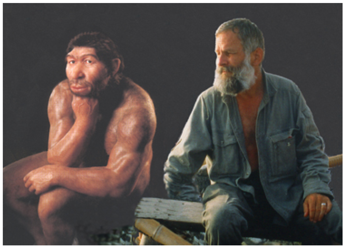
The genetic picture in Africa as well as elsewhere has been found to be far more complicated than the Eve proponents ever envisaged. The much-promoted claims that “Neanderthals” (a term used here only to comply with widespread usage, without endorsing it; Figure 1) were genetically different from modern Europeans, based on very fragmentary DNA sequences, were erroneous, Gutierrez et al. [42] have shown. Their analysis suggests that the pair-wise genetic distance distributions of the two human groups overlap more than claimed, if the high substitution rate variation observed in the mitochondrial D-loop region [43,44,45] and lack of an estimation of the parameters of the nucleotide substitution model are taken into account. Moreover, the results presented from museum specimens, especially “Neanderthal” remains, are probably irrelevant. Pruvost et al. [46] have recently shown that DNA deteriorates rapidly after excavation, up to fifty times as fast as in buried specimens. The various reported “fragmentary DNA sequences” from “Neanderthal” remains stored for up to 150 years need to be considered in that light. A large part, on average 85%, of the genetic material preserved in fossils is lost as a result of treatment by archaeologists and storage in museums, therefore the results disseminated from these specimens and their interpretations may be questioned. More reliable are genetic studies of living populations, which have shown that both Europeans and Africans have retained significant alleles from multiple populations of Robusts [47,48,49]. In fact, the Neanderthal genome seems to include an excess of human-derived single nucleotide polymorphisms [50]. Recent genetic analyses confirm not only that “Neanderthal” genes persist in recent Europeans, Asians and even Papuans [51], but also that “it seems Neandertals interbred with the ancestors of Europeans and Asians, but not with the ancestors of Africans” [52,53]. In the words of Green et al. [51], “[g]iven that the OoA alleles occur at a frequency of much less than 50% in non-Africans (average of 13%, and all less than 30%), the fact that the candidate regions match the Neandertals in 10 of 12 cases (P = 0.019) suggests that they largely derive from Neandertals”. Thus the African Eve model has become an absurdity: it is precisely Late Pleistocene Africans who had the least contact with Europeans. Moreover, even the Green et al. pronouncements are incorrectly expressed: “Neanderthals” did not interbreed with our ancestors; they are our ancestors (Figure 2). If Green et al. wanted to demonstrate that there were genetically very different populations around at the time, they would have to present their genomes’ details also, not only those of the Robusts.
Figure 1.
The author in conference with Mr. T. Neander (on left), who regards archaeological statements about him as defamatory.
Figure 1.
The author in conference with Mr. T. Neander (on left), who regards archaeological statements about him as defamatory.

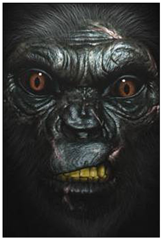
Figure 2.
This very recent rendering of a “Neanderthal”, whose artist does not deserve to be named, reflects the views of many Pleistocene archaeologists.
Figure 2.
This very recent rendering of a “Neanderthal”, whose artist does not deserve to be named, reflects the views of many Pleistocene archaeologists.

The analysis and interpretation of paleogenetic, ancient DNA [54] remains an experimental method and those who over-interpret its results tend to overlook its limitations. Initial results were obtained from a quagga [55], an Egyptian mummy [56], a moa [57], and a cave bear [58], before the genome of Homo sapiens neanderthalensis was tackled [50]. But paleogenetics poses challenges that differ significantly from in vivo studies, because DNA suffers both mechanical and chemical degradation through time and there are high sequencing error and template damage rates [46,59,60,61]. It is certainly easier to template modern DNA than ancient DNA. Results of the polymerase chain reaction (PCR) amplifications, performed by clonage, need to be repeated and three negative controls have to be added to safely detect contamination. Then there is the potential, particularly in moist conditions, of hydrolytic cleavage of phosphodiester bonds between phosphate and sugar [62]. Similarly, sugars and amino groups in proteins and nucleic acids, caused by condensation, can react and lead to errors during PCR. Deamination of cytosine in xanthine, guanine and uracil, or adenine in hypoxanthene can occur, involving the incorporation of nucleotide in the process of PCR amplification. The issues of base substitution [63] and fragmentation of DNA [64] have long been known, and the point is demonstrated, for instance, by the erroneous results obtained from the DNA of insects embedded in amber [65]. Other problems with interpreting or conducting analyses of paleogenetic materials are alterations or distortions through the adsorption of DNA by a mineral matrix, its chemical rearrangement, microbial or lysosomal enzymes degradation, and lesions by free radicals and oxidation [66,67]. These scientific qualifications are generally unheeded in the archaeological folklore established around the “authoritative” DNA data, in much the same way as archaeologists usually fail to heed the reservations of scientists concerning most archaeometric data, e.g., the dating of rock art [68,69,70]. Such results are always grossly simplified, misinterpreted, and over-interpreted, and then embedded in the mythology of mainstream archaeology. In the case of paleogenetic data, they have been eagerly seized by one or another school of thought to support its case or discredit that of the opponents. Yet at no stage do most archaeologists make a concerted effort to appreciate the reservations scientists have concerning scientific data.
For instance there are considerable complexities in the accumulation of base substitutions, or mutations, which are not even relevant to natural selection. The mechanisms governing DNA mutation rates, which are so central to the archaeological claims involving genetics, are not at all well understood. Those mutations that have no selection consequences, “neutral” mutations, are also reflected in DNA mutation rates, which can be estimated by comparing neutrally evolving sequences in species that share a common ancestor. Sequences that are high in pairs of the bases C and G (CpGs) have been positively correlated with mutation rate. However, the chemical modification of CpGs makes them prone to mutation themselves, and with time they are eliminated from neutrally evolving sequences. Walser and Furano [71] have taken advantage of this property to investigate the role of CpGs on the mutation rate of non-CpG DNA by comparing “old” and “young” sequences. They found that CpGs are not only promoting mutations, but they are also influencing how the non-CpG sequences around them are being mutated. In determining the neutral non-CpG mutation rate as a function of CpG content they compared sequence divergence of thousands of pairs of neutrally evolving chimpanzee and human orthologs that differ primarily in CpG content. Both mutation rate and mutational processes are contingent on the local CpG content.
In the absence of any reliability of the many proposed rates of nucleotide changes and the many variables to be accounted for effectively, the contentions by the replacement advocates were unsupported from the beginning of their campaign, and nucleotide recombination renders their views redundant [72]. Instead of unambiguously showing that “anatomically modern humans” originate in one region, sub-Saharan Africa, all the available genetic data suggest that gene flow occurred in Old World hominins throughout much of human evolution [73,74], which is also strongly suggested by all available empirical evidence, both paleoanthropological and archaeological (and had long been predicted by Weidenreich [22]). For instance, the evidence that Homo sapiens neanderthalensis managed to live and subsist at the Arctic Circle, in temperatures that would at times have been below −40 °C [75,76,77], easily dispatches the notion that there were great expanses of habitable land in Europe by the beginning of the Late Pleistocene that remained unoccupied by humans. The Finnish evidence, dating back 135 ka BP, suggests that these innovative people coped with extreme climatic conditions then, and that the demographic modeling of Pleistocene archaeologists [78] must be largely false. If human groups on the margins were forced into regions of truly appalling living conditions, the presence of largely continuous populations can safely be assumed in much of the Old World, and by 50 ka even in Australia.
Comparing the genome of Robusts with that of present-day people, as has been done, is futile; what would need to be compared are the genetic signatures of Robusts and the Graciles contemporary with them, and this has not been attempted. But there are many other unmet conditions to help support the replacement hypothesis. If the Graciles were cognitively and technologically superior to the Robusts, there would need to be distinctive differences in their toolkits, other artifacts and ecological strategies. None are apparent in any of the many regions where people of both somatic forms coexisted, often for very long periods of time. In all such cases, from Spain to Australia, the two populations used very similar or identical technologies, even ornaments. “Neanderthals” produced beads and pendants, and very probably the earliest surviving cave art in Europe [79]. Significantly earlier expressions of symbolism occur in both Asia and Africa [80,81,82]. The advent of the early Upper Paleolithic tool traditions of Eurasia is considered to indicate the arrival of Eve’s progeny there, but they evolved locally and gradually in most parts of the continent. They first appear fairly simultaneously between 45 ka and 40 ka BP, or even earlier, at widely dispersed locations from Spain to Siberia (Makarovo 4/6, Kara Bom, Denisova Cave, Ust’-Karakol, Tolbaga, Kamenka, Khotyk, Podzvonkaya, Tolbor Dorolge [81]). At that time, only Robusts occupied Eurasia (see below). Senftenberg, a clearly Upper Paleolithic (Mode 4) blade industry in the middle of Europe has even been dated to 48,300 ± 2000 (GRO-1217), or a still earlier date, >54,000 years BP (GRO-1771) [83]. The Aurignacian of El Castillo level 18, in Spain, seems to commence well before 40 ka ago [84] (carbon dates of 40,000 ± 2100, 38,500 ± 1800, 37,700 ± 1800 BP). At Abric Romani, the lowest AMS (accelerator mass spectrometry) dates from the Aurignacian average 37 ka BP, but the probably more relevant uranium-series dates point to a sidereal age of 43 ka BP [85]. At El Pendo, the Lower Périgordian (i.e., Châtelperronian) industry, attributed to “Neanderthals” in France, overlies two Early Aurignacian levels [86], a stratigraphic pattern also observed in France, e.g., at Roc de Combe [87] and La Piage [88]. The Châtelperronian at Morín Cave has been dated to about 36,950 carbon-years BP, an antiquity similar to that of the same tradition at French sites (generally 37–33 ka BP). The most recent Middle Paleolithic (Mode 3) occupation known in Spain, however, is at Abric Agut. According to both radiocarbon and U-series dating, it occurred only 13 to 8 ka BP, i.e., straddling the Pleistocene-Holocene interface [89]. Like many other finds, it shows how illusory the separation of the Middle and Upper Paleolithic cultures is [90].
The Iberian pattern of a mosaic and gradually decreasing component of Mode 3 technology in regional EUP (early Upper Paleolithic) lithic industries applies through much of Europe. In southern Italy, variants such as the Uluzzian [91,92], the Uluzzo-Aurignacian, and the Proto-Aurignacian (43–33 ka BP) have been reported [93,94]. The Olschewian of the Alpine region, another Aurignacoid tradition (42–35 ka BP), developed from the final Mousterian [95,96,97,98,99,100,101,102,103,104]. Further east this mosaic includes the Bachokirian of the Pontic region (>43 ka BP), the Bohunician of east-central Europe (44–38 ka BP) [105,106], and various traditions of the Russian Plains. The latter comprise major concentrations of sites in the Prut-Dniester basin and on the middle Don. Some of these industries, such as the Streletsian, Gorodtsovian, and Brynzenian derived clearly from Mousteroid technologies, whereas the Spitzinian or Telmanian are free of Mode 3 bifaces [107]. In parts of Russia, such as regions of the Don River, the Crimea and northern Caucasus, Mode 3 technologies (Mousterian and Eastern Micoquian) continue alongside intermediate and Mode 4 ones and the gradual development from one into the other can be observed at many individual sites. The coexistence of seven accepted tool traditions between 36 ka and 28 ka BP has been reported from the region: the Mousterian, Micoquian, Spitzinian, Streletsian, Gorodtsovian, Eastern Szeletian and Aurignacian (Krems-Dufour variant). The rich mosaic of early Mode 4 industries began before 40 ka BP on the Russian Plain and ended only 24–23 ka BP. In the Crimea, the Middle Paleolithic is thought to have ended only between 20–18 ka BP. Elsewhere in the region, the introduction of a first fully developed Upper Paleolithic tradition (the Kostenkian) appears about 24 ka at the major Kostenki-Borshevo site complex.
A succession of traditions connecting Middle Paleolithic biface technocomplexes, including the late Eastern Micoquian, with typical late Paleolithic ones, continue through the Szeletian of eastern Europe (43–35 ka BP) [108], the Jankovician of Hungary; and the Altmühlian (ca. 38 ka BP), Lincombian (38 ka BP) and Jerzmanovician (38–36 ka BP) further north. Similarly, the gradual development from the Middle Paleolithic at 48 ka BP (with “Neanderthal” footprints of small children) to the Upper Paleolithic is clearly documented in Theopetra Cave, Greece [109,110]. These and numerous other cases of “intermediate” industries or gradual changes all demonstrate the continuity between Mode 3 and Mode 4 technocomplexes in many parts of Europe, but most especially in the east and southeast, the logical entry point of the presumed African invaders. A degree of regionalization precedes this period even in the late Mousterian [78,111,112,113,114], marked by both miniaturization and increasing use of blades, by improved hafting and the use of backed or blunted-back retouch, apparently heralding subsequent developments. German Mode 3 sites have produced backed microliths and evidence of the use of birch resin, and replication experiments suggest that the technology involved in preparing this resin are exceedingly complex. Even in France there is gradual development, both from the Charentian to later Mousterian, and from the “classic Neanderthals” of La Quina and La Chapelle to the more gracile Abri Peyrony specimen. Much the same pertains to western Asia, for instance the Aurignacoid Baradostian tradition of Iran clearly develops in situ from Middle Paleolithic antecedents. The Mousteroid traditions of the Levant also develop gradually into blade industries, e.g., at El Wad, Emireh, Ksar Akil, Abu Halka and Bileni Caves, and that region’s Ahmarian is transitional. The artificial dichotomy between Middle and Upper Paleolithic materials [90] has thus only served to overemphasize differences that mark really gradual changes in technology [115]. The specious separation of Mode 3 and Mode 4 technologies has even less currency in Africa (e.g. the Howieson’s Poort tradition with its microliths, or the Amudian), India [81,116] or China [117]. In Australia the Mode 3 traditions continue until well into the Holocene, and in Tasmania until the arrival of the British, just over two centuries ago.
Perhaps most pertinently, if the Graciles as claimed by the OoA advocates have come from sub-Saharan Africa, and arrived in Europe via the Levant and southeastern Europe, it would be expected that evidence of their presence can be found first in their homeland and later progressively along such a route, in the form of the arrival of a dramatically different technology. No such evidence has been reported, and African Eve advocates have cited none. Not one of the more than twenty perceived EUP stone tool traditions of Europe derives from Africa or the Levant. On the contrary, Aurignacoid or similar traditions arrived in the Levant long after they first arose elsewhere in Eurasia, so they were clearly not introduced through this presumed corridor. Moreover, right across northern Africa, the Mode 3 Middle Stone Age continued up to 20 ka ago, i.e., at 20 or 30 millennia after the advent of Mode 4 technologies across Eurasia. Moreover, there is simply no Mode 4 tradition in sub-Saharan Africa until about 22 ka ago, a glaring inconsistency the advocates of the Eve model have habitually ignored. Nor have they ever explained where the African or Levantine precedents of the Upper Paleolithic art traditions are to be found, if these African invaders were their carriers as claimed. There is no trace of such evidence, nor any proven Pleistocene rock art other than typical Mode 3 productions known from Africa. Even the only demonstrated early mobiliary art from Africa, found in Namibia, is not as claimed of a Mode 3 tradition [118]. The state of available information from the Levant or Arabia indicates much the same along the route the Africans are supposed to have taken to Europe.
Instead of a sudden change of technology in Europe at any time during the period from 50 ka to 25 ka ago, what can be observed is a complex mosaic of regional traditions which, in general, exhibit a gradual change of several variables, such as tool size, knapping method, retouch and reuse. This suggests in all cases in-situ evolution of cultures, rather than the effects of an intrusive tradition. It mirrors precisely the patterns documented in the development in human morphology, as shown next.
3. The Gracilization of Humans
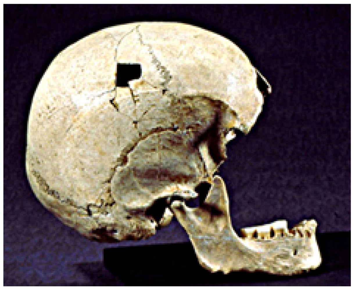
A fundamental error of the replacement advocates [6,7,11,18,19,119,120,121,122,123,124,125,126,127,128] and even others, such as Churchill and Smith [129,130], has been the acceptance of false datings (e.g., those by Protsch) of many European human skeletal remains of the time slot in question. In numerous cases specimens of relatively modern appearance were given ages well in excess of their true antiquities, thus claiming an early appearance of these “modern” features. Examples are the four Stetten specimens from Vogelherd (in the Swabian Jura, southwestern Germany). Although it had always been perfectly transparent to more rigorous commentators that they derived from intrusive Neolithic interments [131,132], the Eve folks had attributed them to the Aurignacian. Direct carbon isotope determinations of samples taken from the mandible of Stetten 1, the cranium of Stetten 2 (Figure 3), a humerus of Stetten 3, and a vertebra of Stetten 4 all agree, falling between 3980 ± 35 BP and 4995 ± 35 BP [133].
Figure 3.
Stetten 2, from the Vogelherd Cave, Germany: “African Eve” advocates believed it to be of the Aurignacian; it is in fact from a Neolithic burial.
Figure 3.
Stetten 2, from the Vogelherd Cave, Germany: “African Eve” advocates believed it to be of the Aurignacian; it is in fact from a Neolithic burial.

Similarly, the Cro-Magnon sample, frequently cited as the “type fossil” of the first “modern” Europeans and derived from four adults and three or four juveniles, had been subjected to so much pseudo-scientific spin that separating it from credible accounts is not readily possible. The group is in reality quite robust, and especially the very pronounced supraorbital torus, projecting occipital bone and other features of cranium 3 are Neanderthaloid rather than gracile. Sonneville-Bordes [134] placed the sample from the Cro-Magnon shelter, just outside Les Eyzies, in the late Aurignacian; Movius [135] suggested an age of about 30 ka BP and preferred an attribution to the Aurignacian 2. Both opinions, and numerous others, are refuted by the direct dating to about 27,760 carbon years BP [136]; it places the Cro-Magnon individuals in the Gravettian rather than the Aurignacian technocomplex.
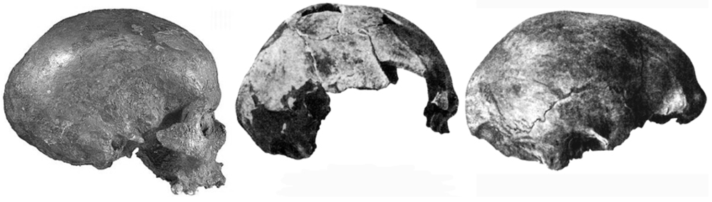
The third set of human remains White [137] cited to contradict Bednarik [138] when he proposed that there is no evidence of the humans of the Early Aurignacian being gracile are the Mladeč specimens from the Czech Republic, often also fielded by other Eve advocates. There is no clear evidence that Pleistocene humans ever walked into this cave, partly excavated about 130 years ago [139]. Most of the macro-faunal remains in it apparently fell through the large shaft in the cave’s roof, and Smyčka [140] proposed that the human remains had also dropped through this chimney, which is probably the case. The literature on this site [141,142,143,144,145,146,147] presents no credible alternative evidence. The recently secured direct dates from some of the human remains [148], from specimens Mladeč 1, 2, 8, 9a and 25c, range from about 26,330 BP (the ulna of 25c) to 31,500 BP. They are therefore, at best, partly of the very final phase of the Aurignacian period with its duration of about 15,000 years. More likely, most or all of the series is of the Gravettian technocomplex. Moreover, there is considerable evidence that the Mladeč specimens were far from “fully modern” [149,150,151,152,153]. Notably, there appears to be pronounced sexual dimorphism, with male crania characterized by thick projecting supraorbital tori, Neanderthaloid posterior flattening, low brain cases and very thick cranial vaults—all typical robust features. As in “Neanderthals”, cranial capacities exceed those of Graciles (1650 cc for Mladeč 5), but there is a reduction in the difference between male and female brain size relative to Neanderthal data. The dimorphism is also expressed in the more inclined forehead in the males, their more angled occipital areas with lambdoidal flattening, broad superior nuchal planes and more prominent inion. The female specimens show similarities with, as well as differences from, accepted Neanderthal females, such as larger cranial vaults, greater prognathism, lack of maxillary notch, a very narrow nose and distinct canine fossa. However, the females are more gracile than the males, while still being more robust than males of later periods (Figure 4). The Mladeč population thus seems to occupy an intermediate position between late Neanderthaloid Homo sapiens, and H. sapiens sapiens, a position it shares with numerous human remains from other Czech sites.
Figure 4.
Mladeč 1, 6 and 5, Czech Republic, showing the striking morphological differences between the two females on the left and the male on the right. (To facilitate comparison, all specimens are shown facing the same direction.)
Figure 4.
Mladeč 1, 6 and 5, Czech Republic, showing the striking morphological differences between the two females on the left and the male on the right. (To facilitate comparison, all specimens are shown facing the same direction.)

This is an important issue to be revisited later in this paper. Suffice it to note here that the material from Pavlov Hill, an important Czech site, is among the most robust available from the European Upper Paleolithic, sharing its approximate age of between 26 and 27 ka with yet another Moravian site of the Gravettian, Předmostí. The more gracile finds from Dolní Vestonice are around 25 ka old and still feature some archaic characteristics (particularly the Neanderthaloid specimen DV16). Another find that has been considered as a very early European “Modern” is the calotte from Podbaba, near Prague, variously described as sapienoid and Neanderthaloid, but undated; it probably belongs to the Mladeč-Předmostí-Pavlov-Dolní Vestonice spectrum. Morphologically similar specimens also come from Cioclovina (Romania), Bacho Kiro levels 6/7 (Bulgaria) and Miesslingtal (Austria), so this is unlikely to be a local phenomenon. Indeed, it needs to be seen in the greater Eurasian context.
Besides the Neolithic human remains from Vogelherd, which the Eve advocates had been all too keen to place at 32 or 35 ka, nearly all of the German fossils claimed to be of the Upper Paleolithic are now thought to be of the Holocene. Of particular interest is the Hahnöfersand calvarium, described as so robust that it was judged to show typical “Neanderthal features” [154] and hailed as the northernmost “Neanderthal” found. It was initially dated to the earliest Upper Paleolithic (Fra-24: 36,300 ± 600 BP; UCLA-2363: 35,000 ± 2000 BP, or 33,200 ± 2990 BP; [154]), which conflicts sharply with results secured by Terberger and Street [155]: P-11493: 7470 ± 100 BP; OxA-10306: 7500 ± 55 BP. The re-dating of the skull fragment from Paderborn-Sande yielded even more dramatic differences. Originally dated at 27,400 ± 600 BP (Fra-15; [156], Terberger and Street report an age of only 238 ± 39 BP (OxA-9879). In fact the skull was so fresh that it emitted a putrid smell when Terberger and Street drilled it for sampling. Then there is the cranial fragment of Binshof near Speyer, dated by R. Protsch in the 1970s as Fra-40 to 21,300 ± 320 BP. According to Terberger and Street it is only 3090 ± 45 carbon years old (OxA-9880). These authors also analyzed two specimens from the Urdhöhle near Döbritz, which had been attributed to the Upper Paleolithic, and found them both to be about 8400 years old. Indeed, of all the German Upper Paleolithic human remains, only one remains safely dated to earlier than 13,000 BP: the interred specimen from Mittlere Klause in Bavaria. A carbon isotope date of 18,200 ± 200 BP (UCLA-1869) from a tibia fragment [8] has been confirmed by Terberger and Street’s date from a vertebra, of 18,590 ± 260 BP (OxA-9856). It has therefore become clear that there are currently no “modern” remains from the first half, if not the first two thirds of the west-central European Upper Paleolithic. In addition, another German key specimen, the skull from Kelsterbach, has mysteriously disappeared from the safe of the Frankfurt institution. It had been dated to 31,200 ± 1600 BP (Fra-5) [9,156], but is also believed to be of the Holocene, perhaps the Metal Ages [155].
Then there are the robust but “modern” hominin remains of the EUP at Velika Pećina, Croatia, close to the Neanderthal site Vindija. This specimen, too, has been a principal support for the replacement advocates, but it has also joined the long list of European humans whose age was grossly overestimated. It is now considered to be only 5045 ± 40 radiocarbon years old (OxA-8294; [157]).
The currently earliest, liminal “intermediate” (between robust and gracile) finds in Europe, the Peştera cu Oase mandible and face from southwestern Romania [158,159], are perhaps about 35,000 radiocarbon years old, but they are without an archaeological context. Although in some aspects “modern”, the “derived Neanderthal features” of the mandible include cross-sectional symphyseal orientation, exceptionally wide ramus, exceptionally large third molars and unilateral mandibular foramen lingular bridging. The partially preserved facial remains, found in a different part of the extensive cave system and apparently from another individual, also combine robust and gracile features. Soficaru et al. [160] have reported six human bones from another Romanian cave, Peştera Muierii, also clearly intermediate between robust and gracile Europeans. Found in 1952, they have now been dated to about 30,000 carbon years and combine a partly modern, partly archaic brain case with a suite of other intermediate features.
The loss of the only relevant Spanish remains, from El Castillo and apparently of the early Aurignacian technocomplex, renders it impossible to determine their anatomy. French contenders for EUP age present a mosaic of unreliable provenience or uncertain age, and direct dating is mostly not available. Like the Vogelherd and other specimens, those from Roche-Courbon [161] and Combe-Capelle (originally attributed to the Châtelperronian levels [162]) are now thought to be of Holocene burials [163,164], and the former is apparently lost. Similar considerations apply to the partial skeleton from Les Cottés, whose stratigraphical position could not be ascertained [165]. Finds from La Quina, La Chaise de Vouthon and Les Roches are too fragmentary to provide diagnostic details. The os frontale and fragmentary right maxilla with four teeth from La Crouzade, the mandible fragment from Isturitz and the two juvenile mandibles from Les Rois, about 28 to 30 ka old [166], range from robust to intermediate [167]. Just as the Cro-Magnon human remains now appear to be of the Gravettian rather than the Aurignacian, so do those from La Rochette. The Fontéchevade parietal bone does lack prominent tori (as do many other intermediate specimens) but the site’s juvenile mandibular fragment is robust.
This pattern of features intermediate between what paleoanthropologists regard as Neanderthals and Moderns is found in literally hundreds of specimens apparently in the order of 45 to 25 ka old (including the large Czech collection lost in the Mikulov Castle fire at the end of World War II). They occur in much of Europe, and intermediate forms between robust Homo sapiens and Homo sapiens sapiens existed also in Asia and Australia. They include examples from right across the breadth of Eurasia, such as those from Largo Velho, Crete, Starosel’e, Rozhok, Akhshtyr’, Romankovo, Samara, Sungir’, Podkumok, Khvalynsk, Skhodnya, Qafzeh, Skhul, as well as Chinese remains such as those from Jinniushan and Tianyuan Cave [168]. Similarly, the African evidence does not, as is often claimed, present “anatomically modern humans” at 150 ka or almost 200 ka. The skulls from Omo Kibish offer some relatively modern features as well as substantially archaic ones; especially Omo 2 is very robust indeed [169]. Their dating, also, is not secure at all, and Omo 2 is a surface find. The much more complete and better dated Herto skull, BOU-VP-16/1, is outside the range of all recent humans in several cranial measurements [170]—and is clearly just as archaic as other specimens of the late Middle Pleistocene, in Africa or elsewhere. The lack of “anatomically modern” humans from sub-Saharan Africa prior to the supposed Exodus is glaring: the Border Cave specimens have no stratigraphic context and are thought to be only around 80 ka old; Omo and Dar es Soltan are obviously not sub-Saharan (and the latter is undated), which leaves the mandibles of Klasies River Mouth, lacking cranial and post-cranial remains. On the other hand, current Australians average a cranial capacity of only 1264 cc (males 1347 cc, females 1181 cc, i.e., well within the range of Homo erectus), while their molars average the size of those of Europeans several hundred millennia ago. And yet they are still considerably smaller than those of fossil Australians, such as the large Kow Swamp sample. So while diminution of molars did occur in Australia, supposedly also settled by Eve’s progeny, it lags greatly behind that of the rest of the world.
With the lack of African fossils of the African Eve “species”, the Eve apostles turned to the Levant for help, and recruited the Mount Carmel finds from Qafzeh Cave and Skhul Shelter as supposed “Moderns”. Yet all of these skulls present prominent tori and receding chins, even Qafzeh 9, claimed to be of the most modern appearance. The distinct prognathism of Skhul 9 matches that of “classic Neanderthals”, and the series of teeth from that cave has consistently larger dimensions than Neanderthaloid teeth. Even Chris Stringer, the principal Eve supporter, concedes that this material is “transitional” or intermediate. Besides, supposedly much later “Neanderthal” burials in nearby Tabun Cave as well as the Qafzeh and Skhul material are all associated with the same Mousterian tools, and the datings of all Mt Carmel sites are far from soundly established, with their many discrepancies. The TL dates from Qafzeh, for instance, clash severely with the amino racemization dates (ranging from 33 to 45 ka), and are in any case plagued by inversion: the lower layer (XXII) averages 87.7 ka, the middle layer (XIX) 90.5, while the uppermost (XVII) averages 95.5. Therefore the claims of 90-ka-old “modern” humans from Mt Carmel, a cornerstone in the Eve notion, are in every respect unsound, and this population is best seen as transitional between robust and gracile forms, from a time when gracilization had commenced elsewhere as well.
This presents an overall picture that is very different from that which the replacement protagonists subscribe to. Their model cannot tolerate such intermediate forms, nor can it allow hybrids, yet in Europe there is a clear continuation of some Neanderthaloid features right up to and into the Holocene. This is demonstrated not only by the Hahnöfersand specimen, but also by others, such as the equally robust Mesolithic skull fragment from Drigge, also from northern Germany, which is about 6,250 years old [171], and numerous other late specimens previously thought to be of the EUP. They range in age from the Magdalenian through the Neolithic, and even younger. One distinctive “Neanderthal” feature is the shape of the mandibular nerve canal, surrounded by a bony ridge in 53% of specimens included in this designation. Its occurrence diminishes during the transition period to 44%, but it is still present in today’s Europeans, at 6% [172]. This feature alone demands the presence of Neanderthal genes in Europeans. The process of gracilization has in fact continued to the present time: even early Mesolithic material is about 10% more robust than modern Europeans. Indeed, Hawks [173] has estimated that at least 25% of the ancestors of later Upper Paleolithic people would need to be Neanderthals to account for the preservation of Neanderthal autapomorphies observed [174,175,176], and the genetic evidence for the Neanderthal ancestry of modern Europeans is overwhelming [51].
This brief review suggests that there are now almost no supposedly modern specimens left as possible contenders for attribution to any Aurignacoid industries. The maxilla from Kent’s Cavern, United Kingdom (~31 14C ka BP), and the Romanian remains from Peştera Cioclovina (~29 14C ka BP) lack secure and diagnostic archaeological association. There are, however, numerous “Neanderthal” remains to fill this void. Of particular interest are the most recent, those from Saint Césaire (~36 ka), Arcy-sur-Cure (~34 ka), Zafarraya Cave (~33.4 ka), Máriaremete Upper Cave (~38 ka), Sungir’ (~25 ka), Trou de l’Abîme (~33 ka), and Vindija Cave (~28 and ~29 ka). This state of affairs has prompted Bednarik [79] to propose that the hypothesis of the EUP people being robust or intermediate is supported by empirical evidence, while the contrary view is without.
At the first of these sites, the “Neanderthal” remains of a burial occur together with clear Châtelperronian artifacts, which until 1979 had been generally assumed to be the work of anatomically modern humans. Arcy-sur-Cure, also in France, yielded numerous ornaments (Figure 5) and portable art objects, again from a Châtelperronian. This prompted various convoluted explanations of how these elaborate pendants could have possibly found their way into a “Neanderthal” assemblage (e.g., [177,178]); a similar argument was used by Karavanic and Smith [179] in explaining the bone points of Neanderthals in Vindija layer G1). It was contended that the primitive Neanderthals must have scavenged these objects from the camps of “Moderns”, as if people lacking the ability to use symbols would have any use for symbolic artifacts. The Jankovichian or Trans-Danubian Szeletian [108] has yielded three mandibular “Neanderthal” teeth from Máriaremete Upper Cave [180]. The Streletsian of Sungir’ in Russia produced a Neanderthaloid tibia from a triple grave of Graciles, and the grave’s complete adult male skeleton exhibits pronounced supraorbital tori [181]; Trou de l’Abîme near Couvin in southern Belgium furnished “Neanderthal” remains together with a typical Aurignacian industry; and there can be no question that the Vindija “late Neanderthals” used EUP tools and technology. Not only has that site supplied some of the most recent Neanderthals found, these are more gracile than Neanderthals of much earlier periods, and they are seen as transitional by some [21,174,182,183]. Vindija Vi-207 is a mandible of 29,080 ± 400 carbon years BP (OxA-8296), Vindija Vi-208 is a parietal of 28,020 ± 360 carbon years BP (OxA-8295) [157, but see 184]. These “late Neanderthals” (or very robust Graciles) exhibit significant reduction in Neanderthaloid features, such as mid-facial prognathism and supraorbital tori. The related tool assemblage includes even apparent bone fabricators [185].
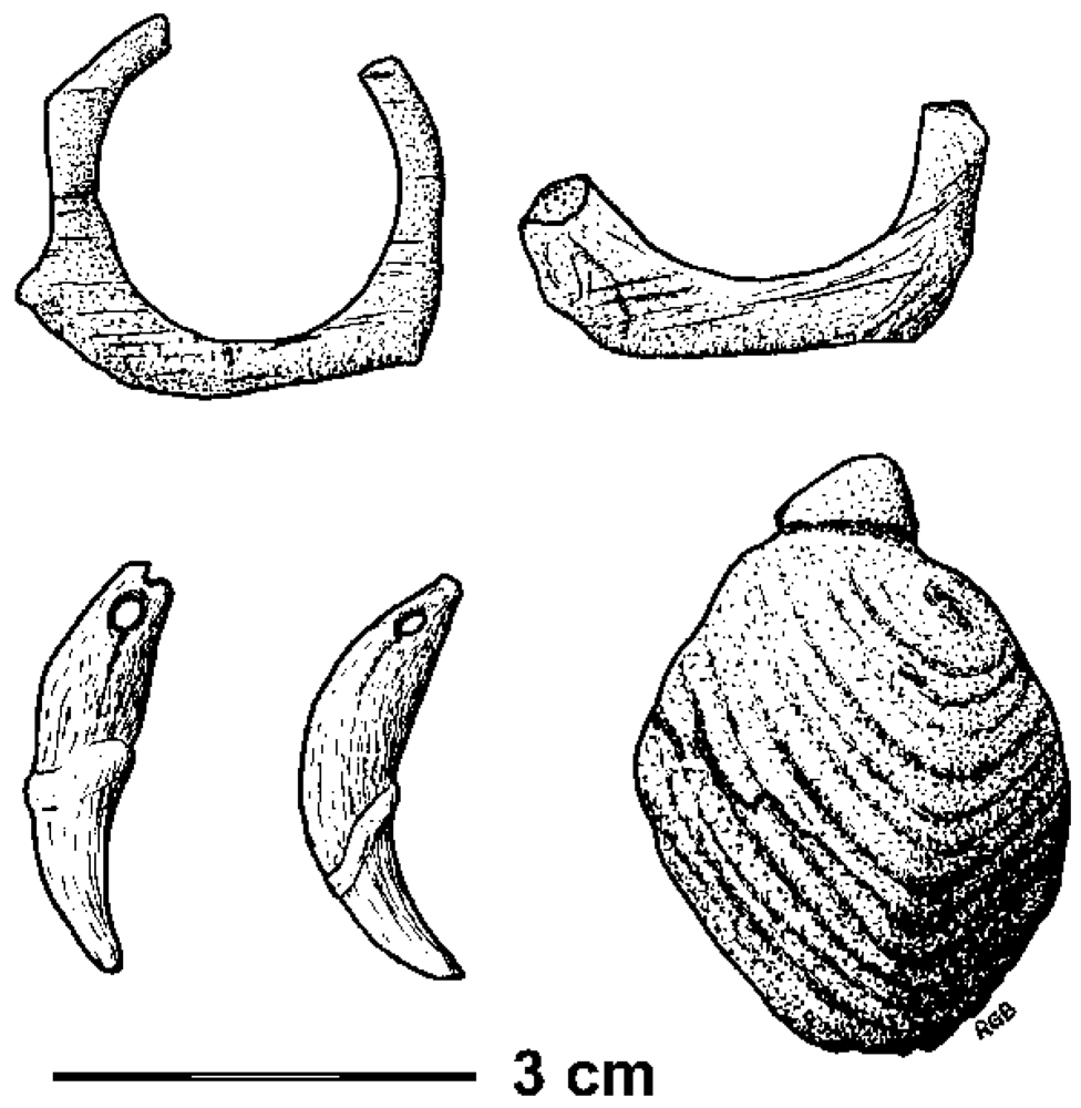
Figure 5.
Ornaments from Grotte du Renne, Arcy-sur-Cure, France, made by “Neanderthals”: two ivory ring fragments, two perforated animal canines, and a fossil shell with an artificial groove for attachment.
Figure 5.
Ornaments from Grotte du Renne, Arcy-sur-Cure, France, made by “Neanderthals”: two ivory ring fragments, two perforated animal canines, and a fossil shell with an artificial groove for attachment.

Ignoring these many significant contradictions to their ideas, the replacement proponents have responded to the recent developments in Germany by contending that the new data bolster their model, because the “Neanderthaloid” Hahnöfersand specimen had been suggested to be a hybrid [154]. Instead of admitting that they have been the victims of a hoax by Protsch, they have hailed each of the very late dates for Neanderthal remains as they appeared in recent years as a confirmation of their hypothesis, gradually painting themselves into a corner. Having strongly contended that a whole spectrum of radical cultural innovations appeared with the beginning of the Aurignacian, they effectively attributed these to their “Neanderthals”, contradicting themselves once again. According to them, the people of the Aurignacian are indistinguishable from us in terms of cognition, behavior and cultural potential. Quite possibly they are right and the late Robusts were behaviorally modern, but that is certainly not what they hoped to show [186]. The period from 45 ka to 28 ka BP has produced dozens of “Neanderthal” specimens, but no securely dated, unambiguously fully modern human remains anywhere in Europe. Hence the available evidence suggests that the people of the first half or even two thirds of the Upper Paleolithic were either robust or intermediate. The replacement hypothesis, obviously, cannot accommodate any intermediate forms; in fact it is decisively refuted by them. As, indeed, it is refuted by the genetic data that present-day non-Africans derive from Neanderthals [51]. This is not even needed to refute the Eve model, which is destroyed by just one single “Neanderthal gene” in the genome of extant Eurasians. Although this model has now been refuted so resoundingly, some of the replacement advocates seem unable to grasp the effects of the new data on their embattled hypothesis [187]. They seem incapable of appreciating that, in science, exceptional claims such as their absurd model require exceptionally persuasive evidence, not the slipshod reasoning and methodological blunders characterizing all presentations of the Eve hypothesis—from Protsch to the present time.
Historically, Pleistocene archaeology has been a series of blunders, hoaxes and mistakes, beginning with the mistreatment of Boucher de Perthes, and the African Eve episode is simply one of the most recent examples of this susceptibility to erroneous consensus views. All present-day humans derive from robust H. sapiens types, who formed a largely continuous population across most of Africa and Eurasia, with the exception of regions that were utterly uninhabitable. Introgression facilitated the travel of genes through this population, just as Weidenreich [22] had predicted in his trellis model (Figure 6). The robust sapienoids were never replaced, they simply became gracile throughout the occupied world, including even in Australia, during the final part of the Late Pleistocene, beginning 40 or 50 ka ago. But what caused this universal gracilization? Since it involved numerous deleterious effects, as will be shown below, it is most unlikely to be the result of any form of natural selection. So how is the origin of anatomically modern humans to be explained?
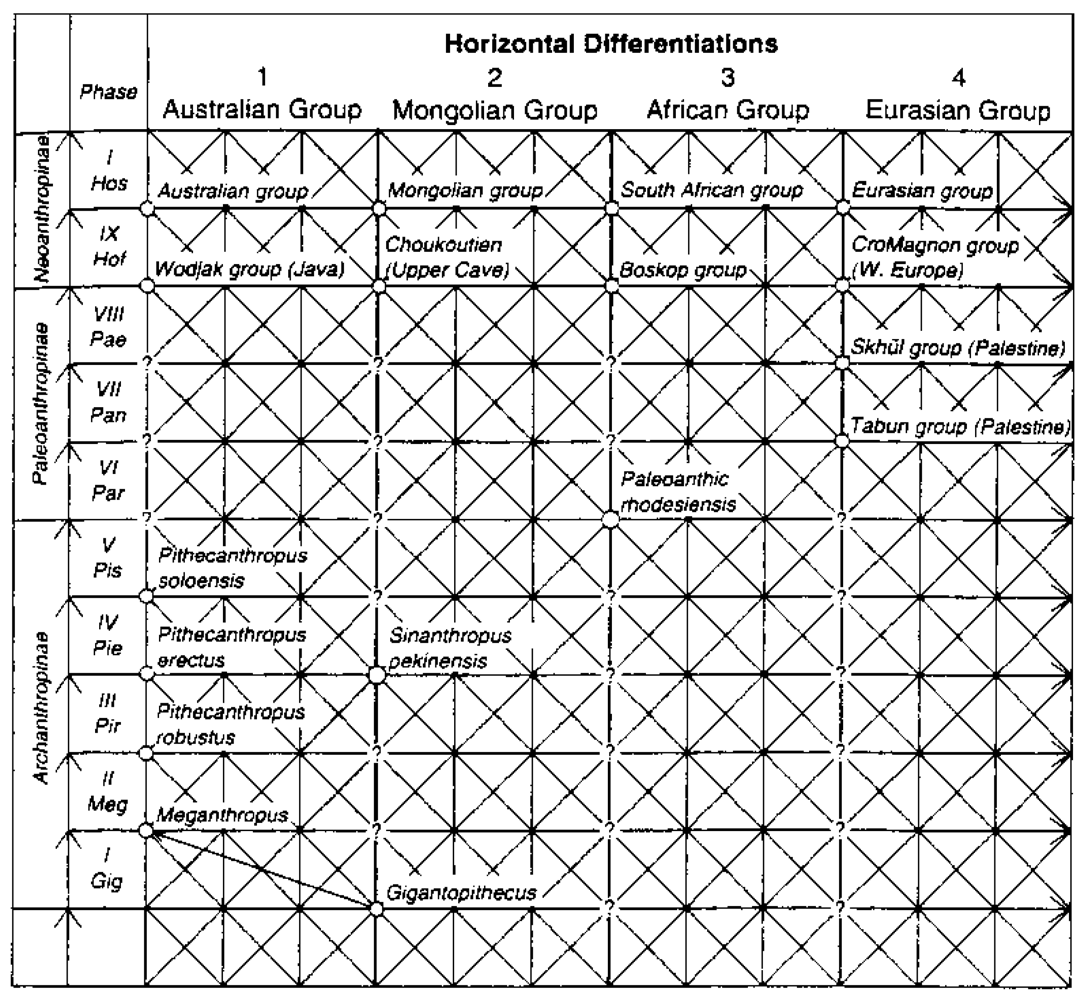
Figure 6.
Franz Weidenreich’s 1946 “trellis” model of polycentric human evolution over three continents.
Figure 6.
Franz Weidenreich’s 1946 “trellis” model of polycentric human evolution over three continents.

4. Alternative Models
Evolutionary theory attributes evolutionary change essentially to two factors, natural selection and sexual selection. In the first, specific phenotypes representing aspects of morphology or behavior are preferentially reproduced across generations of a given population. In the second, phenotypes become over-represented either through mate choice or intra-sexual competition. The emphasis is on genetic inheritance, although the hard evidence for this is not particularly well established.
Over recent years several new directions of inquiry have evolved, challenging simplistic evolutionary theory. Developmental systems theory replaces the overly restrictive focus on the genes with a model of interacting systems [188,189]. While vague overall, it does raise some pertinent points, especially concerning the non-genetic inheritance of traits and the cybernetic feedback from organism-environment systems changing over time. Niche construction has been presented as another major force of evolution [190], operating similar to natural selection. In rather the same way as visual and mental taxonomizing processes and the inclusion of new neural structures becoming available for evolutionary selection in feedback systems [191], niche construction also creates feedback within the evolutionary dynamic. Organisms engaged in it modify the evolutionary pressures acting on them, as well as on other but unrelated populations sharing the same space. Humans are rightly seen as the “ultimate niche constructors”, in which their increasingly complex cultures may play an important role. Laland et al. [192] see much of niche construction as guided by socially learned knowledge and cultural inheritance [193], and Bickerton [3] attributes human language to niche construction.
Evolution has been suggested to encompass also other “dimensions”, termed epigenetic, behavioral and symbolic inheritance systems [41]. All organisms are said to be subject to epigenetic inheritance, which refers to physiological/biological process above the level of DNA. Behavioral inheritance is found in most species, and defines the transference of information or behavior through learning rather than genetically. Symbolic inheritance is apparently found only in humans. The underlying contention of these new ways of thinking is that evolution is not a simple genetic process relying on the appearance of mutations. Evolution does not develop traits for selection; it has no foresight. The idea that human evolution simply cannot be assumed to have been a purely biological process is not at all new [194,195]. It has recently received a new impetus from increasingly sophisticated work, and the notion of a progressive moderation of human evolution by culture is the central plank of the gene-culture co-evolutionary model [196,197]. Most recently, Fuentes [198] has sought to reconcile the pronounced duality of evolutionary biology and socio-cultural anthropology, pointing out that symbolic and other cultural processes influence behavior and potentially physiological and even genetic factors. His demand that behavioral plasticity has a specific role in human behavior runs again counter to neo-Darwinism, but upon reflection it seems impossible to explain hominin development, especially of the Late Pleistocene, without that factor.
These new developments are certainly useful, especially in that they reject the role of genetics in “explaining everything” in hominin evolution. They also express considerable criticism for the self-confirming paradigms of recent decades, critique that is so crucial to a sound epistemology. The debilitating, all-pervading appeal to authority governing archaeology does need to be severely challenged, and this has not occurred adequately. However, there are two significant shortcomings of these various strands of criticisms coming from the sciences. One is that they have not produced an alternative paradigm; they have merely illustrated problems that need to be attended to. The other concerns the lack of relevant empirical evidence, which the sciences simply have no access to because archaeology is either itself unaware of its existence (as far too often appears to be the case); or alternatively it has made great efforts to discredit such evidence in order to uphold its dogma. Therefore the position of the behavioral, cognitive and semiotic sciences is essentially that they have detected the flaws in the dominant model of the emergence of human modernity, but they are not in a position to offer an alternative: archaeology dominates the discourse on hominin evolution, and it determines what may be published in this discipline.
The scenario to account for is that there is a significant change in the physiology of humans during the last 50,000 years in Europe, and modern Europeans differ genetically from robust Europeans 50 ka ago. The same change from Robusts to Graciles occurs in three other continents. Not only do these changes need to be explained, there is another issue which, again, the replacement advocates are completely silent on: the changes that did occur contradict all canons of Darwinian evolution. Without a significant change in their environmental mega-niche, these humans experienced numerous deleterious physiological changes to become gracile. The thickness of their skulls decreased radically, as did the general robusticity of their skeletons. The traces of muscle attachments indicate that physical strength declined markedly, perhaps by as much as 50%. On top of that, their brain shrank by around 200 cc (~13%), and that occurred during a time when the demands on their mental abilities are thought to have increased exponentially. The susceptibility to neurodegenerative diseases developed apparently in this time, and mental illness may have attended gracilization. These changes are certainly dramatic, occurring in fact over just a few tens of millennia. In the history of the human genus, there is no evidence of such rapid changes, and conventional wisdom has it that all previous changes were for the better of the species concerned. That certainly cannot be said about what happened in the most recent history of human evolution, which in many areas looks more like devolution, or evolution in reverse.
So what happened? If it was not a case of invasion by physically (and perhaps even intellectually) inferior Africans of evenly matched technology, what alternative is there? The answer is provided by a combination of two strands of determinants. One is the indisputably very major influence sexual selection has on who passes on their inheritance; the other is the rising power of cultural imperatives over natural. When breeding mate selection becomes moderated by cultural factors (such as cultural constructs of attractiveness, along with perhaps social position, communication ability, body adornment), the laws of evolutionary theory become suspended, and are supplanted by Mendelian laws of inheritance [199], the basis of the discipline of genetics: evolution by natural selection is replaced by breeding, or artificial selection, resulting in domestication. It was in studying artificial selection in pigeons that Darwin detected the similarity with natural selection, and here at last the deliberations seem to come full circle: modern humans are the result of incidental self-domestication. In their fetish of using the purported travel of genes to infer the movement of major populations, Pleistocene archaeologists have ignored that it is not evolutionary genetics that determines inheritance: in the end even Darwin has to defer to Mendel.
This revolutionary alternative has been outlined as a realistic option to the replacement hypothesis [79,200,201,202] and here it is explained in some more detail. The apparently most important question to be asked in this context is this: what could have caused the inherent laws of biological evolution to be suspended for humans during the last fifty millennia or so?
It is particularly important to note that the change seems to have occurred universally and roughly concurrently, in all four continents occupied by hominins by 50 ka BP. Since this enormous geographical range involved numerous climatically and environmentally different niches, from the tropics to the Arctic, it is impossible to explain such largely uniform change from robust to gracile as the result of natural selection. The same rejection of evolutionary dynamics may be implied by the relatively swift conversion, taking only a few tens of millennia. In southern and eastern Europe, one might argue that the Campagnian Ignimbrite Event and subsequent sharp climatic decline almost exactly 40 ka ago [115] may have precipitated demographic and cultural adjustments. Although this environmental bottleneck could have effected genetic or anatomical changes in some parts of Europe, there is no evidence that it did, and the universal human gracilization over the last 50 ka or so demands a universal explanation and precludes a local one. Occurring concurrently in the course of the second half of the Late Pleistocene, in all four continents occupied, this distinctly gradual process needs to be explained if human origins are to be clarified. Robusticity in all humans has continuously decreased over the last 50 ka or so.
In Europe it is best documented by human remains from the central region, particularly in the Czech Republic, from the crucial period of about 31 ka to 26 ka BP, which witnessed distinctive sexual dimorphism. Despite the lack of credible stratigraphic evidence from Mladeč Cave, the recent attempt to provide direct dates from some of its human remains suggests that they represent precisely this interval [148]. As noted above, male crania are characterized by typical robust features. As in “Neanderthals”, cranial capacities exceed those of “anatomically modern humans”, but there is a reduction in the difference between male and female brain size relative to “Neanderthal” data (Figure 7). The female specimens show similarities with, as well as differences from, accepted Neanderthal females, but are far more gracile than the males, while still being more robust than males of later Pleistocene periods. The Mladeč population as well as contemporary others in central Europe (e.g., Pavlov Hill, Předmostí, Dolní Vestonice, Podbaba, Miesslingtal) thus seems to occupy an intermediate position between late Neanderthaloid Homo sapiens and H. sapiens sapiens.
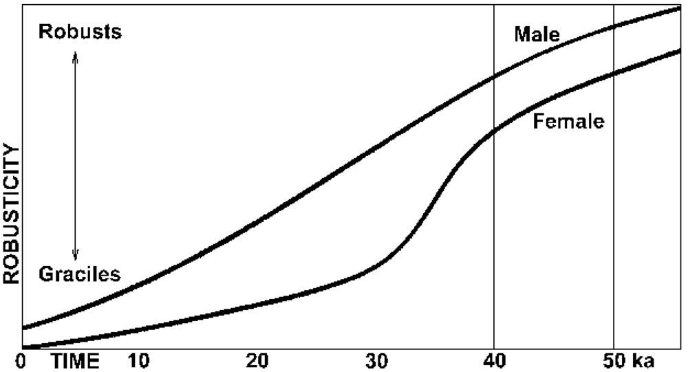
Figure 7.
Schematic depiction of average male and female relative cranial gracility in Europe through time, showing that the decline in robusticity is gradual in males, but accelerated in females between 40 and 30 ka.
Figure 7.
Schematic depiction of average male and female relative cranial gracility in Europe through time, showing that the decline in robusticity is gradual in males, but accelerated in females between 40 and 30 ka.

Gracilization begins typically in females, with males lagging many millennia behind (Figure 7). The process has continued to the Holocene, and reduction in both dimorphism and robusticity is also still active in human evolution today. The face, jaw and teeth of European humans 10 ka ago are in general 10% more robust than those of today’s Europeans and Asians, and those of 30 ka ago are 20–30% more robust. Some modern humans, as noted above, have retained tooth sizes typical of archaic H. sapiens and H. erectus, and other robust features are preserved in many populations or individuals. Neanderthaloid specimens occur in the Mesolithic, through to the Neolithic and even later.
Holocene gracilization could conceivably be explained as a response to changing food-processing techniques or less physically demanding lives. The smallest tooth sizes tend to be found in those areas where food-processing techniques have been used for the longest time. However, this explanation cannot be extended to universal gracilization during the Late Pleistocene. The life style of people 15 ka ago is not thought to have been significantly different from that of 35 ka ago, yet the overall rate of gracilization appears to be have been reasonably uniform over the past 50 ka (Figure 7). As a universal phenomenon it has not been explained, and indeed has been ignored due to the dominance of the replacement model.
Natural selection simply cannot account for a significant reduction in robusticity and reversal of encephalization without any apparent trade-off in evolutionary benefits for the organism in question. No such benefits are apparent, and yet this process seems to have been universal wherever humans existed during the final Pleistocene. It is proposed here that the dimorphism observed during the crucial period of the last twenty or thirty millennia of the Pleistocene presents the key to the most parsimonious explanation. Dimorphism in mammals generally reflects one or both of two selection pressures: competition between males for access to females, or male-female differences in food procuring strategies, with males provisioning females [203,204,205]. In the case of late hominins it has been suggested that physical competition among males may have been diminished radically with the introduction of accurate projectile weapons acting as “equalizers” [206,207]. This is, however, not a satisfactory explanation: effective distance weapons were in use long before the Upper Paleolithic (spears of the Lower Paleolithic were found at seven European sites), together with large game hunting. Thus the “equalizers” had long been in use and they do not explain the gender-specific pattern of later gracilization, nor the extensive fetalization that took place in the final Pleistocene.
5. The Fetalized Human
It is self-evident that practices such as deliberate breeding-mate choice determine procreational success today, so the obvious question to be asked is: at what point in time did it first appear? Other primates (indeed, all other animals) exhibit no preferences in mate selection of youth or specific body ratios, facial features, skin tone or hair; yet in present humans these are deeply entrenched, perhaps “hardwired”. Facial symmetry, seen to imply high immunocompetence [208,209], is also of importance, and in female humans neotenous facial and other features are strongly preferred by males [210,211]. Since this applies undeniably today, the rational way to examine this issue is to consider at what point in human development the influence of non-evolutionary currents can be first detected. It is suggested that around 40 ka ago, cultural practice had become such a determining force in human society that breeding mate selection became increasingly moderated by cultural factors, i.e., by factors attributable to learned behavior. These could have included the application of a variety of cultural constructs in such choices, such as social standing, communication skills, body decoration (which becomes notably prominent 40 ka ago, although existing earlier), and most especially culturally negotiated constructs of physical attractiveness.
In all animals, including all hominins, reproductive success determines phylogenetic direction. If one were to look for evidence of when deliberate sexual selection began to have detectable effects, two strategies spring to mind. One could look for signs that attributes of natural fitness were replaced by attributes that confer no Darwinian survival benefits, or one could look for indications of a culturally mediated preoccupation with female sexuality. One would note that, firstly, gracility, especially of females, develops strongly during the Aurignacian and Gravettian; and secondly, that this very same period is marked by a distinctive preoccupation with female sexual attributes. The latter is found in the common depictions of (mostly) isolated vulvae or pubic triangles [212,213]; at Abris Blanchard, Castanet, Cellier, Le Poisson, Pataud, and La Ferrassie, Laussel and in Chauvet Cave; and the creation of naturalistic female statuettes, often with pronounced sexual aspects (Figure 8), beginning with the Aurignacian [214,215,216]. Therefore the question to be asked is: what cultural preferences could possibly have led to the gracilization of female humans during the second half of the Würm glacial in Europe?
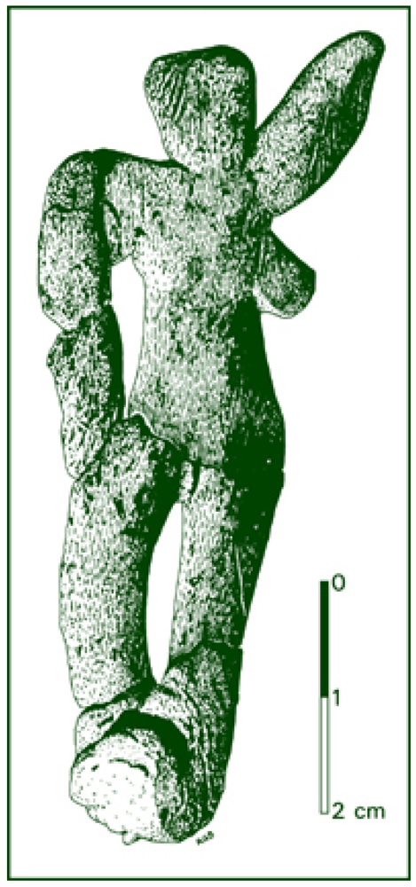
Figure 8.
Serpentine figurine of the Aurignacian, ca. 31,790 carbon years old, Galgenberg near Krems, Austria.
Figure 8.
Serpentine figurine of the Aurignacian, ca. 31,790 carbon years old, Galgenberg near Krems, Austria.

Mating preferences and their genetic results in respect of personality and anatomical traits [217], which could become cultural selection variables, can be modeled by methods of the gene-culture co-evolutionary model [218,219,220,221]. It has been noted that traits selected for can include large female breasts, small feet or male macho behavior, and most certainly physical “attractiveness”. The latter are necessarily informed by cultural constructs of attractiveness, because there is no objective measure of it. The question then becomes: if the recent gracilization of humans were related to fetalization, what would be its anatomical consequences?
Humans resemble chimpanzees anatomically most closely in the latter’s fetal stage [222,223,224]. Both the fetal chimpanzee and the adult human have hair on the top of the head and on the chin, but are otherwise largely naked. In apes, this changes rapidly upon birth, in humans it remains for life. All male adult apes have a penis bone, but it is categorically absent in both fetal chimpanzees and all male humans, from the fetal stage and throughout life. In fact the penis bone of apes is one of the very last parts of the ape fetus to form, shortly before birth, and its atrophy in humans appears to have been compensated for by the significantly increased length and thickness of the penis, relative to apes [225]. Similarly, in female chimpanzees, the labia majora are an infantile feature; in humans they are retained for life. The hymen, too, is present only in the neonate ape, but is retained for life in human females in the absence of penetration. The organs of the lower abdomen, such as rectum, urethra and vagina, are typically aligned with the spine in most adult mammals, including apes; only in fetal apes and humans do they point forward relative to the spine (upright walking appears irrelevant, because fetal apes do not walk). The human ovary reaches full size at the age of five, which is the age of sexual maturity of the apes [223]. Most importantly, the skull of an unborn ape is thin-walled, globular and lacks the prominent tori of the adult ape, thus resembling the cranium of a modern human. Upon birth its robust features develop rapidly. The slow closing of the cranial sutures in humans, on the other hand, is again clearly a neotenous feature, introduced by the recent genes RUNX2 and CBRA1. The face of the ape embryo forms an almost vertical plane, as it does in the modern human all the way through adulthood, which is certainly not the case in mature apes. Even the brains of fetal apes and adult humans are much more similar to each other, in terms of proportion and morphology, than they are to those of adult apes.
These and many other features define the anatomical relationship between ape and man as the latter’s neoteny. The legs of fetal apes are relatively short, while the arms are about as long in relation to the body as in humans. In the apes, the arms become much longer after birth. Human hands and feet resemble those of embryonic apes closely, but differ significantly from both hands and feet of mature apes. In fact the human foot, especially, retains the general structure found in unborn apes, which rather contradicts the hypothesis that it is an adaptation to upright walking. The contrary hypothesis, that upright walk is an adaptation to the neotenous condition of the human foot is in fact more logical, but has never been proposed. Even the shape of the cartilage of the ear in humans is a neotenous feature.
In neoteny, sexual maturity is attained before full somatic development, and juvenile characteristics are retained for life. In an evolutionary perspective, it refers to species whose adults retain juvenile ancestral features. This has also been called fetalization, because in such phylogenic development, fetal characteristics remain into adulthood, and specific processes of anatomical maturation are retarded [223]. Indeed, it is fascinating to note that in human fetalization, biological history seems to be repeating itself: all vertebrates appear to be the result of neoteny in chordates (species having a notochord) hundreds of millions of years ago. The modern human has undergone so much selection in favor of neoteny that this retardation should be seen as rivaling in importance the distinguishing anatomical characteristic of the oversized brain. It therefore needs to be considered here. “But neoteny does not only contribute to the production of large structural change; it is also the cause of the retention of plasticity” or “morphological evolvability” [226]. Adaptively useful novelties supposedly become available as maturation genes are freed by pedomorphosis. This neotenous “retention of plasticity”, also noted by Fuentes [198], could be a key factor in how humans became what they are today.
Encephalization and neoteny in hominin evolution are quite probably related, perhaps through supervenience. It is self-evident that, relative to the neonate ape, the newborn human is not remotely as far developed. For instance, it would find it impossible, for many months after birth, to cling to the fur of a mother for transport. Of course this is related to its excessive brain size, which has caused it to be expelled at a much earlier stage of fetal development. It can be regarded as highly probable that human mothers always had to carry their infants. Indeed, one of the first kinds of artifacts used by early humans was probably some kind of sling or baby carrying bag. The long period during which the human infant was entirely dependent upon the mother, not just for sustenance but also to move with the horde as well as for protection, extended the period for initial learning very significantly. This, obviously, coincided with the continued growth of the brain after birth, which in fact exceeds that of the fetus in man. In the first year after birth, our brain more than doubles in both volume and weight. It continues to grow, approaching adult size by the age of three, but goes on expanding slightly more up to adolescence and even beyond. If this extraordinary development, unheard of in the rest of the animal kingdom, is compared with that of other primates, in simians such as the rhesus monkey and in the gibbon, 70% of adult brain size is achieved at the time of birth, the remaining 30% in the subsequent six months. In the larger apes, the size of the brain approaches adult size after the first year of life. These are very significant differences, and they are all connected with human neoteny.
Another marked difference between humans and most other animals is the abolition of estrus, or periodicity of libido in the female. This almost uniquely human feature has not been explained satisfactorily, but there is a good probability that it is also related to these factors—through one of two alternative scenarios. The excessively long period of infant dependency would have been mirrored in a similar dependency of mothers on the horde, most especially for the meat protein needed for the brain tissue of their unborn [203,227,228,229,230]. It is thought probable that there was strong selection favoring female mutations allowing long periods of sexual receptivity, leading to the abandonment of estrus altogether: those females who were longer or always receptive were favored in the distribution of meat from kills [204,205], in a feedback system facilitating fetal encephalization through better access to animal protein. It has been noted that on occasion, female chimpanzees are only given meat after they have copulated with a successful hunter, and it is logical that such a behavior trait would select in favor of continuously receptive females.
The second alternative explanation for the loss of estrus in humans is simpler and would favor a very late introduction, but may seem no more than a stab into the dark. Domesticated mammalian species lack the seasonal reproduction of their wild ancestors and most can reproduce themselves at almost any time of the year. It is possible that the same effect in humans is the result of their self-domestication.
Be that as it may, the numerous physiological features of human neoteny should suffice to demonstrate that humans are anatomically best defined as a fetalized form of ape. Although the process of selecting in favor of infantile physiology appears to mark much of human history, during the final Pleistocene it suddenly accelerated to an unprecedented rate and resulted in markedly unfavorable mutations, from the perspective of natural selection. Domesticated species and laboratory experiments indicate that strong selection leads to maladaptive consequences, attributed to antagonistic pleiotropy in populations out of equilibrium [231,232]. Worldwide, wherever humans existed 40 or 50 ka ago, possessing as they did Mode 3 technological traditions, they shed all of their robust features in just a few tens of millennia. Their brain size decreased, despite the rapidly growing demands made on their brains. Their muscle bulk waned until their physical strength was perhaps halved, in tandem with significant reductions in bone strength and thickness. The decrease in skull thickness is particularly prominent, as well as the relatively rapid reduction in cranial robusticity. This process occurred so fast that it can be tracked through the millennia. At about 35 ka, partially gracile female specimens are encountered from Europe to Australia. The subsequent skeletal evidence presents a distinctive sexual dimorphism: the female crania, though still much more robust than male crania were toward the end of the Pleistocene, show distinctive gracilization (development of globular crania, reduction or absence of supraorbital tori and occipital projection, significant loss in bone thickness, reduction in prognathism and postorbital constriction, and several other features bringing humans closer to the fetal ape’s morphology). The males, however, remain almost as robust as typical “Neanderthals” (Figure 7). Ten thousand years later, the females have become markedly more gracile, and the robust features of the males have also begun to wane. Toward the end of the Pleistocene, the males begin to catch up with the females, and from there on the loss of robusticity continues right to the present time. These relatively rapid changes are not attributable to genetic drift via a bottleneck, but are the result of selective breeding—unintentional self-domestication.
6. Susceptibility of the Human Brain to Illness
The deleterious effects of human domestication are not limited to the gracilization and neotenization of final Pleistocene humans; there are other significant factors to be considered. For instance extant non-human primates appear to be largely free of the neurodegenerative diseases as well as numerous genetic defects that are so prominent among modern man [233,234,235]. When captive apes and monkeys are deprived of environmental stimulation and the company of conspecifics they often present symptoms resembling obsessive-compulsive behavior [236]. However, such behavior is not attributable to defects of the brain, but to protracted enforced conditions, and variations of it are also evident in many captive non-primate species. Chimpanzees do not experience the significant atrophy in the size of brain and other internal structures that inevitably accompany aging in humans [237]. Consequently, the human susceptibility to neuropathologies such as Alzheimer’s disease is unique in the animal world, and is attributable to the wear and tear of the excessively large brain, most of whose neurons cannot be renewed. While epilepsy and stroke do occur in other primates, other brain disorders have not been reported from natural settings or free-roaming populations [236]. A paper sometimes cited as presenting evidence of neuropathology in non-human primates in fact merely reports the incidence of similar genes in the brains of healthy humans, apes, and monkeys [238]. For instance some of the genes thought to be involved in Alzheimer’s are concordant in humans and other primates, while others are not, but the mere presence of such genes does not imply neuropathology. Moreover, most of the data used in this report derives from another study [239] of specimens that were all free of mental disorders and brain abnormalities detectable by autopsy.
Another possible disadvantage of the cognitive ability of humans is that it may have prompted the reduced apoptosis (the process of “programmed” cell death) of neurons, relative to chimpanzees. This has been proposed to cause the higher risk in humans of cancer and other diseases associated with reduced apoptotic function [240]. A review of the relevant human brain illnesses and their etiologies also suggests that they involve largely the very same areas of the brain that are the phylogenetically most recent, in that they differ most from those of other extant primates [236]. It is these areas that support the much-vaunted perceived advanced cognitive and intellectual characteristics of “modern humans”. It appears that Homo sapiens sapiens is paying a price for the extraordinary abilities acquired. What made us human, especially the neural system supporting language and social cognition [241], has also led to psychosis [225].
Not only have the mental and cognitive developments in our brain rendered humans vulnerable to neurodegenerative diseases as well as frontal lobe connectivity problems—such as those causing autism, schizophrenia, bipolar illness, demyelination—which significantly other primates seem to be free of. They have apparently also facilitated the rise of quite specific personality disorders, such as obsessive compulsive disorder and sociopathic or antisocial personality disorders. Like Rett and Down syndromes and dozens of other known genetic impairments endemic to humans, they could be expected to have been vigorously selected against by natural evolution [242,243,244]. The same applies to such conditions as chronic fatigue syndrome, which, although of unknown etiology, is often related to psychiatric disorders [245]. Here, however, the focus is on neurodegenerative pathologies, of which a considerable variety is being distinguished. Among the better known are Alzheimer’s, Huntington’s and Parkinson’s diseases, frontotemporal dementia and behavioral variant frontotemporal dementia (FTD); but amyotrophic lateral and diffuse myelinoclastic sclerosis, AIDS dementia, Batten disease and neuronal ceroid lipofuscinosis, Creutzfeldt-Jakob disease and many others also pertain. The first listed are briefly reviewed, as well as a few further common human brain illnesses posing evolutionary selection issues.
Alzheimer’s disease (AD) can be diagnosed in people from the 30s onwards, but is primarily a disease of aging. It results from extracellular plaque deposition of beta amyloid and intracellular accumulation of tau, a protein. Tau is the component of intracellular neurofibrillary tangles. These plaques and tangles, clearly visible with MRI (magnetic resonance imaging), are initially found primarily in the hippocampus and entorhinal cortex, later in some areas of the frontal cortex and temporal (medial temporal lobe) and parietal association cortex. Their buildup is related to gene APOE versions e2, e3 and e4 [246]. AD targets the limbic structures [247], including the amygdala, the locus coeruleus and the cholinergic neurons of the nucleus basalis of Meynert. As a result of the plaque deposition neurons and synapses die, axons degenerate and connections are lost; general atrophy of the cortex and brain shrinkage occur [248]. VENs (von Economo neurons, see below) are particularly vulnerable to AD, and about 60% of them may be lost in the anterior cingulate cortex [249].
VENs are also highly implicated in frontotemporal dementia (FTD, also known as Pick’s disease. This group of relatively rare disorders affects primarily the frontal and temporal lobes, associated with personality, behavior and language. In one variant of frontotemporal dementia, known as behavioral variant bvFTD, the anterior cingulate cortex and the orbital frontoinsula both show marked signs of focal degeneration, which is prominent in the right hemisphere [250].
Huntington’s disease derives from cell loss in the basal ganglia and cortex. This movement, cognitive, and behavioral disorder can affect most age groups and occurs in the neostriatum, where marked atrophy of the caudate and putamen is accompanied by selective neuronal loss and astrogliosis. Degrees of atrophy in other regions, including the globus pallidus, thalamus, subthalamic nucleus, substantia nigra, and cerebellum, depend upon the progress of the disease. Its genetic basis involves the expansion of a cysteine-adenosine-guanine (CAG) repeat encoding, a polyglutamine tract in the N-terminus of the protein product called Huntingtin (the function of which remains unknown).
Parkinson’s disease (PD) is associated with a loss of dopaminergic nigrostriatal neurons, which are located in the substantia nigra of the midbrain. By the time a patient is diagnosed with PD, usually at stage three of six defined stages, about 60–70% of the substantia nigra dopamine cells are already lost. By that time the substantia nigra, basal ganglia, amygdala, part of the limbic system, nucleus basalis of Meynert and part of the extended amygdala have all been affected.
Obsessive compulsive disorder (OCD) involves persistent thoughts, feelings and impulses that are attributable to an overactive inferior prefrontal cortex. Appearing first in childhood to early adulthood, this anxiety disorder affects areas of the brain called the “worry circuit” and is connected with an imbalance of the neurotransmitter serotonin [251]. Excessive activity in the inferior prefrontal cortex leads to the development of obsessive stereotypical behaviors. The striatum (caudate nucleus and putamen) is also over-activated in OCD sufferers. Projecting to the striatum, the inferior prefrontal cortex, orbitofrontal cortex and the cingulate cortex cause the caudate to be overactive in the striosome area, thus bringing emotional tones and valences into the experience via the amygdala because it also projects into this same striosome area. Between the matrisome and the striosome areas are the tonically active neurons (TANs), which integrate the input from the inferior orbital frontal cortex via the strisomes with the input from the amygdala and orbitalfrontal region, also via the striosomes. The TANs thus function as a gating mechanism between the matrisome and the striosome regions.
Bipolar or manic-depressive disorder (BD) is characterized by extreme mood swings between alternatively euphoric and depressed states. Cyclothymic disorder is a milder form of this illness [252]. Several genetic regions have been implicated in these conditions, including six specific chromosomes [253]. Since Schildkraut [254] suggested the involvement of the neurotransmitters norephinephrine and serotonin and with the advent of neuroimaging, a number of brain areas have been implicated. This includes the observation of reduced gray matter in the left subgenual prefrontal cortex and amygdala enlargement [255], and decreased neuronal and glial density in association with glial hypertrophy [256]. Significant shape differences have also been observed in caudate and putamen, thus implicating the basal ganglia [257]. MAOA is one of the genes implicated in BD [258].
Schizophrenia afflicts the frontal lobes with connectivity problems, contributing to the appearance of atrophy, and the cingulate cortex, temporal lobes and hippocampus are all adversely affected. The illness involves volumetric changes of gray matter in the right and left middle and inferior temporal gyrus, worsening with chronicity [259]. Hippocampal volume is also reduced in schizophrenia and there is lateral ventricular enlargement [260]. The condition is associated with frontal lobe dysfunction and disconnectivity [261]. These morphometric changes are suggestive of alterations in synaptic, dendritic and axonal organization, a view supported by immunocytochemical and ultrastructural findings. Numerous rare alleles are thought to be involved [262]. Pathology in subcortical structures is not well established apart from the dorsal thalamic nuclei, which are interconnected with the dorsolateral prefrontal cortex (which possesses VENs). The anterior cingulate cortex containing large numbers of VENs is also involved. Reductions in the number of small neurons in layer II and reduced cerebral blood flow in the anterior cingulate have been noted in schizophrenia [263], which has been linked with the genes SLC6A4 [264], NRG1 [265] and NRG3.
Multiple sclerosis (MS) is apparently but not conclusively an autoimmune inflammatory disease of the central nervous system, causing demyelination of axons [266]. It is characterized by multifocal lesions, the MS plaques. Activated mononuclear cells destroy myelin and to some degree oligodendrocytes, the glial cells that produce the myelin in which axons are wrapped. Remaining oligodendrocytes attempt to produce new myelin, but in most cases this pattern of inflammatory reaction subsides only to appear at another location or at another time. The pattern of progression suggests a relative hierarchy of changes over time, involving first frontal and temporal regions [267] and later the pre-central gyrus [268]. MS can impact on any area in the central nervous system although visual areas are commonly affected.
Autism spectrum disorders manifest themselves in early childhood and their etiology remains unknown. Abnormalities have been detected in the frontal and temporal lobes, the cerebellum, the amygdala, and the hippocampus. VENs have been implicated in autism by Allman et al. [269]; but see [270]. Underconnectivity in the brains of children with autism [271] offers a basis for further investigation of this and other pervasive developmental disorders. In some subgroups, cerebellar dysfunction may occur, in others there is dysfunction of the prefrontal cortex and of connections to the parietal lobe. Reduced activation in the fusiform gyrus, the portion of the brain associated with facial recognition, and increased activation of adjacent portions of the brain associated with recognition of objects have been observed. The amygdalas of patients with autism have fewer nerve cells, especially in a subdivision called the lateral nucleus of the amygdala.
Asperger’s syndrome differs from autism in that it lacks the aberrations or delays in language development or cognitive development that are typical of autism. Sufferers may also have normal or even superior intelligence, in contrast to the low IQ associated with autism. However, they share social insensitivity and other characteristics with autism patients. Asperger’s also develops in early childhood, generally after the age of three.
Temporal lobe epilepsy involves the limbic system and may originate in several locations within the temporal lobe, the hippocampus, parahippocampal gyrus, amygdala, etc. [272]. This epileptic condition is included here because of its effects on the limbic system [273], which it shares with many of the other pathologies listed.
Middle cerebral artery stroke, although not related to neurodegenerative conditions, is considered here because it is the leading neurological illness, and evolution would have strongly selected against this susceptibility. Stroke derives from thromboemboli lodged in a cerebral blood vessel; from platelet emboli; or from carotid or vertebral stenosis. The middle cerebral artery is the largest cerebral artery and also the most commonly affected by cerebrovascular incidents. Since it supplies large areas of the brain (most of the outer convex brain surface, nearly all the basal ganglia, and the posterior and anterior internal capsules), infarcts can lead to diverse conditions. These include apraxia (inability to perform previously learned physical task) and dyspraxia (inability to perform a physical task), and Broca’s and Wernicke’s aphasia (expressive and receptive language deficits).
This brief list of brain disorders should suffice to show not only that specific brain regions are selectively affected by them, but also that these are in most cases those very same regions facilitating what are generally defined as the “higher cognitive functions” of the human brain [274]. There is every possible indication that this neurological susceptibility is directly linked to the complexity of the ever-burgeoning human brain. Expressed in simplistic terms, it has given humanity both the genius of its greatest thinkers and artists, and the despair of “losing one’s mind”. This immediately raises a fascinating question: did the human ancestors of early periods of the Pleistocene suffer from neuropathologies? If the two extreme conditions of the human mind are in some measure correlated, the answer might be, these afflictions appeared only as hominin cognition became overly complex, perhaps as if it placed excessive demands on new neural structures. To investigate the origins of mental illnesses involves three closely related issues that have not been investigated in any depth: (1) how did these pathologies initially develop; (2) at what time in our evolution did they appear; and (3) why did evolutionary processes apparently fail to select against the relevant genetic predispositions (the Keller and Miller paradox [243])? Without some appreciation of these issues the diseases concerned have no causal context or explanation; one is merely trying to make sense of end effects at specific loci without an appreciation of how they came about. Science, however, expects some level of causal reasoning from us.
In seeking to determine how humans became human, and how they acquired the neuropathologies this involves, our search would benefit from an initial focus on those rather few characteristics that appear to be uniquely and quintessentially human. Preoccupied with inconclusive word games, the social sciences have not produced much sound empirical data. Neuroscience, on the other hand, has, but that wealth remains largely untapped by those examining hominin cognitive evolution. For instance, one of the major differences between humans and other extant primates is found in Brodmann’s area 10 [275] in the pre-frontal lobe, apparently much more developed in humans than in chimpanzees [276]. This cortical region supports higher cognitive functions, including the extraction of meaning from experience; the organization of mental contents that control creative thinking and language; artistic creation; initiation of, expression of, and planning for future action [277]. Hodgson and Helvenston [278] suggest that area 10 is one of the most likely substrates for the expansion of complex, sustained and focused human consciousness, which is one of the major differences between humans and other extant primates (the other being speech). Details of their neurological disparities suggest that distinctive changes must have taken place during the course of hominin evolution, since the phylogenetic split in the Miocene period. Differences between the brains of humans and members of the pongid clade are essentially in respect of structure and size. The structural dissimilarities are considered first (Figure 9).
VENs (von Economo neurons) occur in both apes (and other mammals) and humans [279,280], but they are larger and far more numerous in the latter, occurring in humans in the anterior cingulate cortex [281,282] and the frontoinsular cortex [283]. These structures are thought to be involved in complex social emotion and cognition. Nerve cells in humans, e.g. in the primary visual cortex, are arranged in far more complex patterns than in apes [284]. The minicolumn, the mammalian brain’s basic information processing structure, in the left planum temporale, is significantly enlarged in the human, relative to the chimpanzee or rhesus monkey [285], containing the axons, dendrites, and synapses that make neural connections [286]. Significantly, the planum temporale is involved in language production. Ullian et al. [287] found that synapses form between neurons only in the presence of astrocytes, neuroglial cells constituting almost half the cells in a human brain (see also [288,289]). These cells secrete the protein thrombospondin, which triggers synapse formation [39] and of which the human brain produces about six times as much as that of chimpanzees or macaques [40]. It also produces about twice as much of THBS4 and THBS2 messenger RNA (mRNA), respectively, in the human cerebral cortex. (RNAs are ribonucleic acids that, like DNA, can carry genetic information.) Thrombospondin expression differences were observed in the forebrain (cortex and caudate), whereas the cerebellum and most non-brain tissues exhibit similar levels of the two mRNAs in humans and chimpanzees. Increased expression of thrombospondins in human brain evolution could result in changes in synaptic organization and plasticity, and contribute to the distinctive cognitive abilities of humans, as well as to the vulnerability to neurodegenerative disease that seems unique to humans [234,235].
Figure 9.
Section (left) and left view of the human brain.

VENs are considered to participate in rapid signal transmissions and are relatively newly evolved in mammals, being present in humans, pongids, sperm and beluga whales, bottlenosed and Risso’s dolphins, and in African and Asian elephants [249,291,292]. This list suggests that VENs may be restricted to relatively large animals with large brains and extensive social networks.
Those areas of the anterior cingulate cortex that contain VENs have been proposed to be a phylogenetically new specialization of the neocortex [293] rather than a more primitive state of cortical evolution, as most other areas of the cingulate cortex are [294]. The anterior cingulate is an area involved with a variety of emotions, both positive and negative, and its VENs are believed to project to Brodmann’s area 10 [281]. While the human frontal lobes are not relatively oversized, parts such as areas 10 and 13 may be enlarged relative to what would be expected from a primate of human body size. The increase in area 10, used in retrieving memories from the individual’s past experience and in planning future action, has been suggested to begin with Homo habilis [295]. Area 13 is part of the limbic system [296] and involved in emotional, motivational and social behavior via its interconnections with other limbic and cortical structures. The cingulate gyrus, a significant part of the limbic lobe, is of substantial size in humans. It subsumes viscero-motor, cognitive-effector, instant emotional experiences, adaptive motor responses, and sensory processing areas [297]. The extended and in some ways unique human limbic system [296] includes the hippocampus and the amygdala, the septum, olfactory nucleus, entorhinal cortex, bed nucleus of the stria terminalis, and the nucleus basalis of Meynert. The amygdala is implicated in a spectrum of social attributions, such as appraisal of the emotional state of others [298], value judgments such as trustworthiness [299], and the emotional tone of memory consolidation and restructuring. The hippocampus receives its input from the entorhinal cortex, which derives its inputs from the associative neocortex—the most recent cortical development and involved in spatial orientation [300]. The entorhinal cortex, hippocampus and amygdala are implicated in memory functioning [301,302]. Since there are few conditions that involve almost exclusively the amygdala (e.g., Urbach-Weithe disease) its now accepted neuroanatomical extension includes the subpallidal region and the bed nucleus of the stria terminalis.
The prefrontal cortex as well as portions of the posterior association areas have in humans become enlarged beyond what would be expected in comparison to primary sensorimotor structures [303]. Preuss and Kaas [304] report that the human Brodmann’s area 17, comprising the primary visual areas, differs from both apes and monkeys in the way information is segregated from the magnocellular and parvocellular layers of the lateral geniculate nucleus. These authors attribute to humans an improved ability of evaluating moving stimuli. Holloway [305,306] notes that the visual striate cortex and lateral geniculate are significantly smaller in a human than expected for an ape of human size and suggests that the variation in the former area began with australopithecines [307]. Another distinctive difference is in the pyramidal motor system, the most recently evolved part of the motor cortex. In particular, the rostral section of the motor cortex is phylogenetically recent. In contrast to apes, where the motor cortex is located on both banks of the central sulcus, it is in humans located in front of the central sulcus. The caudal primary motor area is mediated by corticospinal efferents in the extrapyramidal system, which includes the caudate and putamen, i.e., the striatum. The latter and the globus pallidus form the basal ganglia, thought to integrate emotion and reason for humans, generating motor neuron activity and motor output [308].
Another difference between pongid and human brains concerns the cerebellum, which is smaller in humans than would be expected in an ape of human body size, but larger than expected in the gorilla [276]. It serves fine motor tuning, balance and some aspects of cognition [309], in the routinization of complex cognitive procedures, error detection and language. The ability to predict the actions of others and the preparation for behavioral responses have also been attributed to cerebellar learning of sequences [310].
The parietal association area is larger in humans than in apes, at the expense of the occipital cortices. The planum temporale, presenting a left-right asymmetry favoring the left [311], has been related to language reception, but it is also present in apes [312,313]. This challenges the often-perceived simple relationship between this asymmetry and language. These similarities are perhaps merely homologous in apes and humans, as suggested by the work of Stepniewska et al. [314] with owl monkeys. It showed that when the homologue of Broca’s area is stimulated in that species it produces oral and laryngeal responses. The insula in the anterior Sylvian fissure of the temporal lobe has also been suggested to be large in humans, constituting part of the extended limbic system [296]. The nucleus subputaminalis in the basal forebrain is unique in humans, providing cholinergic innervation to the inferior frontal gyrus where Broca’s area (crucial for speech) is located. Wernicke’s area is specialized in humans for the reception of sounds, especially language comprehension (Figure 9). Located in the posterior temporal lobe, this area has six layers, in contrast to the three-layered allocortex (hippocampus and olfactory cortex) [315].
Having thus considered structural differences between human and ape brains, the question of their respective sizes is reviewed next. Encephalization is one of the two most distinguishing characteristics of humans, the other being neotenization (as discussed above). Averaging 1350 cc, the human brain is significantly larger than that of any other species, relative to body size. This is perhaps best expressed by the encephalization quotient (EQ), introduced by Jerison [316]. It expresses the ratio of actual brain volume to “expected brain volume”, the latter being based on average sizes of living mammals [317]. It predicts an increase of 0.75 in brain size for every unit of body size increase [318]. Based on the cat having an EQ of 1.0, the quotient thus reflects the increase in brain size over and beyond that demanded by body size. The rhesus monkey’s EQ of 2.09 (200 cc brain volume) is not spectacularly smaller than the EQ of the chimpanzee of 2.48, with more than double the brain volume (440 cc). The chimpanzee is our closest living relative, genetically much closer to us than the gorilla, but when it comes to the EQ, it is decidedly dwarfed by our EQ of 7.30.
Since the cell bodies of large brains are more scattered, there is more room in them for interconnections between areas [276]. It is precisely the expansion of association cortices that has made the human brain disproportionately large [303]. The human cortex is ten times larger than that of the macaque and three to four times larger than any ape’s [276]. It had long been assumed that the frontal lobes in humans were much larger than would be expected for a primate of human body size [275,319], but recent evidence suggests that they are just about what should be expected [276,320]. However, as noted above, specific areas (e.g., expanded limbic system, Brodmann’s areas 10 and 13) are larger than would be predicted.
In humans, adult brain size is partly determined by the number of neurons produced and retained during an individual’s ontogeny. Different neural areas differ in the length of the embryonic period of neuronal cytogenesis of precursor cells. The longest periods of cytogenesis apply to the areas of greatest degree of enlargement (cortex), and the shortest apply to the least enlarged brain areas (medulla). This suggests that brain size as well as relative size of specific structures could be the result of developmental timing [321]. A decrease in rates of apoptosis could also account for increased brain size. Rakic and Kornack [322] have suggested that in encephalization, cortical neurons are formed from precursor cells lining the ventricles and migrate radially along glial fascicles to reach their cortical destination. Thus the neurons that are formed first make up the deepest layers of the neocortex, and those formed last are its most superficial. Since the neocortical thickness of the macaque brain is similar to the human brain, it is significant that the surface area of the human cortex is ten times as large as that of the macaque. Surface area is determined by the number of these radial columns, while the number of cells in each column determines cortex thickness. It is suggested, from comparing macaques and mice, that the process is regulated by the numbers of mitotic cell divisions involved in the cytogenesis of neurons prior to or during the early stages of the initial formation of radial columns.
7. Conclusions
The ventromedial or orbital prefrontal cortex has thus been implicated in human cognitive evolution as well as in the attendant pathologies, and the involvement of forebrain-neuroanatomy and neurophysiology of frontal and prefrontal structures in mental illness has been demonstrated [296]. This nexus between emerging advanced cognitive abilities and neurodegenerative susceptibility [236] has considerable implications for the question of the origins of modernity in recent human evolution. On the strength of the evidence presented here, natural selection as such had very limited influence in the most recent direction of this process. That is to be expected, but the timing of the change is to be established: is this a very recent development, related perhaps to urbanization or the “Neolithic revolution”, or did the change from natural to cultural genetic selection already commence in the final Pleistocene? The strongest evidence points to culture favoring deleterious somatic aspects over the last 50 ka, and it also demands some mechanism preventing natural selection acting upon neuropathologies.
The evolution of mental and cognitive faculties in hominins should be assumed to have also involved a gradual change from impulsive toward the obsessive or compulsive range of a spectrum of behavior [323,324]. The apparent absence of neurodegenerative and other mental illnesses in extant non-human primates seems to point in the same direction. Based on judicious archaeological reasoning, a rudimentary sense of perfection appears to have been developed by the Middle Pleistocene, and it can apparently be traced back to Mode 1 technocomplexes [80,90]. The neuroscientific information seems to imply that these ancestors, late H. erectus and early H. sapiens, had developed specific brain structures and tissues that facilitated advanced mental and cognitive performance. As the mental faculties of hominins increased with the rise to the top of the food-chain, individual reliance was delegated to society and to objects, the latter ranging from tools to objects of externally storing symbolic information (“exograms”). But to what extent were our ancestors at the same time rendered progressively more susceptible to neurodegenerative and neurological pathologies?
In some cases first genetic indications are already available that such predisposition may have been limited to “modern” humans. For instance, the DYRK1A gene, implicated in causing Down syndrome, seems absent in robust Homo sapiens. The genes CADPS2 and AUTS2, responsible for autism, also appear to be limited to modern Graciles. Perhaps more dramatic is the proposal that schizophrenia is of late historical origin and might have been introduced by a virus as recently as 200 or 300 years ago [325]. Indeed, there appears to be no earlier mention of the disease, in contrast to other conditions such as bipolar illness [236]. Be that as it may, the NRG3 gene, associated with schizophrenia, also seems to be absent in so-called Neanderthals. Using the human haplotype map to test for selective sweeps in regions associated in genome scans with psychosis, such as 1q21, is promising [326]. Again, such selective sweeps tend to yield relatively recent etiologies, of less than 20 ka, as predicted by the domestication hypothesis. Continuing research is likely to locate more evidence that neurodegenerative illnesses are the burden specifically of “modern” sapienoids, just as other “modern” human genes such as RUNX2 and CBRA1 (causing cleidocranial dysplasia or delayed closure of cranial sutures, malformed clavicles and dental abnormalities) and THADA (associated with type 2 diabetes) are certainly deleterious and would not be expected in robust populations. Allele frequencies may have come to be out of equilibrium, therefore equilibrium-based population-genetic models for explaining standing levels of variation, based on antagonistic pleiotropy or related mechanisms, are irrelevant. In a species fully subject to the canons of natural selection the disadvantageous mutations of Homo sapiens sapiens would tend to be suppressed.
The concept of perfectionism, which can be traced to several Lower Paleolithic features (beads, cupules, hand-axes; [80,82,116,118,138]) certainly has adaptive value in a cultural system, but it does require a level of obsessiveness. There is no sense of perfection apparent in anything extant non-human primates make. Their sleeping nests or tools reveal no compulsion to go beyond the purely functional, and have very probably remained completely unchanged for a long time: no desire to improve is evident. Nor is any such obsession apparent from the tools of australopithecines or early Homo (up to H. erectus). Evidence for these impulses seems to appear roughly mid-way through the Acheulian technocomplex, or close to a million years ago. OCD seems to illustrate that obsessive and neurotic behavior is the price humans pay for their rapid cognitive evolution; it is rather like an unwelcome side effect of it. The argument can be extended to most cultural behavior, which, when viewed pragmatically, is frequently irrational. Seen in a realistic perspective, it is often overwrought and involves countless obsessive aspects, e.g. our regimented behavioral routines or our relentless acquisition of excessive surpluses such as an insurance against the loss of another insurance, which surely would qualify as an obsessive behavior symptom. Our access to “objective reality” (should such a state exist) is severely limited by the residues of irrational ideologies, baseless ontologies and metaphysical straightjackets, the baggage of many millennia of cultural development. Other primates do not share our hankering for something to believe in. Our reliance on “specialists” (shamans, prophets, scientists, medical doctors) ignores that déformation professionnelle is also a distortion in the way the world is perceived. Professional training involves obsessive behavior traits that may be neurophysiologically relatable to compulsive behavior. Indeed, it is almost a tautology to say that without obsessions humans would not have reached the level of cultural sophistication they have: human culture is an outcome of obsessiveness.
By the same token, as that very process appears to be implicated also in the rise of neurodegenerative diseases and other neuropathologies, it needs to be assumed that these, too, developed during the last million years or so. The advantages of the development of the prefrontal cortex must have significantly outweighed its detrimental effects, which might suggest that the latter were negligible initially, otherwise they could have selected against the changes giving rise to cognitive sophistication. The question of when and why natural selection apparently failed to select against unfavorable traits is central to these considerations (the paradox of Keller and Miller [243]), but cannot be answered empirically. However, the hypothesis attributing final Pleistocene human neoteny to cultural selection offers a realistic explanation. Just as evolutionary determinants could not prevent the deleterious changes from robust to gracile forms, because they were overruled by cultural determinants, the mental diseases arising from burgeoning prefrontal cortex complexity may have escaped natural selection in much the same way. The development and persistence of these diseases seems to signal that selection against them was somehow muted. It could also be taken as an indicator that the selective benefits of the Late Pleistocene development of cognition and symboling were of such outstanding effects [205] that the “byproduct” of neuropathologies was tolerated—or rather, not significantly selected against. Perhaps the level of tolerance of these consequences expanded in proportion to the selective benefits of developing cognition.
The principal effect of the rise of symbol-based culture, then, has been its facilitation of changes not dictated by natural evolution, but determined by cultural evolution. This is not to suggest natural selection has been abolished for humans; rather that cultural factors began to co-determine changes to our genome, eventually becoming the prime mover of changes. The gene-culture co-evolutionary model predicts such a development, and some of the evidence and propositions presented in this paper document it. The gracilization marking human development over the past fifty millennia was not an evolutionary process; it was determined by cultural selection. Selection for specific traits results in changes in numerous other traits—changes that are usually deleterious—be they physiological or a range of others. The resulting loss of robusticity involved several reductions in evolutionary fitness, e.g., the size of the brain decreased at a time when demands made of it are thought to have increased dramatically. This development was, however, offset by the increased use of exograms, which eventually reached such extraordinary proportions that today most of our symbolic information resides outside our brain. Self-domestication probably also prompted the suspension of estrus, among other changes.
While the role natural selection played in recent human development has decreased at an exponential rate, the effects of genetic drift may have also become progressively marginalized. Population sizes and densities have increased markedly since the mid-Late Pleistocene, and especially since the advent of agriculture in the early Holocene. Susceptibility to genetic drift is a direct function of population size, and with today’s world population of billions of interbreeding organisms must be assumed to be negligible [327,328,329,330]. However, the final Pleistocene deserves closer scrutiny. To the extent that bottlenecks effect genetic drift within very small and isolated populations, it can reasonably be assumed that these have played major roles on many occasions, ranging from the Campagnian Ignimbrite Event to the many Pleistocene maritime colonization events (Wallacea, Mediterranean, Australia, and more recently Polynesia, New Zealand, to mention a few; Figure 10) [28,331,332,333,334,335,336,337,338,339]. There are, however, other processes that either resemble genetic drift or involve it as an underlying factor. One is niche construction, which can take on many forms, but essentially refers to an organism’s alteration of its environment and the resulting ecological inheritance. As the population adapts to these niches, relevant alleles are preferred, and the changes are neither natural selection-derived nor genetic drift-derived, but rather somewhere between the two.
Figure 10.
Colonizing seafaring began with bamboo rafts almost one million years ago in the islands of Indonesia.
Figure 10.
Colonizing seafaring began with bamboo rafts almost one million years ago in the islands of Indonesia.

Humans, obviously, are the ultimate niche constructors, especially through their culturally determined faculties. Human language, which probably appeared around the beginning of the Pleistocene, has been defined as a niche, and like niche construction it is an autocatalytic process [3]. A much more recent human niche is based on the acquired genetic tolerance to milk sugar. Lactose intolerance is required to ensure that the mammalian mother can feed a new infant. Human lactose tolerance beyond weaning can only be a Holocene development, developing after milk-providing species had become domesticated. A rare mutation on chromosome 2, which in the Pleistocene would have been dysfunctional, gained rapid dominance once animal milk became widely available. Not surprisingly lactose tolerance is greatest among people who have long been milk-consuming herders (>85%), while Chinese are only 7% tolerant, and Native Americans not at all. Again, this is not natural selection in any Darwinian sense, nor is it a stochastic process such as genetic drift. It is, however, highly effective and relatively swift. And it is an example of genetic changes by cultural variables.
In the case of hominins, the most glaring challenge to natural selection derives from the recently developed toleration of maladaptive traits, which range from somatic features universally related to neotenization, to mental disorder susceptibility alleles, and to almost countless other detrimental susceptibilities. Disorder susceptibility alleles have not either fixated, if adaptive, or gone extinct, if maladaptive. Those that are rare, the single-gene Mendelian disorders, may escape selection precisely because they are rare, and the molecular bases of over 1700 of them have now been identified. However, the preservation of the mutations deriving from multiple mutant alleles at different genetic loci involved in the major deleterious etiologies has remained entirely unexplained until now [243]. Not only that, Pleistocene archaeology and paleoanthropology have instead conjured up a fantasy of how humans became “modern” through the complete global replacement of all other hominins by a species mysteriously arising by genetic drift (bottleneck or founder effect)—a veritable Biblical explanation provided by disciplines of Biblical naivety. There can be no doubt that genetic bottlenecks occurred throughout hominin history, and that they had extensive effects on the human genome. But the momentous and rapid changes of the last fifty millennia are largely the result of self-domestication and of cultural ability to tolerate maladaptive susceptibilities.
References
- P.V. Tobias. “The bearing of fossils and mitochondrial DNA on the evolution of modern humans, with a critique of the ‘mitochondrial Eve’ hypothesis.” S. Afr. Archaeol. Bull. 50 (1995): 155–167. [Google Scholar] [CrossRef]
- J.R. Searle. The Construction of Social Reality. London, UK: Allen Lane, 1995. [Google Scholar]
- D. Bickerton. Adam’s Tongue: How Humans Made Language, How Language Made Humans. New York, NY, USA: Hill and Wang, 2010. [Google Scholar]
- M. Schulz. “Die Regeln mache ich.” Der Spiegel 34 (2004): 128–131. [Google Scholar]
- L. Harding. “History of modern man unravels as German scholar is exposed as fraud.” The Guardian; London, UK: Guardian News and Media, 2005. Available online: http://www.guardian.co.uk/science/2005/feb/19/science.sciencenews (accessed on 25 August 2011).
- R.R.P. Von Zieten. “The Dating of Upper-Pleistocene Subsaharan Fossil Hominids and Their Place in Human Evolution: With Morphological and Archaeological Implications.” In Ph.D. Thesis. Los Angeles, CA, USA: University of California, 1973. [Google Scholar]
- R. Protsch. “The absolute dating of Upper Pleistocene sub-Saharan fossil hominids and their place in human evolution.” J. Hum. Evol. 4 (1975): 297–322. [Google Scholar]
- R. Protsch, and H. Glowatzki. “Das absolute Alter des paläolithischen Skeletts aus der Mittleren Klause bei Neuessing, Kreis Kelheim, Bayern.” Anthropol. Anz. 34 (1974): 140–144. [Google Scholar]
- R. Protsch, and A. Semmel. “Zur Chronologie des Kelsterbach-Hominiden.” Eiszeitalter Ggw. 28 (1978): 200–210. [Google Scholar]
- W. Henke, and R. Protsch. “Die Paderborner Calvaria—Ein diluvialer Homo sapiens.” Anthropol. Anz. 36 (1978): 85–108. [Google Scholar]
- R.L. Cann, M. Stoneking, and A.C. Wilson. “Mitochondrial DNA and human evolution.” Nature 325 (1987): 31–36. [Google Scholar]
- H. Curtis, and N.S. Barnes. Biology, 5th ed. New York, NY, USA: Worth Publishers Inc., 1989. [Google Scholar]
- T. Kuhn. The Structure of Scientific Revolutions. Chicago, IL, USA: University of Chicago Press, 1962. [Google Scholar]
- I. Davidson, and W. Noble. “The archaeology of perception: Traces of depiction and language.” Curr. Anthropol. 30 (1989): 125–155. [Google Scholar]
- I. Davidson, and W. Noble. “Tools, humans and evolution—The relevance of the Upper Palaeolithic.” In Tools, Language and Intelligence: Evolutionary Implications. Cascais, Portugal: Wenner-Gren Foundation, 1990, pp. 1–21. [Google Scholar]
- W. Noble, and I. Davidson. Human Evolution, Language and Mind: A Psychological and Archaeological Inquiry. Cambridge, UK: Cambridge University Press, 1996. [Google Scholar]
- F. D’Errico, and A. Nowell. “A new look at the Berekhat Ram figurine: Implications for the origins of symbolism.” Camb. Archaeol. J. 10 (2000): 123–167. [Google Scholar]
- G. Bräuer. “Präsapiens-Hypothese oder Afro-europäische Sapiens-Hypothese? ” Z. Morphol. Anthropol. 75 (1984): 1–25. [Google Scholar]
- C.B. Stringer, and P. Andrews. “Genetic and fossil evidence for the origin of modern humans.” Science 239 (1988): 1263–1268. [Google Scholar]
- V. Eswaran. “A diffusion wave out of Africa.” Curr. Anthropol. 43 (2002): 749–774. [Google Scholar] [CrossRef]
- F.H. Smith, I. Janković, and I. Karavanić. “The assimilation model, modern human origins in Europe, and the extinction of Neandertals.” Quat. Int. 137 (2005): 7–19. [Google Scholar] [CrossRef]
- F. Weidenreich. Apes, Giants, and Man. Chicago, IL, USA: University of Chicago Press, 1946. [Google Scholar]
- J.H. Relethford. “Absence of regional affinities of Neandertal DNA with living humans does not reject multiregional evolution.” Am. J. Phys. Anthropol. 115 (2002): 95–98. [Google Scholar] [CrossRef]
- J.H. Relethford, and L.B. Jorde. “Genetic evidence for larger African population size during recent human evolution.” J. Phys. Anthropol. 108 (1999): 251–260. [Google Scholar] [CrossRef]
- E.H. Bryant, S.A. McComas, and L.M. Combs. “The effect of an experimental bottleneck on quantitative genetic variation in the housefly.” Genetics 114 (1986): 1191–1211. [Google Scholar]
- L.R. Berger, S.E. Churchill, B. de Klerk, and R.L. Quinn. “Small-bodied humans from Palau, Micronesia.” PLoS One 3 (2008): e1780. [Google Scholar]
- J. Hawks, S.-H. Lee, K. Hunley, and M. Wolpoff. “Population bottlenecks and Pleistocene human evolution.” Mol. Biol. Evol. 17 (2000): 2–22. [Google Scholar] [CrossRef]
- R.G. Bednarik, and M. Kuckenburg. Nale Tasih: Eine Floßfahrt in Die Steinzeit. Stuttgart, Germany: Jan Thorbecke, 1999. [Google Scholar]
- M.F. Hammer. “A recent common ancestry for human Y chromosomes.” Nature 378 (1995): 376–378. [Google Scholar] [CrossRef]
- U. Gyllensten, D. Wharton, A. Josefsson, and A.C. Wilson. “Paternal inheritance of mitochondrial DNA in mice.” Nature 352 (1991): 255–257. [Google Scholar]
- P. Awadalla, A. Eyre-Walker, and J. Maynard Smith. “Linkage disequilibrium and recombination in hominid mitochondrial DNA.” Science 286 (1999): 2524–2525. [Google Scholar]
- A.A.M. Morris, and R.N. Lightowlers. “Can paternal mtDNA be inherited? ” Lancet 355 (2000): 1290–1291. [Google Scholar] [CrossRef]
- R.S. Williams. “Another surprise from the mitochondrial genome.” N. Engl. J. Med. 347 (2002): 609–611. [Google Scholar] [CrossRef]
- M. Schwartz, and J. Vissing. “Paternal inheritance of mitochondrial DNA.” N. Engl. J. Med. 347 (2002): 576–580. [Google Scholar]
- F. Rodriguez-Trelles, R. Tarrio, and F.J. Ayala. “Erratic overdispersion of three molecular clocks: GPDH, SOD, and XDH.” Proc. Natl. Acad. Sci. USA 98 (2001): 11405–11410. [Google Scholar] [CrossRef]
- F. Rodriguez-Trelles, R. Tarrio, and F.J. Ayala. “A methodological bias toward overestimation of molecular evolutionary time scales.” Proc. Natl. Acad. Sci. USA 99 (2002): 8112–8115. [Google Scholar] [CrossRef]
- K.K. Kidd, J.R. Kidd, S.A. Pakstis, C.M. Tishkoff, C.M. Castiglione, and G. Strugo. “Use of linkage disequilibrium to infer population histories.” Am. J. Phys. Anthropol. 22 (1996): 138. [Google Scholar]
- A. Gibbons. “Calibrating the mitochondrial clock.” Science 279 (1998): 28–29. [Google Scholar] [CrossRef]
- K. Christopherson, E. Ullian, C. Stokes, C. Mullowney, J. Hill, A. Agah, J. Lawler, D. Mosher, P. Brownstein, and B. Barres. “Thrombospondins are astrocyte-secreted proteins that promote CNS synaptogenesis.” Cell 120 (2005): 421–433. [Google Scholar] [CrossRef]
- M. Cáceres, C. Suwyn, M. Maddox, J.W. Thomas, and T.M. Preuss. “Increased cortical expression of two synaptogenic thrombospondins in human brain evolution.” Cereb. Cortex 17 (2007): 2312–2321. [Google Scholar]
- E. Jablonka, and M. Lamb. Evolution in Four Dimensions: Genetic, Epigenetic, Behavioral, and Symbolic Variation in the History of Life. Cambridge, MA, USA: MIT Press, 2005. [Google Scholar]
- G. Gutierrez, D. Sanchez, and A. Marin. “A reanalysis of the ancient mitochondrial DNA sequences recovered from Neandertal bones.” Mol. Biol. Evol. 19 (2002): 1359–1366. [Google Scholar] [CrossRef]
- M.W. Walberg, and D.A. Clayton. “Sequence and properties of the human KB cell and mouse L cell D-loop regions of mitochondrial DNA.” Nucleic Acids Res. 9 (1981): 5411–5421. [Google Scholar] [CrossRef]
- A. Torroni, M.T. Lott, M.F. Cabell, Y.-S. Chen, L. Lavergne, and D.C. Wallace. “MtDNA and the origin of Caucasians: Identification of ancient Caucasian-specific haplogroups, one of which is prone to a recurrent somatic duplication in the D-loop region.” Am. J. Hum. Genet. 55 (1994): 760–752. [Google Scholar]
- H. Zischler, H. Geisert, A. von Haeseler, and S. Pääbo. “A nuclear ‘fossil’ of the mitochondrial D-loop and the origin of modern humans.” Nature 378 (1995): 489–492. [Google Scholar]
- M. Pruvost, R. Schwarz, B.V. Correia, S. Champlot, S. Braguier, N. Morel, Y. Fernandez-Jalvo, T. Grange, and E.-M. Geigl. “Freshly excavated fossil bones are best for amplification of ancient DNA.” Proc. Natl. Acad. Sci. USA 104 (2007): 739–744. [Google Scholar]
- J. Hardy, A. Pittman, A. Myers, K. Gwinn-Hardy, H.C. Fung, R. de Silva, M. Hutton, and J. Duckworth. “Evidence suggesting that Homo neanderthalensis contributed the H2 MAPT haplotype to Homo sapiens.” Biochem. Soc. Trans. 33 (2005): 582–585. [Google Scholar] [CrossRef]
- D. Garrigan, Z. Mobasher, T. Severson, J.A. Wilder, and M.F. Hammer. “Evidence for archaic Asian ancestry on the human X chromosome.” Mol. Biol. Evol. 22 (2005): 189–192. [Google Scholar]
- A.R. Templeton. “Haplotype trees and modern human origins.” Yearb. Phys. Anthropol. 48 (2005): 33–59. [Google Scholar]
- R.E. Green, J. Krause, S.E. Ptak, A.W. Briggs, M.T. Ronan, J.F. Simons, L. Du, M. Egholm, J.M. Rothberg, M. Paunovic, and S. Pääbo. “Analysis of one million base pairs of Neanderthal DNA.” Nature 444 (2006): 330–336. [Google Scholar]
- R.E. Green, J. Krause, A.W. Briggs, T. Maricic, U. Stenzel, M. Kircher, N. Patterson, H. Li, W. Zhai, Hsi-Yang, and et al. “A draft sequence of the Neandertal genome.” Science 328 (2010): 710–722. [Google Scholar]
- A. Gibbons. “Close encounters of the prehistoric kind.” Science 328 (2010): 680–684. [Google Scholar]
- M. Krings, A. Stone, R.W. Schmitz, H. Krainitzki, M. Stoneking, and S. Pääbo. “Neandertal DNA sequences and the origin of modern humans.” Cell 90 (1997): 19–30. [Google Scholar] [CrossRef]
- S. Pääbo. “Extraction, characterization, molecular cloning, and enzymatic amplification.” Proc. Natl. Acad. Sci. USA 86 (1989): 1939–1943. [Google Scholar] [CrossRef]
- R. Higuchi, B. Bowman, M. Freiberger, O.A. Ryder, and A.C. Wilson. “DNA sequences from the quagga, and extinct member of the horse family.” Nature 312 (1984): 282–284. [Google Scholar]
- S. Pääbo. “Molecular cloning and ancient Egyptian mummy DNA.” Nature 314 (1985): 644–645. [Google Scholar]
- A. Cooper, C. Mourer-Chauvirp, G.K. Chambers, A. von Haeseler, A.C. Wilson, and S. Pääbo. “Independent origins of New Zealand moas and kiwis.” Proc. Natl. Acad. Sci. USA 89 (1992): 8741–8744. [Google Scholar]
- J.P. Noonan, M. Hofreiter, D. Smith, J.R. Priest, N. Rohland, G. Rabeder, J. Krause, J.C. Detter, S. Pääbo, and E.M. Rubin. “Genomic sequencing of Pleistocene cave bears.” Science 309 (2005): 597–599. [Google Scholar]
- S. Pääbo, H. Poinar, D. Serra, V. Jaenicke-Després, J. Hebler, N. Rohland, M. Kuch, J. Krause, L. Vigilant, and M. Hofreiter. “Genetic analyses from ancient DNA.” Annu. Rev. Genet. 38 (2004): 645–679. [Google Scholar] [CrossRef]
- M. Pruvost, T. Grange, and E.-M. Geigl. “Minimizing DNA contamination by using UNG-coupled quantitative real time PCR on degraded DNA samples: Application to DNA studies.” BioTechniques 38 (2005): 569–575. [Google Scholar]
- L. Orlando, and C. Hänni. “Du nouveau pour l’AND ancien.” Méd. Sci. 8-9 (2008): I-XVI. [Google Scholar]
- J.P. Jolivet, and M. Henry. De la Solution à L’oxyde, Condensation des Cations en Solution Aqueuse, Chimie de Surface des Oxides. Paris, France: InterEditions CNRS, 1994. [Google Scholar]
- T. Lindhal, and B. Nyberg. “Rate depurination of native deoxyribonucleic acid.” Biochemistry 11 (1972): 3610–3618. [Google Scholar]
- E.M. Golenberg, A. Bickel, and P. Weihs. “Effect of highly fragmented DNA on PCR.” Nucleic Acids Res. 24 (1996): 5026–5033. [Google Scholar] [CrossRef]
- G. Gutierrez, and A. Marin. “The most ancient DNA recovered from amber-preserved specimen may not be as ancient as it seems.” Mol. Biol. Evol. 15 (1998): 926–929. [Google Scholar] [CrossRef]
- E.-M. Geigl. “Why ancient DNA research needs taphonomy.” In Biosphere to Lithosphere: New Studies in Vertebrate Taphonomy. Edited by T. O’Connor. Oxford, UK: Oxbow Books, 2005, pp. 79–86. [Google Scholar]
- L. Carlier, J. Couprie, A. le Maire, L. Guilhaudis, I. Milazzo, M. Gondry, D. Davoust, B. Gilquin, and S. Zinn-Justin. “Solution structure of the region 51-160 of human KIN17 reveals an atypical winged helix domain.” Protein Sci. 16 (2007): 2750–2755. [Google Scholar] [CrossRef]
- R.G. Bednarik. “Only time will tell: A review of the methodology of direct rock art dating.” Archaeometry 38 (1996): 1–13. [Google Scholar] [CrossRef]
- R.G. Bednarik. “The dating of rock art: A critique.” J. Archaeol. Sci. 29 (2002): 1213–1233. [Google Scholar] [CrossRef]
- A. Watchman. “A universal standard for reporting the ages of petroglyphs and rock paintings.” In Dating and the Earliest Known Rock Art. Edited by M. Strecker and P. Bahn. Oxford, UK: Oxbow Books, 1999, pp. 1–3. [Google Scholar]
- J. Walser, and A. Furano. “The mutational spectrum of non-CpG DNA varies with CpG content.” Genome Res., 2010. [Google Scholar] [CrossRef]
- E. Strauss. “Can mitochondrial clocks keep time? ” Science 283 (1999): 1435–1438. [Google Scholar] [CrossRef]
- A.R. Templeton. “Gene lineages and human evolution.” Science 272 (1996): 1363. [Google Scholar]
- A. Templeton. “Out of Africa again and again.” Nature 416 (2002): 45–51. [Google Scholar] [CrossRef]
- H.-P. Schulz. “The lithic industry from layers IV-V, Susiluola Cave, western Finland, dated to the Eemian interglacial.” Préhistoire Européenne 16-17 (2002): 7–23. [Google Scholar]
- H.-P. Schulz, B. Eriksson, H. Hirvas, P. Huhta, H. Jungner, P. Purhonen, P. Ukkonen, and T. Rankama. “Excavations at Susiluola Cave.” Suomen Mus. 109 (2002): 5–45. [Google Scholar]
- P. Pavlov, J.I. Svendsen, and S. Indrelid. “Human presence in the European Arctic nearly 40,000 years ago.” Nature 413 (2001): 64–67. [Google Scholar] [CrossRef]
- C. Gamble. The Palaeolithic Societies of Europe. Cambridge, UK: Cambridge University Press, 1999. [Google Scholar]
- R.G. Bednarik. “Antiquity and authorship of the Chauvet rock art.” Rock Art Res. 24 (2007): 21–34. [Google Scholar]
- R.G. Bednarik. “Palaeoart and archaeological myths.” Camb. Archaeol. J. 2 (1992): 27–43. [Google Scholar] [CrossRef]
- R.G. Bednarik. “The Pleistocene art of Asia.” J. World Prehistory 8 (1994): 351–375. [Google Scholar] [CrossRef]
- R.G. Bednarik. “The earliest evidence of palaeoart.” Rock Art Res. 20 (2003): 89–135. [Google Scholar]
- F. Felgenhauer. “Das Paläolithikum von Willendorf in der Wachau, Niederösterreich. Vorbericht über die monographische Bearbeitung.” Forsch. Fortschr. 33 (1959): 152–155. [Google Scholar]
- V. Cabrera Valdés, and J. Bischoff. “Accelerator 14C dates for early Upper Palaeolithic (basal Aurignacian) at El Castillo Cave (Spain).” J. Archaeol. Sci. 16 (1989): 577–584. [Google Scholar] [CrossRef]
- J.L. Bischoff, K.R. Ludwig, J.F. Garcia, E. Carbonell, M. Vaquero, T.W. Stafford, and A.J.T. Jull. “Dating of the basal Aurignacian sandwich at Abric Romani (Catalunya, Spain) by radiocarbon and uranium series.” J. Archaeol. Sci. 21 (1994): 541–551. [Google Scholar] [CrossRef]
- J. González Echegaray, L.G. Freeman, I. Barandiaran, J.M. Apellaniz, K. Butzer, C. Fuentes Vidarte, B. Madariaga, J.A. Gonzalez Morales, and A. Leroi-Gourhan. La Cueva de El Pendo. Madrid, Spain: Bibliotheca Praehistorica Hispana, 1980. [Google Scholar]
- F. Bordes, and J. Labrot. “La stratigraphie du gisement de Roc de Combe (Lot) et ses implications.” Bull. Soc. Préhist. Franç. 64 (1967): 15–28. [Google Scholar]
- F. Champagne, and R. Espitalié. La Piage, Site Préhistorique Sur Lot; No. 15. Paris, France: Mémoires de la Société Préhistorique Française, 1981. [Google Scholar]
- M. Vaquero, M. Esteban, E. Allué, J. Vallverdú, E. Carbonell, and J.L. Bischoff. “Middle Palaeolithic refugium, or archaeological misconception? A new U-series and radiocarbon chronology of Abric Agut (Capellades, Spain).” J. Archaeol. Sci. 29 (2002): 953–958. [Google Scholar] [CrossRef]
- R.G. Bednarik. “Traces of cultural continuity in Middle and Upper Palaeolithic material evidence.” Origini 18 (1995): 47–67. [Google Scholar]
- A. Palma di Cesnola. “Le leptolithique archaïque en Italie.” In Périgordien et Gravettien en Europe. Edited by B. Klíma. Nice, France: Congrès IX, Colloque XV; UISPP, 1976, pp. 66–99. [Google Scholar]
- A. Palma di Cesnola. “L’Uluzzien: Faciès italien du Leptolithique archaïque.” L’Anthropologie 93 (1989): 783–811. [Google Scholar]
- S.L. Kuhn, and A. Bietti. “The late middle and early Upper Paleolithic in Italy.” In The Geography of Neandertals and Modern Humans in Europe and the Greater Mediterranean. Edited by O. Bar-Yosef and D. Pilbeam. Cambridge, MA, USA: Peabody Museum of Archaeology and Ethnology, 2000, pp. 49–76. [Google Scholar]
- S.L. Kuhn, and M.C. Stiner. “The antiquity of hunter-gatherers.” In Hunter-Gatherers: An Interdisciplinary Perspective. Edited by C. Panter-Brick, R.H. Layton and P. Rowley-Conwy. Cambridge, UK: Cambridge University Press, 2001, pp. 99–142. [Google Scholar]
- O. Abel. “Das Lebensbild der eiszeitlichen Tierwelt der Drachenhöhle bei Mixnitz.” In Die Drachenhöhle bei Mixnitz. Edited by O. Abel and G. Kyrle. Vienna, Austria: Speläologische Monographien, 1931, Volume 7-9, pp. 885–920. [Google Scholar]
- E. Bächler. Das Alpine Paläolithikum der Schweiz. Basle, Switzerland: Monographien zur Ur- und Frühgeschichte der Schweiz, 1940, Volume 2. [Google Scholar]
- J. Bayer. “Wildkirchlikultur.” Eiszeit und Urgeschichte 6 (1929): 142. [Google Scholar]
- R.G. Bednarik. “Wall markings of the cave bear.” Stud. Speleol. 9 (1993): 51–70. [Google Scholar]
- S. Brodar. “Zur Frage der Höhlenbärenjagd und des Höhlenbärenkults in den paläolithischen fundstellen Jugoslawiens.” Quartär 9 (1957): 147–159. [Google Scholar]
- H. Cramer. “Der Lebensraum des eiszeitlichen Höhlenbären und die ‘Höhlenbärenjagdkultur’.” Z. Dtsch. Geol. Ges. 93 (1941): 181–196. [Google Scholar]
- G. Kyrle. “Die Höhlenbärenjägerstation.” In Die Drachenhöhle bei Mixnitz. Edited by O. Abel and G. Kyrle. Vienna, Austria: Speläologische Monographien, 1931, pp. 804–962, Band 7-9. [Google Scholar]
- M. Malez. “Das Paläolithikum der Veternicahöhle und der Bärenkult.” Quartär 11 (1959): 171–188. [Google Scholar]
- L. Vértes. “Die Rolle des Höhlenbären im ungarischen Paläolithikum.” Quartär 11 (1959): 151–170. [Google Scholar]
- L.F. Zotz. Altsteinzeitkunde Mitteleuropas. Stuttgart, Germany: F. Enke, 1951. [Google Scholar]
- J. Svoboda. “The Bohunician.” In La Mutation. Edited by J.K. Kozłowski. Liège, Belgium: ERAUL, 1990, pp. 169–192. [Google Scholar]
- J. Svoboda. “The complex origin of the Upper Paleolithic in the Czech and Slovak Republics.” In Before Lascaux: The Complete Record of the Early Upper Paleolithic. Edited by H. Knecht, A. Pike-Tay and R. White. Boca Raton, FL, USA: CRC Press, 1993, pp. 23–36. [Google Scholar]
- M. Anikovich. “Early Upper Paleolithic cultures of eastern Europe.” J. World Prehistory 6 (2005): 205–245. [Google Scholar] [CrossRef]
- P.L. Allsworth-Jones. “The Szeletian: Main trends, recent results, and problems for resolution.” In The Pleistocene Perspective. Edited by M. Day, R. Foley and R. W. London, UK: Allen and Unwin, 1986, pp. 1–25. [Google Scholar]
- N. Kyparissi-Apostolika. Theopetra Cave. Twelve Years of Excavation and Research 1987-1998. Athens, Greece: Institute for Aegean Prehistory, 2000. [Google Scholar]
- Y. Facorellis, N. Kyparissi-Apostolika, and Y. Maniatis. “The cave of Theopetra, Kalambaka: Radiocarbon evidence for 50,000 years of human presence.” Radiocarbon 43 (2001): 1029–1048. [Google Scholar]
- J.K. Kozłowski. “A multiaspectual approach to the origins of the Upper Palaeolithic in Europe.” In The Emergence of Modern Humans. An Archaeological Perspective. Edited by P. Mellars. Edinburgh, UK: Edinburgh University Press, 1990, pp. 419–438. [Google Scholar]
- M.C. Stiner. Honor Among Thieves. A Zooarchaeological Study of Neandertal Ecology. Princeton, NJ, USA: Princeton University Press, 1994. [Google Scholar]
- S.L. Kuhn. Mousterian Lithic Technology. An Ecological Perspective. Princeton, NJ, USA: Princeton University Press, 1995. [Google Scholar]
- J. Riel-Salvatore, and G.A. Clark. “Grave markers. Middle and Early Upper Paleolithic burials and the use of chronotypology in contemporary Paleolithic research.” Curr. Anthropol. 42 (2001): 449–479. [Google Scholar] [CrossRef]
- F.G. Fedele, and B. Giaccio. “Paleolithic cultural change in western Eurasia across the 40,000 BP timeline: Continuities and environmental forcing.” In Exploring the Mind of Ancient Man. Festschrift to Robert G. Bednarik. Edited by P. Chenna Reddy. New Delhi, India: Research India Press, 2007, pp. 292–316. [Google Scholar]
- R.G. Bednarik, G. Kumar, A. Watchman, and R.G. Roberts. “Preliminary results of the EIP project.” Rock Art Res. 22 (2005): 147–197. [Google Scholar]
- X. Gao, and C.J. Norton. “A critique of the Chinese ‘Middle Palaeolithic’.” Antiquity 76 (2002): 397–412. [Google Scholar]
- P.B. Beaumont, and R.G. Bednarik. “On a search for ancestral rock art in the southeastern Kalahari, South Africa.” In Pré actes du congrès, IFRAO Ariège 2010, Pleistocene art of Africa, DVD. Lacombe, Tarascon-sur-Arège, France, 2010. (in press) [Google Scholar]
- G. Bräuer. “New evidence of the transitional period between Neanderthal and modern man.” J. Hum. Evol. 10 (1981): 467–474. [Google Scholar]
- G. Bräuer. “A craniological approach to the origin of anatomically modern Homo sapiens in Africa and implications for the appearance of modern Europeans.” In The Origins of Modern Humans: A World Survey of the Fossil Evidence. Edited by F.H. Smith and F. Spencer. New York, NY, USA: Alan R. Liss, 1984, pp. 327–410. [Google Scholar]
- G. Bräuer. “The evolution of modern humans: A comparison of the African and non-African evidence.” In The Human Revolution: Behavioural and Biological Perspectives on the Origins of Modern Humans. Edited by P. Mellars and C. Stringer. Edinburgh, UK: Edinburgh University Press, 1989, pp. 123–154. [Google Scholar]
- C.B. Stringer. “Human evolution and biological adaptation in the Pleistocene.” In Hominid Evolution and Community Ecology: Prehistoric Human Adaptation in Biological Perspective. Edited by R. Foley. London, UK: Academic Press, 1984, pp. 55–83. [Google Scholar]
- C.B. Stringer. “The fate of the Neanderthals.” Nat. Hist. 93 (1984): 6–12. [Google Scholar]
- C.B. Stringer. “Middle Pleistocene hominid variability and the origin of Late Pleistocene humans.” In Ancestors: The Hard Evidence. Edited by E. Delson. New York, NY, USA: Alan R. Liss, 1985, pp. 289–295. [Google Scholar]
- C.B. Stringer. “The origin of early modern humans: A comparison of the European and non-European evidence.” In The Human Revolution: Behavioural and Biological Perspectives on the Origins of Modern Humans. Edited by P. Mellars and C. Stringer. Edinburgh, UK: Edinburgh University Press, 1989, pp. 232–244. [Google Scholar]
- P. Mellars, and C. Stringer. Introduction. In The Human Revolution: Behavioural and Biological Perspectives on the Origins of Modern Humans. P. Mellars, and C. Stringer. Edinburgh, UK: Edinburgh University Press, 1989, pp. 1–14. [Google Scholar]
- J. Wainscoat. “Out of the garden of Eden.” Nature 325 (1987): 13. [Google Scholar] [CrossRef]
- J.S. Wainscoat, A.V.S. Hill, A.L. Boyce, J. Flint, M. Hernandez, S.L. Thein, J.M. Old, J.R. Lynch, A.G. Falusi, D.J. Weatherall, and et al. “Evolutionary relationships of human populations from an analysis of nuclear DNA polymorphisms.” Nature 319 (1986): 491–493. [Google Scholar]
- S.E. Churchill, and F.H. Smith. “A modern human humerus from the early Aurignacian of Vogelherdhöhle (Stetten, Germany).” Am. J. Phys. Anthropol. 112 (2000): 251–273. [Google Scholar] [CrossRef]
- S.E. Churchill, and F.H. Smith. “Makers of the early Aurignacian of Europe.” Am. J. Phys. Anthropol. 113 (2000): 61–115. [Google Scholar] [CrossRef]
- A. Czarnetzki. “Zur entwicklung des Menschen in Südwestdeutschland.” In Urgeschichte in Baden-Württemberg. Edited by H. Müller Beck. Stuttgart, Germany: Konrad Theiss, 1983, pp. 217–240. [Google Scholar]
- W. Gieseler. Die Fossilgeschichte des Menschen. Stuttgart, Germany: Konrad Theiss, 1974. [Google Scholar]
- N.J. Conard, P.M. Grootes, and F.H. Smith. “Unexpectedly recent dates for human remains from Vogelherd.” Nature 430 (2004): 198–201. [Google Scholar] [CrossRef]
- D. Sonneville-Bordes. “De Position-stratigraphique et chronologique relative des restes humains du Palaéolithique supérieur entre Loire et Pyrénées.” Ann. Paléontol. 45 (1959): 19–51. [Google Scholar]
- H.L. Movius. “The Abri of Cro-Magnon, Les Eyzies (Dordogne) and the probable age of the contained burials on the basis of the nearby Abri Pataud.” Anu. Estud. Atl. 15 (1969): 323–344. [Google Scholar]
- D. Henry-Gambier. “Les fossiles de Cro-Magnon (Les-Eyzies-de-Tayac, Dordogne): Nouvelles données sur leur position chronologique et leur attribution culturelle.” Bull. Mém. Soc. D’Anthropol. Paris 14 (2002): 89–112. [Google Scholar]
- R. White. “Comment on Bednarik, R.G., ‘Concept-mediated markings in the Lower Palaeolithic’.” Curr. Anthropol. 36 (1995): 623–625. [Google Scholar]
- R.G. Bednarik. “Concept-mediated marking in the Lower Palaeolithic.” Curr. Anthropol. 36 (1995): 605–634. [Google Scholar]
- J. Szombathy. “Die diluvialen Menschenreste aus der Fürst-Johanns-Höhle bei Lautsch in Mähren.” Die Eiszeit 2 (1925): 1-34, 73-95. [Google Scholar]
- J. Smyčka. “Litovel a okolí za pravĕku.” Pravĕk 3 (1907): 140–150. [Google Scholar]
- J. Knies. “Nový nález diluviálního človĕka u Mladče na Moravĕ.” Vĕstník klubu přírodovĕdného v Prostĕjovĕ 7 (1906): 1–19. [Google Scholar]
- J. Smyčka. “Nálezy diluviálního člověka v Mladči u Litovle na Moravě.” Obzor Prehistorický 1 (1922): 111–120. [Google Scholar]
- J. Smyčka. “Kdy přišel první člověk do litovelského kraje.” In Vlastivědný sborník střední a severní Moravy III. Litovel, Czech Republic: příloha Olomoucko a Litovelsko, 1925, pp. 6–7. [Google Scholar]
- E. Weiser. Reise und Wanderbuch, 2nd ed. Freudenthal, Germany: Ernst Feld, 1928. [Google Scholar]
- J. Jelínek. “Historie, identifikace a význam mladečských antropologických nálezů z počátku mladého paleolitu.” Anthropologie 25 (1987): 51–69. [Google Scholar]
- J.A. Svoboda, J. van der Plicht, and V. Kuzelka. “Upper Palaeolithic and Mesolithic human fossils from Moravia and Bohemia (Czech Republic): Some new 14C dates.” Antiquity 76 (2002): 957–962. [Google Scholar]
- R.G. Bednarik. “The cave art of Mladeč cave, Czech Republic.” Rock Art Res. 23 (2006): 207–216. [Google Scholar]
- E.M. Wild, M. Teschler-Nicola, W. Kutschera, P. Steier, E. Trinkaus, and W. Wanek. “Direct dating of early Upper Palaeolithic human remains from Mladeč.” Nature 435 (2005): 332–335. [Google Scholar]
- F.H. Smith. “Upper Pleistocene hominid evolution in south-central Europe: A review of the evidence and analysis of trends.” Curr. Anthropol. 23 (1982): 667–686. [Google Scholar]
- F.H. Smith. “Continuity and change in the origin of modern Homo sapiens.” Z. Morphol. Anthropol. 75 (1985): 197–222. [Google Scholar]
- D.W. Frayer. “Cranial variation at Mladeč and the relationship between Mousterian and Upper Palaeolithic hominids.” Anthropologie 23 (1986): 243–256. [Google Scholar]
- E. Trinkaus, and M. Le May. “Occipital bunning among Later Pleistocene hominids.” Am. J. Phys. Anthropol. 57 (1982): 27–35. [Google Scholar] [CrossRef]
- J. Jelínek, M.H. Wolpoff, and D.W. Frayer. “Evolutionary significance of the quarry cave specimens from Mladeč.” Anthropologie 43 (2005): 215–228. [Google Scholar]
- G. Bräuer. “Die morphologischen Affinitäten des jungpleistozänen Strinbeins aus dem Elbmündungsgebiet bei Hahnöfersand.” Z. Morphol. Anthropol. 71 (1980): 1–42. [Google Scholar]
- T. Terberger, and M. Street. “Jungpaläolithische Menschenreste im westlichen Mitteleuropa und ihr Kontex.” In Erkenntnisjäger: Kultur und Umwelt des Frühen Menschen. Edited by J.M. Burdukiewicz, L. Fiedler, W.-D. Heinrich, A. Justus and E. Brühl. Halle, Germany: Veröffentlichungen des Landesamtes für Archäologie Sachsen-Anhalt–Landesmuseum für Vorgeschichte, 2003, Volume 57/2, pp. 579–591. [Google Scholar]
- W. Henke, and H. Rothe. Paläoanthropologie. Berlin, Germany: Wiley-VCH Verlag GmbH, 1994. [Google Scholar]
- F.H. Smith, E. Trinkaus, P.B. Pettitt, I. Karavanić, and M. Paunović. “Direct radiocarbon dates for Vindija G1 and Velika Pećina Late Pleistocene hominid remains.” Proc. Natl. Acad. Sci. USA 96 (1999): 12281–12286. [Google Scholar] [CrossRef]
- E. Trinkaus, O. Moldovan, ş. Milota, A. Bîlgar, L. Sarcina, S. Athreya, S.E. Bailey, R. Rodrigo, G. Mircea, T. Higham, and et al. “An early modern human from the Peştera cu Oase, Romania.” Proc. Natl. Acad. Sci. USA 100 (2003): 11231–11236. [Google Scholar]
- R. Rodrigo, M. Gherase, L. Sarcină, O. Moldovan, R.G. Constantin, C. Franciscus, P.E. Zollikofer, M. Ponce de León, and et al. “Peştera cu Oase 2 and the cranial morphology of early modern Europeans.” Proc. Natl. Acad. Sci. USA 104 (2007): 1165–1170. [Google Scholar]
- A. Soficaru, A. Doboş, and E. Trinkaus. “Early modern humans from the Peştera Muierii, Baia de Fier, Romania.” Proc. Natl. Acad. Sci. USA 103 (2006): 17196–171201. [Google Scholar] [CrossRef]
- P. Geay. “Sur la découverte d’un squelette aurignacien en Charente-Maritime.” Bull. Soc. Préhistoroque Française 54 (1957): 193–197. [Google Scholar] [CrossRef]
- H. Klaatsch, and O. Hauser. “Homo Aurignaciensis Hauseri.” Prähistorische Z. 1 (1910): 273–338. [Google Scholar] [CrossRef]
- M. Perpère. “L’aurignacien en Poitou-Charentes (Étude des Collections D’industries Lithiques).” Ph.D. Thesis, University of Paris, Paris, France, 1971. [Google Scholar]
- G. Asmus. “Kritische Bemerkungen und neue Gesichtspunkte zur jungpaläolithischen Bestattung von Combe-Capelle, Périgord.” Eiszeitalter Ggw. 15 (1964): 181–186. [Google Scholar]
- M. Perpère. “Les grands gisements aurignaciens du Poitou.” L’Anthropologie 77 (1973): 683–716. [Google Scholar]
- F.V. Ramirez Rozzi, F. d’Errico, M. Vanhaeren, P.M. Grootes, B. Kerautret, and V. Dujardin. “Cutmarked human remains bearing Neandertal features and modern human remains associated with the Aurignacian at Les Rois.” J. Anthropol. Sci. 87 (2009): 153–185. [Google Scholar]
- E. Trinkaus. “European early modern humans and the fate of the Neandertals.” Proc. Natl. Acad. Sci. USA 104 (2007): 7367–7372. [Google Scholar] [CrossRef]
- H. Shang, H. Tong, S. Zhang, F. Chen, and E. Trinkaus. “An early modern human from Tianyuan Cave, Zhoukoudian, China.” Proc. Natl. Acad. Sci. USA 104 (2007): 6573–6578. [Google Scholar]
- I. McDougall, F.H. Brown, and J.G. Fleagle. “Stratigraphic placement and age of modern humans from Kibish, Ethiopia.” Nature 433 (2005): 733–736. [Google Scholar]
- T.D. White, B. Asfaw, D. DeGusta, H. Gilbert, G.D. Richards, G. Suwa, and F.C. Howell. “Pleistocene Homo sapiens from Middle Awash, Ethiopia.” Nature 425 (2003): 742–747. [Google Scholar]
- T. Terberger. “Endmesolithische Funde von Drigge, Lkr. Rügen—Kannibalen auf Rügen? ” Jahrb. Bodendenkmalpflege Mecklenburg-Vorpommern 46 (1998): 7–44. [Google Scholar]
- R. Lewin. Human Evolution: An Illustrated Introduction. Oxford, UK: Blackwell Publishing, 2005. [Google Scholar]
- J. Hawks. “Have Neanderthals left us their genes? ” In Human Evolution: Abstracts of Papers Presented at the 1997 Cold Spring Harbor Symposium on Human Evolution Arranged by L. L. Cavalli-Sforza and J. D. Watson. Edited by L. Cavalli-Sforza. Cold Spring Harbor, NY, USA: Cold Spring Harbor Laboratory, 1997, p. 81. [Google Scholar]
- D.W. Frayer. “Evolution at the European edge: Neanderthal and Upper Paleolithic relationships.” Préhist. Européenne 2 (1993): 9–69. [Google Scholar]
- D.W. Frayer. “Perspectives on Neandertals as ancestors.” In Conceptual Issues in Modern Human Origins Research. Edited by G.A. Clark and C.M. Willermet. New York, NY, USA: Aldine de Gruyter, 1998, pp. 220–234. [Google Scholar]
- D.W. Frayer, M.H. Wolpoff, A.G. Thorne, F.H. Smith, and G.G. Pope. “Getting it straight.” Am. Anthropol. 96 (1994): 424–438. [Google Scholar]
- R. White. “Technological and social dimensions of Aurignacian-age body ornaments across Europe.” In Before Lascaux: The Complex Record of the Early Upper Palaeolithic. Edited by H. Knecht, A. Pike-Tay and R. White. Boca Raton, FL, USA: CRC Press, 1993, pp. 277–299. [Google Scholar]
- J.-J. Hublin, F. Spoor, M. Braun, F. Zonneveld, and S. Condemi. “A late Neanderthal associated with Upper Palaeolithic artefacts.” Nature 381 (1996): 224–226. [Google Scholar]
- I. Karavanic, and H. Smith. “The Middle/Upper Palaeolithic interface and the relationship of Neanderthals and early modern humans in the Hrvatsko Zagorje, Croatia.” J. Hum. Evol. 34 (1998): 223–248. [Google Scholar] [CrossRef]
- V. Gábori-Csánk. Le Jankovichien: Une Civilisation Paléolithiques en Hongrie. Liège, Belgium: ERAUL 53, 1993. [Google Scholar]
- O.N. Bader. Sungir: Verkhnepaleoliticheskaya Stoyanka. Moscow, Russia: Izdatel’stvo “Nauka”, 1978. [Google Scholar]
- F.H. Smith, and G. Ranyard. “Evolution of the supraorbital region in Upper Pleistocene fossil hominids from south-central Europe.” Am. J. Phys. Anthropol. 53 (1980): 589–610. [Google Scholar] [CrossRef]
- M. Wolpoff. Paleoanthropology, 2nd ed. New York, NY, USA: McGraw-Hill, 1999. [Google Scholar]
- T. Higham, C.B. Ramsey, I. Karavanić, F.H. Smith, and E. Trinkaus. “Revised direct radiocarbon dating of the Vindija G1 Upper Paleolithic Neandertals.” Proc. Natl. Acad. Sci. USA 103 (2006): 553–557. [Google Scholar]
- J.C.M. Ahern, I. Karavanic, M. Paunović, I. Janković, and F.H. Smith. “New discoveries and interpretations of fossil hominids and artifacts from Vindija Cave, Croatia.” J. Hum. Evol. 46 (2004): 25–65. [Google Scholar]
- F. D’Errico. “Comment on Bednarik, R.G., ‘Concept-mediated markings of the Lower Palaeolithic’.” Curr. Anthropol. 36 (1995): 618–620. [Google Scholar]
- P. Mellars. “The impossible coincidence. A single-species model for the origins of modern human behavior in Europe.” Evol. Anthropol. 14 (2005): 12–27. [Google Scholar] [CrossRef]
- S. Oyama. Evolution’s Eye: A Systems View of the Biology-Culture Divide. Durham, NC, USA: Duke University Press, 2000. [Google Scholar]
- S. Oyama, P.E. Griffiths, and R.D. Gray. Cycles of Contingency: Developmental Systems and Evolution. Cambridge, MA, USA: MIT Press, 2001. [Google Scholar]
- F.J. Odling-Smee, K.N. Laland, and M.W. Feldman. Niche Construction: The Neglected Process in Evolution. Princeton, NJ, USA: Princeton University Press, 2003. [Google Scholar]
- R.G. Bednarik. “On the cognitive development of hominids.” Man Environ. 15 (1990): 1–7. [Google Scholar]
- K. Laland, J. Odling-Smee, and M.W. Feldman. “Niche construction, biological evolution and cultural change.” Behav. Brain Sci. 23 (2000): 131–175. [Google Scholar] [CrossRef]
- J.B. Silk. “Social component of fitness in primate groups.” Science 317 (2007): 1347–1351. [Google Scholar] [CrossRef]
- T. Dobzhansky. Mankind Evolving: The Evolution of the Human Species. New Haven, CT, USA: Yale University Press, 1962. [Google Scholar]
- T. Dobzhansky. “On the evolutionary uniqueness of man.” In Evolutionary Biology. Edited by T. Dobzhansky, M.K. Hecht and W.C. Steere. New York, NY, USA: Appleton-Century-Crofts, 1972, pp. 415–430. [Google Scholar]
- R. Boyd, and P.J. Richerson. The Origin and Evolution of Cultures. New York, NY, USA: Oxford University Press, 2005. [Google Scholar]
- P.J. Richerson, and R. Boyd. Not by Genes Alone: How Culture Transformed Human Evolution. Chicago, IL, USA: University of Chicago Press, 2005. [Google Scholar]
- A. Fuentes. Evolution of Human Behavior. New York, NY, USA; Oxford, UK: Oxford University Press, 2009. [Google Scholar]
- J.G. Mendel. “Versuche über Pflanzen-Hybriden.” Verh. Naturforsch. Ver. Brünn 4 (1866): 3–47. [Google Scholar]
- R.G. Bednarik. “The mythical moderns.” J. World Prehistory 21 (2008): 85–102. [Google Scholar] [CrossRef]
- R.G. Bednarik. “Children as Pleistocene artists.” Rock Art Res. 25 (2008): 173–182. [Google Scholar]
- R.G. Bednarik. “The domestication of humans.” Anthropologie 46 (2008): 1–17. [Google Scholar]
- L.C. Aiello, and P. Wheeler. “The expensive-tissue hypothesis: The brain and the digestive system in human and primate evolution.” Curr. Anthropol. 36 (1995): 199–221. [Google Scholar]
- M. Biesele. Women Like Meat. The Folklore and Foraging Ideology of the Kalahari Ju/’Hoan. Johannesburg, South Africa: Witwatersrand University Press, 1993. [Google Scholar]
- T. Deacon. The Symbolic Species. New York, NY, USA: Norton, 1997. [Google Scholar]
- C. Boehm. “Egalitarian society and reverse dominance hierarchy.” Curr. Anthropol. 34 (1993): 227–254. [Google Scholar]
- C. Boehm. Hierarchy of the Forest: The Evolution of Egalitarian Behavior. Cambridge, MA, USA: Harvard University Press, 1999. [Google Scholar]
- K. Grammer, and R. Thornhill. “Human facial attractiveness and sexual selection: The role of symmetry and averageness.” J. Comp. Psychol. 108 (1994): 233–242. [Google Scholar] [CrossRef]
- T.K. Shackelford, and R.J. Larsen. “Facial asymmetry as an indicator of psychological, emotional, and physiological distress.” J. Pers. Soc. Psychol. 72 (1997): 456–466. [Google Scholar] [CrossRef]
- D.M. Jones. “Sexual selection, physical attractiveness and facial neoteny: Cross-cultural evidence and implications.” Curr. Anthropol. 36 (1995): 723–748. [Google Scholar]
- D.M. Jones. “An evolutionary perspective on physical attractiveness.” Evol. Anthropol. 5 (1996): 97–109. [Google Scholar] [CrossRef]
- B. Delluc, and G. Delluc. “Les manifestations graphiques aurignaciens sur support rocheux des environs des Eyzies (Dordogne).” Gallia Préhistoire 21 (1978): 213–438. [Google Scholar]
- L. Chiotti, B. Delluc, and G. Delluc. “Art et parure aurignaciens de l’abri Pataud (Les Eyzies-de-Tayac, Dordogne, France) dans le contexte aurignacien Périgord.” In Les Chemins de L’art Aurignacien en Europe—Das Aurignacien und die Anfänge der Kunst in Europa. Edited by H. Floss and N. Rouquerol. Aurignac, France: Éditions Musée-forum Aurignac, 2007, pp. 171–186. [Google Scholar]
- R.G. Bednarik. “The Galgenberg figurine from Krems, Austria.” Rock Art Res. 6 (1989): 118–125. [Google Scholar]
- N.J. Conard. “A female figurine from the basal Aurignacian of Hohle Fels cave in southwestern Germany.” Nature 459 (2009): 248–252. [Google Scholar] [CrossRef]
- P. Mellars. “Origins of the female image.” Nature 459 (2009): 176–177. [Google Scholar] [CrossRef]
- K.N. Laland. “Sexual selection with a culturally transmitted mating preference.” Theor. Popul. Biol. 45 (1994): 1–15. [Google Scholar] [CrossRef]
- L.L. Cavalli-Sforza, and M.W. Feldman. “Cultural vs. biological inheritance.” Am. J. Hum. Genet. 25 (1973): 618–637. [Google Scholar]
- M.W. Feldman, and L.L. Cavalli-Sforza. “On the theory of evolution under genetic and cultural transmission with application to the lactose absorption problem.” In Mathematical Evolutionary Theory. Edited by M.W. Feldman. Princeton, USA, NJ: Princeton University Press, 1989, pp. 145–173. [Google Scholar]
- K. Aoki, and M.W. Feldman. “Recessive hereditary deafness, assortative mating, and persistence of a sign language.” Theor. Popul. Biol. 39 (1991): 358–372. [Google Scholar] [CrossRef]
- W.H. Durham. Coevolution: Genes, Culture, and Human Diversity. Stanford, CA, USA: Stanford University Press, 1991. [Google Scholar]
- J.B.S. Haldane. The Causes of Evolution. Edited by London Longmans Green & Co.:. New York, NY, USA: Harper Brothers, 1932. [Google Scholar]
- G.R. De Beer. Embryos and Ancestors. Oxford, UK: Oxford University Press, 1940. [Google Scholar]
- M.F. Ashley Montagu. An Introduction to Physical Anthropology. Springfield, IL, USA: Thomas, 1960. [Google Scholar]
- C.R. Badcock. The Psychoanalysis of Culture. Oxford, UK: Basil Blackwell, 1980. [Google Scholar]
- G.R. De Beer. Embryology and Evolution. Oxford, UK: Oxford University Press, 1930. [Google Scholar]
- W.R. Leonard. “Food for thought: Dietary change was a driving force in human evolution.” Sci. Am. 287 (2002): 106–115. [Google Scholar] [CrossRef]
- W.R. Leonard, and M.L. Robertson. “Nutritional requirements and human evolution: A bioenergetics model.” Am. J. Hum. Biol. 4 (1992): 179–195. [Google Scholar] [CrossRef]
- W.R. Leonard, and M.L. Robertson. “Evolutionary perspectives on human nutrition: The influence of brain and body size on diet and metabolism.” Am. J. Hum. Biol. 6 (1994): 77–88. [Google Scholar] [CrossRef]
- W.R. Leonard, and M.L. Robertson. “Comparative primate energetics and hominid evolution.” Am. J. Phys. Anthropol. 102 (1997): 265–281. [Google Scholar] [CrossRef]
- P. Andolfatto. “Adaptive hitchhiking effects on genome variability.” Curr. Opin. Genet. Dev. 11 (2001): 635–641. [Google Scholar] [CrossRef]
- J. Lu, T. Tang, H. Tang, J. Huang, S. Shi, and C.-I. Wu. “The accumulation of deleterious mutations in rice genomes: A hypothesis on the cost of domestication.” Trends Genet. 22 (2006): 126–131. [Google Scholar] [CrossRef]
- D.C. Rubinsztein, W. Amos, J. Leggo, S. Goodburn, R.S. Ramesar, J. Old, R. Dontrop, R. McMahon, D.E. Barton, and M.A. Ferguson-Smith. “Mutational bias provides a model for the evolution of Huntington’s disease and predicts a general increase in disease prevalence.” Nat. Genet. 7 (1994): 525–530. [Google Scholar] [CrossRef]
- L.C. Walker, and L.C. Cork. “The neurobiology of aging in nonhuman primates.” In Alzheimer’s Disease, 2nd. Edited by R.D. Terry, R. Katzman, K.L. Bick and S.S. Sisodia. New York, NY, USA: Lippincott Williams and Wilkins, 1999, pp. 233–243. [Google Scholar]
- M.V. Olson, and A. Varki. “Sequencing the chimpanzee genome: Insights into human evolution and disease.” Nat. Rev. Genet. 4 (2003): 20–28. [Google Scholar] [CrossRef]
- R.G. Bednarik, and P.A. Helvenston. “The nexus between neurodegeneration and advanced cognitive abilities.” Anthropos 106 (in press). (2011). [Google Scholar]
- C.C. Sherwood, A.D. Gordon, J.S. Allen, K.A. Phillips, J.M. Erwin, P.R. Hof, and W.D. Hopkins. “Aging of the cerebral cortex differs between humans and chimpanzees.” Proc. Natl. Acad. Sci. USA, 2011. [Google Scholar] [CrossRef]
- M. Marvanová, J. Ménager, E. Bezard, R.E. Bontrop, L. Pradier, and G. Wong. “Microarray analysis of nonhuman primates: Validation of experimental models in neurological disorders.” FASEB J. 17 (2003): 929–931. [Google Scholar]
- W. Enard, P. Khaitovich, J. Klose1, F. Heissig, S. Zöllner, P. Giavalisco, K. Nieselt-Struwe, E. Muchmore, A. Varki, R. Ravid, and et al. “Intra- and interspecific variation in primate gene expression patterns.” Science 296 (2002): 340–343. [Google Scholar]
- G. Arora, N. Polavarapu, and J.F. McDonald. “Did natural selection for increased cognitive ability in humans lead to an elevated risk for cancer? ” Med. Hypotheses 73 (2009): 453–436. [Google Scholar]
- D. Horrobin. The Madness of Adam and Eve: How Schizophrenia Shaped Humanity. London, UK: Bantam Press, 2002. [Google Scholar]
- S.W. Gangestad, and R.W. Yeo. “Behavioral genetic variation, adaptation and maladaptation: An evolutionary perspective.” Trends Cognit. Sci. 1 (1997): 103–108. [Google Scholar] [CrossRef]
- M.C. Keller, and G. Miller. “Resolving the paradox of common, harmful, heritable mental disorders: Which evolutionary genetic models work best? ” Beha. Brain Sci. 29 (2006): 385–452. [Google Scholar]
- M.C. Keller. “The evolutionary persistence of genes that increase mental disorders risk.” Curr. Dir. Psychol. Sci. 17 (2008): 395–399. [Google Scholar] [CrossRef]
- M.M. Brown, C. Kaplan, L.A. Jason, and C.B. Keys. “Subgroups of chronic fatigue syndrome based on psychiatric disorder onset and current psychiatric status.” Health 2 (2010): 90–96. [Google Scholar] [CrossRef]
- J.M. Castellano, J Kim, F.R. Stewart, H. Jiang, R.B. DeMattos, B.W. Patterson, A.M Fagan, J.C. Morris, K.G. Mawuenyega, C. Cruchaga, and et al. “Human apoE isoforms differentially regulate blain amyloid-β peptide clearance.” Sci. Transl. Med. 3 (2011): 89. [Google Scholar] [CrossRef]
- B.T. Hyman, G.W. van Hoesen, A.R. Damasio, and C.L. Barnes. “Alzheimer’s disease: Cell-specific pathology isolates the hippocampal formation.” Science 225 (1984): 1168–1170. [Google Scholar]
- D.A. Smith. “Imaging the progression of Alzheimer pathology through the brain.” Proc. Natl. Acad. Sci. USA 99 (2002): 4135–4137. [Google Scholar]
- W.W. Seeley, D.A. Carlin, and J.M. Allman. “Early frontotemporal dementia targets neurons unique to apes and humans.” Ann. Neurol. 60 (2006): 660–667. [Google Scholar]
- W.W. Seeley, J.M. Allman, D.A. Carlin, R.K. Crawford, M.N. Macedo, M.D. Greicius, S.J. Dearmond, and B.L. Miller. “Divergent social functioning in behavioral variant frontotemporal dementia and Alzheimer disease: Reciprocal networks and neuronal evolution.” Alzheimer Dis. Assoc. Disord. 21 (2007): S50–S57. [Google Scholar] [CrossRef]
- J.M. Schwartz, and S. Begley. The Mind and the Brain: Neuroplasticity and the Power of Mental Force. New York, NY, USA: HarperCollins, 2002. [Google Scholar]
- F.K. Goodwin, and K.R. Jamison. Manic Depressive Illness. Oxford, UK: Oxford University Press, 1990. [Google Scholar]
- N. Craddock, and I. Jones. “Genetics of bipolar disorder: Review article.” J. Med. Genet. 26 (1999): 585–594. [Google Scholar]
- J.J. Schildkraut. “The catecholamine hypothesis of affective disorders: A review of supporting evidence.” Am. J. Psychiatry 122 (1965): 509–528. [Google Scholar]
- M.P. Vawter, W.J. Freed, and J.E. Kleinman. “Neuropathology of bipolar disorder.” Biol. Psychiatr. 48 (2000): 486–504. [Google Scholar] [CrossRef]
- G. Rajkowska. “Reductions in neuronal and glial density characterize the dorsolateral prefrontal cortex in bipolar disorder.” Biol. Psychiatr. 49 (2009): 741–752. [Google Scholar] [CrossRef]
- J. Hwang, I.K. Lyoo, S.R. Dager, S.D. Friedman, J.S. Oh, J.Y. Lee, S.J. Kim, D.L. Dunner, and P.F. Renshaw. “Basal ganglia shape alterations in bipolar disorder.” Am. J. Psychiatr. 163 (2006): 276–285. [Google Scholar] [CrossRef]
- M. Preisig, F. Ferrero, and A. Malafosse. “Monoamine oxidase a and tryptophan hydroxylase gene polymorphisms: Are they associated in bipolar disorder? ” Am. J. Pharmacogenet. 5 (2005): 45–52. [Google Scholar] [CrossRef]
- N. Kuroki, M.E. Shenton, D.F. Salisbury, Y. Hirayasu, T. Onitsuka, H. Ersner-Hershfield, D. Yurgelun-Todd, R. Kikinis, F.A. Jolesz, and R.W. McCarley. “Middle and inferior temporal gyrus gray matter volume abnormalities in first-episode schizophrenia: An MRI study.” Am. J. Psychiatr. 163 (2006): 2103–2110. [Google Scholar] [CrossRef]
- P.J. Harrison. “The neuropathology of schizophrenia: A critical review of the data and their interpretation.” Brain 122 (1999): 593–624. [Google Scholar] [CrossRef]
- D.H. Mathalon, and J.M. Ford. “Divergent approaches converge on frontal lobe dysfunction in schizophrenia.” Am. J. Psychiatr. 154 (2008): 944–948. [Google Scholar] [CrossRef]
- J.M. McClellan, E. Susser, and M.C. King. “Schizophrenia: A common disease caused by multiple rare alleles.” Br. J. Psychiatr. 190 (2007): 194–199. [Google Scholar] [CrossRef]
- C.A. Tamminga, G.K. Thaker, R. Buchanan, B. Kirkpatrick, L.D. Alphs, T.N. Chase, and W.T. Carpenter. “Limbic system abnormalities identified in schizophrenia using PET with fluorodeoxyglucose.” Arch. Gen. Psychiatr. 49 (1992): 522–530. [Google Scholar] [CrossRef]
- H.J. Cho, I. Meira-Lima, Q. Cordeiro, L. Michelon, P.C. Sham, H. Vallada, and D.A. Collier. “Population-based and family-based studies on the serotonin transporter gene polymorphisms and bipolar disorder: A systematic review and meta-analysis.” Mol. Psychiatr. 10 (2005): 771–781. [Google Scholar] [CrossRef]
- D. Li, D.A. Collier, and L. He. “Meta-analysis shows strong positive association of the neuregulin 1 (NRG1) gene with schizophrenia.” Hum. Mol. Genet. 15 (2006): 1995–2002. [Google Scholar] [CrossRef]
- M. Sailor, B. Fischl, D. Salat, C. Tempelmann, E. Busa, N. Bodammer, H.-J. Heinzel, and A. Dale. “Focal cortical thinning of the cerebral cortex in multiple sclerosis.” Brain 126 (2003): 1734–1744. [Google Scholar] [CrossRef]
- C.E. Lumsden. “The neuropathology of multiple sclerosis.” In Handook of Clinical Neurology. Edited by P.J. Vinken and G.W. Bruyn. Amsterdam, The Netherlands: North-Holland, 1970, Volume 9, pp. 217–309. [Google Scholar]
- C. Wegner, and P.M. Mathews. “A new view of the cortex, new insights into multiple sclerosis.” Brain 126 (2003): 1810–1821. [Google Scholar]
- J.M. Allman, K.K. Watson, N.A. Tetreault, and A.Y. Hakeem. “Intuition and autism: A possible role for von Economo neurons.” Trends Cognit. Sci. 9 (2005): 367–373. [Google Scholar] [CrossRef]
- D.P. Kennedy, K. Semendeferi, and E. Courchesne. “No reduction of spindle neuron number in the frontoinsular cortex in autism.” Brain Cognit. 64 (2007): 124–129. [Google Scholar] [CrossRef]
- C. Hughes, I. Soares-Boucaud, J. Hochmann, and U. Frith. “Social behaviour in pervasive developmental disorders: Effects of informant, group and ‘theory-of-mind’.” Eur. Child Adolesc. Psychiatr. 6 (1997): 191–198. [Google Scholar]
- D.F. Benson, and D. Blumer. Psychiatric Aspects of Neurologic Disease. New York, NY, USA: Grune and Stratton, 1975. [Google Scholar]
- F.O. Volle, and P.A. Heron-Helvenston. Epilepsy and You. Springfield, IL, USA: Charles C. Thomas Publishing Company, 1979. [Google Scholar]
- A.R. Damasio, D. Tranel, and H. Damasio. “Individuals with sociopathic behavior caused by frontal damage fail to respond autonomically to social stimuli.” Behav. Brain Res. 41 (1990): 81–94. [Google Scholar] [CrossRef]
- K. Brodmann. “Neue Ergebnisse über die vergleichende histologische Lokalisation der Grosshirnrinde mit besonderer Berücksichtigung des Stirnhirns.” Anat. Anz. 41 (1912): 157–216. [Google Scholar]
- K. Semendeferi. “Advances in the study of hominoid brain evolution: Magnetic resonance imaging (MRI) and a 3-D reconstruction.” In Evolutionary Anatomy of the Primate Cerebral Cortex. Edited by D. Falk and K.R. Gibson. Cambridge, UK: Cambridge University Press, 2001. [Google Scholar]
- A.R. Damasio. “The frontal lobes.” In Clinical Neuropsychology. Edited by K. Heilman and H. Valenstein. Oxford, UK: Oxford University Press, 1985, pp. 339–375. [Google Scholar]
- D. Hodgson, and P.A. Helvenston. “The emergence of the representation of animals in palaeoart: Insights from evolution and the cognitive, limbic and visual systems of the human brain.” Rock Art Res. 23 (2006): 3–40. [Google Scholar]
- E.A. Nimchinsky, E. Gilissen, J.M. Allman, D.P. Perl, J.M. Erwin, and P. Hof. “A neuronal morphologic type unique to humans and great apes.” Proc. Natl. Acad. Sci. USA 96 (1999): 5268–5273. [Google Scholar]
- K.K. Watson, T.K. Kones, and J.M. Allman. “Dendritic architecture of the von Economo neurons.” Neuroscience 141 (2006): 1107–1112. [Google Scholar] [CrossRef]
- J. Allman, A. Hakeem, and K. Watson. “Two phylogenetic specializations in the human brain.” Neuroscientist 4 (2002): 335–345. [Google Scholar]
- M. Hayashi. “Spindle neurons in the anterior cingulate cortex of humans and great apes.” In Cognitive Development in Chimps. Edited by T. Matsuzawa, M. Tomonaga and M. Tanaka. Tokyo, Japan: Springer-Verlag, 2006, pp. 64–74. [Google Scholar]
- D. Sridharan, D.J. Levitin, and V. Menon. “A critical role for the right fronto-insular cortex in switching between central-executive and default-mode networks.” Proc. Natl. Acad. Sci. USA 105 (2008): 12569–12574. [Google Scholar] [CrossRef]
- T.M. Preuss, and G.Q. Coleman. “Human-specific organization of primary visual cortex: Alternating compartments of dense Cat-301 and calbindin immunoreacitivity in layer 4A.” Cereb. Cortex 12 (2002): 671–691. [Google Scholar] [CrossRef]
- D. Buxhoeveden, and M.F. Casanova. “The minicolumn and evolution of the brain.” Brain Behav. Evol. 60 (2002): 125–151. [Google Scholar] [CrossRef]
- C.C. Sherwood, J.K. Rilling, R.L. Holloway, and P.R. Hof. “Evolution of the brain in humans: Specializations in a comparative perspective.” In Encyclopedia of Neuroscience. Edited by M.D. Binder, N. Hirokawa and U. Windhorst. Berlin, Germany: Springer, 2009, pp. 1334–1338. [Google Scholar]
- E.M. Ullian, S.K. Sapperstein, K.S. Christopherson, and B. Barres. “Control of synapse number by glia.” Science 291 (2001): 569–570. [Google Scholar]
- E.M. Ullian, B.T. Harris, A. Wu, J.R. Chan, and B.A. Barres. “Schwann cells and astrocytes induce synapse formation by spinal motor neurons in culture.” Mol. Cell. Neurosci. 25 (2004): 241–251. [Google Scholar] [CrossRef]
- B.A. Barres. “The mystery and magic of glia: A perspective on their roles in health and disease.” Neuron 60 (2008): 430–440. [Google Scholar] [CrossRef]
- P.R. Hof, and E. van der Gucht. “Structure of the cerebral cortex of the humpback whale. Megaptera novaeangliae (Cetacea, Mysticeti, Balaenopteridae).” Anat. Rec. 290 (2007): 1–31. [Google Scholar] [CrossRef]
- C. Butti, C.C. Sherwood, A.Y. Hakeem, and J.M. Allman. “Total number and volume of von Economo neurons in the cerebral cortex of cetaceans.” J. Comp. Neurol. 515 (2009): 243–259. [Google Scholar] [CrossRef]
- A.Y. Hakeem, C.C. Sherwood, C.J. Bonar, C. Butti, P.R. Hof, and J.M. Allman. “Von Economo neurons in the elephant brain.” Anat. Rec. 292 (2009): 242–248. [Google Scholar] [CrossRef]
- J. Allman, A. Hakeem, J.M. Erwin, E. Nimchinsky, and P. Hof. “The anterior cingulate cortex: The evolution of an interface between emotion and cognition.” Ann. New York Acad. Sci. 935 (2001): 107–117. [Google Scholar]
- E.A. Nimchinsky, B.A. Vogt, J.H. Morrison, and P.R. Hof. “Spindle neurons of the human anterior cingulate cortex.” J. Comp. Neurol. 355 (1995): 27–37. [Google Scholar] [CrossRef]
- K. Semendeferi. “Evolution of the Hominoid Prefrontal Cortex: A Quantitative and Image Analysis of Areas 13 and 10.” Ph.D. Dissertation, University of Iowa, Iowa City, IA, USA, 1994. [Google Scholar]
- L. Heimer, G.W. Van Hoesen, M. Trimble, and D.S. Zahm. Anatomy of Neuropsychiatry: The New Anatomy of the Basal Forebrain and Its Implications for Neuropsychiatric Illness. Burlington, NJ, USA: Academic Press, 2007. [Google Scholar]
- M.S. Mega, and J. Cummings. “The cingulate and cingulate syndromes.” In Contemporary Behavioural Neurology. Edited by M.R. Trimble and J.L. Cummings. Oxford, UK: Butterworths, 1997, pp. 189–214. [Google Scholar]
- R. Adolphs, D. Tranel, H. Damasio, and A.R. Damasio. “Impaired recognition of emotion in facial expression following bilateral damage in the human amygdala.” Nature 372 (1994): 669–672. [Google Scholar] [CrossRef]
- R.D. Adolphs, D. Tranel, S. Hamann, A.W. Young, A.J. Calder, E.A. Phelps, A. Anderson, G.P. Lee, and A.R. Damasio. “Recognition of facial emotion in nine subjects with bilateral amygdala damage.” Neuropsychologia 37 (1999): 1111–1117. [Google Scholar] [CrossRef]
- L.M. Frank, E.N. Brown, and M.A. Wilson. “Trajectory encoding in the hippocampus and entorhinal cortex.” Neuron 27 (2000): 169–178. [Google Scholar] [CrossRef]
- P. Gloor. “Experiential phenomena of temporal lobe epilepsy.” Brain 113 (1990): 1673–1694. [Google Scholar] [CrossRef]
- P. Gloor. “Role of the amygdala in temporal lobe epilepsy.” In The Amygdala. Edited by A.P. Aggleton. New York, NY, USA: Wiley-Liss Press, 1992. [Google Scholar]
- T.M. Preuss. “The discovery of cerebral diversity: An unwelcome scientific revolution.” In Evolutionary Anatomy of the Primate Cerebral Cortex. Edited by D. Falk and K.R. Gibson. Cambridge, MA, USA: Cambridge University Press, 2001, pp. 138–164. [Google Scholar]
- T.M. Preuss, and J.H. Kaas. “Human brain evolution.” In Fundamental Neuroscience. Edited by F.E. Bloom, S.C. Landes, J.L. Robert, L.R. Squire and M.J. Zigmond. San Diego, CA, USA: Academic Press, 1999, pp. 1283–1311. [Google Scholar]
- R.L. Holloway. “Toward a synthetic theory of human brain evolution.” In Origins of the Human Brain. Edited by J.P. Changeaux and J. Chavillon. Oxford, UK: Clarendon Press, 1995, pp. 42–55. [Google Scholar]
- R.L. Holloway. “Evolution of the human brain.” In Handbook of Human Symbolic Evolution. Edited by A. Lock and C. Peters. Oxford, UK: Clarendon Press, 1996, pp. 74–116. [Google Scholar]
- R.L. Holloway. “Revisiting australopithecine visual striate cortex: Newer data from chimpanzee and human brains suggest it could have been reduced during australopithecine times.” In Evolutionary Anatomy of the Primate Cerebral Cortex. Edited by D. Falk and K.R. Gibson. New York, NY, USA: Cambridge University Press, 2001, pp. 177–186. [Google Scholar]
- R.-A. Rathelot, and P.L. Strick. “Subdivisions of primary motor cortex based on cortico-motoneuronal cells.” Proc. Natl. Acad. Sci. USA 106 (2009): 918–923. [Google Scholar] [CrossRef]
- H.C. Leiner, A.L. Leiner, and R.S. Dow. “Human brain map.” In The Cerebellum and Cognition. Edited by J.D. Schmahmann. San Diego, CA, USA: Academic Press, 1995, p. 244. [Google Scholar]
- R.-A. Mueller, and E. Courchesne. “The cerebellum: So much more.” Science 181 (1998): 879–880. [Google Scholar]
- N. Geschwind, and W. Levitsky. “Human brain: Left-right asymmetries in temporal speech region.” Science 161 (1968): 186–187. [Google Scholar]
- P.J. Gannon, R.L. Holloway, D.C. Broadfield, and A.R. Braun. “Asymmetry of chimpanzee planum temporale: Humanlike pattern of Wernicke’s brain language area homolog.” Science 279 (1998): 220–222. [Google Scholar] [CrossRef]
- P.J. Gannon, N.M. Kheck, and P.R. Hof. “Language areas of the hominoid brain: A dynamic communicative shift on the upper east side planum.” In Evolutionary Anatomy of the Primate Cerebral Cortex. Edited by D. Falk and K.R. Gibson. New York, NY, USA: Cambridge University Press, 2001, pp. 216–240. [Google Scholar]
- I. Stepniewska, T.M. Preuss, and J.H. Kaas. “Architectonics, somatotopic organization and ipsilateral cortical connections of the primary motor area (M1) of owl monkeys.” J. Comp. Neurol. 330 (1993): 238–271. [Google Scholar] [CrossRef]
- D. Buxhoeveden, W. Lefkowitz, P. Loats, and E. Armstrong. “The linear organization of cell columns in human and nonhuman anthropoid Tpt cortex.” Anat. Embryol. 194 (1996): 23–36. [Google Scholar]
- H.J. Jerison. Evolution of the Brain and Intelligence. New York, NY, USA: Academic Press, 1973. [Google Scholar]
- B. Kolb, and I.Q. Whishaw. Fundamentals of Human Neuropsychology, 6th ed. New York, NY, USA: Worth Publishing Co., 2008. [Google Scholar]
- R. Martin. “Scaling of the mammalian brain: The maternal energy hypothesis.” News Physiol. Sci. 11 (1996): 149–156. [Google Scholar]
- S.M. Blinkov, and I.I. Glezer. The Human Brain in Figures and Tables: A Quantitative Handbook. New York, NY, USA: Basic Books, Inc., Plenum Press, 1968. [Google Scholar]
- K. Semendeferi. “The evolution of the frontal lobes: A volumetric analysis based on three-dimensional reconstruction’s of magnetic resonance scans of human and ape brains.” J. Hum. Evol. 32 (1997): 375–388. [Google Scholar] [CrossRef]
- P.M. Kaskan, and B.L. Finley. “Encephalization and its developmental structure: How many ways can a brain get big.” In Evolutionary Anatomy of the Primate Cerebral Cortex. Edited by D. Falk and K.R. Gibson. New York, NY, USA: Cambridge University Press, 2001, pp. 14–29. [Google Scholar]
- P. Rakic, and D.R. Kornack. “Neocortical expansion and elaboration during primate evolution: A view from neuroembryology.” In Evolutionary Anatomy of the Primate Cerebral Cortex. Edited by D. Falk and K.R. Gibson. New York, NY, USA: Cambridge University Press, 2001, pp. 30–56. [Google Scholar]
- R. Ochse. Before the Gates of Excellence: The Determinants of Creative Genius. Cambridge, UK: Cambridge University Press, 1990. [Google Scholar]
- R.G. Bednarik. “Neurophysiology and palaeoart.” AURA Newsl. 25 (2008): 6–16. [Google Scholar]
- E.H. Hare. “Schizophrenia as a recent disease.” Br. J. Psychiatr. 153 (1988): 521–531. [Google Scholar] [CrossRef]
- B.F. Voight, S. Kudaravalli, X. Wen, and J.K. Pritchard. “A map of recent positive selection in the human genome.” PLoS Biol. 4 (2006): e72. [Google Scholar] [CrossRef]
- S. Wright. “The evolution of dominance.” Am. Nat. 63 (1929): 556–561. [Google Scholar]
- M. Kimura. “Evolutionary rate at the molecular level.” Nature 217 (1968): 624–626. [Google Scholar] [CrossRef]
- M. Kimura. The Neutral Theory of Molecular Evolution. Cambridge, UK: Cambridge University Press, 1983. [Google Scholar]
- D.T. Suzuki, A.J.F. Griffiths, J.H. Miller, and R.C. Lewontin. An Introduction to Genetic Analysis, 4th ed. New York, NY, USA: W. H. Freeman, 1989. [Google Scholar]
- R.G. Bednarik. “The initial peopling of Wallacea and Sahul.” Anthropos 92 (1997): 355–367. [Google Scholar]
- R.G. Bednarik. “The origins of navigation and language.” Artefact 20 (1997): 16–56. [Google Scholar]
- R.G. Bednarik. “The earliest evidence of ocean navigation.” Int. J. Nautic. Archaeol. 26 (1997): 183–191. [Google Scholar]
- R.G. Bednarik. “Maritime navigation in the Lower and Middle Palaeolithic.” C. R. L’Académie Sci. Series IIA 328 (1999): 559–563. [Google Scholar]
- R.G. Bednarik. “Pleistocene seafaring in the Mediterranean.” Anthropologie 37 (1999): 275–282. [Google Scholar]
- R.G. Bednarik. “Bednarik, R.G.” Quartär 49/50 (1999): 95–109. [Google Scholar]
- R.G. Bednarik. “Replicating the first known sea travel by humans: The Lower Pleistocene crossing of Lombok Strait.” Hum. Evol. 16 (2001): 229–242. [Google Scholar] [CrossRef]
- R.G. Bednarik. “Seafaring in the Pleistocene.” Camb. Archaeol. J. 13 (2003): 41–66. [Google Scholar] [CrossRef]
- R.G. Bednarik. “Early seafaring.” In Encyclopaedia of the History of Science, Technology, and Medicine in Non-Western Cultures. Edited by H. Selin. Dortrecht, The Netherlands: Springer, 2008, pp. 1978–1983. [Google Scholar]
© 2011 by the authors; licensee MDPI, Basel, Switzerland. This article is an open-access article distributed under the terms and conditions of the Creative Commons Attribution license (http://creativecommons.org/licenses/by/3.0/).
Share and Cite
MDPI and ACS Style
Bednarik, R.G. The Origins of Human Modernity. Humanities 2012, 1, 1-53. https://doi.org/10.3390/h1010001
AMA Style
Bednarik RG. The Origins of Human Modernity. Humanities. 2012; 1(1):1-53. https://doi.org/10.3390/h1010001
Chicago/Turabian StyleBednarik, Robert G. 2012. "The Origins of Human Modernity" Humanities 1, no. 1: 1-53. https://doi.org/10.3390/h1010001