Review

From Genome to Phenotype: An Integrative Approach to Evaluate the Biodiversity of Lactococcus lactis

by
Valérie Laroute
1,
Hélène Tormo
2,
Christel Couderc
2,
Muriel Mercier-Bonin
3,
Pascal Le Bourgeois
1,4,
Muriel Cocaign-Bousquet
1 and
Marie-Line Daveran-Mingot
1,4,*
1
LISBP, Université de Toulouse, CNRS, INRA, INSA, Toulouse, France
2
Département des Sciences Agronomiques et Agroalimentaire, équipe Agroalimentaire et Nutrition, Université de Toulouse, INP-Purpan, Toulouse, France
3
Toxalim (Research Centre in Food Toxicology), Université de Toulouse, INRA, ENVT, INP-Purpan, UPS, Toulouse, France
4
Université de Toulouse III, Université Paul Sabatier, F-31062 Toulouse, France
*
Author to whom correspondence should be addressed.
Microorganisms 2017, 5(2), 27; https://doi.org/10.3390/microorganisms5020027
Submission received: 23 March 2017 / Revised: 9 May 2017 / Accepted: 12 May 2017 / Published: 19 May 2017

Abstract

:
Lactococcus lactis is one of the most extensively used lactic acid bacteria for the manufacture of dairy products. Exploring the biodiversity of L. lactis is extremely promising both to acquire new knowledge and for food and health-driven applications. L. lactis is divided into four subspecies: lactis, cremoris, hordniae and tructae, but only subsp. lactis and subsp. cremoris are of industrial interest. Due to its various biotopes, Lactococcus subsp. lactis is considered the most diverse. The diversity of L. lactis subsp. lactis has been assessed at genetic, genomic and phenotypic levels. Multi-Locus Sequence Type (MLST) analysis of strains from different origins revealed that the subsp. lactis can be classified in two groups: “domesticated” strains with low genetic diversity, and “environmental” strains that are the main contributors of the genetic diversity of the subsp. lactis. As expected, the phenotype investigation of L. lactis strains reported here revealed highly diverse carbohydrate metabolism, especially in plant- and gut-derived carbohydrates, diacetyl production and stress survival. The integration of genotypic and phenotypic studies could improve the relevance of screening culture collections for the selection of strains dedicated to specific functions and applications.

Graphical Abstract

1. Introduction

Lactic acid bacteria (LAB) contain a variety of industrially important genera including Enterococcus, Lactobacillus, Lactococcus, Leuconostoc, Oenococcus, Pediococcus, and Streptococcus. Among them, the Lactococcus genus, belonging to the phylum Firmicutes, is closely related to the Streptococcus genus, both members of the Streptococcaceae family, and has 11 species: L. lactis, L. raffinolactis, L. garviae, L. plantarum, L. piscium, L. chungengensis, L. fujiensis, L. taiwanensis, L. formosensis and two newly identified species L. hircilactis and L. laudensis [1]. To date, L. lactis is the best known lactococcal species. It is one of the most frequently used microorganisms in the dairy industry and its use has the “generally recognized as safe” (GRAS) status. L. lactis is involved in the manufacture of various dairy products, both artisanal and industrial ones, such as (soft) cheese, buttermilk and sour cream. Its major role in dairy as dairy starter culture is to provide lactic acid at an efficient rate during milk fermentation. In addition to its role in the first acidification step, L. lactis contributes to the flavor of dairy products, notably due to its capacity to produce diacetyl and acetoin. L. lactis is also involved in microbial safety, with a high production of lactic acid but also of anti-microbial agents such as bacteriocins [2,3]. Until recently, L. lactis strains routinely used in food fermentation have been selected according to their technological properties (acidification rate, phages resistance…) and their capability to produce diacetyl, an aroma well-known for its buttery taste. However, the increasing demand for products with a wide range of new organoleptic properties has boosted investigations regarding the biodiversity of the species.
The aim of this review is to highlight the biodiversity of L. lactis by using an integrated approach. Three levels of diversity are explored: genetic, genomic and functional characteristics. Defining this diversity will enable rational selection of optimized candidates not only for dairy products but also for non-food applications, including white biotechnology or health issues [4].

2. Main Characteristics of the L. lactis Species

2.1. Taxonomic Features

Lactococcus lactis is a Gram-positive, non-sporulating, aerotolerant bacteria belonging to the Streptococcaceae family. The species is divided into four subspecies, lactis, cremoris, hordniae and tructae [5,6], and one Diacetylactis biovar. It is a part of mesophilic microorganisms involved in dairy fermented products, but only subsp. lactis and subsp. cremoris are of industrial interest. Before the development of molecular methods, phenoptypic features were classically used to discriminate the two subspecies. The ability to grow in 4% NaCl (m/v) at 40 °C and pH 9.2 and to degrade arginine were the main characteristics of L. lactis subsp. lactis, whereas L. lactis subsp. cremoris did not share these features.
In the early 1980s, genotyping methods replaced phenotypic characterization. The 16S rRNA sequence became the gold standard for species delineation, and differentiated the two subspecies with as little as 0.7% of nucleotide divergence. However, distinguishing between the two subspecies is of special importance in the elaboration of dairy products, particularly in cheese. Numerous molecular methods, including Amplified Ribosomal DNA Restriction Analysis (ARDRA) [7,8] and Southern blot hybridization of branched chain amino acid biosynthesis genes were then proposed [9]. Various subspecies-specific PCR have been developed. As a result, the mosaic structure of the histidine biosynthesis operon has been exploited. In contrast to lactis subspecies, a 200-bp insertion in the hisZ gene was reported in the cremoris subspecies, resulting in amplicons of different sizes [10,11]. Similarly, the polymorphism of the gad operon was used to design a subspecies-specific PCR [12]. This operon encodes glutamate decarboxylase (gadB) and its transporter (gadC) and is involved in the conversion of glutamate into γ-aminobutyric acid (GABA) [13]. The presence of deletions in the 3′ untranslated region of gadB was only observed for the cremoris subspecies. A PCR fragment spanning the 3′-UTR allowed for distinguishing between the two subspecies. Moreover, the presence or absence of an AseI restriction site inside this amplicon has been correlated to the GAD+ or GAD− phenotype, respectively corresponding to Lactis and Cremoris phenotypes.
With the increasing amount of available data on bacterial genome sequences, the use of average nucleotide identity (ANI) is now a valuable tool for accurate species classification [14]. Cavanagh et al. [15] determined ANIb values (ANI calculated using the BLAST algorithm) of a set of 19 L. lactis genome sequences. In the same subspecies, ANIb values were comprised between 96.53% and 99.96%. In contrast, strains classified in different subspecies shared between 85.54% and 87.45% ANIb values. These values were below the threshold for species circumscription (<95%) [14] and could justify considering the lactis and cremoris subspecies as two different species.
Phenotypic and genotypic identifications are not always correlated. If the lactis genotype encompasses strains sharing the same phenotypic traits described above (termed the “Lactis” phenotype), the cremoris genotype is quite phenotypically heterogeneous. Although the two reference strains MG1363 and SK11 belong to the cremoris genotype, one displays the Lactis phenotype and the other the Cremoris phenotype [16,17]. This led to some confusion in the characterization of the two subspecies. Thus, genetic classification alone may not represent the phenotypic diversity, and both genotypic and phenotypic studies should be performed in tandem to accurately represent the multifaceted potential of a strain. However, our recent data based on Multi-Locus Sequence Typing (MLST) analysis of the cremoris genotype revealed two genetic lineages, one corresponding to strains with the Lactis phenotype and one with strains harboring the Cremoris phenotype.

2.2. Ecological Niches

L. lactis can colonize very different biotopes. The species is occasionally recovered as the subdominant population from traditional sourdoughs, brought to this complex ecosystem via the raw material [18,19]. Indeed, plant material is considered as the natural habitat of the subspecies lactis, where it usually occurs as an early colonizer, and is later replaced by species that are more tolerant to low pH values. Kelly et al. [20] found this bacterium in seeds prior to sprouting. The occurrence of bacteriocin producers probably favors the dominance of these strains. Among LAB, Lactococcus is one of main epiphytic and endophytic bacteria [21]. More specifically, L. lactis can inhabit different parts of plants including the stems of Eucalyptus [22], corn, peas [23], and the leaves of sugar cane [24]. The strains associated with plants rapidly grow and reach high cell densities in leaf tissue lysates due to their capacity to consume a broad range of carbohydrates and to their fewer amino acid auxotrophies [25]. Moreover, some strains could have positive effects on plant growth via their ability to solubilize or mineralize phosphate [26]. Although humans and animals are not a common host, they may contain L. lactis [6,27]. However, it is generally accepted that L. lactis originates from plant material. For example, the L. lactis subsp. cremoris Mast36 strain isolated from bovine mastitis possesses genes of plant origin and a cluster of genes associated with pathogenicity [28].
Raw milk is a well-known source of L. lactis, but it is plausible that milk is not its natural habitat but was rather colonized by the species after contact with dairy environment and plants. The animal’s environment appeared to play a role in the balance between the dominance of L. lactis and enterococci in goat milk [29]. The possible inoculation of the milk by L. lactis originating from hay was discussed. Milking machines were an important source of inoculation with microorganisms from milk including L. lactis [30,31]. Analysis of raw milk showed a reduction in the level of lactococci over time [32,33]. This was probably linked to sanitation practices and, particularly, the milking machines [30,31,34,35]. These results suggest that milking machines are a major reservoir of L. lactis.
Although the subspecies lactis has been found in various environments from plants to cattle and milk, in most cases, the strains displaying a cremoris genotype and a Cremoris phenotype (that can be regarded as the “true” subspecies cremoris) are only present in milk. Indeed, growth at 40 °C in 4% NaCl (m/v) at pH 9.2 and the capacity to utilize arginine are not required in milk, contrary to more stringent habitats. More detailed phylogenetic studies may be able to confirm the possible reductive evolution of this subspecies for its adaptation to milk. Due to its ability to colonize various biotopes, Lactococcus lactis subsp. lactis is generally considered to be genetically more diverse than the subspecies cremoris.

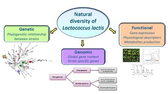
3. Lactococcus lactis: Multiple Levels of Diversity

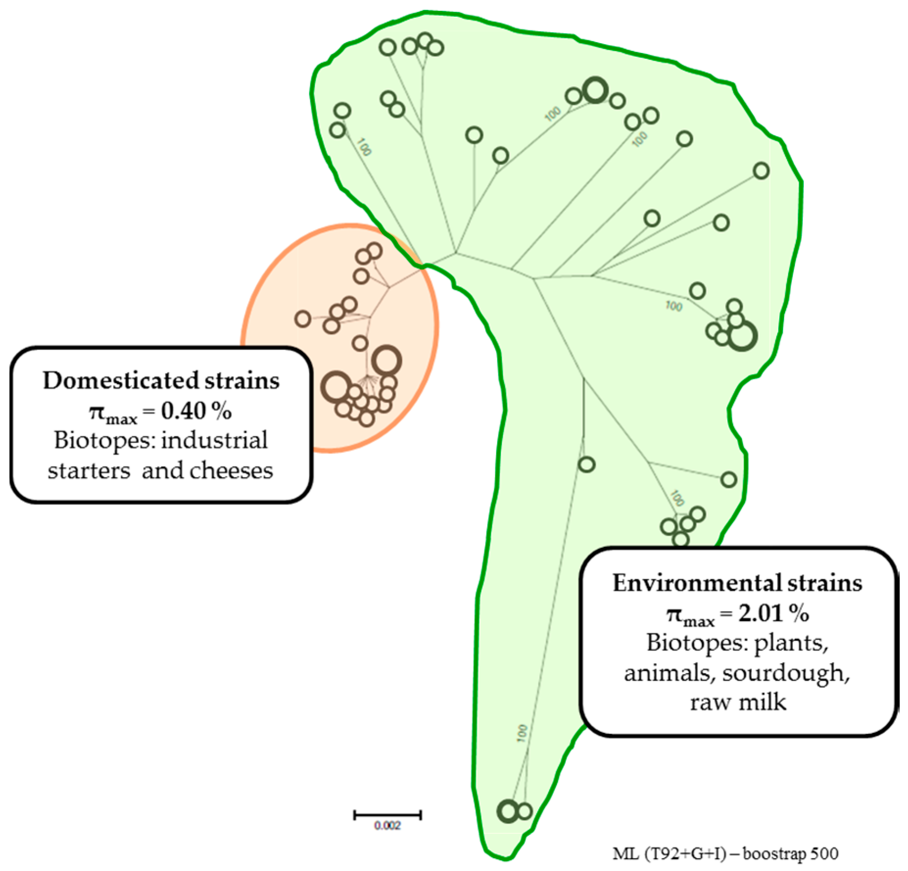
3.1. Genetic Structure of L. lactis

The development of next generation sequencing (NGS) technologies has provided numerous complete or draft lactococcal genomes. The sequence of the L. lactis subsp. lactis IL1403 strain was released in 2001 [36]. Of the 83 L. lactis sequences available on public databases, 50 were published in 2015 and 2016 (Table 1). However, a robust phylogenetic analysis of the species from the core genome derived from these data has not yet been conducted. Up to now, the extent of genetic diversity has been explored by Multi Locus Sequence Typing (MLST) studies. MLST is a powerful technique based on the sequencing of a limited number of genes in the core genome [37]. It provides information on the population structure, the long-term epidemiology and the evolutionary history of the species. Indeed, the concatenated sequences of the set of genes represent one “signature” of the core genome and are used in phylogenetic analysis.
Several MLST studies were conducted with different sets of strains from both subspecies and from different origins. Rademaker et al. [38] used a subset of 89 strains of the two subspecies lactis and cremoris of dairy and non-dairy origin. Five genes were used for phylogenetic analysis. This revealed two major, distinct genomic lineages within the species. These lineages did not correlate with the phenotypic characterization of the two subspecies but did correlate with the genotypic identification. One genomic lineage consists of the lactis genotype and phenotype strains including biovar Diacetylactis. The second lineage encompasses isolates with the cremoris genotype but with the two Cremoris and Lactis phenotypes, underlining the fact that only identifying the genotype reflects the evolutionary history. However, this analysis was unable to differentiate the strains based on their origins. The same results were obtained by Fermandez et al. [39] using the same MLST scheme but a different set of strains (mainly isolated from traditional cheeses and raw milk). To increase the discriminatory potential of this method, Xu et al. [40] analyzed a partial sequence of 12 genes from 197 L. lactis strains isolated from natural homemade yogurt. As was the case in the previous studies, the two major lineages corresponding to the two subspecies were revealed. Despite the increased number of genes, the genetic distance between the two subspecies did not enable the distinction of clusters within the lineages. As suggested, these two subspecies could be considered as two different species, but the method lacks accuracy to distinguish the L. lactis species considered as a whole. A more precise phylogenetic study has been proposed for the L. lactis subsp. lactis [41]. The πMAX, defined as maximum nucleotide diversity, was chosen as an indicator of the diversity within the subspecies because it is not directly sensitive to the size of the sample. A phylogenetic tree was constructed from the concatenated sequences of the six loci targeted in this new MLST scheme. Two of them, glyA and recN, belonged to the gene set identified as the most reliable predictor of whole genome relatedness [42]. The computed πMAX for the subspecies lactis was 2.01%, a value within the range of values calculated for several species. The genetic structure clearly clustered the 36 L. lactis subsp. lactis strains of the study in two groups with different diversity level (Figure 1). The first group, with low genetic diversity, had a πMAX of 0.4%, and clustered strains isolated from dairy starters or fermented products and involved in industrial milk processing. These strains could be considered as “domesticated”. The second group, with high diversity, had a πMAX of 2.01%, and was identical to that of the whole subspecies. Clearly, it is the main contributor of the diversity of the subspecies with “environmental” strains isolated from various natural sources such as plants, animals and raw milk. According to the structure of the population described by this analysis, Passerini et al. [41] proposed classifying the subspecies lactis strains in “domesticated” versus “environmental” strains instead of dairy and non-dairy, as it is usually the case. The “domesticated” strains emerged more recently, probably due to the selective pressure of industrial processing. An alternative hypothesis would be that actual “domesticated” strains originate from very few strains isolated and used as commercial starters for standardized cheese production in the early 20th century. The “environmental” strains appeared first and their high genetic diversity explains their ubiquitous presence in various natural environments. The “environmental” status of strains isolated from raw milk reflects their plant origins.

3.2. Genomic Diversity

In addition to the allelic variation between genes conserved among strains (i.e., core genes), a second level of biodiversity related to the gene content shared by a few strains or specific to one strain (i.e., accessory genes) can be described. This accessory genome provides a given strain with wide adaptability and extending capacities such as the ability to colonize different ecological niches. The increasing amount of lactococcal genome sequences is undoubtedly a powerful tool to highlight these capacities. In the NCBI genome database (Table 1), the mean genome size of the two subspecies is quite similar (2504 kb ranging from 2245 kb to 2744 kb for the subspecies lactis and 2537 kb ranging from 2000 kb to 2862 kb for the subspecies cremoris). It should be noted that about a 30% difference between the smallest and the largest genomes has been found for the cremoris subspecies, indicating high fluctuation in strain-to-strain coding capacity. This difference is bigger than that found in the lactis subspecies (20%) and might reflect the large plasmid content in some strains of the cremoris subspecies. For a set of strains, the chromosome size has been obtained from the sequence data. The average chromosome sizes are 2491 kb and 2467 kb with about 10% to 15% difference between the extremes, for the subspecies lactis and cremoris, respectively. This variation in chromosomal length is similar to that found in natural isolates of E. coli [43,44] and classifies L. lactis among bacterial species with high genome diversity.
Similar results were obtained by Passerini et al. [41] in a Pulsed-Field Gel Electrophoresis (PFGE) study of 36 strains of the subspecies lactis isolated from diverse habitats. However, the distribution of chromosome sizes did not correlate with the MLST-based phylogeny. Indeed, marked differences in chromosome size were observed in both the domesticated and the environmental lineages. This suggests that “domestication” does not automatically reduce the genome in this subspecies as a consequence of its adaptation to growth in milk. In contrast, Kelly et al. [45] observed the influence of the origin of the strain on the length of the chromosome. This study comprised 80 strains and the smallest chromosome sizes were found in dairy strains, among which, those of the “true” subspecies cremoris (cremoris genotype and Cremoris phenotype) had the smallest chromosomes. This reductive chromosome evolution might be a consequence of adaptation of the subspecies cremoris to milk environments.
Plasmids contribute significantly to the genome content of L. lactis as they can account for 4.7% and 8.4% of the genome within the lactis and cremoris subspecies respectively. Recent sequence data from NCBI identified 96 completely sequenced lactococcal plasmids. Examination of the plasmid content of 150 dairy starters revealed a mean of seven plasmids per strain [45]. Genomic analysis of the L. lactis subsp. cremoris UC509.9 strain revealed that extrachromosomal DNA represents more than 200 kb distributed in eight plasmids [46]. In several cases, plasmids have been shown to be transmissible by conjugation, a natural process of DNA transfer that could be exploited in the dairy industry to improve strains by conferring new desirable phenotypes. In contrast, in non-dairy isolates, the average was only two plasmids per strain with larger plasmids (>10 kb) [45]. Plasmid-encoded genes can harbor important technological traits such as proteinase activity, lactose utilization, bacteriophage resistance and bacteriocin production [47,48,49,50,51,52]. Some enhance flavor via citrate utilization. The citP gene, which encodes a citrate permease involved in this pathway, is often located on a plasmid [53]. Glutamate dehydrogenase encoding genes are also of interest in the dairy industry. Indeed, this activity stimulates amino acid catabolism in LAB by supplying the 2-oxoglutarate required for amino acid transamination, which is the first step in the conversion of amino acid into aroma compounds. This gene has been observed on plasmid sequences derived from plant and raw milk isolates [54,55]. Natural plasmids harbor food-grade selectable markers, such as copper or cadmium resistance, which are the subject of considerable interest [49,54,56]. Besides these technological properties, plasmids appear to harbor genes that are beneficial for the colonization of specific niches. This is the case for exopolysaccharides (EPS), which play an important role in plant surface attachment and biofilm formation. Furthermore, the capacity to adhere to intestinal epithelial cells and mucins was described in the plant-derived strain TIL448 by Meyrand et al. [57] and Le et al. [58], respectively. A cluster of genes encoding typical genetic biosynthetic machinery for pili formation was found on a plasmid, thereby conferring adhesion and muco-adhesion capacity to the strain. Recently, a novel 12 kb plasmid pSH74 from NCDO 712 was found to contain a new type of pilus gene cluster. Overexpression of the pilus gene cluster led to the formation of appendices on the cell surface [56]. Although plasmid localization for useful properties can be considered as a particular benefit, since transfer via conjugation is possible, it should be kept in mind that this might be also a major disadvantage due to instability and easy loss of plasmids [59].

3.3. Functional Diversity

Lactococcus lactis is known to display a variety of phenotypes. This phenotypic richness provides a third level of diversity. Generally, there is no correlation between phenotype and genetic lineage and genetically closely related strains do not necessarily share the same phenotypes. For instance, the lacE gene, associated with the capacity to consume the lactose, is widely distributed among both domesticated and environmental strains of the subsp. lactis [41]. Likewise, diversity in robustness during heat or oxidative stress has been reported in 39 L. lactis strains isolated from diverse habitats [60] and does not appear to be related to a specific lactis or cremoris genotype. This phenotypic robustness is associated with the absence/presence of pattern genes in a collection of strains. For example, the presence of genes encoding a cellobiose transporter (yidB), a signal recognition particle receptor protein (ftsY) and two hypothetical proteins (ymgH and ymgI) were associated with the ability to survive oxidative stress. Similarly, the presence/absence of a gene encoding a manganese transporter (mtsC) was correlated with resistance of the strains to heat stress. A more exhaustive genomic analysis would have been able to explain this functional diversity, at least to a certain extent. Within the L. lactis subsp. lactis A12 and KF147 genomes, additional genes encode a range of functions related to the utilization of plant sugars [61,62]. In contrast to industrial dairy starters, these strains efficiently metabolize arabinose and raffinose, and the raffinose-metabolism associated pathway differs in the two strains (cf. Section 4.1).
Besides the presence/absence of genes (related to the accessory genome), inactivation or differential regulation of conserved genes may be the basis of phenotypic diversity. To compare the biochemical properties of 20 strains belonging either to lactis or cremoris subspecies, Fernandez et al. [39] identified the enzymatic activities of 20 enzymes using the API ZYM and API 20 Strep systems (bioMérieux, Montalieu-Vercieu, France). For all of the strains, a lack of activity was observed in 10 out of the 20 enzymes studied, reflecting either the absence of related genes or their inactivation. For the other enzymes, activity levels for a given enzyme varied considerably among strains and between genotypes. Overall, the level of activity of the cremoris genotype was higher than that of the lactis genotype. These results could be correlated to a more extensive analysis of strain-specific variations in the activities of the enzymes [63]. Eighty-four L. lactis strains from diverse origins were chosen to quantify the specific activity of five enzymes known for their impact on flavor formation (aminopeptidase N,X-propyl-dipeptidyl aminopeptidase, branched-chain aminotransferase, hydroxyisocaproic acid dehydrogenase and esterase). Two types of media were used to assess the extent of conservation of the regulatory mechanisms between closely related strains. The authors defined the environment-dependent and strain-specific variations of enzymes activities as “the regulatory phenotype”. This term encompasses the cumulative effects of key mechanisms including transcription, translation or any other allosteric factors that influence enzymatic activity. The data revealed that four out of the five activities measured produced very diverse regulatory responses, clearly showing that regulation differed according to the environmental conditions in a strain-specific manner. This suggests very diverse regulatory characteristics in individual strains and highlights the possibility to reveal various phenotypes. Thus, in a phenotypic screening, the conditions used must be taken into account, and, in addition, conditions used should be as close as possible to those encountered in the process of interest.
The multi-phenotypes expressed by a strain in a specific environment may be a way to differentiate genetically closely related strains. To this end, Dhaisne et al. [64] selected 82 variables as important dairy features, including physiological indicators of the milking process (growth, acidification) and extracellular metabolites, some of which are involved in flavor. These authors tested the variables in nine L. lactis subsp. lactis strains belonging to the domesticated group with low genetic diversity and the ability to grow in milk. Twenty variables were identified as phenotypic markers that would make it possible to clearly discriminate between strains and to demonstrate their phenotypic uniqueness in this environment. These phenotypic markers were linked to glycolysis, proteolysis and lipolysis, three metabolic pathways involved in flavor production, and highlight the strain-dependent regulation of these pathways.

4. From Genome to Phenotype: Original Functions Explained Using an Integrated Approach

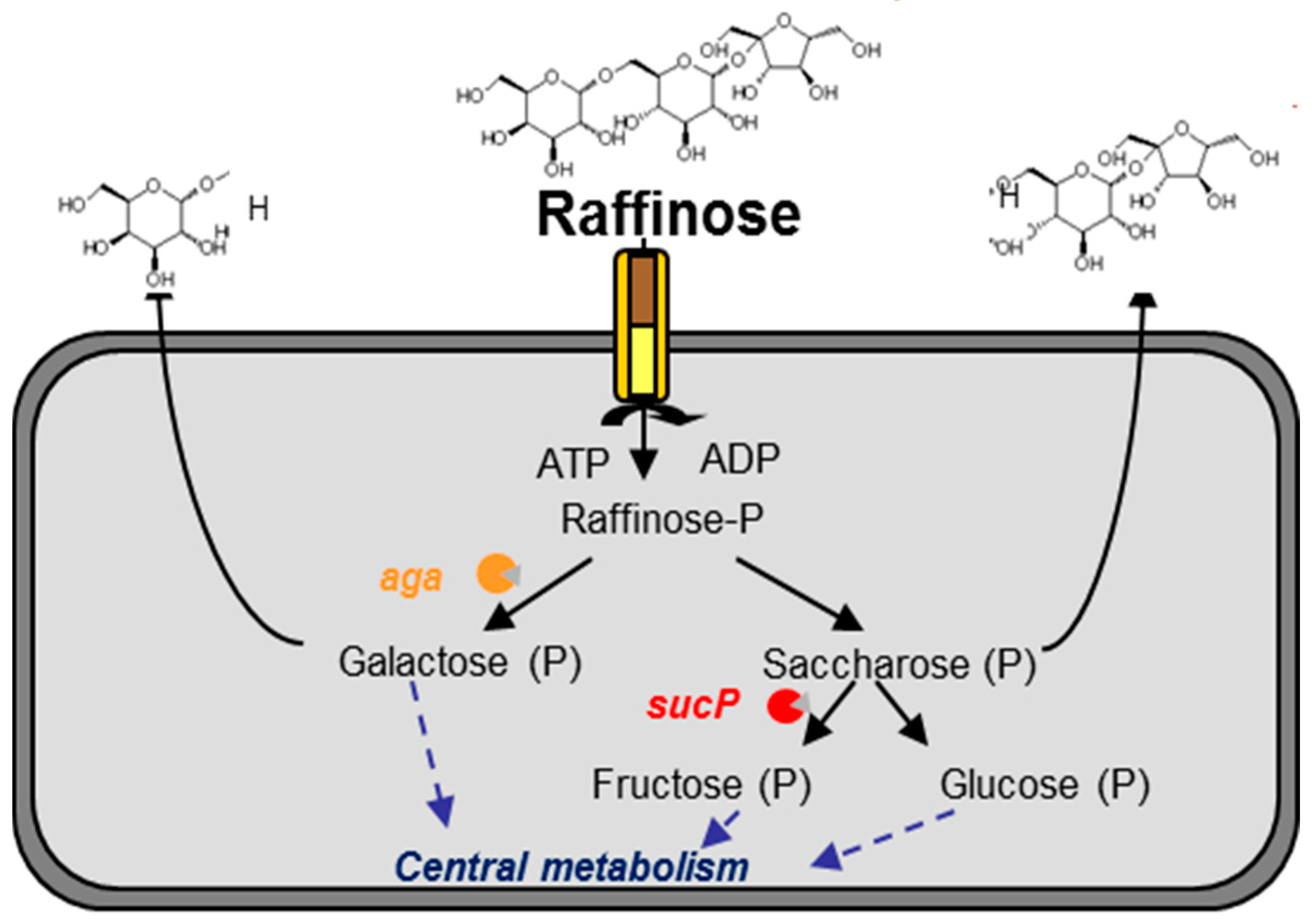
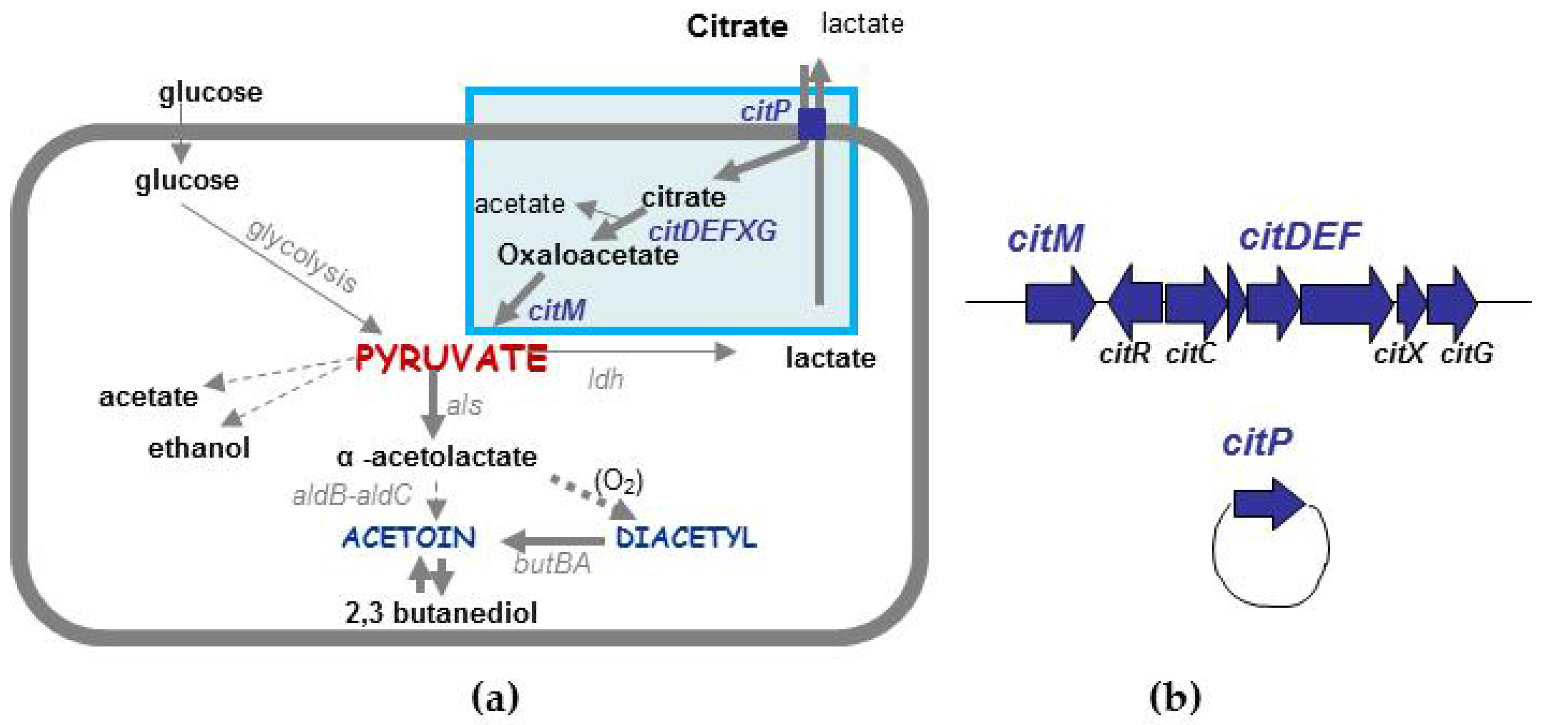
4.1. Range of Raffinose Metabolism

The original habitat of L. lactis is believed to be plants because environmental strains have the capacity to metabolize many plant-derived carbohydrates while the domesticated ones cannot. Several studies have highlighted the ability of environmental strains to use arabinose, xylose, maltose, galacturonate and α-galactosides including melibiose, stachyose and raffinose [39,48]. In the case of raffinose metabolism, genetic features associated with this phenotype are strain dependent. Indeed, the two environmental strains KF147 and A12 metabolize raffinose in two different ways. In the genome of the KF147 strain, a gene cluster for α-galactoside uptake, breakdown and D-galactose conversion via the Leloir pathway has been described: fbp-galR-aga-galK-galT-purH-agaRCBA-sucP [65]. Part of this α-galactoside gene cluster, fbp-galR-aga-galK-galT, closely resembles (90 to 94% nucleotide identity) that of Lactococcus raffinolactis ATCC 43920 [66], a species that naturally degrades raffinose. The cluster is located on a 51 kb transposon, which could be transferred to the MG1363 strain via conjugation, conferring the capacity to use α-galactosides [67]. In contrast, in the A12 strain, genes related to raffinose metabolism differ from those in KF147 [18]. Firstly, these genes are duplicated on the genome, one copy being hosted by a 42 kb plasmid and the other by a 69 kb plasmid. Secondly, α-galactosidase (aga) and sucrose phosphorylase (sucP) are present but only share, respectively, 52% and 65% nucleotide identity with those of KF147, suggesting an xenologous origin. Thirdly, a putative transporter has been described upstream of these genes. The original structure of the transporter, corresponding to a translational fusion of permease and kinase domains, differs from that of the putative raffinose ABC transporter (encoded by agaA, agaB and agaC in the KF147 strain) and the putative PTS system in the L. raffinolactis ATCC 43920.
Genomic analysis associated with physiological data allowed the A12 raffinose pathway to be partly elucidated (Figure 2): the transporter is hypothesized to manage both the uptake and the phosphorylation of raffinose. According to this hypothesis, raffinose is cleaved into galactose and saccharose by α-galactosidase. Only 50% of galactose is hypothesized to be consumed by the cell via the Leloir pathway, the remaining 50% being excreted into the medium. Saccharose is more efficiently used by the cell than galactose since only 30% is released into the medium and 70% would be intracellularly cleaved into fructose and glucose by the sucrose phosphorylase. If the physiological data clearly demonstrated the existence of the α-1,6 hydrolysis of raffinose, further investigations are required to propose a potential β-1,2 hydrolysis of raffinose into melibiose and fructose. This particular metabolism would confer a competitive advantage to this strain and enable trophic links with other members of the natural ecosystem.

4.2. Different Types of Diacetyl/Acetoin Production

Thanks to their creamy and buttery flavor notes, diacetyl and acetoin are essential components of dairy products. In L. lactis subsp. lactis, aroma production is associated with the capacity to metabolize citrate, and diacetyl production is proportional to citrate consumption in aerobiosis [68]. The Diacetylactis biovar encompasses strains that have this pathway and this metabolism has been exhaustively described in the dairy strain CRL264 isolated from cheese [69]. Citrate is transported by the plasmid-encoded citrate permease CitP, while genes encoding its intracellular metabolism are located in a large chromosomal cluster (Figure 3b). After its uptake in the cell, citrate is cleaved into acetate and oxaloacetate by the citrate lyase (CitDEF) and its auxiliary proteins (CitC, CitX and CitG). Oxaloacetate is subsequently decarboxylated to pyruvate by the oxaloacetate decarboxylase, CitM. Citrate utilization leads to the accumulation of pyruvate that can be rerouted to two alternative pathways: one generates acetate and/or ethanol and formate, the other generates diacetyl and acetoin. In the second case, two molecules of pyruvate are condensed into α-acetolactate, which is either converted into diacetyl (spontaneous oxydative decarboxylation) or into acetoin (Figure 3a) [70]. In the dairy industry, to rapidly assess the potential of a strain for the production of aroma compounds, citrate utilization is investigated, either by growth on the Kempler and McKay (KMK) medium [71] or by PCR amplification of genes related to the citrate pathway, such as citP. Indeed, the citrate plasmid is systematically associated with the presence of the chromosomal cluster [72]. These strains are mostly clustered in the “domesticated” ecotype with low genetic diversity, limiting the diversification of starters. Using an integrated approach, Passerini et al. [72] demonstrated that most of both domesticated and environmental strains can produce diacetyl/acetoin. This expands the extent of the biovar Diacetylactis. Depending on the rate of pyruvate synthesis, the kinetics and the amounts of aroma compounds differ among strains. The presence of the citrate pathway, which actually delineates the Diacetylactis biovar, is related to the rapid accumulation of aroma. In such a case, the name “Citrate” biovar might be more appropriate. Other inefficient-citrate-consuming strains can produce as much aroma but through a slower metabolism. In this case, production depends on their glucose fermenting capacity and pyruvate rerouting towards fermentation end products and is strain-dependent, suggesting different modes of regulation. Thus, only considering genomic features does not fully account for the aromatic potential of a strain. Revealing metabolic differences would be easier by analyzing phenotypes than by analyzing the subtle genomic differences, likely responsible for metabolic heterogeneity.

5. Technical and Specific Properties of Environmental Strains for New Applications

The diversity of phenotypes expressed by L. lactis, and the technological traits associated with environmental strains could be exploited in dairy fermentation. In addition to acidification, the diversity of metabolic pathways and their end-products such as volatile compounds [23] can be used for the development of new starters with original flavor profiles. From a food safety point of view, many environmental strains produce bacteriocins and bacteriocin-like compounds [73].
Emerging evidence suggests that transient food-borne bacteria play a significant role in host health and gut microbiota, as recently illustrated for L. lactis [74]. The authors hypothesized that the ingested strain L. lactis CNCM I-1631 could either grow in vivo, adhere to the intestinal wall, or both. To support the second option, the functionality of L. lactis cell wall proteins was assessed in vivo using a ∆srtA mutant [74]. Consistent with these data, the presence of various gene clusters associated with pili biogenesis, their efficient expression—for instance—in the plant TIL448 strain [57] and the ability to adhere to mucin, also conferred by the joint expression of a mucus-binding protein [58], reinforce lactococcal adhesion as a pivotal factor in transient persistence of L. lactis in the gut. Intestinal growth of L. lactis may also be a key parameter for increased fitness in the intestine. It requires carbon sources such as mucin-derived carbohydrates and particularly N-acetylglucosamine and mannose, which are made available in the gut distal part by commensal mucus degrading bacteria and are then appropriate sugars for exogenously applied bacteria [75]. In mono-associated mice, L. lactis was shown to colonize and thrive in the digestive tract, notably through a shift in the gut distal part in lactococcal metabolism from lactose catabolism to N-acetylglucosamine and the utilization of mannose [76]. In line with these findings, 151 strains from diverse origins and belonging to the lactis and cremoris subspecies were screened for their ability to degrade mucin-derived carbohydrates, including fucose, galactose, N-acetylglucosamine, N-acetylgalactosamine and mannose (vs. lactose and glucose). Interestingly, 10% and 90% of strains were able to metabolize N-acetylgalactosamine and galactose, respectively, and none were able to grow on fucose and 100% efficiently degraded N-acetylglucosamine and mannose (unpublished data). Moreover, using the same L. lactis collection, a wide range of gamma-aminobutyric acid (GABA) production was observed (unpublished data). GABA, a product of glutamate decarboxylation by the glutamic acid decarboxylase, has positive effects on human health such as reducing blood pressure [77,78], psychological stress reducing action [79], and modulating renal function [80]. These phenotypic features associated with technological traits provide a challenging basis for exploiting selected L. lactis strains in the development of health-promoting dairy products enriched in GABA.

6. Conclusions

The purpose of this review was to highlight the natural diversity of L. lactis. If phenotypic differences extensively contribute to this diversity, genetic and genomic variability provide an additional level of diversity that is of primary importance in the development of starters. Indeed, mixing strains with different genotypic characteristics will ensure that the many potentialities encoded by the “pan-genome” of the species are covered. Even though some genetic features are not expressed under specific process conditions, they may be of relevance for other processes or applications. To assess this overall diversity, the use of a multi-scale integrated approach spanning from genotype to phenotype is indispensable.

Acknowledgments

This work was financially supported in part via the BLaMI project (Bactéries Lactiques et MICI) by Syndifrais-CNIEL (Syndicat National des Fabricants de Produits Frais/Centre National Interprofessionnel de l’Economie Laitière) (Paris, France).

Conflicts of Interest

The authors declare no conflict of interest.

References

  1. Meucci, A.; Zago, M.; Rossetti, L.; Fornasari, M.E.; Bonvini, B.; Tidona, F.; Povolo, M.; Contarini, G.; Carminati, D.; Giraffa, G. Lactococcus hircilactis sp. nov. and Lactococcus laudensis sp. nov., isolated from milk. Int. J. Syst. Evol. Microbiol. 2015, 65, 2091–2096. [Google Scholar] [CrossRef] [PubMed]
  2. Bali, V.; Panesar, P.S.; Bera, M.B.; Kennedy, J.F. Bacteriocins: Recent trends and potential applications. Crit. Rev. Food Sci. Nutr. 2016, 56, 817–834. [Google Scholar] [CrossRef] [PubMed]
  3. Guinane, C.M.; Cotter, P.D.; Hill, C.; Ross, R.P. Microbial solutions to microbial problems; lactococcal bacteriocins for the control of undesirable biota in food. J. Appl. Microbiol. 2005, 98, 1316–1325. [Google Scholar] [CrossRef] [PubMed]
  4. Mazzoli, R.; Bosco, F.; Mizrahi, I.; Bayer, E.A.; Pessione, E. Towards lactic acid bacteria-based biorefineries. Biotechnol. Adv. 2014, 32, 1216–1236. [Google Scholar] [CrossRef] [PubMed]
  5. Schleifer, K.H.; Kraus, J.; Dvorak, C.; Kilpper-Bälz, R.; Collins, M.D.; Fischer, W. Transfer of Streptococcus lactis and related streptococci to the genus Lactococcus gen. nov. Syst. Appl. Microbiol. 1985, 6, 183–195. [Google Scholar] [CrossRef]
  6. Pérez, T.; Balcázar, J.L.; Peix, A.; Valverde, A.; Velázquez, E.; de Blas, I.; Ruiz-Zarzuela, I. Lactococcus lactis subsp. tructae subsp. nov. isolated from the intestinal mucus of brown trout (Salmo trutta) and rainbow trout (Oncorhynchus mykiss). Int. J. Syst. Evol. Microbiol. 2011, 61, 1894–1898. [Google Scholar] [CrossRef] [PubMed]
  7. Ward, L.J.H.; Brown, J.C.S.; Davey, G.P. Two methods for the genetic differentiation of Lactococcus lactis ssp. lactis and cremoris based on differences in the 16S rRNA gene sequence. FEMS Microbiol. Lett. 1998, 166, 15–20. [Google Scholar] [CrossRef] [PubMed]
  8. Pu, Z.Y.; Dobos, M.; Limsowtin, G.K.Y.; Powell, I. Integrated polymerase chain reaction-based procedures for the detection and identification of species and subspecies of the Gram-positive bacterial genus Lactococcus. J. Appl. Microbiol. 2002, 93, 353–361. [Google Scholar] [CrossRef] [PubMed]
  9. Godon, J.-J.; Delorme, C.; Ehrlich, S.D.; Renault, P. Divergence of genomic sequences between Lactococcus lactis subsp. lactis and Lactococcus lactis subsp. cremoris. Appl. Environ. Microbiol. 1992, 58, 4045–4047. [Google Scholar] [PubMed]
  10. Corroler, D.; Desmasures, N.; Gueguen, M. Correlation between polymerase chain reaction analysis of the histidine biosynthesis operon, randomly amplified polymorphic DNA analysis and phenotypic characterization of dairy Lactococcus isolates. Appl. Microbiol. Biotechnol. 1999, 51, 91–99. [Google Scholar] [CrossRef] [PubMed]
  11. Beimfohr, C.; Ludwig, W.; Schleifer, K.-H. Rapid genotypic differentiation of Lactococcus lactis subspecies and biovar. Syst. Appl. Microbiol. 1997, 20, 216–221. [Google Scholar] [CrossRef]
  12. Nomura, M.; Kobayashi, M.; Okamoto, T. Rapid PCR-based method which can determine both phenotype and genotype of Lactococcus lactis subspecies. Appl. Environ. Microbiol. 2002, 68, 2209–2213. [Google Scholar] [CrossRef] [PubMed]
  13. Sanders, J.W.; Leenhouts, K.; Burghoorn, J.; Brands, J.R.; Venema, G.; Kok, J. A chloride-inducible acid resistance mechanism in Lactococcus lactis and its regulation. Mol. Microbiol. 1998, 27, 299–310. [Google Scholar] [CrossRef] [PubMed]
  14. Richter, M.; Rossello-Mora, R. Shifting the genomic gold standard for the prokaryotic species definition. Proc. Natl. Acad. Sci. USA 2009, 106, 19126–19131. [Google Scholar] [CrossRef] [PubMed]
  15. Cavanagh, D.; Casey, A.; Altermann, E.; Cotter, P.D.; Fitzgerald, G.F.; McAuliffe, O. Evaluation of Lactococcus lactis iolates from non-dairy sources with potential dairy applications reveals extensive phenotype-genotype disparity and implications for a revised species. Appl. Environ. Microbiol. 2015, 81, 3961–3972. [Google Scholar] [CrossRef] [PubMed]
  16. Cavanagh, D.; Fitzgerald, G.F.; McAuliffe, O. From field to fermentation: The origins of Lactococcus lactis and its domestication to the dairy environment. Food Microbiol. 2015, 47, 45–61. [Google Scholar] [CrossRef] [PubMed]
  17. Wegmann, U.; O’Connell-Motherway, M.; Zomer, A.; Buist, G.; Shearman, C.; Canchaya, C.; Ventura, M.; Goesmann, A.; Gasson, M.J.; Kuipers, O.P.; et al. Complete genome sequence of the prototype lactic acid bacterium Lactococcus lactis subsp. cremoris MG1363. J. Bacteriol. 2007, 189, 3256–3270. [Google Scholar] [CrossRef] [PubMed]
  18. Passerini, D.; Coddeville, M.; Le Bourgeois, P.; Loubière, P.; Ritzenthaler, P.; Fontagné-Faucher, C.; Daveran-Mingot, M.-L.; Cocaign-Bousquet, M. The carbohydrate metabolism signature of Lactococcus lactis strain A12 reveals its sourdough ecosystem origin. Appl. Environ. Microbiol. 2013, 79, 5844–5852. [Google Scholar] [CrossRef] [PubMed]
  19. Robert, H.; Gabriel, V.; Fontagné-Faucher, C. Biodiversity of lactic acid bacteria in French wheat sourdough as determined by molecular characterization using species-specific PCR. Int. J. Food Microbiol. 2009, 135, 53–59. [Google Scholar] [CrossRef] [PubMed]
  20. Kelly, W.J.; Davey, G.P.; Ward, L.J. Characterization of lactococci isolated from minimally processed fresh fruit and vegetables. Int. J. Food Microbiol. 1998, 45, 85–92. [Google Scholar] [CrossRef]
  21. Minervini, F.; Celano, G.; Lattanzi, A.; Tedone, L.; Mastro, G.D.; Gobbetti, M.; Angelis, M.D. Lactic acid bacteria in durum wheat flour are endophytic components of the plant during its entire life cycle. Appl. Environ. Microbiol. 2015, 81, 6736–6748. [Google Scholar] [CrossRef] [PubMed]
  22. Procopio, R.E.L.; Arajo, W.L.; Maccheroni, W., Jr.; Azevedo, J.L. Characterization of an endophytic bacterial community associated with Eucalyptus spp. Genet. Mol. Res. 2009, 8, 1408–1422. [Google Scholar] [CrossRef] [PubMed]
  23. Alemayehu, D.; Hannon, J.A.; McAuliffe, O.; Ross, R.P. Characterization of plant-derived lactococci on the basis of their volatile compounds profile when grown in milk. Int. J. Food Microbiol. 2014, 172, 57–61. [Google Scholar] [CrossRef] [PubMed]
  24. Serna Cock, L.; Rodriguez de Stouvenel, A. Lactic acid production by a strain of Lactococcus lactis subsp. lactis isolated from sugar cane plants. Electron. J. Biotechnol. 2006, 9, 40–45. [Google Scholar] [CrossRef]
  25. Golomb, B.L.; Marco, M.L. Lactococcus lactis metabolism and gene expression during growth on plant tissues. J. Bacteriol. 2015, 197, 371–381. [Google Scholar] [CrossRef] [PubMed]
  26. De Lacerda, J.R.M.; da Silva, T.F.; Vollú, R.E.; Marques, J.M.; Seldin, L. Generally recognized as safe (GRAS) Lactococcus lactis strains associated with Lippia sidoides Cham. are able to solubilize/mineralize phosphate. SpringerPlus 2016, 5, 828. [Google Scholar] [CrossRef] [PubMed]
  27. Gao, Y.; Lu, Y.; Teng, K.-L.; Chen, M.-L.; Zheng, H.-J.; Zhu, Y.-Q.; Zhong, J. Complete genome sequence of Lactococcus lactis subsp. lactis CV56, a probiotic strain isolated from the vagina of healthy women. J. Bacteriol. 2011, 193, 2886–2887. [Google Scholar] [CrossRef] [PubMed]
  28. Plumed-Ferrer, C.; Gazzola, S.; Fontana, C.; Bassi, D.; Cocconcelli, P.-S.; von Wright, A. Genome sequence of Lactococcus lactis subsp. cremoris Mast36, a strain isolated from bovine mastitis. Genome Announc. 2015, 3, e00449-15. [Google Scholar] [CrossRef] [PubMed]
  29. Tormo, H.; Ali Haimoud Lekhal, D.; Roques, C. Phenotypic and genotypic characterization of lactic acid bacteria isolated from raw goat milk and effect of farming practices on the dominant species of lactic acid bacteria. Int. J. Food Microbiol. 2015, 210, 9–15. [Google Scholar] [CrossRef] [PubMed]
  30. RMT Fromages de Terroir Group. The Microbiology of Raw Milk. Towards a Better Understanding of the Microbial Ecosystems of Milk and the Factors that Affect Them; Bronwen Percival: London, UK, 2015; ISBN 978-0993419409. [Google Scholar]
  31. Mallet, A.; Guéguen, M.; Kauffmann, F.; Chesneau, C.; Sesboué, A.; Desmasures, N. Quantitative and qualitative microbial analysis of raw milk reveals substantial diversity influenced by herd management practices. Int. Dairy J. 2012, 27, 13–21. [Google Scholar] [CrossRef]
  32. Panoff, J.-M.; Desmasures, N.; Thammavongs, B.; Guéguen, M. In situ protection of microbiodiversity is under consideration. Microbiology 2002, 148, 625. [Google Scholar] [CrossRef] [PubMed]
  33. Salama, M.S.; Musafija-Jeknic, T.; Sandine, W.E.; Giovannoni, S.J. An ecological study of lactic acid bacteria: Isolation of new strains of Lactococcus including Lactococcus lactis subspecies cremoris. J. Dairy Sci. USA 1995, 78, 1004–1017. [Google Scholar] [CrossRef]
  34. Montel, M.-C.; Buchin, S.; Mallet, A.; Delbes-Paus, C.; Vuitton, D.A.; Desmasures, N.; Berthier, F. Traditional cheeses: Rich and diverse microbiota with associated benefits. Int. J. Food Microbiol. 2014, 177, 136–154. [Google Scholar] [CrossRef] [PubMed]
  35. Tormo, H.; Delacroix-Buchet, A.; Lopez, C.; Ali Haimoud Lekhal, D.; Roques, C. Farm management practices and diversity of the dominant bacterial species in raw goat’s milk. Int. J. Dairy Sci. 2011, 6, 29–43. [Google Scholar] [CrossRef]
  36. Bolotin, A.; Wincker, P.; Mauger, S.; Jaillon, O.; Malarme, K.; Weissenbach, J.; Ehrlich, S.D.; Sorokin, A. The complete genome sequence of the lactic acid bacterium Lactococcus lactis ssp. lactis IL1403. Genome Res. 2001, 11, 731–753. [Google Scholar] [CrossRef] [PubMed]
  37. Maiden, M.C.J.; Bygraves, J.A.; Feil, E.; Morelli, G.; Russell, J.E.; Urwin, R.; Zhang, Q.; Zhou, J.; Zurth, K.; Caugant, D.A.; et al. Multilocus sequence typing: A portable approach to the identification of clones within populations of pathogenic microorganisms. Proc. Natl. Acad. Sci. USA 1998, 95, 3140–3145. [Google Scholar] [CrossRef] [PubMed]
  38. Rademaker, J.L.W.; Herbet, H.; Starrenburg, M.J.C.; Naser, S.M.; Gevers, D.; Kelly, W.J.; Hugenholtz, J.; Swings, J.; van Hylckama Vlieg, J.E.T. Diversity analysis of dairy and non-dairy Lactococcus lactis isolates, using a novel multilocus sequence analysis scheme and (GTG)5-PCR fingerprinting. Appl. Environ. Microbiol. 2007, 73, 7128–7137. [Google Scholar] [CrossRef] [PubMed]
  39. Fernández, E.; Alegría, Á.; Delgado, S.; Martín, M.C.; Mayo, B. Comparative phenotypic and molecular genetic profiling of wild Lactococcus lactis subsp. lactis strains of the L. lactis subsp. lactis and L. lactis subsp. cremoris genotypes, isolated from starter-free cheeses made of raw milk. Appl. Environ. Microbiol. 2011, 77, 5324–5335. [Google Scholar] [CrossRef]
  40. Xu, H.; Sun, Z.; Liu, W.; Yu, J.; Song, Y.; Lv, Q.; Zhang, J.; Shao, Y.; Menghe, B.; Zhang, H. Multilocus sequence typing of Lactococcus lactis from naturally fermented milk foods in ethnic minority areas of China. J. Dairy Sci. 2014, 97, 2633–2645. [Google Scholar] [CrossRef] [PubMed]
  41. Passerini, D.; Beltramo, C.; Coddeville, M.; Quentin, Y.; Ritzenthaler, P.; Daveran-Mingot, M.-L.; Bourgeois, P.L. Genes but not genomes reveal bacterial domestication of Lactococcus lactis. PLoS ONE 2010, 5, e15306. [Google Scholar] [CrossRef] [PubMed]
  42. Zeigler, D.R. Gene sequences useful for predicting relatedness of whole genomes in bacteria. Int. J. Syst. Evol. Microbiol. 2003, 53, 1893–1900. [Google Scholar] [CrossRef] [PubMed]
  43. Lukjancenko, O.; Wassenaar, T.M.; Ussery, D.W. Comparison of 61 sequenced Escherichia coli genomes. Microb. Ecol. 2010, 60, 708–720. [Google Scholar] [CrossRef] [PubMed]
  44. Bergthorsson, U.; Ochman, H. Distribution of chromosome length variation in natural isolates of Escherichia coli. Mol. Biol. Evol. 1998, 15, 6–16. [Google Scholar] [CrossRef] [PubMed]
  45. Kelly, W.J.; Ward, L.J.H.; Leahy, S.C. Chromosomal diversity in Lactococcus lactis and the origin of dairy starter cultures. Genome Biol. Evol. 2010, 2, 729–744. [Google Scholar] [CrossRef] [PubMed]
  46. Ainsworth, S.; Zomer, A.; de Jager, V.; Bottacini, F.; van Hijum, S.A.F.T.; Mahony, J.; van Sinderen, D. Complete genome of Lactococcus lactis subsp. cremoris UC509.9, host for a model lactococcal P335 bacteriophage. Genome Announc. 2013, 1, e00119-12. [Google Scholar] [CrossRef] [PubMed]
  47. Mills, S.; McAuliffe, O.E.; Coffey, A.; Fitzgerald, G.F.; Ross, R.P. Plasmids of lactococci-genetic accessories or genetic necessities? FEMS Microbiol. Rev. 2006, 30, 243–273. [Google Scholar] [CrossRef] [PubMed]
  48. Siezen, R.J.; Bayjanov, J.R.; Felis, G.E.; van der Sijde, M.R.; Starrenburg, M.; Molenaar, D.; Wels, M.; van Hijum, S.A.F.T.; van Hylckama Vlieg, J.E.T. Genome-scale diversity and niche adaptation analysis of Lactococcus lactis by comparative genome hybridization using multi-strain arrays. Microb. Biotechnol. 2011, 4, 383–402. [Google Scholar] [CrossRef] [PubMed]
  49. Fallico, V.; Ross, R.P.; Fitzgerald, G.F.; McAuliffe, O. Novel conjugative plasmids from the natural isolate Lactococcus lactis subspecies cremoris DPC3758: a repository of genes for the potential improvement of dairy starters. J. Dairy Sci. 2012, 95, 3593–3608. [Google Scholar] [CrossRef] [PubMed]
  50. Siezen, R.J.; Renckens, B.; van Swam, I.; Peters, S.; van Kranenburg, R.; Kleerebezem, M.; de Vos, W.M. Complete sequences of four plasmids of Lactococcus lactis subsp. cremoris SK11 reveal extensive adaptation to the dairy environment. Appl. Environ. Microbiol. 2005, 71, 8371–8382. [Google Scholar] [CrossRef] [PubMed]
  51. Ainsworth, S.; Stockdale, S.; Bottacini, F.; Mahony, J.; van Sinderen, D. The Lactococcus lactis plasmidome: Much learnt, yet still lots to discover. FEMS Microbiol. Rev. 2014, 38, 1066–1088. [Google Scholar] [CrossRef] [PubMed]
  52. Górecki, R.K.; Koryszewska-Bagińska, A.; Gołębiewski, M.; Żylińska, J.; Grynberg, M.; Bardowski, J.K. Adaptative potential of the Lactococcus lactis IL594 strain encoded in its 7 plasmids. PLoS ONE 2011, 6, e22238. [Google Scholar] [CrossRef] [PubMed]
  53. Sesma, F.; Gardiol, D.; de Ruiz Holgado, A.P.; de Mendoza, D. Cloning of the citrate permease gene of Lactococcus lactis subsp. lactis biovar Diacetylactis and expression in Escherichia coli. Appl. Environ. Microbiol. 1990, 56, 2099–2103. [Google Scholar] [PubMed]
  54. Tanous, C.; Chambellon, E.; Yvon, M. Sequence analysis of the mobilizable lactococcal plasmid pGdh442 encoding glutamate dehydrogenase activity. Microbiology 2007, 153, 1664–1675. [Google Scholar] [CrossRef] [PubMed]
  55. Fallico, V.; McAuliffe, O.; Fitzgerald, G.F.; Ross, R.P. Plasmids of raw milk cheese isolate Lactococcus lactis subsp. lactis biovar Diacetylactis DPC3901 suggest a plant-based origin for the strain. Appl. Environ. Microbiol. 2011, 77, 6451–6462. [Google Scholar] [CrossRef] [PubMed]
  56. Tarazanova, M.; Beerthuyzen, M.; Siezen, R.; Fernandez-Gutierrez, M.M.; de Jong, A.; van der Meulen, S.; Kok, J.; Bachmann, H. Plasmid complement of Lactococcus lactis NCDO712 reveals a novel pilus gene cluster. PLoS ONE 2016, 11, e0167970. [Google Scholar] [CrossRef] [PubMed]
  57. Meyrand, M.; Guillot, A.; Goin, M.; Furlan, S.; Armalyte, J.; Kulakauskas, S.; Cortes-Perez, N.G.; Thomas, G.; Chat, S.; Péchoux, C.; et al. Surface proteome analysis of a natural isolate of Lactococcus lactis reveals the presence of pili able to bind human intestinal epithelial cells. Mol. Cell. Proteom. 2013, 12, 3935–3947. [Google Scholar] [CrossRef] [PubMed]
  58. Le, D.T.L.; Tran, T.-L.; Duviau, M.-P.; Meyrand, M.; Guérardel, Y.; Castelain, M.; Loubière, P.; Chapot-Chartier, M.-P.; Dague, E.; Mercier-Bonin, M. Unraveling the role of surface mucus-binding protein and pili in muco-adhesion of Lactococcus lactis. PLoS ONE 2013, 8, e79850. [Google Scholar] [CrossRef] [PubMed]
  59. Wegmann, U.; Overweg, K.; Jeanson, S.; Gasson, M.; Shearman, C. Molecular characterization and structural instability of the industrially important composite metabolic plasmid pLP712. Microbiology 2012, 158, 2936–2945. [Google Scholar] [CrossRef] [PubMed]
  60. Dijkstra, A.R.; Setyawati, M.C.; Bayjanov, J.R.; Alkema, W.; van Hijum, S.A.F.T.; Bron, P.A.; Hugenholtz, J. Diversity in robustness of Lactococcus lactis strains during heat stress, oxidative stress, and spray drying stress. Appl. Environ. Microbiol. 2014, 80, 603–611. [Google Scholar] [CrossRef] [PubMed]
  61. Guellerin, M.; Passerini, D.; Fontagné-Faucher, C.; Robert, H.; Gabriel, V.; Loux, V.; Klopp, C.; Loir, Y.L.; Coddeville, M.; Daveran-Mingot, M.-L.; et al. Complete genome sequence of Lactococcus lactis subsp. lactis A12, a strain isolated from wheat sourdough. Genome Announc. 2016, 4, e00692-16. [Google Scholar] [CrossRef] [PubMed]
  62. Siezen, R.J.; Bayjanov, J.; Renckens, B.; Wels, M.; van Hijum, S.A.F.T.; Molenaar, D.; van Hylckama Vlieg, J.E.T. Complete genome sequence of Lactococcus lactis subsp. lactis KF147, a plant-associated lactic acid bacterium. J. Bacteriol. 2010, 192, 2649–2650. [Google Scholar] [CrossRef] [PubMed]
  63. Bachmann, H.; Starrenburg, M.J.C.; Dijkstra, A.; Molenaar, D.; Kleerebezem, M.; Rademaker, J.L.W.; van Hylckama Vlieg, J.E.T. Regulatory phenotyping reveals important diversity within the species Lactococcus lactis. Appl. Environ. Microbiol. 2009, 75, 5687–5694. [Google Scholar] [CrossRef] [PubMed]
  64. Dhaisne, A.; Guellerin, M.; Laroute, V.; Laguerre, S.; Cocaign-Bousquet, M.; Le Bourgeois, P.; Loubiere, P. Genotypic and phenotypic analysis of dairy Lactococcus lactis biodiversity in milk: Volatile organic compounds as discriminating markers. Appl. Environ. Microbiol. 2013, 79, 4643–4652. [Google Scholar] [CrossRef] [PubMed]
  65. Siezen, R.J.; Starrenburg, M.J.C.; Boekhorst, J.; Renckens, B.; Molenaar, D.; van Hylckama Vlieg, J.E.T. Genome-scale genotype-phenotype matching of two Lactococcus lactis Isolates from plants identifies mechanisms of adaptation to the plant niche. Appl. Environ. Microbiol. 2008, 74, 424–436. [Google Scholar] [CrossRef] [PubMed]
  66. Boucher, I.; Vadeboncoeur, C.; Moineau, S. Characterization of genes involved in the metabolism of α-galactosides by Lactococcus raffinolactis. Appl. Environ. Microbiol. 2003, 69, 4049–4056. [Google Scholar] [CrossRef] [PubMed]
  67. Machielsen, R.; Siezen, R.J.; van Hijum, S.A.F.T.; van Hylckama Vlieg, J.E.T. Molecular description and industrial potential of Tn6098 conjugative transfer conferring alpha-galactoside metabolism in Lactococcus lactis. Appl. Environ. Microbiol. 2011, 77, 555–563. [Google Scholar] [CrossRef] [PubMed]
  68. Aymes, F.; Monnet, C.; Corrieu, G. Effect of alpha-acetolactate decarboxylase inactivation on alpha-acetolactate and diacetyl production by Lactococcus lactis subsp. lactis biovar Diacetylactis. J. Biosci. Bioeng. 1999, 87, 87–92. [Google Scholar] [CrossRef]
  69. García-Quintáns, N.; Repizo, G.; Martín, M.; Magni, C.; López, P. Activation of the diacetyl/acetoin pathway in Lactococcus lactis subsp. lactis bv. Diacetylactis CRL264 by acidic growth. Appl. Environ. Microbiol. 2008, 74, 1988–1996. [Google Scholar] [CrossRef] [PubMed]
  70. Drider, D.; Bekal, S.; Prévost, H. Genetic organization and expression of citrate permease in lactic acid bacteria. Genet. Mol. Res. 2004, 3, 273–281. [Google Scholar] [PubMed]
  71. Kempler, G.M.; McKay, L.L. Improved medium for detection of citrate-fermenting Streptococcus lactis subsp. diacetylactis. Appl. Environ. Microbiol. 1980, 39, 926–927. [Google Scholar] [PubMed]
  72. Passerini, D.; Laroute, V.; Coddeville, M.; Le Bourgeois, P.; Loubière, P.; Ritzenthaler, P.; Cocaign-Bousquet, M.; Daveran-Mingot, M.-L. New insights into Lactococcus lactis diacetyl- and acetoin-producing strains isolated from diverse origins. Int. J. Food Microbiol. 2013, 160, 329–336. [Google Scholar] [CrossRef] [PubMed]
  73. Ayad, E.H.E.; Verheul, A.; Wouters, J.T.M.; Smit, G. Antimicrobial-producing wild lactococci isolated from artisanal and non-dairy origins. Int. Dairy J. 2002, 12, 145–150. [Google Scholar] [CrossRef]
  74. Zhang, C.; Derrien, M.; Levenez, F.; Brazeilles, R.; Ballal, S.A.; Kim, J.; Degivry, M.-C.; Quéré, G.; Garault, P.; van Hylckama Vlieg, J.E.T.; et al. Ecological robustness of the gut microbiota in response to ingestion of transient food-borne microbes. ISME J. 2016, 10, 2235–2245. [Google Scholar] [CrossRef] [PubMed]
  75. Fabich, A.J.; Jones, S.A.; Chowdhury, F.Z.; Cernosek, A.; Anderson, A.; Smalley, D.; McHargue, J.W.; Hightower, G.A.; Smith, J.T.; Autieri, S.M.; et al. Comparison of carbon nutrition for pathogenic and commensal Escherichia coli strains in the mouse intestine. Infect. Immun. 2008, 76, 1143–1152. [Google Scholar] [CrossRef] [PubMed]
  76. Roy, K.; Meyrand, M.; Corthier, G.; Monnet, V.; Mistou, M.-Y. Proteomic investigation of the adaptation of Lactococcus lactis to the mouse digestive tract. Proteomics 2008, 8, 1661–1676. [Google Scholar] [CrossRef] [PubMed]
  77. Inoue, K.; Shirai, T.; Ochiai, H.; Kasao, M.; Hayakawa, K.; Kimura, M.; Sansawa, H. Blood-pressure-lowering effect of a novel fermented milk containing γ-aminobutyric acid (GABA) in mild hypertensives. Eur. J. Clin. Nutr. 2003, 57, 490–495. [Google Scholar] [CrossRef] [PubMed]
  78. Hayakawa, K.; Kimura, M.; Kasaha, K.; Matsumoto, K.; Sansawa, H.; Yamori, Y. Effect of a γ-aminobutyric acid-enriched dairy product on the blood pressure of spontaneously hypertensive and normotensive Wistar–Kyoto rats. Br. J. Nutr. 2004, 92, 411. [Google Scholar] [CrossRef] [PubMed]
  79. Nakamura, H.; Takishima, T.; Kometani, T.; Yokogoshi, H. Psychological stress-reducing effect of chocolate enriched with γ-aminobutyric acid (GABA) in humans: Assessment of stress using heart rate variability and salivary chromogranin A. Int. J. Food Sci. Nutr. 2009, 60, 106–113. [Google Scholar] [CrossRef] [PubMed]
  80. Kim, H.Y.; Yokozawa, T.; Nakagawa, T.; Sasaki, S. Protective effect of γ-aminobutyric acid against glycerol-induced acute renal failure in rats. Food Chem. Toxicol. 2004, 42, 2009–2014. [Google Scholar] [CrossRef] [PubMed]
Figure 1. The two phylogenetic groups of the Lactococcus lactis subsp. lactis. The unrooted maximum likelihood tree (bootstrap 500, Tamura 3-parameter model) was constructed from the concatenated sequences of the six loci of MLST scheme from [41]. Open circles correspond to the different sequence type (ST). The size of the circles is proportional to the number of strains.
Microorganisms 05 00027 g001
Figure 2. Putative raffinose metabolism in the environmental L. lactis subsp. lactis A12 strain.
Microorganisms 05 00027 g002
Figure 3. Main contributors to diacetyl/acetoin production. (a) pathways involved in citrate metabolism and aroma production. Pyruvate is a key intermediate. (b) chromosomal citrate operon and plasmidic citP gene involved in citrate transport.
Microorganisms 05 00027 g003
Table 1. Eighty-three available Lactococcus lactis genomes. The data was collected from National Center for Biotechnology Information (NCBI); accessed 18 January 2017 (https://www.ncbi.nlm.nih.gov/genome/genomes/156?); N. D.: not determined
StrainSubspeciesDateGenome Size (Mb)Chrom. Size (Mb)Number of PlasmidsProteinIsolation Source
IL1403lactis20012.365592.3655902277Cheese starter culture
KF147lactis20092.635652.5981412445Mung bean sprouts
CNCM I-1631lactis20112.51133 N.D.2403Fermented milk
CV56lactis20112.518742.3994652378Vaginal flora
IO-1lactis20122.421472.4214702229Water in kitchen sink drain pit
Dephy 1lactis20132.60355 N.D.2459N.D.
KLDS 4.0325lactis20132.595492.5892532448Homemade koumiss
LD61lactis bv. diacetylactis20132.59924 N.D.2490Starter culture for dairy fermentation
TIFN2lactis bv. diacetylactis20132.50507 N.D.2296Cheese starter
TIFN4lactis bv. diacetylactis20132.55039 N.D.2349Cheese starter
YF11lactis20132.52731 N.D.2328Dairy
511lactis20142.48081 N.D.2304N.D.
1AA59lactis20142.57654 N.D.2406Artisanal cheese
AI06lactis20142.398092.3980902178Acai pulp
Bpl1lactis20142.3057 N.D.2092Wild flies
CECT 4433lactis20142.57915 N.D.2290Cheese
GL2lactis bv. diacetylactis20142.33892 N.D.2135Dromedary milk
NCDO 2118lactis20142.592262.554612382Frozen peas
S0lactis20142.48872.488702311Fresh raw milk
ATCC 19435lactis20152.54729 N.D.2373Dairy starter
CRL264lactis bv. diacetylactis20152.57372 N.D.2446Cheese
DPC6853lactis20152.50715 N.D.2116Corn
E34lactis20152.37566 N.D.2217Silage
K231lactis20152.33604 N.D.2178White kimchii
K337lactis20152.44552 N.D.2263White kimchii
KF134lactis20152.4634 N.D.2282Alfalfa and radish sprouts
KF146lactis20152.57452 N.D.2408Alfalfa and radish sprouts
KF196lactis20152.44589 N.D.2282Japanese kaiwere shoots
KF201lactis20152.37639 N.D.2222Sliced mixed vegetables
KF24lactis20152.61922 N.D.2483Alfalfa sprouts
KF282lactis20152.65125 N.D.2471Mustard and cress
KF67lactis20152.6843 N.D.2514Grapefruit juice
KF7lactis20152.36676 N.D.2209Alfalfa sprouts
Li-1lactis20152.47593 N.D.2303Grass
LMG 7760lactis20152.24545 N.D.2072N.D.
LMG 14418lactis20152.41093 N.D.2275Bovine milk
LMG 8520lactis20152.43558 N.D.2060Leaf hopper
LMG 8526lactis20152.47749 N.D.2304Chinese radish seeds
LMG 9446lactis20152.4884 N.D.2324Frozen peas
LMG 9447lactis20152.70754 N.D.2552Frozen peas
M20lactis20152.67432 N.D.2535Soil
ML8lactis20152.52187 N.D.2373Dairy starter
N42lactis20152.74392 N.D.2540Soil and grass
NCDO895lactis20152.47306 N.D.2319Dairy starter
UC317lactis20152.49842 N.D.2357Dairy starter
A12lactis20162.730622.603942487Sourdough
DRA4lactis bv. diacetylactis20162.45755 N.D.2283Dairy starter
JCM 7638lactis20162.39386 N.D.-N.D
Ll1596lactis20162.39296 N.D.2237Teat canal
NBRC 100933lactis20162.54762 N.D.2406N.D
RTB018lactis20162.48665 N.D.2168Intestinal content of rainbow trout
NBRC 100931hordniae20162.42828 N.D.2079Leaf hopper
SK11cremoris20062.598352.4385952412Dairy
MG1363cremoris20072.529482.5294802400Dairy
NZ9000cremoris20102.530292.5302902404Dairy
A76cremoris20112.57712.4526242382Cheese production
UC509.9cremoris20122.457352.2504382188Irish Dairy
KW2cremoris20132.427052.4270502223Fermented corn
TIFN1cremoris20132.67978 N.D.2285Cheese starter
TIFN3cremoris20132.72521 N.D.2291Cheese starter
TIFN5cremoris20132.54151 N.D.2232Cheese starter
TIFN6cremoris20132.59151 N.D.2334Cheese starter
TIFN7cremoris20132.63409 N.D.2505Cheese starter
A17cremoris20142.67994 N.D.2367Taiwan fermented cabbage
GE214cremoris20142.80103 N.D.2603Cheese
HP(T)cremoris20142.26951 N.D.2042Mixed strain dairy starter culture
DPC6856cremoris20152.86238 N.D.2606Bovine rumen
DPC6860cremoris20152.60744 N.D.2261Grass
Mast36cremoris20152.60534 N.D.2414Milk from a cow with mastitis
AM2cremoris20162.48157 N.D.2254Dairy starter
B40cremoris20162.49846 N.D.2220Dairy starter
FG2cremoris20162.58614 N.D.2260Dairy starter
HPcremoris20162.39396 N.D.2132Dairy starter
IBB477cremoris20162.850352.6421752653Raw milk
KW10cremoris20162.36102 N.D.2177Kaanga Wai
LMG 6897cremoris20162.3672 N.D.2101Cheese starter
N41cremoris20162.61571 N.D.2410Soil and grass
NBRC 100676cremoris20162.34409 N.D.2093N.D.
NCDO763cremoris20162.48569 N.D.2331Dairy starter
P7266cremoris20162.00015 N.D.1984Litter on pastures
SK110cremoris20162.46761 N.D.2241Dairy starter
V4cremoris20162.54895 N.D.2344Raw sheep milk
WG2cremoris20162.54251 N.D.2306Cheese

