Article

Squalicorax Chips a Tooth: A Consequence of Feeding-Related Behavior from the Lowermost Navesink Formation (Late Cretaceous: Campanian-Maastrichtian) of Monmouth County, New Jersey, USA

by
Martin A. Becker
1 and
John A. Chamberlain, Jr.
2,*
1
Department of Environmental Science, William Paterson University, Wayne, NJ 07470, USA
2
Department of Earth & Environmental Sciences, Brooklyn College, Brooklyn, NY 11210, USA
*
Author to whom correspondence should be addressed.
Geosciences 2012, 2(2), 109-129; https://doi.org/10.3390/geosciences2020109
Submission received: 6 March 2012 / Revised: 20 April 2012 / Accepted: 23 May 2012 / Published: 30 May 2012
(This article belongs to the Special Issue Paleontology and Geo/Biological Evolution)

Abstract

:
Chipped and broken functional teeth are common in modern sharks with serrated tooth shape. Tooth damage consists of splintering, cracking, and flaking near the cusp apex where the enameloid is broken and exposes the osteodentine and orthodentine. Such damage is generally viewed as the result of forces applied during feeding as the cusp apex impacts the skeletal anatomy of prey. Damage seen in serrated functional teeth from sharks Squalicorax kaupi [1] and Squalicorax pristodontus [1] from the late Cretaceous lowermost Navesink Formation of New Jersey resembles that occurring in modern sharks and suggests similar feeding behavior. Tumbling experiments using serrated modern and fossil functional shark teeth, including those of Squalicorax, show that teeth are polished, not cracked or broken, by post-mortem abrasion in lowermost Navesink sediment. This provides further evidence that chipped and broken Squalicorax teeth are feeding-related and not taphonomic in origin. Evolution of rapid tooth replacement in large sharks such as Squalicorax ensured maximum functionality after feeding-related tooth damage occurred. Serrated teeth and rapid tooth replacement in the large sharks of the Mesozoic and Cenozoic afforded them competitive advantages that helped them to achieve their place as apex predators in today’s ocean.

1. Introduction

Sharks with serrated teeth commonly number among the largest apex predators in modern marine ecosystems. This is particularly true for white sharks, Carcharodon carcharias [2], bull sharks, Carcharhinus leucas [3], and tiger sharks, Galeocerdo cuvier [4] which can achieve lengths up to 6 meters and weights in excess of 2000 kilograms [5,6,7,8,9]. Such large sharks with serrated tooth shape are known to feed upon a range of prey including: marine mammals, turtles, birds, large osteichthyans and other chondrichthyans [10,11,12,13,14,15,16,17,18,19].
Sharks with serrated tooth shape can apply enormous bite forces during feeding [20,21,22,23,24]. As a result, such sharks commonly break teeth in the functional position during feeding. Examination of jaws from large, modern sharks with serrated teeth often reveals teeth in the functional position with splintering, cracking and flaking along the cusp apices. These damaged teeth are rapidly replaced, usually in less than one week, by new teeth, which we refer to here as pre-functional teeth, developing in files within the epithelial tissue and attached to the dental membrane [25,26,27,28]. Pre-functional teeth continuously rotate in conveyor belt fashion into the functional position at the forward edge of the jaw. This insures maximum tooth sharpness and overall jaw functionality as pristine new teeth replace old functional teeth broken during feeding.
In the fossil record, recovery of shark jaws with teeth is an exceedingly rare event due to the poor preservation potential of cartilaginous jaw anatomy [29,30,31]. However, teeth are comprised of extremely durable and highly insoluble biogenic apatite and are continuously replaced throughout a shark’s lifetime [32,33]. This replacement process produces hundreds, even thousands of teeth, from a single shark with a high probability of some becoming fossils. While missing tooth elements may have their origin in taphonomic processes, in many instances splintering, and cracks and chips in tooth cusps in fossil teeth bear striking similarities to those seen in morphologically similar modern functional teeth still contained in the jaws and thus not solely subject to taphonomic processes or fossilization.
One well-known Upper Cretaceous shark with serrated teeth and global distribution is the genus Squalicorax [34]. To date, at least five North American species of Squalicorax have been identified on the basis of teeth associated with preserved jaw cartilage, and from artificial tooth sets, i.e., from dental series reconstructed from the teeth of many different individuals. The most common Campanian-Maastrichtian species are assigned to Squalicorax kaupi [1] and Squalicorax pristodontus [1,32,35,36,37,38]. Many details of skeletal anatomy and paleoecology of Squalicorax have been worked out over the last decade from skeletons as well as teeth directly and indirectly associated with prey [35,39]. Prey items are sometimes large animals with robust skeletal anatomy [39,40,41].
In this study, we analyze tooth cusp damage seen in an assemblage of Squalicorax kaupi and Squalicorax pristodontus from the lowermost Navesink Formation in Monmouth County, New Jersey. We demonstrate from this assemblage that: (1) isolated teeth that occupied the functional position in Squalicorax kaupi and Squalicorax pristodontus can be distinguished from pre-functional teeth that were developing in an individual tooth file; (2) modern shark jaws with serrated functional teeth have feeding-related damage along their cusp apices similar to that seen in some Squalicorax kaupi and Squalicorax pristodontus functional teeth; (3) taphonomic damage simulated by tumbling experiments on serrated modern functional teeth as well as Squalicorax kaupi and Squalicorax pristodontus functional teeth is random and can be distinguished from feeding-related damage; (4) damage seen in Squalicorax kaupi and Squalicorax pristodontus functional teeth is the product of feeding behavior; and, (5) similar cusp damage to Squalicorax kaupi and Squalicorax pristodontus functional teeth occurs in other chondrichthyan fossil localities in North America and is also feeding-related.

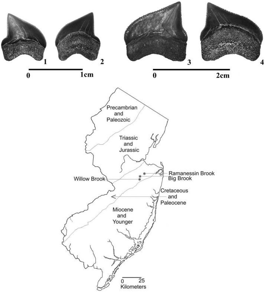
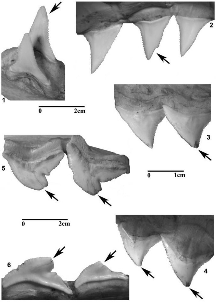
2. Teeth of Squalicorax

Teeth from the anacoracid Squalicorax are abundant and well-known from the Cenomanian through the Maastrichtian in North America [33,39,42,43,44,45,46,47,48,49,50,51]. The blade-like tooth shape, serrated edges, anaulacorhizous, bilobate root and absence of cusplets readily distinguishes teeth belonging to Squalicorax from other Cretaceous chondrichthyans. Figure 1 shows examples of pristine lateral teeth from Squalicorax kaupi and Squalicorax pristodontus from the lowermost Navesink Formation of Monmouth County, New Jersey, which we utilized in this study.
Figure 1. Squalicorax kaupi and Squalicorax pristodontus functional teeth (upper panel) and location of Ramanessin, Big and Willow Brooks, Monmouth County, New Jersey (lower panel). 1 and 2, Squalicorax kaupi; 3 and 4, Squalicorax pristodontus. Differences between Squalicorax kaupi and Squalicorax pristodontus are based on the well-defined distal notch seen in Squalicorax kaupi and the overall larger size of Squalicorax pristodontus. Scale bars: 1,2 = 1.0 cm and 3,4 = 2.0 cm. Tooth orientations: 1,3 = labial face; 2,4 = lingual face.
Geosciences 02 00109 g001
Previous studies compare the similarities of Squalicorax to the modern tiger shark Galeocerdo cuvier, particularly the serrated mesial and distal edges of their teeth and vertebral centra which lack radial lamellae [35,39]. While these features suggest a carcharhiniform evolutionary history for Squalicorax, tooth histology from this genus is that of an osteodont, and consistent with teeth belonging to lamniform sharks [52]. Both modern carcharhiniforms and lamniforms are known have rapid rates of tooth replacement and by analogy to fossil jaw examples this is also thought to have occurred in Squalicorax [35,53,54,55]. Reconstructions from naturally occurring and artificial tooth sets demonstrate that both these species are monognathic homodonts [31,33,35,36]. Differences between Squalicorax kaupi and Squalicorax pristodontus are subtle and based primarily on the well-defined distal notch seen in Squalicorax kaupi and the overall larger size of Squalicorax pristodontus (See Figure 1).

3. Materials and Methods

3.1. Modern Jaws and Teeth

Fifty modern shark jaws with associated teeth belonging to Carcharodon carcharias, Carcharhinus leucas and Galeocerdo cuvier were examined in the collection of the Department of Ichthyology of the American Museum of Natural History (AMNH). These species were selected based on serrated tooth shape that is generally similar to Squalicorax kaupi and Squalicorax pristodontus. Additionally, both these modern and fossil species of sharks are known to prey upon animals with robust skeletal anatomy. Similar prey should ideally result in equivalent damage to tooth cusps in functional teeth during feeding.
Four varieties of functional tooth cusp damage were identified among these fifty modern jaws: (A) labial damage on the tooth cusp apex; (B) labial damage on the mesial edge; (C) lingual damage on the tooth cusp apex; and, (D) lingual damage on the mesial edge (Figure 2). In some cases, observed functional tooth damage includes combinations of these four different varieties as seen on both the labial and lingual cusp faces. It is important to note that no developing (pre-functional teeth within individual tooth files) in these modern jaws had tooth cusp damage of the four varieties listed above. The majority of developing teeth along with their roots are protected within the epithelial jaw tissue from direct impact with prey during feeding.
Teeth in the functional position have fully-developed, bilobate roots, convex lingual cusp faces and concave labial cusp faces. The root and cusp are also well-mineralized, thick, and the serrations well-defined along the mesial and distal edges. Developing teeth lack all these features, particularly the well-developed, bilobate roots and convex lingual cusp faces. We also note that the roots in developing teeth are only partially permineralized and appear lighter in color than fully developed teeth. Developing teeth are lost upon the death of the animal or in extreme cases of feeding related injury. Table 1 lists the number of teeth in these categories and upon which this study is based.
Figure 2. Modern shark jaws displaying functional teeth with feeding-related damage of the following types: (A) labial damage on the tooth cusp apex; (B) labial damage on the mesial or distal tooth edge; (C) lingual damage on the tooth cusp apex; and (D) lingual damage on the mesial or distal edge. 1, labial view of an anterior tooth in the lower jaw of Carcharodon carcharias (AMNH 53095) with type A cusp damage; 2, lingual view of a lateral tooth in the upper jaw of Carcharodon carcharias (AMNH 53095) with type D cusp damage; 3, labial view of a lateral tooth in the upper jaw of Carcharhinus leucas (AMNH 225868D) with type A cusp damage; 4, lingual view of a lateral tooth in the upper jaw of Carcharhinus leucas (AMNH 225868D) with type C cusp damage; 5, labial view of a lateral tooth in the lower jaw of Galeocerdo cuvier (AMNH 37950SD) with type A and B cusp damage; 6, labial view of a lateral tooth in the upper jaw of Galeocerdo cuvier (AMNH 37950SD) with type A and B cusp damage; Scale bars for 1,2,5,6 = 2.0 cm; 3,4 = 1.0 cm.
Geosciences 02 00109 g002
Table 1. Sample populations of S. kaupi and S. pristodontus functional and pre-functional teeth from the lowermost Navesink Formation. Individual teeth are designated as functional or pre-functional according to the degree of cusp and root development as described in the text.
Species Functional TeethPre-Functional TeethIndeterminate or Broken During CollectingTotal Number of Teeth Studied
UndamagedFeeding DamageTaphonomic DamageUndamagedPathologically Deformed1
Squalicorax kaupi1873616144842371928
Squalicorax pristodontus132030326187477
Note: 1. as defined in [56].

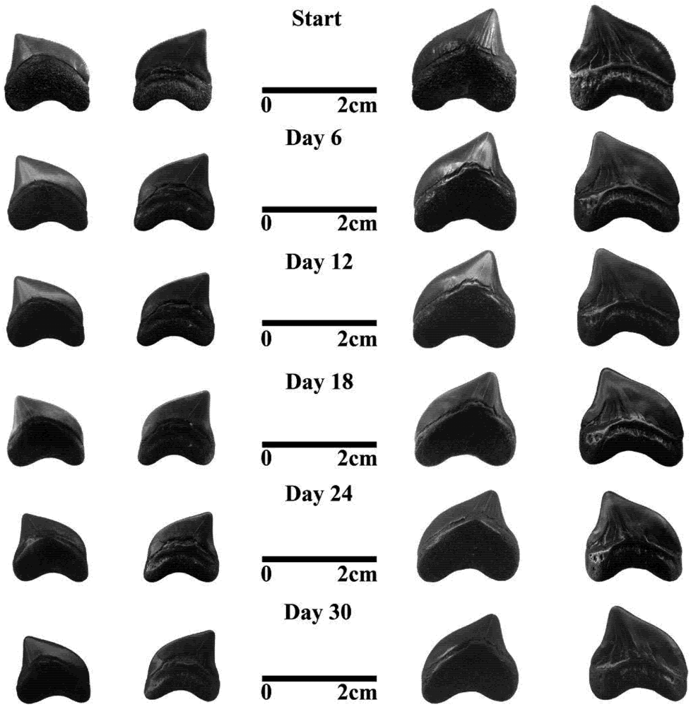
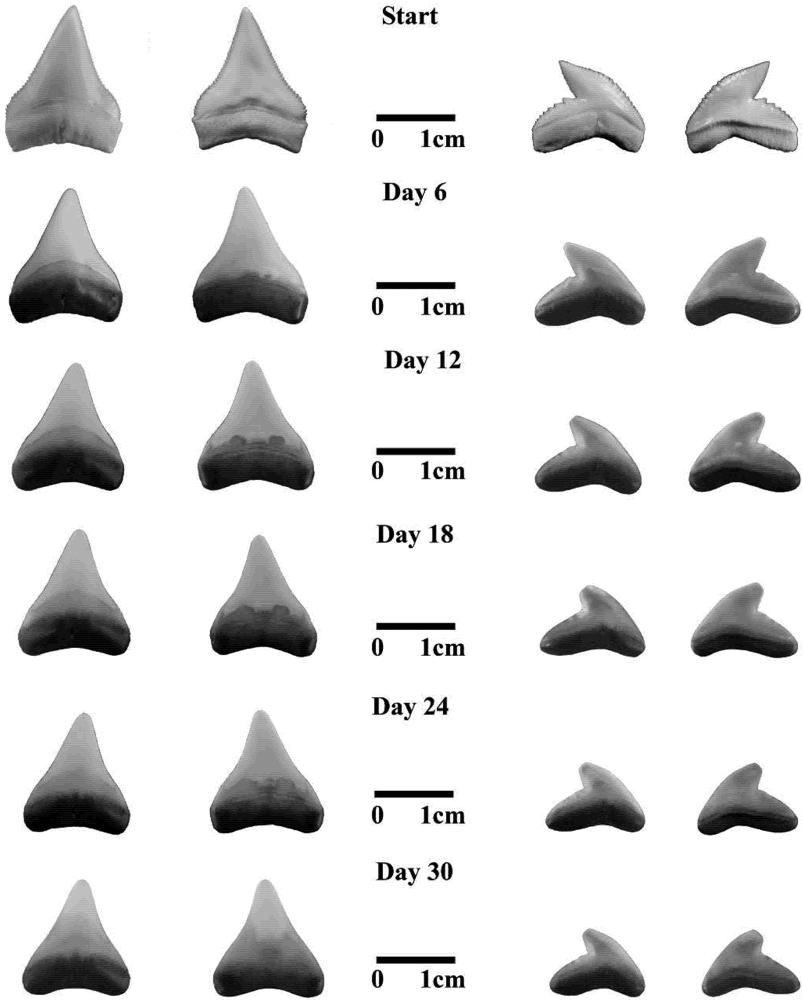
3.2. Fossil Teeth and Tumbler Experiments
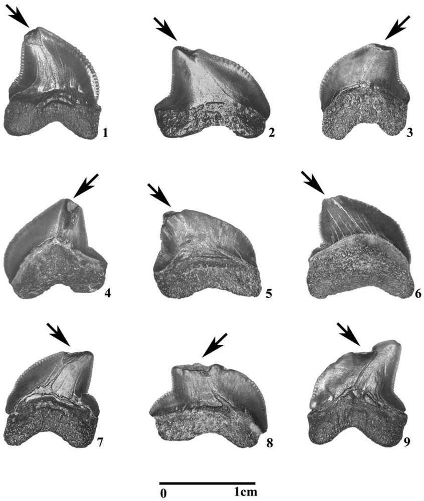
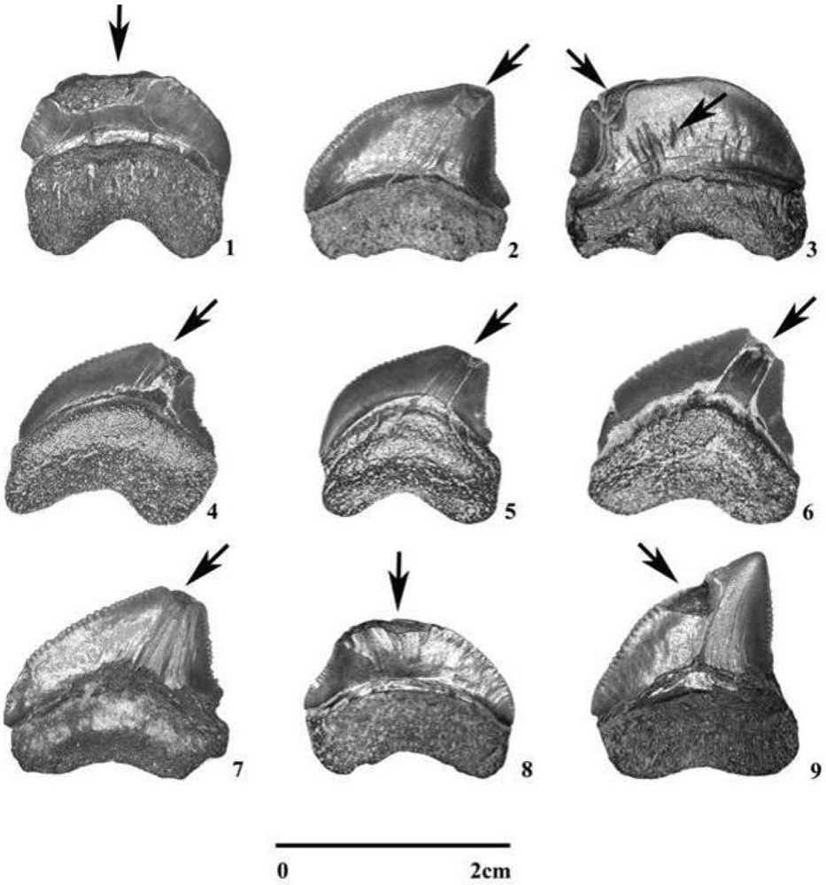
Squalicorax kaupi and Squalicorax pristodontus teeth utilized in this study are housed in the William Paterson University (WPU) paleontological collections. These teeth were recovered from a lag deposit that drapes a disconformity and comprises the lowermost Navesink Formation [57] along Ramanessin, Big and Willow Brooks, Monmouth County, New Jersey. This lag consists of silty, micaceous marl with uncommon quartz pebbles up to 2.0 centimeters in diameter and ranges from 10–30 centimeters thick. Such lag deposits are common in the Late Cretaceous of North America and represent complex taphonomic histories that include multiple episodes of exhumation and reburial associated with sea level cyclicity [45,49,58,59].
An industrial grade rock tumbler was used to simulate taphonomic effects and distinguish them from feeding-related damage among functional teeth of Squalicorax kaupi and Squalicorax pristodontus. Although pre-functional teeth were also exposed to similar taphonomic effects, these teeth are not exposed directly to feeding-related impact along their cusp apices and were excluded from further consideration in our taphonomic simulation experiments. Five pristine, functional teeth of Squalicorax kaupi and Squalicorax pristodontus respectively were placed in 5.0 kg of sediment from the lowermost Navesink Formation and water was added to cover the top of the sediment. Teeth were tumbled for one month and removed at six day intervals for inspection and photography. The same procedure was utilized for five pristine, functional teeth from each of the three modern test species: Carcharodon carcharias, Carcharhinus leucas and Galeocerdo cuvier. All specimens were taken from WPU paleontological collections.

4. Results

4.1. Sample Populations

Details of Squalicorax kaupi and Squalicorax pristodontus tooth sample population are compiled in Table 1. Of the 1926 Squalicorax kaupi teeth analyzed, 239 teeth were determined to occupy the functional position and 1448 teeth were determined to be pre-functional based on the degree of cusp and root development. Of the 477 Squalicorax pristodontus teeth analyzed, 63 teeth were determined to occupy the functional position and 326 teeth were determined to be pre-functional based on the degree of cusp and root development using the criteria described above.
Teeth broken during field excavation and sieving along with those where not enough of the cusp and root were preserved to determine the exact location in the file constituted 12% of our total Squalicorax kaupi sample population or 237 teeth, and 18% of our total Squalicorax pristodontus sample population or 87 teeth. Figure 3 and Figure 4 show nine Squalicorax kaupi and nine Squalicorax pristodontus functional teeth respectively that display damage analogous to that seen in the modern AMNH jaws and determined to be feeding-related. This sample population also contained four pathological Squalicorax kaupi teeth and one pathological Squalicorax pristodontus tooth with highly deformed cusps and roots similar to pathological teeth from the Navesink Formation that we described earlier [53].
Figure 3. Feeding related damage in Squalicorax kaupi teeth from the lowermost Navesink; Formation, Monmouth County, New Jersey. Types A and B damage: 13, 5, 8 and 9; Types C and D damage: 4, 6 and 7. Scale bar: 1–9 = 1.0 cm. Tooth orientations: 1–3, 5, 8, 9 show labial face. 4, 6, 7 show lingual face.
Geosciences 02 00109 g003
Figure 4. Feeding related damage in Squalicorax pristodontus teeth from the lowermost Navesink Formation, Monmouth County, New Jersey. Types A and B damage: 13, 79; Types C and D damage: 46. Scale bar: 1–9 = 2.0 cm. Tooth orientations: 1–3, 7–9 show labial face; 4–6 show lingual face. Note in 3 additional tooth damage on labial cusp face.
Geosciences 02 00109 g004

4.2. Tumbling Experiments

The results of tumbling experiments conducted on Squalicorax kaupi and Squalicorax pristodontus functional teeth are shown in Figure 5. After six days of tumbling, serrated edges of the teeth of both Squalicorax kaupi and Squalicorax pristodontus were completely removed.
This was followed by thinning and polishing of the enameloid and root on both the lingual and labial cusp faces. Near the cusp base after one month of tumbling, the enameloid was completely removed exposing the osteodentine and orthodentine, particularly on the labial cusp face. Squalicorax kaupi and Squalicorax pristodontus lost 14.4 and 14.2% of their original mass after one month of tumbling, respectively (Table 2). No splintering, cracking and flaking along the tooth cusp similar to that seen in the modern shark jaws illustrated in Figure 2, or in Squalicorax kaupi and Squalicorax pristodontus teeth (Figure 3 and Figure 4) were observed.
A similar pattern of material loss due to tumbling was observed in teeth of modern Carcharodon carcharias, Carcharhinus leucas, and Galeocerdo cuvier (Figure 6 and Figure 7). The teeth of these three sharks tested in the tumbler are clearly polished as indicated in Figure 7 and Figure 8, and show no indication of significant cracking, chipping, or other forms of breakage. One can also observe in Figure 6 and Figure 7 that the test specimens, particularly the roots, become darker in color as tumbling time increases. The roots of the modern teeth, and to a lesser extent the enameloid, incorporate iron staining from the host sediment in the tumbler almost immediately. At the end of one month, tooth roots and cusp edges were heavily iron-stained. Carcharodon, Carcharhinus and Galeocerdo teeth lost 42.6, 49.4 and 48.3 % of their original mass after one month of tumbling (Table 2). We attribute the smaller reduction in mass seen in the fossil teeth to increased durability associated with permineralization undergone by these teeth during fossilization and diagenesis.
Figure 5. Squalicorax kaupi and Squalicorax pristodontus functional teeth (lingual and labial views) during one month in an industrial tumbler with 5.0 kg of host sediment from the lowermost Navesink Formation. Individual teeth were removed at six day intervals and photographed. Note overall reduction in size, loss of serrations, thinning of enameloid, osteodentine and orthodentine.
Geosciences 02 00109 g005
Table 2. Results of tumbling experiments conducted on functional teeth of S. kaupi, S. pristodontus, C. carcharodon, C. leucas and G. cuvier. The average mass of five teeth from each species was used before and after tumbling in calculating the percentage of lost mass.
SPECIESMass Before Testing1 (gm)Mass After Testing2 (gm)% Loss3
FOSSILSqualicoras kaupi0.410.3514.6
Squalicorax pristodontus1.271.0914.2
MODERNCarcharodon carcharias2.491.4342.6
Carcharhinus leucas0.770.3949.4
Galeocerdo cuvier0.290.1543.3
Notes:1. Average mass of 5 teeth for each species prior to tumbling for 30 days;2. Average mass after tumbling for 30 days;3. Average loss of mass after tumbling for 30 days.
Figure 6. Carcharodon carcharias functional tooth (lingual and labial views) during one month in an industrial tumbler with 5.0 kg of host sediment from the lowermost Navesink Formation. Individual teeth were removed at six day intervals and photographed. All scale bars = 2.0 cm. Note iron staining in root and along cusp edges, and physical changes similar to those seen in Figure 5 and Figure 6.
Geosciences 02 00109 g006
Figure 7. Functional teeth of Carcharhinus leucas (left two columns) and Galeocerdo cuvier (right two columns) (lingual and labial views) during one month in an industrial tumbler with 5.0 kg of host sediment from the lowermost Navesink Formation. Individual teeth were removed at six day intervals and photographed. All scale bars =1.0 cm. Note iron staining in root and along cusp edges, and physical changes similar to those seen in Figure 5 and Figure 6.
Geosciences 02 00109 g007
Figure 8. Squalicorax kaupi tooth in labial view. 1, Labial feeding-related damage on the tooth cusp apex. (Type A damage); 2, 70X SEM close-up of cusp apex. Note rounded edges adjacent to missing enameloid and exposure of osteodentine and orthodentine. Scale bar: 1 = 1.0 cm; 2 = 1.0 mm.
Geosciences 02 00109 g008

5. Discussion

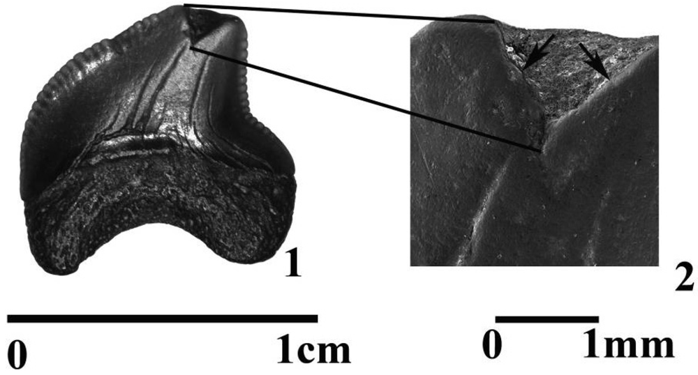
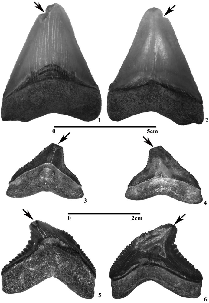
5.1. Taphonomic Versus Feeding-Related Tooth Damage

All Squalicorax kaupi and Squalicorax pristodontus teeth utilized in this study were recovered from a lag deposit and therefore must have experienced some taphonomic transport and reworking. The central focus of this paper is to determine whether such reworking is the cause of the tooth damage we observe, or if, in contrast, damage can be attributed primarily to feeding injury. During feeding, maximum force is concentrated in teeth along the point of impact, often the cusp apex. In loading tests this force rapidly dissipates across the mesial and distal cutting edges toward the root [23,24]. This concentrated force on cutting surfaces results in the type of repeated tooth breaks of cusp and cutting surfaces, seen in modern sharks with serrated tooth shape (see Figure 2) and is the product of the cusp apices impacting bony anatomy in prey [60,61,62,63]. Comparing the broken Squalicorax kaupi and Squalicorax pristodontus teeth in Figure 3 and Figure 4 to the feeding damaged teeth of our modern analogues illustrated in Figure 2, indicates that the characteristics of the breaks in the fossil teeth, and in particular their association with cusp apex and cutting edges, are essentially the same as those of feeding damaged teeth in the modern examples. Moreover, the results of the tumbler experiment illustrated in Figure 5, Figure 6 and Figure 7, show that both modern and fossil teeth are polished by abrasion in the tumbler; they do not suffer major breaks. Because the test teeth were tumbled with lowermost Navesink sediment, we infer that post-mortem transport and reworking were not a factor in producing the major breaks in the fossil teeth seen in Figure 4 and Figure 5. Based on these observations, we suggest that the broken, chipped, and cracked teeth discussed here are the result of damage induced during feeding.
When a tooth damaged in feeding falls out of the jaw it becomes a sedimentary clast. It can then undergo transport, reworking, and the associated abrasion that acts on all such materials. As a result, fossil chondrichthyan teeth can be expected to show both types of damage, i.e., major breaks associated with feeding, and rounding and smoothing of edges and surfaces associated with taphonomic abrasion. The interplay of these two modes of tooth damage should be visible in many, if not most, damaged teeth. This viewpoint is reinforced by examination of the cusp apex of a Squalicorax kaupi tooth using an EVEX-3000 Scanning Electron Microscope. The distinct break of this tooth (Figure 8.1) is similar to feeding damage seen in Squalicorax kaupi and Squalicorax pristodontus (Figure 3 and Figure 4) as well as in modern teeth (Figure 2). However, at 70× magnification (Figure 8.2) the broken enameloid tooth edges and ridges in the surface of the exposed osteodentine are observed to be strongly rounded. They are not sharp or angular as would be expected in a tooth freshly damaged during feeding. This suggests that the cusp apex was broken prior to taphonomic reworking and more than likely occurred during a feeding-related impact. Rounding of the exposed edges and surfaces occurred after the tooth was shed or its owner died and the tooth was released onto the substrate as a sedimentary particle.
The types of cusp breaks seen in Squalicorax kaupi and Squalicorax pristodontus and the modern jaws with serrated teeth from the AMNH can also be observed in Cenozoic sharks. Three Miocene-Recent serrated fossil teeth from the Peace River in Hardee County, Florida are shown in Figure 9. These teeth were eroded out of the river banks and incorporated with stream gravels along the base of the river. These Cenozoic teeth have cusp breaks along both the lingual and labial faces analogous to those seen in the feeding damaged teeth of Squalicorax kaupi and Squalicorax pristodontus (Figure 3 and Figure 4) and the modern AMNH teeth (Figure 2).
Figure 9. Fossil functional teeth displaying Types A–D feeding-related damage from the Bone Valley Formation (Miocene-Recent) Peace River, Hardee County, Florida, (WPU paleontological collections). 1 and 2, Carcharocles megalodon; 2 and 3, Carcharhinus leucas; 3 and 4, Galeocerdo cuvier. Scale bars: 1–4 = 5.0 cm; 5–6 = 2.0 cm. Tooth orientations-lingual face: 1, 3, 5. labial face: 2, 4, 6.
Geosciences 02 00109 g009

5.2. Modern Versus Fossil Functional Teeth

Examination of the jaws of modern Carcharodon carcharias, Carcharhinus leucas and Galeocerdo cuvier demonstrates that it would not be uncommon to find as many as 300 teeth in the lower and upper jaws of an individual animal of these species with approximately 50 teeth, or seventeen percent, in the functional position [5,60,61]. This percentage compares very favorably to the fossil teeth in our sample population, where 14% of Squalicorax kaupi teeth and 16% of Squalicorax pristodontus teeth, were determined to have been in the functional position when the animals from which they derive were alive. Teeth broken during field collection or those where location in the tooth file could not be determined, 12% and 18% for Squalicorax kaupi and Squalicorax pristodontus respectively, may slightly alter the percentage of functional teeth involved in this study. However, the addition of a few functional or pre-functional teeth to this sample population does not drastically compromise our sorting techniques and ability to recognize functional and pre-functional teeth when compared to modern analogues.
Damaged functional teeth in the modern AMNH jaws determined to be the product of feeding-related behavior range from a few percent to as much as 10%. Our fossil population was determined to have 15% of Squalicorax kaupi, and 32% of Squalicorax pristodontus, functional teeth broken due to feeding-related behavior. While the exact cause of this larger percentage of broken teeth relative to the modern AMNH jaws remains unknown at this time, one of the more interesting possibilities is that Squalicorax kaupi and Squalicorax pristodontus may have had slightly slower rates of tooth production and replacement. This slightly slower rate of tooth replacement would have resulted in a greater residence time for any broken functional tooth and a higher percentage of broken teeth in the jaws at any one time. Similar results for slower tooth replacement have been previously identified in some Late Paleozoic and Mesozoic sharks [64,65,66,67,68,69]. Evidence from these studies includes large wear facets related to feeding on the tooth cusps and suggested slower rates of tooth replacement occurred in these ancestral sharks [69].

5.3. Functional and Pre-Functional Teeth

By analogy to modern sharks, teeth in the functional position in the jaws of fossil sharks were fully formed, fully biomineralized and fully ready for use. These teeth would thus have been extremely hard and highly resistant to taphonomically induced abrasion occurring within the depositional environmental prior to final burial and fossilization. Pre-functional teeth further back in the tooth files of fossil forms, like such teeth in modern sharks, would have been less mineralized and comprised of smaller amounts of enameloid, osteodentine and orthodentine. Such teeth released by death into the sediment before they are fully formed and shed by a live animal would be far more susceptible to taphonomically induced abrasion. As a consequence of such differential resistance, pre-functional teeth would be preferentially destroyed as recognizable fossils faster and by less abrasion then functional teeth. In a general way, therefore, the fossil record of shark teeth should primarily be a record of functional and late pre-functional teeth. Early pre-functional teeth should be relatively rare.

5.4. Bite Force and Prey in Modern and Fossil Sharks with Serrated Tooth Shape

The bite force of modern sharks (e.g., taxa with serrated teeth) were analyzed in several studies and indicate forces of several thousands of Newton [21,22,23,24,70,71]. Serrated teeth form an effective cutting dentition that together with the tearing and sawing action produced as the shark shakes its head from side to side can remove large pieces from its prey [20,53,72]. Modeling based on tooth and jaw shape has also been utilized in the fossil sharks Carcharocles megalodon [1], Squalicorax kaupi and Squalicorax pristodontus to infer bite force [21,35,73,74]. By applying isometric scaling, Wroe et al. [21] estimated the bite force for Carcharocles megalodon at an incredible 93,127 N to 182,201 N. Shimada and Cicimurri [35] compared Meckel’s cartilage in the modern tiger shark, Galeocerdo cuvier, to Squalicorax kaupi and Squalicorax pristodontus. Their results indicate that Squalicorax had a larger surface area for the attachment of the quadratomandibularis muscle. This larger attachment surface implied a larger muscle, which led them to conclude that the biting force of Squalicorax was significantly greater than that of an equivalent sized Galeocerdo cuvier [35].

5.5. Diet of Fossil and Extant Sharks with Serrated Teeth

Large and mature sharks with serrated tooth shape such as Carcharodon carcharias, Carcharhinus leucas and Galeocerdo cuvier are opportunistic feeders which actively hunt and scavenge the world’s oceans. They also migrate seasonally to specific locations to take advantage of abundant food sources, such as pinnipeds and albatross [11,15]. Carcharodon carcharias, Carcharhinus leucas and Galeocerdo cuvier have also been documented scavenging marine mammals including whales and dolphins as well as turtles and large fish [10,14,17,75]. Bones recovered from the sea floor in regions where such sharks congregate often bear teeth marks created by the sawing and cutting behavior of serrated teeth [76,77].
Direct and indirect fossil evidence suggests similar feeding behavior among fossil sharks with serrated tooth shape, particularly in Carcharocles megalodon, Squalicorax pristodontus and Squalicorax kaupi. In locations where Carcharocles megalodon teeth are abundant, vertebral centra and ribs from marine mammals including whales, dolphin and dugongs bear serrated teeth marks from predation orscavenging [63,78,79,80,81]. In fact, the evolution of tooth shape and size in Carcharocles megalodon is thought to have occurred contemporaneously with the appearance in the Cenozoic of large mammals such as seals and whales [61,82]. Squalicorax kaupi and Squalicorax pristodontus teeth have been found embedded in mosasaur vertebral centra. This association provides direct evidence of predation or scavenging by these sharks on contemporary large marine reptiles [83]. Serrated teeth from Squalicorax kaupi and Squalicorax pristodontus leave a distinct and unmistakable striated tooth pattern on prey [39,40,84,85] which is similar to bite marks made by modern sharks with serrated tooth shape.
Turtle bones with striated tooth marks occur in the same stratigraphic horizon as the Squalicorax kaupi and Squalicorax pristodontus teeth utilized in the present study [86]. Additionally, this horizon contains pathologically deformed Squalicorax kaupi and Squalicorax pristodontus teeth caused by feeding damage to developing teeth [56] and spiral coprolites contains containing bone fragments [87]. Such associations provide additional evidence that the above described feeding behaviors also occurred at the Monmouth County collection sites.

5.6. Serrated Teeth Replacement as an Evolutionary Advantage

The ability to rapidly replace teeth is one of the most significant evolutionary advantages achieved by neoselachians throughout the Mesozoic [34,70,88,89,90,91]. Rapid tooth replacement would have ensured maximum functionality of the dentition, particularly after feeding-related tooth damage occurred. Serrated teeth such as those seen in Squalicorax kaupi and Squalicorax pristodontus also provided an evolutionary blueprint for similar teeth seen in some of the larger carcharhiniforms and lamniforms throughout the Cenozoic. This type of tooth shape helped make possible successful scavenging and predation on marine mammals, turtles, birds, large osteichthyans and other chondrichthyans.

Acknowledgments

We thank A. Koney and A. O’Brien for assistance with specimen collection and analysis. We also thank Stephanie Klug, University of Bristol, and an anonymous reviewer for this journal for their comments on an earlier version of this paper. This research was supported in part by Assigned Release Time and Center for Research grants from William Paterson University to MAB and by Professional Staff CongressCity University of New York research awards to JAC.

References

  1. Agassiz, L. Recherches sur les Poissons Fossils, Volumes 1–5; Imprimerie de Neuchâtel: Soleure, Switzerland, 1833–1844; Volume 1, p. 420. [Google Scholar]
  2. Linnaeus, C. Systema Naturae per Regna Trianaturae, Secundum Classes, Ordines, Genera, Species cum Characteribus, Differentiis, Synonymis, Locis; Laurentius Salvius: Stockholm, Sweden, 1758; p. 824. [Google Scholar]
  3. Müller, J.; Henle, J. Systematische Beschreibung der Plagiostomen; Berlin, Germany, 1838–1841. [Google Scholar]
  4. Müller, J.; Henle, J. On the generic characters of cartilaginous fishes with descriptions of new genera. Mag. Nat. Hist. 1837, 2, 1–91. [Google Scholar]
  5. Compagno, L.J.V. Sharks of the World. An Annotated and Illustrated Catalogue of Shark Species Known to Date; Food and Agriculture Organization (FAO) Fisheries Synopsis: Rome, Italy, 1984; Volume 4, Part 1-Hexanchiformes to Lamniformes, pp. 1–250; Part 2-Carcharhiniformes, pp. 251–633. [Google Scholar]
  6. Mollet, H.; Cailliet, G.; Klimley, A.; Ebert, D.; Testi, A.; Compagno, L.V.G. A review of length validation methods and protocols to measure large white sharks. In Great White Sharks: The Biology of Carcharodon Carcharias; Klimley, A.P., Ainley, D.G., Eds.; Academic Press: San Diego, CA, USA, 1996; pp. 91–107. [Google Scholar]
  7. Shimada, K. The relationship between the tooth size and total body length in the white shark, Carcharodon carcharias (Lamniformes: Lamnidae). J. Fossil Res. 2003, 35, 28–33. [Google Scholar]
  8. Cruiz-Martinez, A.; Chiappa-Carrar, X.; Arenas-Fuentes, V. Age and growth of the Bull Shark, Carcharhinus leucas, from southern Gulf of Mexico. J. Northwest Atl. Fish. Sci. 2004, 35, 367–374. [Google Scholar]
  9. Goldman, K.J. Age and growth in elasmobranch fishes. In Management Techniques for Elasmobranch Fisheries; FAO Fisheries Technical Paper 474; Musick, J.A., Bonfil, R., Eds.; Food and Agricultural Organization of the United Nations: Rome, Italy, 2004; pp. 97–132. [Google Scholar]
  10. Tuma, R.E. An investigation of the feeding habits of the bull shark, Carcharias Leucas, in the Lake Nicaragua-Rio San Juan system. In Investigations of the Ichthyofauna of Nicaraguan Lakes; Thorson, T.B., Ed.; University of Nebraska Press: Lincoln, NE, USA, 1976; pp. 533–538. [Google Scholar]
  11. Ainley, D.G.; Strong, C.S.; Huber, H.R.; Lewis, T.J.; Morrell, S.H. Predation by sharks on pinnipeds at the Farallon Islands. Fish. B-NOAA 1981, 78, 941–945. [Google Scholar]
  12. Tricas, T.C.; Taylor, L.R.; Naftel, G. Diel behavior of the tiger shark, Galeocerdo cuvier, at French Frigate Shoals, Hawaiian Islands. Copeia 1981, 4, 904–908. [Google Scholar]
  13. Tricas, T.C.; McCosker, J.E. Predatory behavior of the White Shark Carcharodoncarcharias) with notes on its behavior. Proc. Calif. Acad. Sci. 1984, 42, 221–238. [Google Scholar]
  14. Long, D. Apparent predation by a White Shark Carcharodon carcharias on a pygmy sperm whale Kogia breviceps. Fish B-NOAA 1991, 89, 538–540. [Google Scholar]
  15. Rauzon, M.J. Sharks and albatross on laysan island. Ocean Realm 1993, 27–30. [Google Scholar]
  16. Dudley, S.F.J.; Anderson-Reade, M.D.; Thompson, G.S.; McMullen, P.B. Concurrentscavenging off a whale carcass by great white sharks, Carcharodon carcharias, and tiger sharks, Galeocerdo cuvier. Fish B-NOAA 2000, 98, 646–649. [Google Scholar]
  17. Fergusson, I.K.; Compagno, L.J.V.; Marks, M.A. Predation by white sharks Carcharodon carcharias (Chondrichthyes: Lamnidae) upon chelonians, with new records from the Mediterranean Sea and a first record of the ocean sunfish Mola mola (Osteichthyes: Molidae) as stomach contents. Environ. Biol. Fish. 2000, 58, 447–453. [Google Scholar] [CrossRef]
  18. Heithaus, M.R.; Dill, L.M.; Marshall, G.J.; Buhleier, B. Habitat use and foraging behavior of tiger sharks (Galeocerdo cuvier) in a seagrass ecosystem. Mar. Biol. 2002, 140, 237–249. [Google Scholar] [CrossRef]
  19. Hiethaus, M.R.; Frid, A.; Dill, L.M. Shark-inflicted injury frequencies, escape ability, and habitat use of green and loggerhead turtles. Mar. Biol. 2002, 140, 229–236. [Google Scholar] [CrossRef]
  20. Frazzetta, T.H. The mechanics of cutting and the form of shark teeth (Chondrichthyes, Elasmobranchii). Zoomorphology 1988, 108, 93–107. [Google Scholar] [CrossRef]
  21. Wroe, S.; Huber, D.R.; Lowry, M.; McHenry, C.; Moreno, K.; Clausen, P.; Ferrara, T.L.; Cunningham, E.; Dean, M.N.; Summers, A.P. Three-dimensional computer analysis of white shark jaw mechanics: How hard can a great white bite? J. Zool. 2008, 276, 336–342. [Google Scholar] [CrossRef]
  22. Huber, D.R.; Claes, J.M.; Mallefet, J.; Herrel, A. Is extreme bite performance associated with extreme morphologies in sharks? Physiol. Biochem. Zool. 2009, 82, 20–28. [Google Scholar] [CrossRef]
  23. Whitenack, L.B.; Simkins, D.C., Jr.; Motta, P.J. Biology meets engineering: The structural mechanics of fossil and extant shark teeth. J. Morphol. 2010, 272, 169–179. [Google Scholar]
  24. Whitenack, L.B.; Simkins, D.C., Jr.; Motta, P.J.; Hirai, M.; Kumar, A. Young’s modulus and hardness of shark tooth biomaterials. Arch. Oral Biol. 2010, 55, 203–209. [Google Scholar] [CrossRef]
  25. Breder, C.M., Jr. The shedding of teeth by Carcharias littoralis (Mitchill). Copeia 1942, 1, 42–44. [Google Scholar] [CrossRef]
  26. Reif, W.E.; McGill, D.; Motta, P. Tooth replacement rates of the sharks Triakissemifasciata and Ginglymostoma cirratum. Zool. Jahrb. 1978, 99, 151–156. [Google Scholar]
  27. Luer, C.A.; Blum, P.C.; Gilbert, P.W. Rate of tooth replacement in the nurse shark Ginglymostoma cirratum. Copeia 1990, 1, 182–191. [Google Scholar]
  28. Overstrom, N.A. Estimated tooth replacement in captive sand tiger sharks (Carchariastaurus Rafinesque 1810). Copeia 1991, 1, 525–526. [Google Scholar] [CrossRef]
  29. Shimada, K. Skeletal anatomy of the Late Cretaceous lamniform shark, Cretoxyrhinamantelli, from the Niobrara Chalk in Kansa. J. Vert. Paleontol. 1997, 17, 642–652. [Google Scholar] [CrossRef]
  30. Shimada, K. Skeletal and dental anatomy of lamniform shark, Cretalamna appendiculatafrom Upper Cretaceous Niobrara Chalk of Kansas. J. Vert. Paleontol. 2007, 27, 584–602. [Google Scholar] [CrossRef]
  31. Shimada, K.; Cicimurri, D.J. Skeletal anatomy of the Late Cretaceous shark, Squalicorax (Neoselachii: Anacoracidae). Palaeontol. Z. 2005, 79, 241–261. [Google Scholar]
  32. Welton, B.J.; Farish, R.F. The Collector’s Guide to Fossil Sharks and Rays from the Cretaceous of Texas; Before Time: Lewisville, TX, USA, 1993; pp. 1–204. [Google Scholar]
  33. Kent, B. Fossil Sharks of the Chesapeake Bay Region; Egan Rees and Boyer: Baltimore, MD, USA, 1994; pp. 1–146. [Google Scholar]
  34. Cappetta, H. Chondrichthyes II, Mesozoic and Cenozoic Elasmobranchii. In Handbook of Paleoichthyology; Schultze, H.P., Ed.; Gustav Fischer Verlag: Stuttgart, Germany, 1987; Volume 3B, pp. 1–193. [Google Scholar]
  35. Shimada, K.; Cicimurri, D.J. Skeletal anatomy of the Late Cretaceous shark, Squalicorax (Neoselachii: Anacoracidae). Palaeontol. Z. 2005, 79, 241–261. [Google Scholar]
  36. Shimada, K.; Cicimurri, D.J. The oldest record of the Late Cretaceous anacoracid shark, Squalicorax pristodontus (Agassiz) from the Western Interior, with comments on Squalicorax phylogeny. B. New Mexico Mus. Nat. Hist. Sci. 2006, 35, 177–184. [Google Scholar]
  37. Siverson, M.; Lindgren, J.; Kelley, L.S. Anacoracid sharks from the Albian (Lower Cretaceous) Pawpaw Shale of Texas. Palaeontology 2007, 50, 939–950. [Google Scholar] [CrossRef]
  38. Shimada, K. New anacoracid shark from Upper Cretaceous Niobrara Chalk of western Kansas, USA. J. Vertebr. Paleontol. 2008, 28, 1189–1194. [Google Scholar] [CrossRef]
  39. Schwimmer, D.R.; Stewart, J.D.; Williams, G.D. Scavenging by sharks of the Genus Squalicorax in the Late Cretaceous of North America. Palaios 1997, 12, 71–83. [Google Scholar] [CrossRef]
  40. Everhart, M.J. Late Cretaceous interaction between predators and prey: Evidence of feeding by two species of shark on a mosasaur. PalArch’s J. Vertebr. Palaeontol. 2004, 1, 1–7. [Google Scholar]
  41. Shimada, K.; Everhart, M.J. Shark-bitten Xiphactinus audax (Teleostei, Ichthyodectiformes) from the Niobrara Chalk (Upper Cretaceous) of Kansas. Mosasaur 2004, 4, 41–46. [Google Scholar]
  42. Cappetta, H.; Case, G.R. Contribution à l’étude des Sélaciens du groupe Monmouth (Campanien-Maestrichtien) du New Jersey. Palaeontogr. Abt. A 1975, 151, 1–46. [Google Scholar]
  43. Case, G.R. A new selachian fauna from the Judith River Formation (Campanian) of Montana. Palaeontogr. Abt. A 1978, 160, 76–205. [Google Scholar]
  44. Case, G.R. A new selachian fauna from the Late Campanian of Wyoming (Teapot Sandstone Member, Mesaverde Formation, Big Horn Basin). Palaeontogr. Abt. A 1987, 197, 1–37. [Google Scholar]
  45. Case, G.R.; Schwimmer, D.R. Late Cretaceous fish from the Blufftown Formation (Campanian) in western Georgia. J. Paleontol. 1988, 62, 290–301. [Google Scholar]
  46. Case, G.R.; Cappetta, H. A new selachian fauna from the Late Maastrichtian of Texas (Upper Cretaceous/Navarro Group; Kemp Formation). München Geowissenschaftliche Abh. A 1997, 34, 131–189. [Google Scholar]
  47. Robb, A.J., III. The upper cretaceous (campanian, black creek formation) fossil fish fauna of phoebus landing, Bladen County, North Carolina. Mosasaur 1989, 4, 75–92. [Google Scholar]
  48. Manning, E.M.; Dockery, D.T. A guide to the Frankstown vertebrate fossillocality (Upper Cretaceous), Prentiss County, Mississippi. Miss. Dept. Environ. Qual. Off. Geol. Circ. 1992, 4, 1–43. [Google Scholar]
  49. Becker, M.A.; Slattery, W.; Chamberlain, J.A., Jr. Mixing of santonian and campanian chondrichthyan and ammonite macrofossils along a transgressive lag deposit, Greene County, Western Alabama. Southeast. Geol. 1998, 37, 205–216. [Google Scholar]
  50. Becker, M.A.; Chamberlain, J.A., Jr.; Wolf, G. Chondrichthyans from the Arkadelphia formation (Upper Cretaceous: Upper Maastrichtian) of hot spring county Arkansas. J. Paleontol. 2006, 80, 700–716. [Google Scholar] [CrossRef]
  51. Hartstein, E.; Decina, L.; Keil, R. A late cretaceous (severn formation) vertebrate assemblage from Bowie, Maryland. Mosasaur 1999, 6, 17–24. [Google Scholar]
  52. Becker, M.A.; Chamberlain, R.B.; Chamberlain, J.A., Jr. Large carcharhinoid-type shark vertebrae in the Upper Cretaceous of New Jersey: Evidence for an anacoracid origin. Northeast. Geol. Environ. Sci. 2008, 30, 118–129. [Google Scholar]
  53. Moss, S.A. Feeding mechanisms in sharks. Am. Zool. 1977, 17, 355–364. [Google Scholar]
  54. Compagno, L.J.V. Sharks of the Order Carcharhiniformes; Princeton University Press: Princeton, NJ, USA, 1988; pp. 1–486. [Google Scholar]
  55. Shimada, K. Dental homologies in lamniform sharks (Chondrichthyes: Elasmobranchii). J. Morphol. 2002, 251, 38–72. [Google Scholar] [CrossRef]
  56. Becker, M.A.; Chamberlain, J.A., Jr.; Stoffer, P.W. Pathological tooth deformities in modern and late Cretaceous chondrichthyans: A consequence of feeding related injury. Lethaia 2000, 36, 1–16. [Google Scholar]
  57. Becker, M.A.; Slattery, W.; Chamberlain. J.A., Jr. Reworked Campanian and Maastrichtian macrofossils in a sequence bounding, transgressive lag deposit, Monmouth County, New Jersey. Northeast. Geol. Environ. Sci. 1996, 18, 234–252. [Google Scholar]
  58. Becker, M.A.; Wellner, R.W.; Mallery, C.S.; Chamberlain, J.A., Jr. Chondrichthyans from the lower ferron sandstone member of the mancos shale (upperCretaceous: Middle Turonian) of emery and carbon counties, Utah, USA. J. Paleontol. 2010, 84, 248–266. [Google Scholar] [CrossRef]
  59. Shimada, K.; Schumacher, B.A.; Parkin, J.A.; Palermo, J.M. Fossil marine vertebrates from the lowermost Greenhorn Limestone (Upper Cretaceous: Middle Cenomanian) in southeastern Colorado. J. Paleontol. 2006, 80, 1–45. [Google Scholar]
  60. Budker, P.; Whitehead, P. The Life of Sharks; Columbia University Press: New York, NY, USA, 1971; pp. 1–222. [Google Scholar]
  61. Hubble, G. Using tooth structure to determine the evolutionary history of the white shark. In Great White Sharks: The Biology ofCarcharodon carcharias; Klimley, A.P., Ainley, D.G., Eds.; Academic Press: San Diego, CA, USA, 1996; pp. 9–18. [Google Scholar]
  62. Shimada, K. Paleoecological relationships of the late Cretaceous lamniform shark, Cretoxyrhina mantelli (Agassiz). J. Paleontol. 1997, 71, 926–933. [Google Scholar]
  63. Renz, M. Megalodon: Hunting the Hunter; Paleo Press: Lehigh Acres, FL, USA, 2002; pp. 1–159. [Google Scholar]
  64. Zangerl, R. Chondrichthyes I: Paleozoic Elasmobranchii. In Handbook of Paleoichthyology; Schultze, H.P., Ed.; Gustav Fischer Verlag: New York, NY, USA, 1981; pp. 1–115. [Google Scholar]
  65. Maisey, J.G. Anatomical revision of the fossil shark, Hybodus fraasi (Chondrichthyes: Elasmobranchii). Am. Mus. Novit. 1986, 2857, 1–16. [Google Scholar]
  66. Maisey, J.G. Discovering Fossil Fishes; Henry Holt and Company: New York, NY, USA, 1996; p. 223. [Google Scholar]
  67. Williams, M.E. Tooth retention in cladodont sharks: With comparison between primitive grasping and swallowing, and modern cutting and gouging feeding mechanisms. J. Vertebr. Paleontol. 2001, 21, 214–226. [Google Scholar] [CrossRef]
  68. Botella, H. The oldest fossil evidence of dental lamina in sharks. J. Vertebr. Paleontol. 2006, 26, 1002–1003. [Google Scholar] [CrossRef]
  69. Botella, H.; Valenzuela-Ríos, J.I.; Martínez-Pérez, C. Tooth replacement rates in early chondrichthyans: A qualitative approach. Lethaia 2009, 42, 365–376. [Google Scholar] [CrossRef]
  70. Motta, P.J.; Wilga, C.D. Advances in the study of feeding behaviors, mechanisms, and mechanics of sharks. Environ. Biol. Fish. 2001, 60, 131–156. [Google Scholar] [CrossRef]
  71. Iscan, M.Y.; McCabe, B.Q. Analysis of human remains recovered from a shark. Forensic Sci. Int. 1995, 72, 15–23. [Google Scholar] [CrossRef]
  72. Randall, J.E. Review of the biology of the tiger shark (Galeocerdo cuvier). Aust. J. Mar. Fresh. Res. 1992, 43, 21–31. [Google Scholar] [CrossRef]
  73. Applegate, S.P.; Espinosa-Arrubarrena, L. The fossil history of Carcharodon and its possible ancestor, Cretolamna: A study in tooth identification. In Great White Sharks: The Biology of Carcharodon Carcharias; Klimley, A.P., Ainley, D.G., Eds.; Academic Press: San Diego, CA, USA, 1996; pp. 19–36. [Google Scholar]
  74. Gottfried, M.D.; Compagno, L.J.V.; Bowman, S.C. Size and skeletal anatomy of the giant “Megatooth” shark Carcharodon Megalodon. In Great White Sharks: The Biology of Carcharodon Carcharias; Klimley, A.P., Ainley, D.G., Eds.; Academic Press: San Diego, CA, USA, 1996; pp. 55–66. [Google Scholar]
  75. Klimley, A.P. The predatory behavior of the white shark. Am. Sci. 1994, 82, 122–133. [Google Scholar]
  76. Martin, R.A.; Hammerschlag, N.; Collier, R.S.; Fallows, C. Predatory behaviour of white sharks (Carcharodon carcharias) at Seal Island, South Africa. J. Mar. Biol. Assoc. UK 2005, 85, 1121–1135. [Google Scholar] [CrossRef]
  77. Randall, B.M.; Randall, R.M.; Compagno, L.V.J. Injuries to jackass penguins (Spheniscus demersus): Evidence for shark involvement. J. Zool. 1988, 214, 589–599. [Google Scholar] [CrossRef]
  78. Demere, T.A.; Cerutti, R.A. A Pliocene shark attack on a cethotheriid whale. J. Paleont. 1982, 56, 1480–1482. [Google Scholar]
  79. Cigala-Fulgosi, F. Predation (or possible scavenging) by a great white shark on an extinct species of bottlenosed dolphin in the Italian Pliocene. Tertiary Res. 1990, 12, 17–36. [Google Scholar]
  80. Ehret, D.J.; MacFadden, B.J.; Salas-Gismondi, R. Caught in the act: Trophic interactions between a 4-million year old white shark (Carcharodon) and a Mysticete whale from Peru. Palaios 2009, 24, 329–333. [Google Scholar] [CrossRef]
  81. Bianucci, G.; Sorce, G.B.; Storai, T.; Landini, W. Killing in the Pliocene: Shark attack on a dolphin from Italy. Palaeontology 2010, 53, 457–470. [Google Scholar] [CrossRef]
  82. Purdy, R.W. Paleoecology of fossil white sharks. In Great White Sharks. The Biology of Carcharodon carcharias; Klimley, A.P., Ainley, D., Eds.; Academic Press: San Diego, CA, USA, 1996; pp. 133–139. [Google Scholar]
  83. Schwimmer, D.R.; Stewart, J.D.; Williams, G.D. Scavenging by sharks of the genus Squalicorax in the Late Cretaceous of North America. Palaios 1997, 12, 71–83. [Google Scholar] [CrossRef]
  84. Neumann, C. Evidence of predation on Cretaceous sea stars from north-west Germany. Lethaia 2000, 33, 65–70. [Google Scholar] [CrossRef]
  85. Shimada, K.; Hooks, G.E. Shark-bitten Protostegid turtles from the Upper Cretaceous Mooreville Chalk, Alabama. J. Paleontol. 2004, 78, 205–210. [Google Scholar] [CrossRef]
  86. Becker, M.A.; Chamberlain, J.A., Jr.; Goldstein, L. Evidence for a shark-bitten turtle costal from the lowermost Navesink Formation (Campanian-Maastrichtian) Monmouth County, New Jersey. Northeast. Geol. Environ. Sci. 2006, 28, 174–181. [Google Scholar]
  87. Becker, M.A.; Meier, J.; Slattery, W. Spiral coprolites from the Upper Cretaceous Wenonah-Mt. Laurel and Navesink formations in the northern coastal plain of New Jersey. Northeast. Geol. Environ. Sci. 1999, 21, 181–187. [Google Scholar]
  88. Reif, W.E. Evolution of dermal skeleton and dentition invertebrates: The odontoderegulation theory. Evol. Biol. 1982, 15, 287–368. [Google Scholar]
  89. Cuny, G. Primitive neoselachian sharks: A survey. Oryctos 1998, 1, 3–21. [Google Scholar]
  90. Kriwet, J.; Klug, S. Diversity and biogeography patterns of Late Jurassicneoselachians (Chondrichthys: Elasmobranchii). Geol. Soc. Lond. Spec. Publ. 2008, 295, 55–70. [Google Scholar] [CrossRef]
  91. Underwood, C.J. Diversification of the Neoselachii (Chondrichthyes) during the Jurassic and Cretaceous. Palaeobiology 2006, 32, 215–235. [Google Scholar] [CrossRef]

