Wood Petrifaction: A New View of Permineralization and Replacement


Geology Department, Western Washington University, Bellingham, WA 98225, USA
Geosciences 2017, 7(4), 119; https://doi.org/10.3390/geosciences7040119
Submission received: 21 October 2017
/
Revised: 13 November 2017
/
Accepted: 16 November 2017
/
Published: 20 November 2017
Abstract
:Petrified wood has traditionally been divided into two categories based on preservation processes: permineralization (where tissues are entombed within a mineral-filled matrix) and replacement (where organic anatomical features have been replicated by inorganic materials). New analytical evidence suggests that for most petrified wood, permineralization and replacement are not independent processes; instead, both processes may occur contemporaneously during diagenesis. Infiltration of mineral-bearing groundwater may initially cause permineralization of cellular tissues, but the wood is undergoing gradual degradation. The degree of anatomical preservation thus depends on the relative rates of mineral precipitation and tissue destruction. Rapid rates of mineralization under relatively mild Eh and pH conditions favor the preservation of organic matter. These conditions appear to be more common for calcium carbonate deposition than for silicification, based on observations of fossil woods from many localities. Because of these preservational complexities, “mineralization” and “mineralized” are more accurate as general descriptive terms than “permineralization” and “permineralized”.
1. Introduction
The popular description of petrified wood as “wood turned to stone” does not address the question of how buried plant tissue becomes mineralized. Petrifaction of wood has long been described as being the result of either of two very different mineralogic pathways. Permineralization is a phenomenon where the original cellular material becomes entombed when open spaces become filled with silica, calcium carbonate, or other minerals. These anatomical voids include cell lumen, vessel lumen, and intercellular spaces. Mineral-filled spaces may also include fractures, rot pockets, or insect galleries. Replacement is a process where the original organic constituents degrade during fossilization, allowing minerals to occupy spaces formerly occupied by cell walls. With replacement, anatomical features may remain visible because of color or textural variations in the mineral phase [1]. A third variant occurs when wood is completely destroyed prior to mineralization, producing an empty cavity. Later the filling of this mold with mineral matter results in a cast that preserves only the external surface morphology.
Belief that wood petrifaction may result either from permineralization or replacement has long been perpetuated by teachers and textbook authors, but these explanations of wood silicification are seldom accompanied by supporting evidence. If permineralization is the mode of petrifaction, relict organic materials should be readily detectable; scarcity of organic matter in mineralized wood is evidence of replacement. This paper describes the use of optical microscopy, scanning electron microscopy, and thermal gravimetric analysis to investigate these phenomena. The results indicate that wood petrifaction commonly results from a combination of processes, and can seldom be accurately described using terminology that implies a single pathway. Permineralization is well documented for plant tissues preserved in calcareous “coal balls”, modern hot spring sinter, and some fossiliferous cherts, and in these specimens significant amounts of relict organic matter may be preserved. In contrast, true permineralization is relatively rare in silicified wood, where visible evidence of cellular anatomy is the result of inorganic mineralization rather than actual tissue preservation.
This paper focuses on wood petrified with silica and calcium carbonate; mineralization with calcium phosphate, pyrite, limonite, hematite, and other minerals will be considered in a later publication, but the basic hypotheses of permineralization and replacement apply to all petrified wood, regardless of mineralogy.
2. Historical Background
The belief that wood petrifaction results from duel processes dates to the 1800s; early references were summarized by St. John [2]. The concept of replacement requires the destruction of the original wood, accompanied by deposition of minerals that preserve visual evidence of the cellular architecture. This explanation is complicated by the impossibility of molecule-for-molecule substitution of small silicic acid molecules for large organic molecules; the destruction of organic wood constituents accompanied by the simultaneous precipitation of inorganic minerals is a more complex process. In contrast, a more simple explanation can be made for permineralization, where the original tissue is entombed by precipitation of minerals. Stewart and Rothwell [3] provide this description: “Permineralization occurs when the soluble silicates, carbonates, iron compounds and so on infiltrate cells and spaces between them. The process is analogous to the embedding of plant and animal tissues when preparing them for sectioning and microscopic examination.”
This description echoes declarations made in earlier paleobotany textbooks. In 1941, C.A. Arnold wrote: “Although the deposition of mineral substances within plant tissues is dependent upon the release of certain substances from the cell walls, there is no evidence of direct replacement of any of the unaltered organic constituents of the plant by the petrifying tissues.” [4]. Andrews later made a similar claim in 1961: “In the case of some fossil woods, the mineral matter may be dissolved out, using hydrofluoric acid with silicified specimens, leaving the original wood intact” [5]. The problem with these conjectures is the scarcity of supporting evidence.
Petrifaction of plant tissues via cellular permineralization is well documented for calcareous coal balls, siliceous lagerstätten, and silica-encrusted wood in modern hot springs, as well as evidence from laboratory simulations. These phenomena are discussed in a later section of this paper. Otherwise, general acceptance of permineralization as the most popular explanation for wood petrifaction can be traced to a few early investigations.
In 1927, St. John [2] attempted to analyze the role of replacement versus permineralization (which she referred to as “impregnation”) during wood petrifaction. She subjected sawn specimens of petrified wood to treatment with hydrofluoric acid, using optical microscopy to determine the possible presence of intact cellular tissues. Her published report described results obtained from four “typical” silicified specimens. Intact cellular tissue was observed in a specimen of black Eocene wood from the Yellowstone National Park, Wyoming, USA; no relict tissue was visible in silicified alder (Alnus) from an unknown locality, or from unidentified fossil wood from New Mexico, USA; two taxonomically unidentified samples from unknown locations both showed cellular preservation in some areas, but not in other portions of the same specimen. A fifth specimen, from Kansas, USA, was mineralized with calcite; treatment with hydrochloric acid revealed cellular tissue. St. John interpreted her experimental results to mean wood petrifaction could result from impregnation, replacement, or a combination of the two.
Arnold [4] questioned the concept of “molecule by molecule replacement”, basing his skepticism on examination of Devonian Callixylon wood. He concluded that “a considerable amount of the original substance might remain, with silica merely present as a preserving medium filling the cell spaces and the intercellular spaces”. He observed that treatment of several small pieces of fossil wood with hydrofluoric acid resulted in residual fragments of very soft material. When this material was consolidated with collodion, microtome slices revealed a well-preserved cell structure. Although Arnold’s study involved only fossil wood from a single locality, the 300 million year age of the specimens suggests that organic matter can be preserved even after prolonged burial. Indeed, Devonian Callixylon is considered to be the oldest-known petrified wood (Figure 1).
One of Arnold’s objections to the replacement model for wood petrifaction was his assertion that silica does not have an affinity for organic compounds; he believed that if such an affinity existed, fossil wood would contain organosilicates rather than SiO2. Arnold also asserted that cellular material would remain preserved forever because the primary structural components, particularly cellulose, are extremely insoluble [4].
In later years, Arnold’s subjective assumptions have been contradicted by experimental and observational evidence. Under laboratory conditions, cellulose and lignin are soluble only under rather extreme chemical conditions, but in nature these wood components are susceptible to microbial degradation. Also, contrary to Arnold’s views, cell wall constituents have been demonstrated to have a strong affinity for dissolved silica. This process is now commonly referred to as “organic templating”. Following sections of this paper present various lines of evidence relevant to the wood silicification process.
If simple permineralization, as envisioned by Arnold, is a valid concept, quantitative analysis of relict organic matter in silicified wood should reveal a high percentage of the original organic matter. As a further confirmation, microscopic examination should show the presence of intact cell walls, with silica-filled cell lumen and intercellular spaces. The permineralization model also presumes that the first step in petrifaction is the precipitation of silica in these voids. Abundant evidence exists to show that many petrified wood specimens do not exhibit these characteristics.
3. Organic Templating as First Step in Permineralization
Historical interpretations of permineralization assumed that petrifaction occurs when minerals are precipitated in cell lumina and intercellular spaces, entombing intact cell walls. The modern concept of organic templating conflicts with this hypothesis. Experimental studies by Leo and Barghoorn [6], reveal that initial silica deposition begins within cell walls rather than in cell lumina. Their laboratory studies revealed that initial silica precipitation involves the affinity of silicic acid for hydroxyl groups in hollocelluloses and lignins, the primary structural components of plant vascular tissue. This experimental evidence is in marked contrast to Arnold’s assertion [4] that there is no chemical affinity between silica and organic compounds present in wood. The silicification sequence described by Leo and Barghoorn [6] is the basis for the hypothesis of “organic templating”. Observations of silicified wood provide abundant support for this concept. This discovery has significant relevance for the present study: if cell walls are the initial site for silica deposition, anatomical details may be preserved even though the original organic materials were later lost. In this situation, fossil wood can be considered permineralized during the incipient stages of petrifaction, when silica is infiltrating porous cell walls. However, the final product is typically silicified wood that contains only a very small proportion of the original material. Thus, a fossilization process that began as permineralization ultimately produces a petrifaction that could perhaps more accurately be described as replacement.
4. SEM Evidence
During wood petrifaction, where silica precipitation may occur in several stages, with different polymorphs deposited in separate episodes [7,8,9], these silica minerals may undergo diagenetic transformation, e.g., amorphous opal opal-A transforms to microcrystalline opal-Ct, which may convert to chalcedony. When wood has been fully petrified, mineralization involves all the anatomical features, producing the dense, homogeneous material favored by collectors because the material can be polished to high luster. However, in specimens that contain incomplete mineralization, fractured surfaces may contain open spaces that show the anatomical architecture in three dimensions; these specimens are ideally suited for examination by scanning electron microscopy (SEM). Methods are described in Appendix A.
SEM photomicrographs from petrified wood show that mineralization commonly begins with silica deposition in cell walls (Figure 2 and Figure 3), consistent with the organic templating hypothesis of Leo and Barghoorn [6]. In many silicified wood specimens, cell lumina remain empty, and cell walls show no visible presence of original tissue. In specimens that show deposition of silica within cell lumina (Figure 4), this deposition typically occurs after cell walls have already been replaced by earlier mineral polymorphs. Intercellular spaces may remain open even after cell lumina have become mineral-filled (Figure 4a,b). In summary, SEM images of silicified wood seldom support the classic permineralization hypothesis, where minerals entomb the original cellular material.
5. Effect of Hydrofluoric Acid Treatment
As noted in the introduction, a common assertion has been that hydrofluoric acid treatment of silicified wood dissolves permineralizing silica to reveal original wood cells. The weakness of this hypothesis is that prior to the current study, it had seldom been tested. St. John [2] reported variable results from her HF experiments, but the fragility of the relict tissue hindered accurate microscopic observations. My own experiments involved treatment of silicified wood chips that were cemented to aluminum sample holders, allowing SEM images to be obtained at high magnification on HF-treated specimen surfaces that were not damaged by subsequent handling. Under these analytical conditions, cell walls were commonly observed to have been replaced by silica minerals, with no visible evidence of relict tissue (Figure 5). As noted later in this report, amounts of relict organic matter were determined by a thermal gravimetric method.
6. Analyses of Relict Organic Matter
A limitation of St. John’s 1927 study [2] is that using hydrofluoric acid to dissolve a large volume of silicate rock to reveal a small amount of relict cellular matter yields qualitative visual images, but no quantitative information in regards to the organic content. Mustoe [10] used an alternate strategy that provides semi-quantitative data. In this method, a known volume of powdered fossil wood is heated at 450 °C to burn away relict organic matter. The amount of original carbon is estimated from the density of nearest modern relatives, and the percentage of relict organic matter can be calculated based on the loss on ignition. Results for various silicified wood samples are shown in Table 1. In nearly every case, the percentage of wood remaining after petrifaction is very small. These data are consistent with a generalized estimate by Leo and Barghoorn [6] that average abundance of organic tissue in petrified wood is on the order of 10% of the original amount.
7. Examples of Permineralization
Data presented in the preceding section of this report confirms that siliceous petrified woods commonly preserve details of cellular anatomy even though only scant amounts of relict organic matter remain, contrary to the permineralization hypothesis. However, true permineralization can be observed in plant materials that were mineralized in several different geologic environments, e.g., “coal balls” and other calcareous concretions, silicified chert, modern siliceous hot spring sinter, and laboratory simulations.
7.1. Calcareous Mineralizaton, Including Coal Balls
First described in 1855 [11], coal balls are calcareous concretions that widely occur in Carboniferous coal beds. A summary of the extensive literature can be found in [12]. Preservation of plant remains is variable, but many specimens show excellent anatomical detail. The common method of study involves treating a flat surface of the concretion with 5% HCl to expose cellular tissue, which is then replicated as an impression made when a thin sheet of cellulose acetate is pressed to the surface after it has been moistened with a solvent (Figure 6). Coal balls may provide clues for understanding petrifaction of woods where calcite is the principal mineral constituent.
Other calcified wood specimens have been observed to yield acetate peels that show anatomical detail (Figure 6, Figure 7 and Figure 8). Precipitation of calcium carbonate requires geochemical conditions that are very different from the conditions that favor silica precipitation, so inferences drawn from coal balls are not necessarily relevant for interpreting the origin of most petrified wood.
The above examples are evidence that calcareous mineralization may preserve large amounts of original tissue, consistent with the permineralization concept. However, in some deposits wood mineralized with calcium carbonate may contain only small amounts of organic matter (Figure 8). This variation suggests that the organic matter: calcium carbonate ratio reflects the rate of mineralization versus the rate of tissue degradation, the same phenomenon that occurs during wood silicification.
7.2. Siliceous Lagerstätten
Silica precipitated in peat bogs may entomb plant remains, providing examples of true permineralization. Two of the best-known examples are the Devonian Rhynie Chert in Scotland, and the Eocene Princeton Chert in British Columbia, Canada. At both localities, fossilization involved silicification of ancient peat bogs, but geochemical conditions at the two sites were very different. The Rhynie Chert involved silica precipitation in a hot spring environment [15,16,17]. The Princeton Chert has been interpreted to result from cyclic fluvial processes that ensued from episodes of tectonic tilting of a floodplain [18]. At both localities, plant tissues are preserved in exquisite detail, Cellular architecture can be revealed using the acetate peel method, using hydrofluoric acid to etch the surface of chert slabs, evidence of the existence of permineralized tissue. Fossilized tissues include foliage, stem tissue (Figure 9), seeds, and fruit, but petrified wood is scarce.
Conifer wood in the Lower Eocene lignite beds in Mississippi, USA provides an unusual example of permineralization. This site contains wood that is mostly mummified (i.e., original tissue), with small amounts of silica (Figure 10), as well as petrified specimens that have silica-filled lumina (Figure 11). These specimens are examples of permineralization, where original cells are preserved
7.3. Modern Hot Springs
Hot springs that produce siliceous sinter may cause wood to become encrusted and infiltrated with amorphous opal. Examples include logs, limbs and other plant materials that are transported into the hot spring by wind or gravity, but silica deposition may also occur in trunks of living trees that inhabit the geothermal area. Dissolved silica concentrations may be very high in hot spring water, and silica precipitation rates may be very rapid. For example, at the sinter apron bordering Cistern Spring in Yellowstone National Park, USA, silica is deposited at a rate of ~5 cm/year [22]. Windfall causes trunks and branches of Lodgepole Pine (Pinus contorta) to be abundant at Cistern Spring, and this wood undergoes rapid silicification [23]. Similar wood occurs at many Yellowstone hot springs (Figure 12). At another site, silica deposition on wood was reported as 0.1–4.0 mm/year [24]. Rapid silicification of modern wood has also been reported from hot springs in Japan [25,26]. In addition to natural occurrences, several researchers have placed wood samples in hot springs to observe the process of rapid silicification [26,27].
Consideration of hot springs for fossilization of wood has an important caveat. Hot spring sinter aprons are characterized by rapid silica precipitation that may result in encrustation and incipient permineralization of wood, but these deposits are not good analogs for the formation of petrified wood. The reason is that the hydrothermal environments are either strongly oxidizing, with pH that is either very high (alkaline chloride hot springs), or strongly reducing, with very low pH (acid sulfide hot springs). In these harsh environments, organic matter is rapidly destroyed. As a result, twigs or woody debris falling into the spring may initially become encrusted and infiltrated with silica, but silicified wood is seldom preserved as the sinter ages. Plant fossils are most likely to be preserved only as casts or molds rather than mineralized tissues.
7.4. Laboratory Simulations
Recipes for rapid wood petrifaction date to 16th century alchemists; more than 500 years later researchers are still perfecting methods for “turning wood to stone”. A comprehensive overview of these experiments appears in a recent article [28]. Several basic strategies have been employed, the simplest approach being to infiltrate silica-bearing solutions into permeable wood, often using elevated temperatures to speed the rate of silica precipitation. The result is wood that is permineralized, retaining virtually all of the original cellular tissue. A modification of this method is to use boiling water or chemical solvents to remove low-molecular weight components, leaving a lignin/cellulose framework [6,29]. At the completion of silica deposition, organic matter is removed by wet-ashing with strong mineral acids, or high temperature combustion. A modern trend has been to use wood as a template for deposition of ceramic materials such as silicon carbide [28,29,30,31], or zeolites [32]. The goal of these studies has been to achieve porous materials that have industrial value, rather than to duplicate natural silicification. Despite these experimental variations, the initial steps in these processes all involve permineralization, where silica precipitates in open spaces within the intact wood (Figure 13).
8. Discussion
Examples of wood petrifaction can be found that neatly fall into categories of permineralization and replacement, but more commonly fossil wood results from a combination of these processes. Infiltration of mineral-bearing groundwater may initially cause permineralization of cellular tissues, but the wood is also undergoing gradual degradation. Thus, wood petrifaction commonly involves simultaneous degradation and mineralization. Permineralization and replacement are not independent processes; instead, they commonly occur concurrently. The fidelity of anatomical preservation depends on the relative rates of these processes. Rapid rates of mineral deposition under relatively mild Eh and pH conditions favor the preservation of organic matter. These conditions appear to be more common for calcium carbonate deposition than for silicification, as evidenced by observations of permineralized wood specimens from many localities.
SEM images and HF etched specimens show silicification commonly begins at cell walls, but subsequent mineralization may occur via multiple pathways [7,8,9,34,35]. The preservation of relict organic matter is highly variable, but commonly very low. Because of these complexities, as general descriptive terms, “mineralization” and “mineralized” are more accurate than “permineralization” and “permineralized”.
Acknowledgments
I thank Graham Beard for providing specimens and acetate peels for Vancouver Island, BC, Canada fossil wood, David Lang for allowing me to study Eocene wood from his study site in Mississippi, USA. Other specimens were contributed by Les & Geri Maxwell, Jim Mills, and John Church. Photos were generously provided by Brian Atkinson, Chris Ballhaus, Stephen McLoughlin, Ana Sagasti, and Hans Steur.
Conflicts of Interest
The author declares no conflict of interest.
Appendix A. Analytical Methods
Electron photomicrographs were made using a VEGA Tescan scanning electron microscope (www.tescan-usa.com), with specimens mounted on aluminum stubs with epoxy adhesive and sputter coated with Au. Optical photomicrographs were obtained using a 1960s vintage Zeiss petrographic microscope equipped with an inexpensive Cnscope 5 megapixel CMOS digital microscope camera (http://stores.ebay.com/globalseller25?_trksid=p2047675.l2563). Images were captured via USB connection using Amcap software; scale bars for optical and SEM photomicrographs were added using Adobe Illustrator. Quantitative thermal gravimetric methods were described in detail in a previous publication [10]. Mineral compositions of specimens shown in Figure 2, Figure 3, Figure 4, Figure 5, Figure 6, Figure 7, Figure 8, Figure 9, Figure 10, Figure 11 and Figure 12 were determined by Energy Dispersive Spectroscopy (EDS) analysis for SEM specimens, and by polarized light microscopy for thin sections. In most instances, the composition of bulk specimens used to prepare microscope samples were first verified using X-ray diffraction (Rigaku Geigerflex diffactometer using Ni-filtered K-α radiation). Specimens used in this investigation are archived in the author’s research collection at Western Washington University, Bellingham, WA, USA.
References
- Mustoe, G.E.; Acosta, M. Origin of petrified wood color. Geosciences 2016, 6, 25. [Google Scholar] [CrossRef]
- St. John, R.N. Replacement vs. impregnation in petrified wood. Econ. Geol. 1927, 22, 729–739. [Google Scholar] [CrossRef]
- Stewart, W.N.; Rothwell, G.W. Paleontology and the Evolution of Plants, 2nd ed.; Cambridge University Press: Cambridge, UK, 1993; p. 9. [Google Scholar]
- Arnold, C.A. Introduction to Paleobotany; McGraw-Hill: New York, NY, USA, 1947; p. 35. [Google Scholar]
- Andrews, H.N., Jr. Studies in Palaeobotany; John Wiley & Sons: New York, NY, USA, 1961; p. 14. [Google Scholar]
- Leo, R.F.; Barghoorn, E.S. Silicification of wood. Harv. Univ. Bot. Mus. Leafl. 1976, 25, 1–47. [Google Scholar]
- Mustoe, G.E. Mineralogy and geochemistry of late Eocene silicified wood from Florissant Fossil Beds National Monument, Colorado. In Paleontology of the Upper Eocene Florissant Formation, Colorado; Meyer, H.W., Smith, D.M., Eds.; Geological Society of America: Boulder, CO, USA, 2008; Volume 435, pp. 127–140. [Google Scholar]
- Mustoe, G.E. Late Tertiary petrified wood from Nevada, USA: Evidence of multiple silicification pathways. Geosciences 2015, 5, 286–309. [Google Scholar] [CrossRef]
- Viney, M.; Dietrich, D.; Mustoe, G.; Link, P.; Lampke, T.; Gőtze, J.; Rőßler, R. Multi-stage silicification of Pliocene wood: Re-examination on an 1895 discovery from Idaho, USA. Geoscience 2016, 6, 21. [Google Scholar] [CrossRef]
- Mustoe, G.E. Density and loss on ignition as indicators of the fossilization of silicified wood. IAWA J. 2016, 37, 98–111. [Google Scholar] [CrossRef]
- Hooker, J.D.; Binney, E.W. On the structure of certain limestone nodules enclosed in seams of bituminous coal, with a description of some trigonocarpons contained in them. Philos. Trans. R. Soc. 1855, 145, 149–156. [Google Scholar] [CrossRef]
- Scott, A.C.; Rex, G.M. The formation and significance of Carboniferous coal balls. Philos. Trans. R. Soc. Lond. B Biol. Sci. 1985, 311, 123–137. [Google Scholar] [CrossRef]
- Atkinson, B.; Rothwell, G.; Stockey, R.A. Hubbardiastrobus cunninghamiodes gen. et sp. nov, evolution of a lower Cretaceous diversification of Cunninghamioid Cupressaceae. Int. J. Plant Sci. 2015, 175, 256–259. [Google Scholar] [CrossRef]
- Bomfleur, B.; McLoughlin, S.; Vajda, V. Fossilized nuclei and chromosomes reveal 180 million years of genomic stasis in royal ferns. Science 2014, 343, 1376–1377. [Google Scholar] [CrossRef] [PubMed]
- Rice, C.M.; Ashcroft, W.A.; Batten, D.J.; Boyce, A.J.; Caufield, J.B.D.; Fallick, A.E. A Devonian auriferous hot spring, Rhynie, Scotland. J. Geol. Soc. Lond. 1995, 159, 229–250. [Google Scholar] [CrossRef]
- Rice, C.M.; Trewin, N.H.; Anderson, L.I. Geological setting of the Early Devonian Rhynie cherts, Aberdeenshire, Scotland: An early terrestrial hot spring system. J. Geol. Soc. Lond. 2002, 159, 203–214. [Google Scholar] [CrossRef]
- Trewin, N.H.; Fayers, S.R.; Kelman, R. Subaqueous silicification of the contents of small ponds in and Early Devonian hot-spring complex, Rhynie, Scotland. Can. J. Earth Sci. 1967, 40, 1697–1712. [Google Scholar] [CrossRef]
- Mustoe, G.E. Cyclic sedimentation in the Eocene Allenby Formation of south-central British Columbia and the origin of the Princeton Chert fossil beds. Can. J. Earth Sci. 2011, 48, 25–43. [Google Scholar] [CrossRef]
- Kidston, R.; Lang, W.H. On Old Red Sandstone plants showing structure, from the Rhynie chert bed, Aberdeenshire. Part I. Rhynia gwynne-vaughani Kidston & Lang. Trans. R. Soc. Edinb. 1917, 51, 761–784. [Google Scholar]
- Edwards, D.S. Evidence for the sporophytic status of the Lower Devonian plant Rhynia Gwynne-vaughanii Kidston and Lang. Rev. Palaeobot. Palynol. 1980, 29, 177–188. [Google Scholar] [CrossRef]
- Sagasti, A.J.; Massini, J.G.; Escapa, I.H.; Guido, D.M.; Channing, A. Millerocaulis zamunerae sp. nov. (Osmundaceae) from Jurassic, geothermally influenced, wetland environments of Patagonia, Argentine. Alcheringa Aust. J. Paleontol. 2016, 40, 451–474. [Google Scholar]
- White, D.E.; Hutchinson, R.A.; Keith, T.E.C. The Geology and Remarkable Thermal Activity of Norris Geyser Basin, Yellowstone National Park, Wyoming; U.S. Geological Survey Professional Paper: Reston, VA, USA, 1988; p. 84. [Google Scholar]
- Hellawell, J.; Ballhaus, C.; Gee, C.T.; Mustoe, G.E.; Nagel, T.J.; Wirth, R.; Rethemeyer, J.; Tomaschek, F.; Geisler, T.; Greef, K.; et al. Incipient silicification of recent conifer wood at a Yellowstone hot spring. Geochim. Cosmochim. Acta 2015, 149, 79–87. [Google Scholar] [CrossRef]
- Allen, E.T.; Day, A.L. Hot Springs of the Yellowstone National Park; Carnegie Institution of Washington Publication: Washington, DC, USA, 1935; pp. 161–163. [Google Scholar]
- Akahane, H.; Furuno, T. Recent silicified woods in the Tatyama Hot Spring (Shin-yu), Toyama Prefecture. J. Geol. Soc. Jpn. 1993, 99, 457–466. [Google Scholar] [CrossRef]
- Akahane, H.; Furuno, T.; Miyama, H.; Yoshikawa, T.; Yamamoto, S. Rapid wood silicification in hot spring water: An explanation of silicification of wood during the Earth’s history. Sediment. Geol. 2004, 169, 219–228. [Google Scholar] [CrossRef]
- Channing, A.; Edwards, D. Experimental taphonomy: Silicification of plants in Yellowstone hot-spring environments. Trans. R. Soc. Edinb. 2003, 94, 503–521. [Google Scholar] [CrossRef]
- Dietrich, D.; Viney, M.; Lampke, T. Petrifactions and wood-templated ceramics: Comparisons between natural and artificial silicifications. IAWA J. 2015, 36, 167–185. [Google Scholar] [CrossRef]
- Goëtze, J.; Möckel, R.; Langhof, N.; Hengst, M.; Klinger, M. Silicification of wood in the laboratory. Silik. Ceram. 2008, 52, 268–277. [Google Scholar]
- Shin, Y.; Jun, L.; Jeong, H.C.; Zimin, N.; Exharos, G.J. Hierarchically ordered ceramics through surfactant-templated sol-gel mineralization of biological cellular structures. Adv. Mater. 2001, 13, 728–732. [Google Scholar] [CrossRef]
- Shin, Y.; Wang, C.; Exharos, G.J. Synthesis of SiC ceramics by carbothermal reduction of mineralized wood with silica. Adv. Mater. 2005, 17, 71–73. [Google Scholar] [CrossRef]
- Dong, A.; Yajun, W.; Yi, T.; Nan, R.; Yahong, Z.; Yinhong, Y.; Zi, G. Zeolitic tissue through wood cell templating. Adv. Mater. 2002, 14, 926–929. [Google Scholar] [CrossRef]
- Ballhaus, C.; Gee, C.T.; Bockrath, C.; Greef, K.; Mansfeldt, T.; Rhede, D. The silicification of trees in volcanic ash—An experimental study. Geochim. Cosmochim. Acta 2012, 84, 62074. [Google Scholar] [CrossRef]
- Mustoe, G.E.; Viney, M. Mineralogy of Paleocene petrified wood from Cherokee Ranch Fossil Forest, central Colorado, USA. Geosciences 2017, 7, 23. [Google Scholar] [CrossRef]
- Saminpanya, S.; Sutherland, F.L. Silica phase-transformations during diagenesis within petrified woods found in fluvial deposits from Thailand-Myanmar. Sediment. Geol. 2013, 290, 15–26. [Google Scholar] [CrossRef]
Figure 1.
Callixylon whiteanum, petrographic thin section, transverse orientation. Devonian, Woodford Shale, OK, USA. This new thin section is similar to the specimen described in 1947 by Arnold [4], showing dark carbonaceous cell walls enclosing silica-filled cell lumina.
Figure 1.
Callixylon whiteanum, petrographic thin section, transverse orientation. Devonian, Woodford Shale, OK, USA. This new thin section is similar to the specimen described in 1947 by Arnold [4], showing dark carbonaceous cell walls enclosing silica-filled cell lumina.
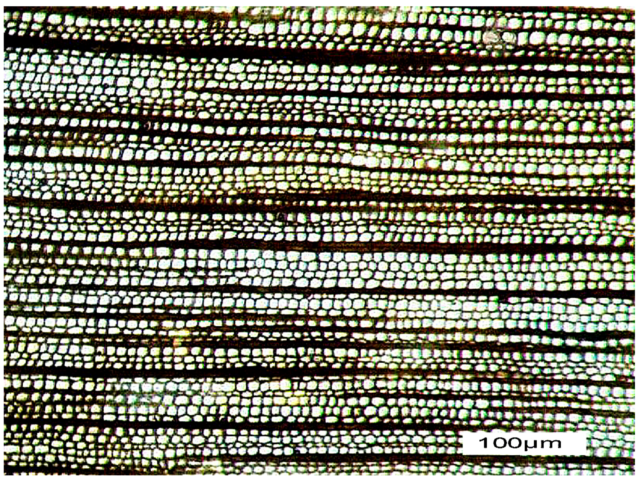
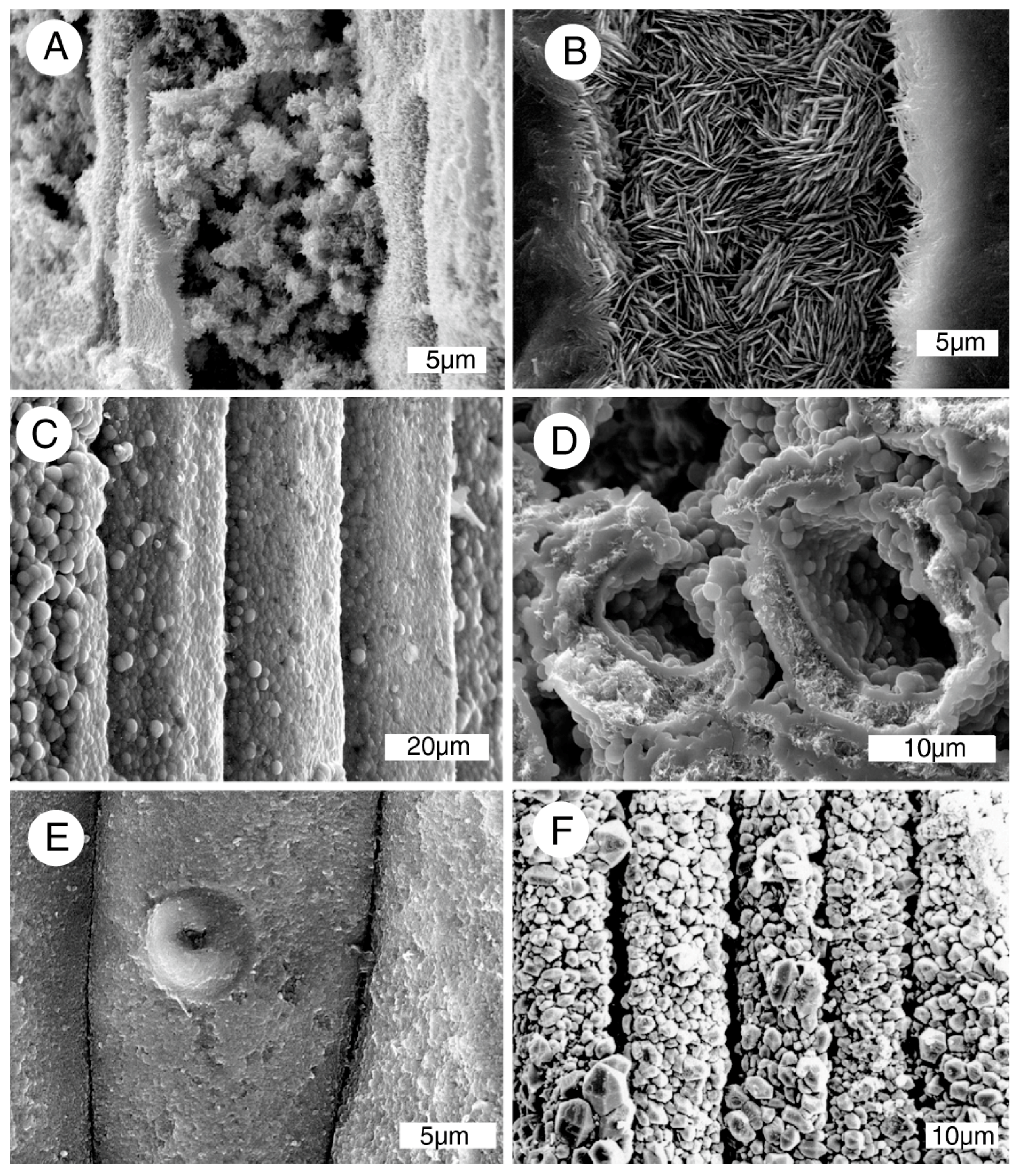
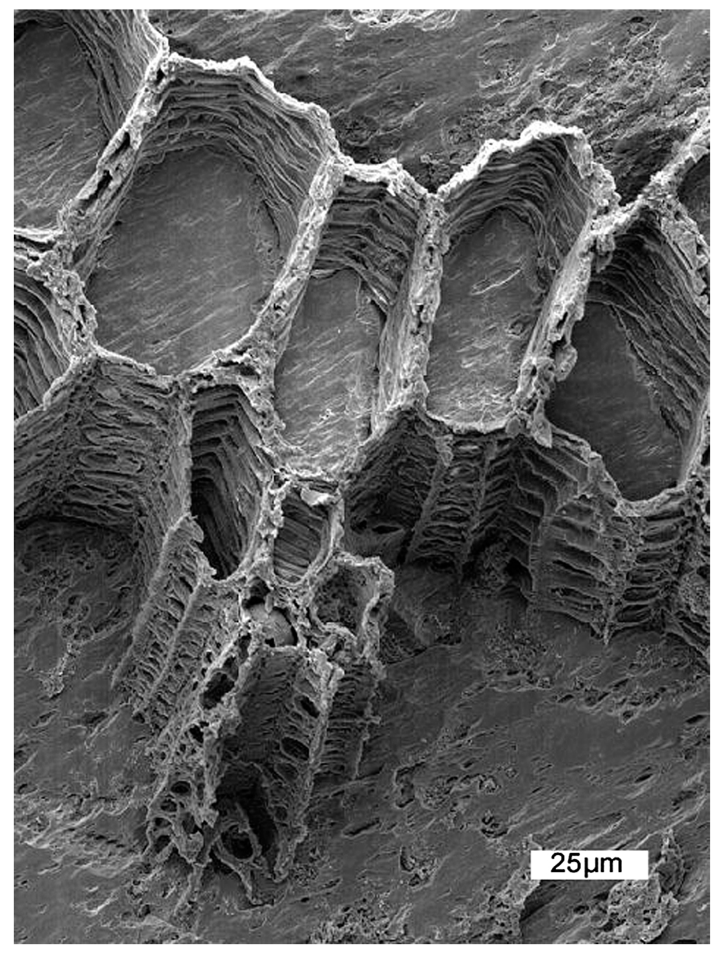
Figure 2.
Scanning electron microscopy (SEM) images of silicified woods with mineralized cell walls and empty lumina (some examples are marked with star symbols). (A) Miocene conifer wood showing a tracheid with opaline walls and botryoidal opal-A layer on interior surface, Lyon County, NV, USA; (B) Miocene opalized wood from Lovelock, NV, USA, Radial orientation showing silicified cell walls and empty lumina; (C) Miocene wood from Norita Opal Mine, Virgin Valley, NV, USA. Transverse view shows cell walls containing an interior lining of microcrystalline opal-CT; (D) Tertiary opalized wood from Soccorro, NM, USA. Radial view showing detailed anatomical preservation; (E) Transverse orientation, Miocene wood from Cessily Anne Opal Mine, Virgin Valley, NV, USA. Opal replacement of cell walls obliterated the original multi-layered architecture. Lumina are lined with botryoidal opal-A and microcrystalline opal-CT; (F) Miocene wood from Washoe County, NV, USA, transverse orientation. Cell walls are replaced by opal-A, with botryoidal textures on interior surfaces.
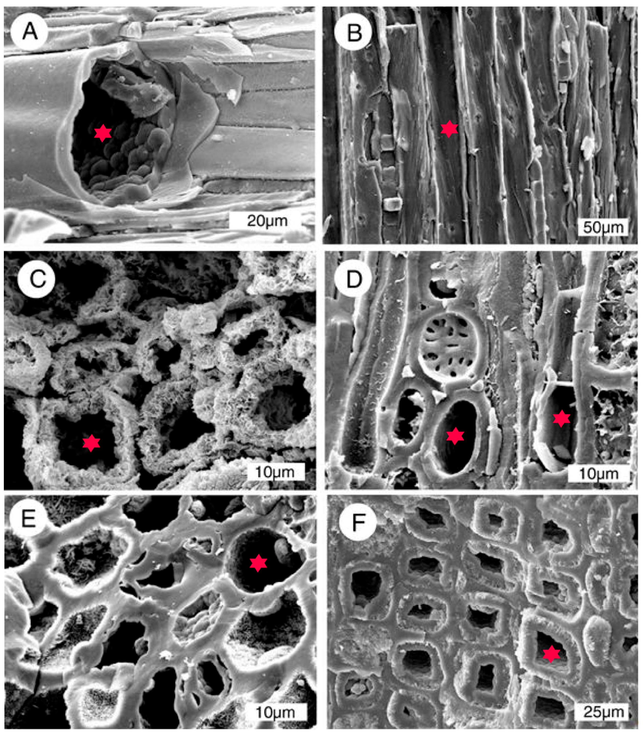
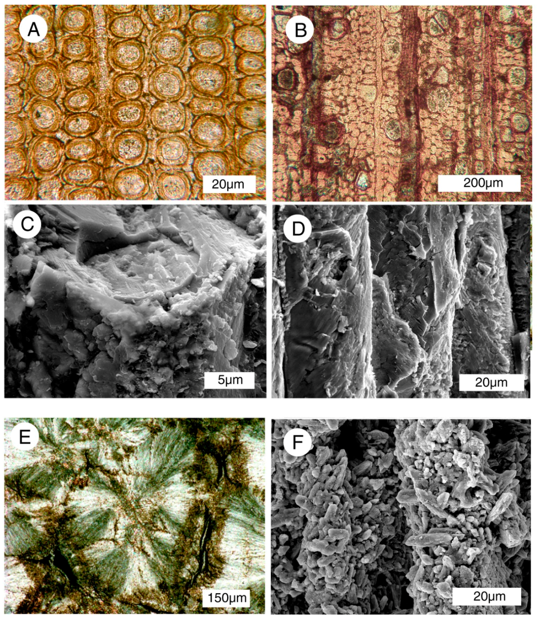
Figure 3.
SEM images of silicified woods showing cell walls where anatomical features have been replicated by silica minerals. (A) Miocene wood from Schurz, NV, USA, radial orientation showing cells walls replaced by opal-CT; (B) Miocene oak, Clover Creek, ID, USA. Radial orientation, interior view of a tracheid replaced by opal-CT; (C) Miocene wood, Washoe County, NV, USA. Radial orientation, tracheid walls mineralized with opal-A; (D) Miocene wood from Section 27 claim, Virgin Valley, NV, USA, Cell walls show multi-layered architecture, consisting of porous opal-A with surface layer of botryoidal opal-A; (E) Eocene wood from Yellowstone River, near Billings, MT, USA. Radial orientation, tracheids architecture mimicked by microcrystalline quartz; (F) Cretaceous conifer wood, Trinity County, TX, USA. Radial orientation, tracheids replicated with crystalline quartz.

Figure 4.
Mineralized cells, Miocene specimens showing opaline silica filling cell lumina and as a replacement for cell walls. (A) Bonanza Opal Mine, Virgin Valley, NV, USA. Transverse orientation, cell walls and lumina mineralized with opal-CT; (B) Norita Opal Mine, Virgin Valley, NV, USA. Transverse orientation, cells and lumina filled with opal-CT, intercellular spaces remain open; (C) Washoe County, NV, USA, oblique transverse orientation, cell walls mineralized with opal-A, lumen filled with porous opal; (D) Washoe County, NV, USA, oblique transverse orientation, cell walls mineralized with opal-A, lumina lined with amorphous porous opal, central area partially filled with botryoidal opal-A; (E,F) Hazen, NV, USA, radial orientation, cells mineralized with opal-A lepispheres. These lepispheres partially fill cell lumina, rather than replicating actual tissue.
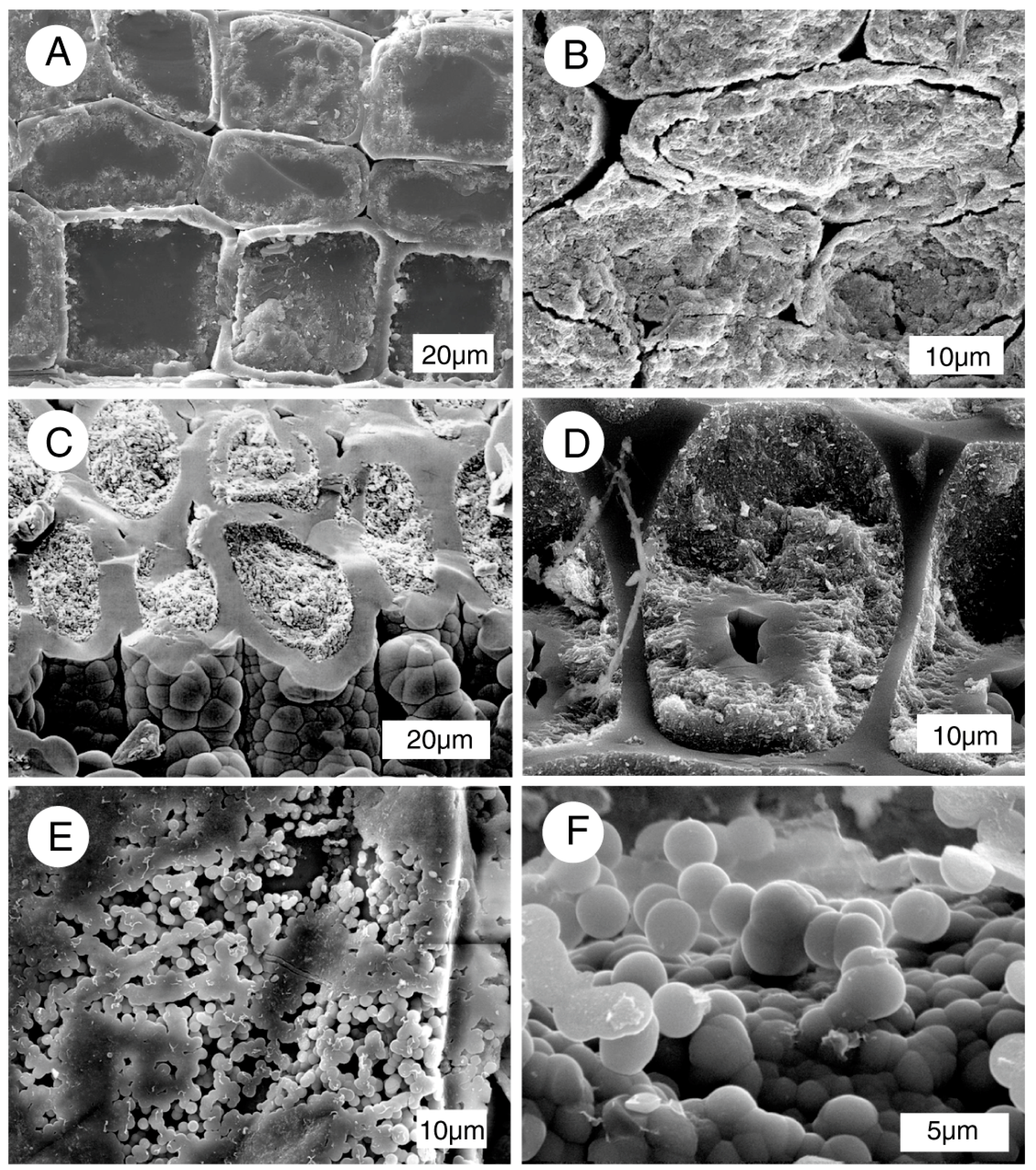
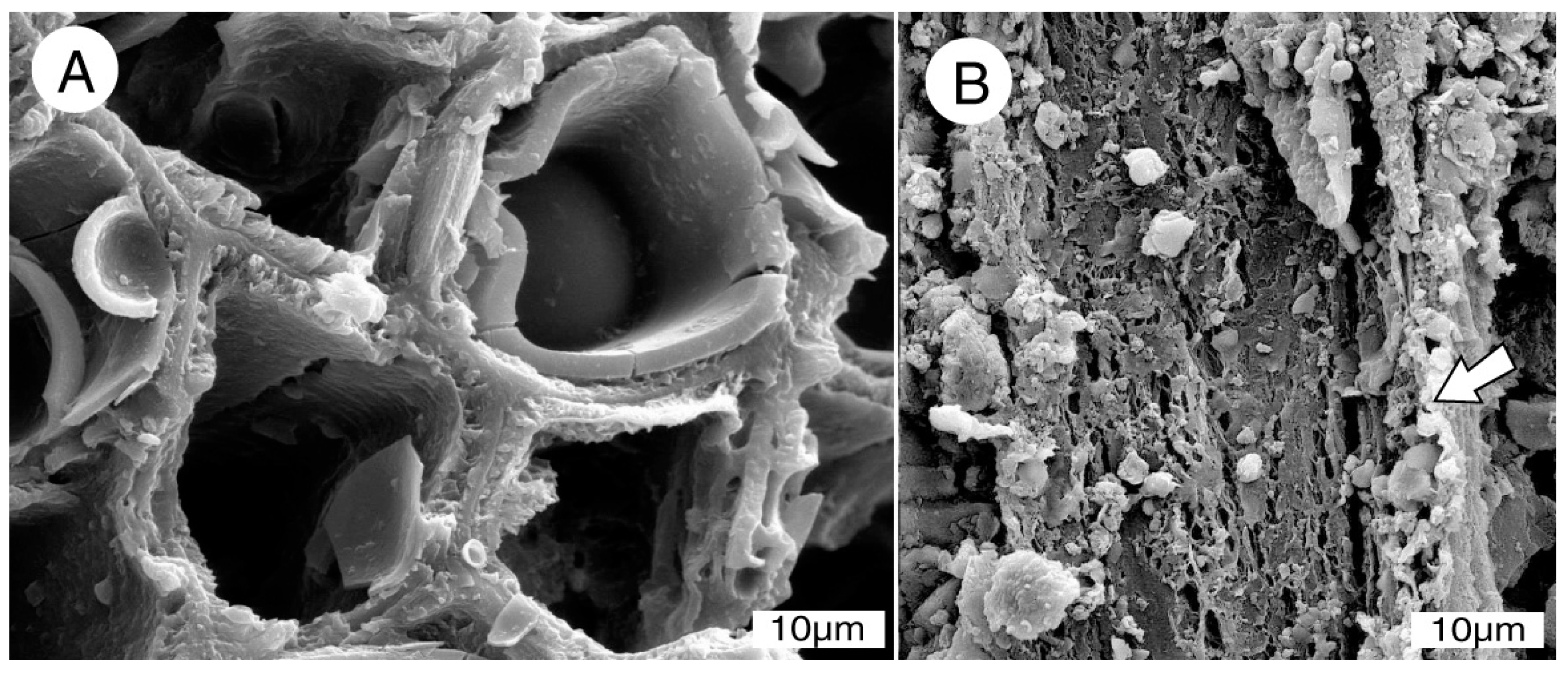
Figure 5.
SEM images of Miocene woods after 15 seconds of treatment with 40% hydrofluoric acid. Relict tissue was present during early stages of mineralization, but no relict organic matter is visible in these specimens. The end result meets the definition of replacement, not permineralization. (A,B). Specimen from Cedarville, CA, USA. (A) Radial orientation, (B) transverse orientation. Cell walls are mineralized with fibrous chalcedony. Lumina contain subhedral quartz crystals. (C,D) radial views of single tracheids, showing cell walls mineralized with a mosaic of interlocking microcrystals of chalcedony, preserving intervessel pit architecture. Lumina remain empty. (C) Saddle Mountain, Grant County, WA, USA; (D) Yakima Canyon, Yakima County, WA, USA, showing chalcedony-replaced cell walls with discernable circular pits.
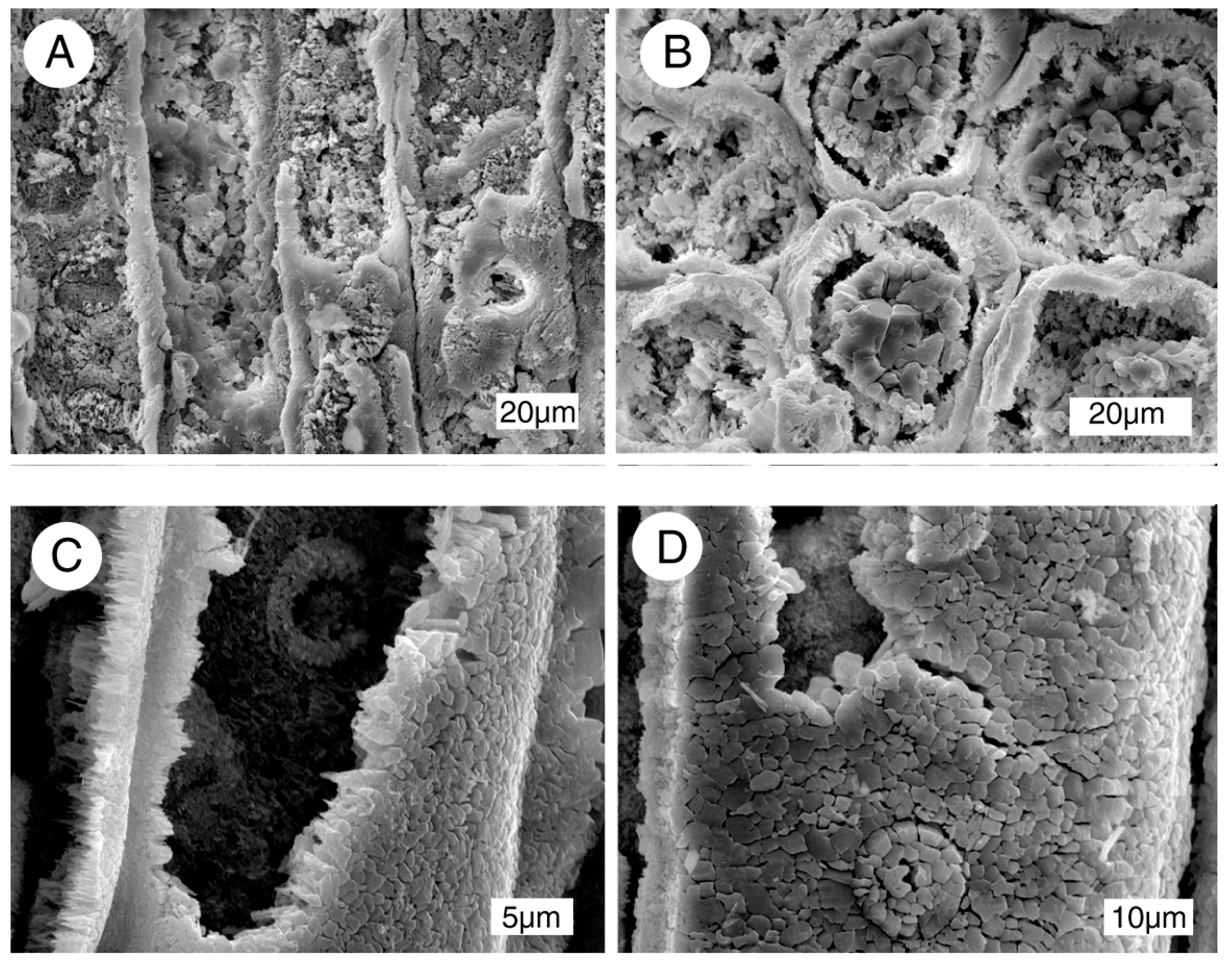
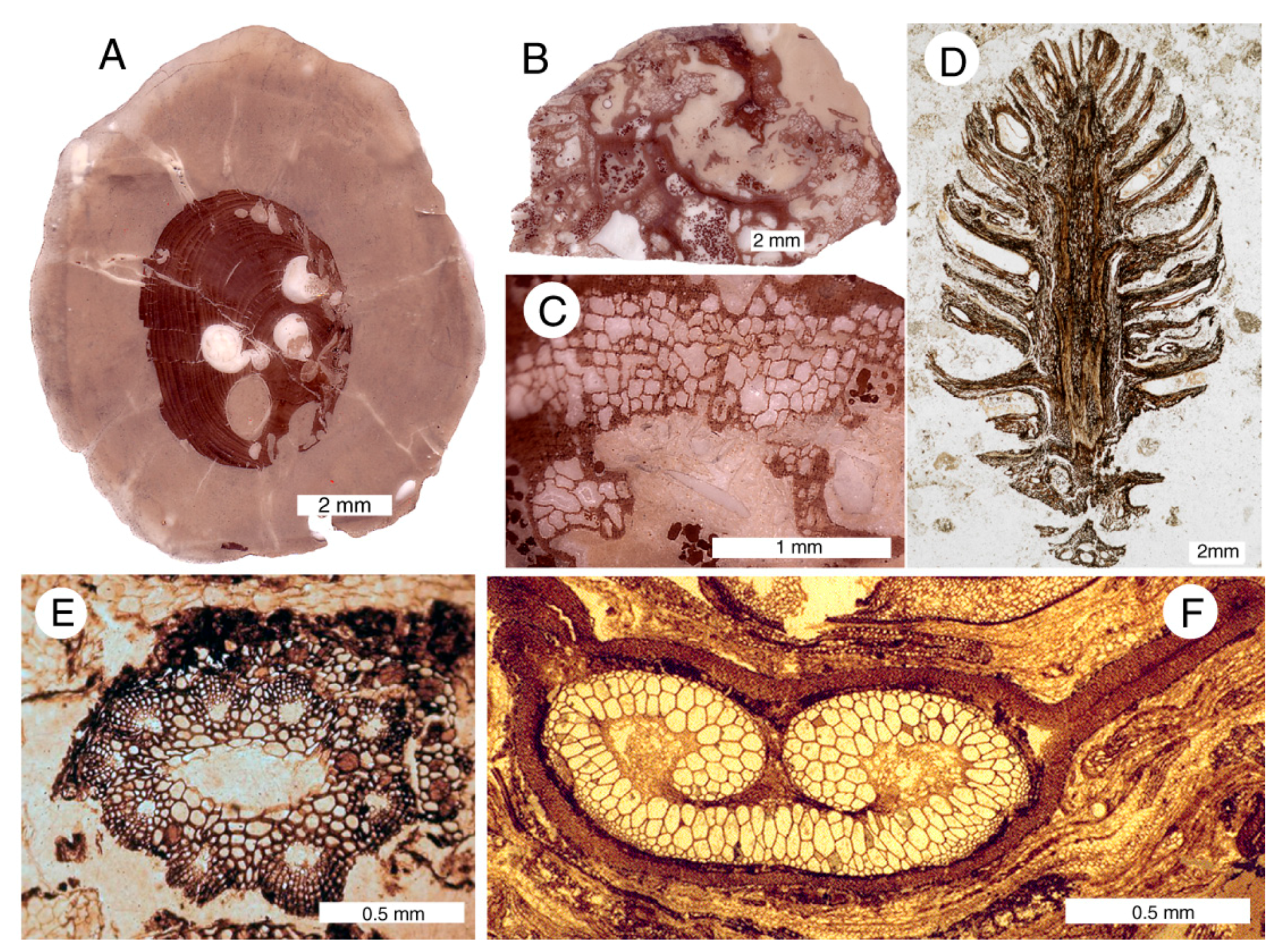
Figure 6.
Woods permineralized with calcite. (A–C) acetate peels of teredo-bored wood in a calcareous concretion, Lower Cretaceous, Apple Bay, Vancouver Island, BC, Canada; (D) Longitudinal section of apical region of ovulate cone of Hubbardiastrobus cunninghamiodes from Apple Bay [13] (photo courtesy of Brian A. Atkinson; (E) Calamites stem in Pennsylvanian coal ball, Lancashire coal field, England. Photo courtesy of Hans Steur; (F) Anacheropteris involuta preserved in Upper Pennsylvanian coal ball, IL, USA. Photo courtesy of University of Mūnster Paleobotany Research Center.

Figure 7.
Permineralization: cell walls of Lower Jurassic fern Osmundastrum pulchellum from Korsarőd, Sweden, revealed by HCl dissolution of calcareous matrix to reveal original cell walls [14]. Photo courtesy of Stephen McLoughlin.
Figure 7.
Permineralization: cell walls of Lower Jurassic fern Osmundastrum pulchellum from Korsarőd, Sweden, revealed by HCl dissolution of calcareous matrix to reveal original cell walls [14]. Photo courtesy of Stephen McLoughlin.

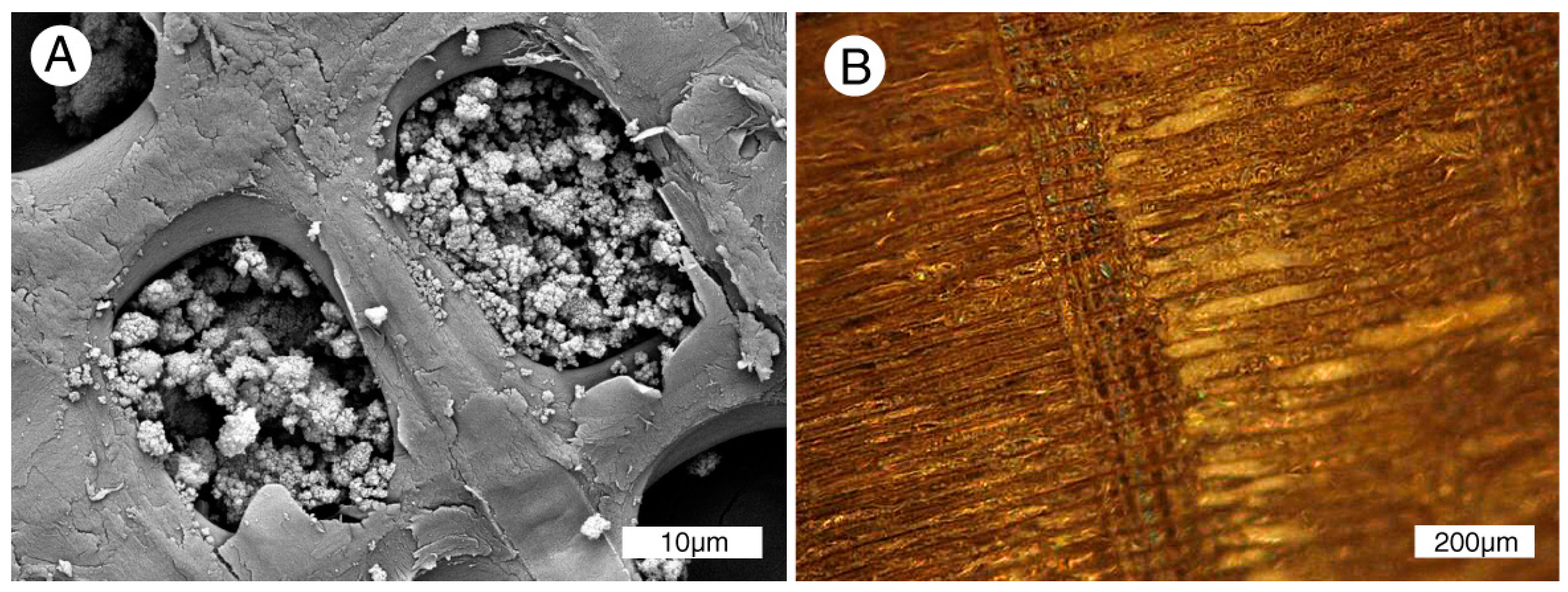
Figure 8.
Calcite-mineralized woods showing weak or no evidence of permineralization. (A–E) are from the Cretaceous Nanaimo Group, Vancouver Island, BC, Canada. (A) Specimen from Puntledge River, transverse orientation. Relict organic matter is present as very thin cell borders; (B) Specimen from Collishaw Point, Hornby Island, transverse orientation. Relict organic matter causes thin walls to be brownish red; (C) SEM view of a single tracheid of Puntledge River wood. Cell wall appears thick because of microcrystalline calcite. Lumen is filled with more coarsely crystalline calcite; (D) SEM view, specimen from Port Moody, tangential orientation. Relict smooth surfaces of cell walls are mineralized with calcite; (E) Port Hardy specimen, transverse orientation. Polarized light illumination shows radiating calcite crystals that have disrupted cellular tissue; preserve smooth texture; (F) Neogene wood from Glades County, FL, USA. Tangential orientation, showing tracheid walls mineralized with coarsely-crystalline calcite.

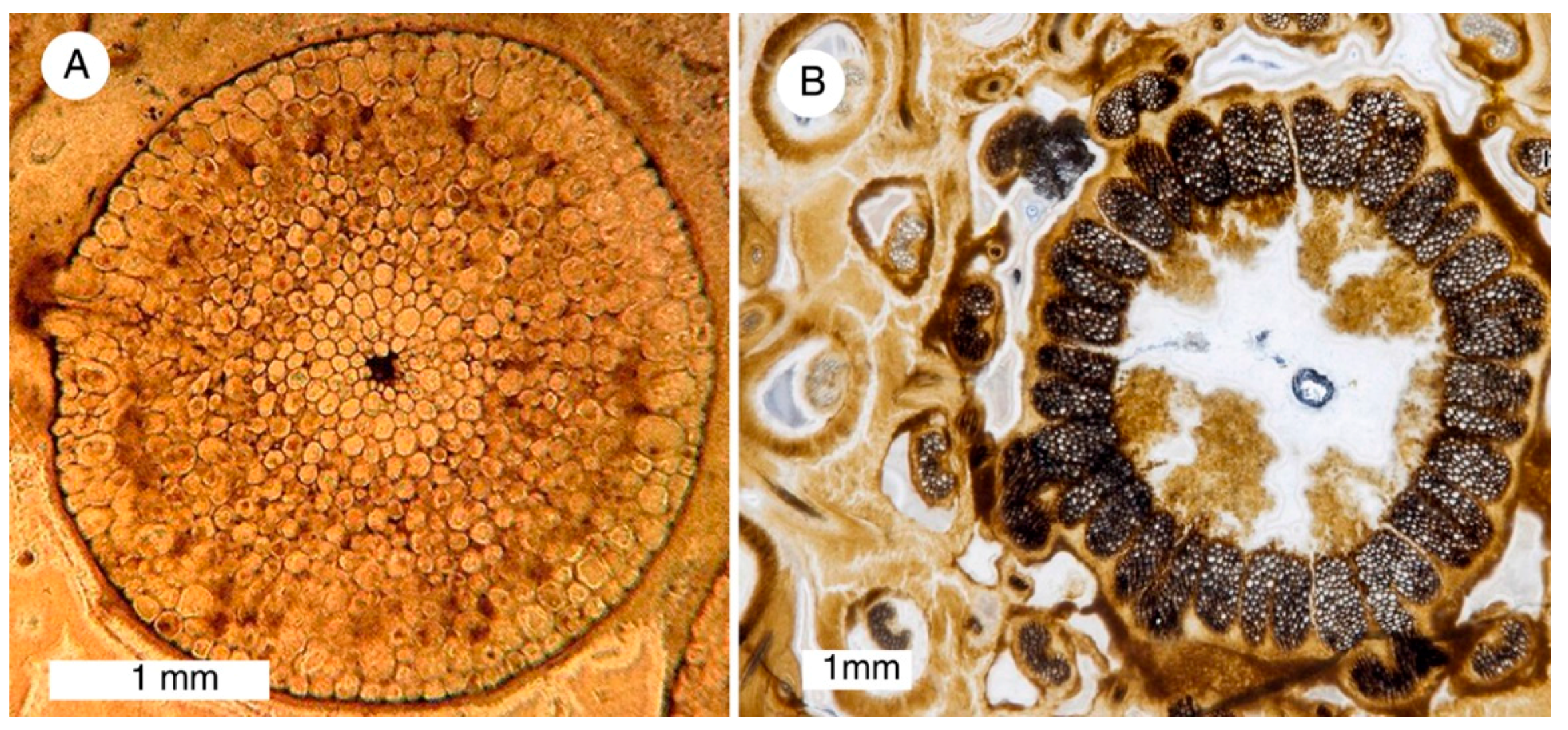
Figure 9.
Siliceous permineralization as revealed in acetate peels. A. Rhynia Gwynne-vaughanii, transverse view of stem, Devonian, Rhynie chert, Scotland [19,20]. Photo courtesy of Paleobotany Research Center University of Münster; B. Millerocaulis zammerae, transverse view of stem, Jurassic, Patagonia, Argentina [21]. Photo courtesy of Ana Sagasti).

Figure 10.
Eocene conifer wood from Red Hills Lignite Mine, Choctaw County, AL, USA. (A) Mummified wood, oblique transverse orientation, showing preservation of multi-layered cell walls; (B) Partially silicified wood, tangential orientation, showing lace-like degradation of cell wall, and Si precipitation in f outer wall (arrow).

Figure 11.
Permineralized Lower Eocene conifer wood, Red Hills Lignite Mine, Choctaw County, MS, USA. (A,B) Transverse orientation, showing organic mummified cell walls with lumina and intercellular space filled with silica.
Figure 11.
Permineralized Lower Eocene conifer wood, Red Hills Lignite Mine, Choctaw County, MS, USA. (A,B) Transverse orientation, showing organic mummified cell walls with lumina and intercellular space filled with silica.

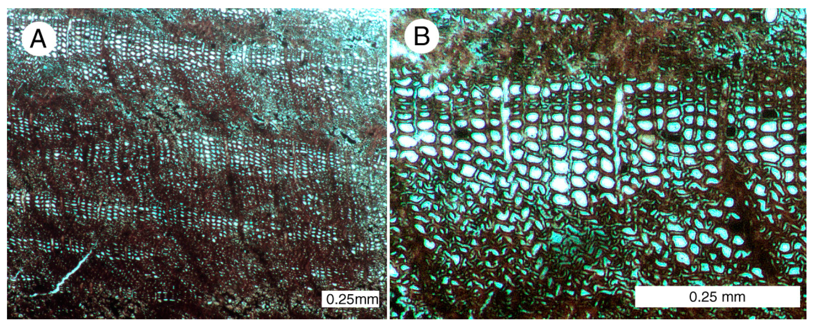
Figure 12.
Incipient permineralization of modern Pinus contorta (Lodgepole Pine) wood can be observed at hot springs in Yellowstone Park, WY, USA. (A,B) silica encrusted woody debris on hot spring sinter apron; (C) Distribution of silica as mapped by X-ray microanalysis of transverse specimen. Warm, bright colors represent areas of elevated silica. Incipient silcification has occurred near exterior surfaces and along fractures [23]; (D) Radial orientation, showing cells encrusted with amorphous silica (opal-A); (E) Transverse view, tracheids showing multilayered unmineralized cell walls, with opal-A precipitated on interior surfaces; (F) High magnification shows thin silica zone (arrow) as an interlayer within tracheid cell walls; (G) Opal-A lepispheres encrust a pit aperture plate.
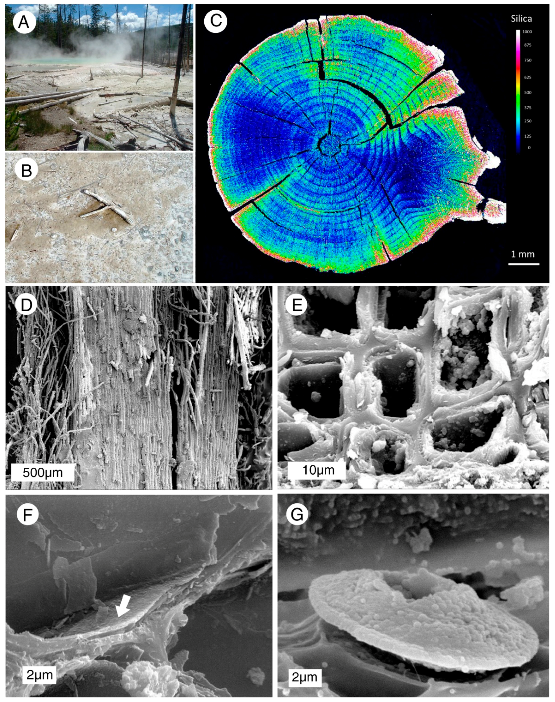
Figure 13.
Experimental silica permineralization of modern conifer wood, Pseudotsuga menziesii. Samples were exposed to H4SiO4 saturated solutions for ~90 days at 90 °C, with obsidian powder as the silica source [33]. (A) Transverse orientation, showing tracheid lumina filled with silica gel aggregate; (B) reflected light image of a longitudinal section cut parallel to the tracheids using a scalpel, showing lumina permineralized with silica gel. Photos courtesy of Chris Ballhaus, Bonn University.

Table 1.
Preservation of relict organic matter in silicified wood.
| Age | Location | Genus | Mineralogy * | Density g/cm3 | % LOI 450° ** | Estimated Original Density *** | Calculated % Original Wood |
|---|---|---|---|---|---|---|---|
| Devonian | Murray, OK | Callixylon | chalcedony | 2.49 | 0.41 | - | - |
| Triassic | Holbrook, AZ | Araucarioxylon | chalcedony | 2.62 | 0.19 | 0.52 | 0.96 |
| Cretaceous | Montague Co., TX | Cupressinoxylon | quartz | 2.53 | 0.55 | 0.45 | 3.09 |
| Eocene | Leesville, LA | Palmoxylon | chalcedony | 2.58 | 0.14 | 0.56 | 0.65 |
| Eocene | Eden Valley, WY | Palmoxylon | chalcedony | 2.50 | 1.39 | 0.56 | 6.21 |
| Eocene | Watertree River, SC | Palmoxylon | chalcedony | 2.32 | 2.20 | 0.56 | 9.11 |
| Oligocene | Panama | Palmoxylon | chalcedony | 2.57 | 1.24 | 0.56 | 5.69 |
| Paleocene | North Dakota | Metasequoia | chalcedony | 2.60 | 0.33 | 0.45 | 1.91 |
| Oligocene | Rapid City, SD | Metasequoia | chalcedony | 2.62 | 0.27 | 0.45 | 1.57 |
| Eocene | Florissant, CO | Sequoioxylon | chalcedony | 2.53 | 0.36 | 0.45 | 2.02 |
| Eocene | Florissant, CO | Sequoioxylon | chalcedony | 2.41 | 0.43 | 0.45 | 2.30 |
| Eocene | Gallatin Co., MT | Sequoioxylon | quartz | 2.48 | 3.94 | 0.45 | 21.71 |
| Miocene | Yakima, WA | Platanus | chalcedony | 2.37 | 0.83 | 0.56 | 3.51 |
| Miocene | Yakima, WA | Ulmus | opal-CT | 1.95 | 2.18 | 0.60 | 7.09 |
| Miocene | Madras, OR | Quercinium | opal-CT | 2.01 | 2.80 | 0.74 | 7.61 |
| Miocene | Swartz Canyon, OR | Quercinium | chalcedony | 2.58 | 0.78 | 0.74 | 2.72 |
| Miocene | Bliss Co., Idaho | Quercinium | opal-CT | 1.93 | 1.65 | 0.74 | 4.30 |
| Pleistocene | Florida | Taxodium | chalcedony | 2.54 | 0.55 | 0.48 | 2.91 |
| Eocene | Cache Creek, BC | unknown | opal-CT | 2.07 | 4.49 | 0.45 | 20.65 |
| Miocene | Miller Mtn., NV | unknown | opal-CT | 2.07 | 3.75 | 0.45 | 17.25 |
| Miocene | Yakima Co., WA | Cupressinoxylon | opal-CT | 1.99 | 3.28 | 0.45 | 14.50 |
| Miocene | Washoe County, NV | unknown | opal-CT | 1.86 | 2.77 | 0.45 | 11.45 |
| Miocene | Rawhide, NV | unknown | opal-CT | 1.85 | 4.68 | 0.45 | 19.24 |
| Miocene | Lake Tahoe, CA | unknown | opal-CT | 1.90 | 3.25 | 0.45 | 13.72 |
| Neogene | Columbia | unknown | opal-CT | 1.90 | 2.66 | 0.45 | 11.23 |
| Miocene | Nye County, NV | conifer | opal-CT | 1.90 | 1.61 | 0.45 | 6.80 |
* Mineralogy determined by X-ray diffraction and scanning electron microscopy; ** Loss on ignition after heating at 450 °C; *** Density estimated from extant relatives.
© 2017 by the author. Licensee MDPI, Basel, Switzerland. This article is an open access article distributed under the terms and conditions of the Creative Commons Attribution (CC BY) license (http://creativecommons.org/licenses/by/4.0/).
Share and Cite
MDPI and ACS Style
Mustoe, G.E. Wood Petrifaction: A New View of Permineralization and Replacement. Geosciences 2017, 7, 119. https://doi.org/10.3390/geosciences7040119
AMA Style
Mustoe GE. Wood Petrifaction: A New View of Permineralization and Replacement. Geosciences. 2017; 7(4):119. https://doi.org/10.3390/geosciences7040119
Chicago/Turabian StyleMustoe, George E. 2017. "Wood Petrifaction: A New View of Permineralization and Replacement" Geosciences 7, no. 4: 119. https://doi.org/10.3390/geosciences7040119
Note that from the first issue of 2016, this journal uses article numbers instead of page numbers. See further details here.