Formulation Strategies to Improve Oral Bioavailability of Ellagic Acid
by
 , , , and
Guendalina Zuccari
* ,
,
Sara Baldassari
 ,
,
Giorgia Ailuno
,
Federica Turrini
,
Silvana Alfei
 and
and
Gabriele Caviglioli
Department of Pharmacy, Università di Genova, 16147 Genova, Italy
*
Author to whom correspondence should be addressed.
Appl. Sci. 2020, 10(10), 3353; https://doi.org/10.3390/app10103353
Submission received: 6 April 2020
/
Revised: 26 April 2020
/
Accepted: 8 May 2020
/
Published: 12 May 2020
(This article belongs to the Special Issue Functional Foods and Food Supplements)
Abstract
:Featured Application
An updated description of pursued approaches for efficiently resolving the low bioavailability issue of ellagic acid.
Abstract
Ellagic acid, a polyphenolic compound present in fruit and berries, has recently been the object of extensive research for its antioxidant activity, which might be useful for the prevention and treatment of cancer, cardiovascular pathologies, and neurodegenerative disorders. Its protective role justifies numerous attempts to include it in functional food preparations and in dietary supplements, and not only to limit the unpleasant collateral effects of chemotherapy. However, ellagic acid use as a chemopreventive agent has been debated because of its poor bioavailability associated with low solubility, limited permeability, first pass effect, and interindividual variability in gut microbial transformations. To overcome these drawbacks, various strategies for oral administration including solid dispersions, micro and nanoparticles, inclusion complexes, self-emulsifying systems, and polymorphs were proposed. Here, we listed an updated description of pursued micro and nanotechnological approaches focusing on the fabrication processes and the features of the obtained products, as well as on the positive results yielded by in vitro and in vivo studies in comparison to the raw material. The micro and nanosized formulations here described might be exploited for pharmaceutical delivery of this active, as well as for the production of nutritional supplements or for the enrichment of novel foods.
1. Introduction
Pomegranate, Punica granatum, is well-known as a traditional medicinal fruit mentioned in the Old Testament of the Bible, the Qur’an, the Jewish Torah, and the Babylonian Talmud as a sacred fruit harbinger of fertility, abundance, and luck. Historically and currently, pomegranate has been used for various purposes [1]. The bioactive compound mainly responsible for the health effects of pomegranate is ellagic acid (EA), as it represents one of the most potent dietary antioxidants.
EA is a polyphenol compound which derives from ellagitannins (ET), a family of molecules in which hexahydroxydyphenic acid residues are esterified with glucose or quinic acid. Following a hydrolytic process, the hexahydroxydyphenic acid group is released, dehydrates and spontaneously lactonizes, forming EA. Punicalagin isomers are the most representative of total tannins extracted from pomegranate (Figure 1).
Present also in a large variety of tropical fruit, nuts, berries (strawberries, raspberries, blackberries, cranberries, goji berries) and in a type of edible mushroom (Fistulina epatica), EA has recently attracted deep interest, since many research studies showed the role of oxidative stress in different pathologic conditions such as cancer, diabetes, cardiovascular and autoimmune diseases, obesity, and neurodegenerative disorders [2,3,4,5,6,7]. In this regard, EA may be a useful adjunct in Parkinson’s disease treatment, as it proved to protect dopamine from oxidation and, by reducing inflammation and oxidative stress, to exert a neuroprotective effect [8]. Besides being able to reduce the pathological levels of NF-kB, EA may play a beneficial role in diseases associated with CNS inflammation, such as multiple sclerosis. EA has also been proposed as antidepressant and anxiolytic drug for its capability to modulate the monoaminergic system and to increase the endogenous levels of brain-derived neurotrophic factors [6]. In the last decade, several studies investigated EA effects on many types of cancer, showing its ability to arrest cell cycle progression, modulate pathways linked to cell viability, and inhibit angiogenesis via inactivating metalloproteinases [9,10,11,12,13,14,15,16,17,18]. For example, EA resulted in inducing apoptosis and modulating gene expression in different colon cancer cell lines at the concentrations achievable in the intestinal lumen from the diet, suggesting a potential role in cancer chemoprevention [19]. Moreover, EA may counterbalance the dangerous side effects of antitumoral drugs such as cisplatin, doxorubicin, and cyclosporine [20,21,22]. In addition, EA proved to possess antimicrobial properties inhibiting the growth of methicillin-resistant Staphylococcus aureus and Salmonella [23]. These properties were explained through the ability of EA of either coupling with proteins of the bacterial wall or inhibiting gyrase activity, which cleaves the DNA strand during bacterial replication. Furthermore, EA seemed to counteract the HIV-1 virus replication, by binding envelope proteins and inhibiting reverse transcriptase [24]. Pomegranate extract has been traditionally used for treating gastrointestinal bleeding and various types of wounds, due to EA’s ability to promote blood coagulation by the activation of the Hageman factor (Factor XII) [25]. Moreover, preliminary results suggest potential anti-inflammatory properties [26,27] and an important role in prevention of cardiovascular diseases [28,29,30]. Finally, EA finds application also in cosmetic and nutraceutical industries, mainly for antiaging and prophylactic purposes respectively [31,32,33]. An overview of EA activities is shown in Figure 2.
However, though EA has the potential to become an efficacious agent in the prevention and therapy of several diseases, as supported by several papers present in the literature, the working efficacy of EA dietary and therapeutic formulations have been mainly examined at preclinical level. To date, few clinical studies have been performed to evaluate EA beneficial effects in humans, with often a limited number of patients, and most of them concern the administration of pomegranate juice or extract in cancer patient diet or deal with the evaluation of the enhancement of cognitive/functional recovery after stroke [34,35]. It was suggested that EA’S low water solubility and rapid metabolism might hinder the progress towards translational research. In our opinion, further studies focused on the application of micro and nanotechnology could provide more encouraging data, opening the path to new strategies able to overcome EA’s poor biopharmaceutical characteristics. Hence, the goal of this review is to provide to scientists, embarking on the research involving EA, a scenario of all pursued formulation attempts. Therefore, starting from a description of EA bioavailability and solubility, a summary of micro and nanosystem-based strategies suitable for improving orally administered EA efficacy is provided.
2. EA Chemical Structure and Solubility
EA is a chromene-dione derivative (2,3,7,8-tetrahydroxy-chromeno [5,4,3-cde]chromene-5,10-dione), encompassing both a hydrophilic moiety with 4 hydroxyl groups and 2 lactone groups, and a planar lipophilic moiety with 2 biphenyl rings. This particular structure has both hydrogen bonding acceptor (lactone) and donor (–OH) sites (Figure 1). Due to the weak acidic nature of its four phenolic groups (pKa1 = 5.6 at 37 °C), around neutral pH it is mainly deprotonated on positions 8 and 8′, while above pH 9.6 lactone rings open to give a carboxyl derivative [36]. EA low oral bioavailability is mostly due to its poor water solubility (9.7 µg/mL), which increases with pH, as well as the antioxidant action [37]. However, in basic solutions, phenolic compounds lack stability as these molecules, under ionic form, undergo extensive transformations or are converted to quinones as a result of oxidation. A stability study on pomegranate fruit peel extract demonstrated that EA content significantly decreases in few weeks regardless of the pH of the solution, due to the hydrolysis of the ester group with hexahydroxydiphenic acid formation, suggesting that EA should not be stored in aqueous medium [38]; this aspect reinforces the need to develop novel systems also for EA stabilization.
Prior to facing the route of the development of a new delivery system, the knowledge of solvents, co-solvents and substances in which EA could be consistently soluble and stable represents an indispensable step for designing a good formulation. In this regard, with the aim of providing a useful guidance for further research studies, Table 1 gathers published data concerning EA solubility. Concerning organic solvents, EA is slightly soluble in methanol, soluble in DMSO and shows maximum solubility in N-methyl-2-pyrrolidone (NMP), confirming the effect of basic pH on EA dissolution. An analogous trend is also observable in aqueous solutions, where results implied that the solubility of EA depends on the pH values of the media. While EA is almost insoluble in acidic media and distilled water, its water solubility is significantly improved by basic pH. As highlighted further ahead, one of the mostly exploited vehicles is polyethylene glycol (PEG) 400, as it is endowed with satisfactory biocompatibility and, at the same time, is miscible with both aqueous and organic solvents. EA solubility in oils and surfactants is also provided, helpful for developing emulsifying-based techniques.
3. EA Dietary Assumption
In foods and beverages, EA is present in several forms, such as unmodified, glycosylated and/or acetylated or inside the structure of hydrolysable ET polymers, usually esterified with glucose moieties. However, only a small fraction of free EA is absorbed in the stomach, since ET are resistant to acidic pH. ET hydrolysis occurs in the small intestine, leading to the release of EA, which can be absorbed mainly by passive diffusion, although an in vitro experiment on Caco-2 cells monolayer model suggested the involvement of a protein-mediated transport [39]. Once reached the systemic circulation, EA undergoes a massive first pass effect, being transformed in methyl esters, dimethyl esters or glucuronides, detectable in human plasma from 1 to 5 h after ET ingestion [40]. Meanwhile, unabsorbed ET and EA fractions are mostly converted to a family of metabolites called urolithins by colon microflora. Urolithins contain a common structure of dibenzopyranone and are more lipophilic, showing higher absorption rate across colon epithelium compared to EA, thus resulting 25–80 times more bioavailable [41]. Naturally, there is huge variability in microbial metabolism of EA among individuals depending on differences in gut microbiota composition. According to the metabotype (M-0, M-A and M-B), humans may produce no urolithins, highly active urolithins or less active urolithin, so that EA consumption may not exert equal health benefits in all subjects [42,43,44].
EA oral bioavailability was studied in human subjects after 180 mL of pomegranate juice consumption. In this study, the maximum EA blood concentration (Cmax) was 32 ng/mL which was rapidly metabolized in the next 4 h, confirming the extremely poor intestinal absorption [41]. Other pharmacokinetic studies supported the hypothesis that higher EA-to-ET ratio in the oral dose corresponds to higher EA plasma concentrations, but, intriguingly, no enhancement in bioavailability was observed increasing the dose of free EA up to 524 mg, suggesting that a saturation condition in the absorption process was achieved [45]. In addition to these unfavorable conditions, it must be considered that polyphenols are not consumed in sufficient quantities among population in Western Europe and developing countries, because of inadequate fruit and vegetable intake, though an increased awareness of the benefits of a balanced diet is fostering the assumption of dietary supplements [46].
4. Formulation Strategies for Improving EA Oral Bioavailability
The Biopharmaceutics Classification System (BCS) is a scientific framework for classifying drug substances based on their aqueous solubility and intestinal permeability. It is a drug-development tool that divides drugs into high/low solubility and permeability classes. Based on its low aqueous solubility and low permeability (apparent permeability coefficient = 0.13 × 10⁻⁶ cm/s [47]), EA is classified as class IV drug according to the BCS, i.e., endowed with low solubility and low permeability, features that limit a lot its clinical application. Micro and nanotechnology approaches widely demonstrated great potential in modifying pharmacokinetic, bioavailability and stability of several drugs, including phytochemicals used in cancer chemoprevention or in dietary supplements, contributing to preserve the properties of the active ingredients or to mask bad tastes and odors [48,49,50]. From a formulation point of view, a good EA dosage form would grant easier handling, transport and storage, increase its light resistance, hamper undesirable reactions such as oxidation and hydrolysis, and enhance solubility.
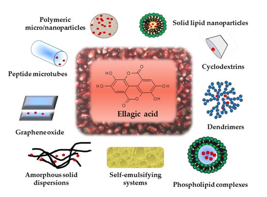
Focusing on preparative methods, features and biological results of the developed EA-loaded formulations, an exhaustive description of the employed strategies is thereafter presented. The various approaches were divided according to the kind of the dosage form and reported in Table 2 and Table 3 depending on the micro or nanosize of the final product. Moreover, in the final paragraph, examples of fixed combinations containing EA were also described and discussed.
4.1. Micronized EA (m-EA)
According to Noyes-Whitney law, an effective way to increase dissolution rate and oral absorption of a substance is the size reduction of powder particles [51]. For this purpose, anti-solvent precipitation may represent a valid processing technique. In comparison to more popular micro and nanocarriers that will be discussed in the following sections, this approach presents several advantages, such as easy realization and rapidity. In addition, the process allows the reduction of organic solvents use and the one-step processing favors scalability. The anti-solvent precipitation of a poorly water-soluble substance is simply obtained by dissolving it in a solvent, followed by rapid mixing with a solvent-miscible anti-solvent. After mixing, the substance precipitates in micro-sized particles, since a supersaturated solution forms. The critical step of this method is to produce crystal nuclei rapidly, avoiding their excessive growth. For process optimization, precipitation time and temperature, addition speed of the solution to the anti-solvent, their volume ratio, drug concentration and stirring intensity are the keys parameters that have to be checked.
Li et al. set the best conditions to produce m-EA by anti-solvent precipitation with an accurate preformulative study [52]. An EA solution in NMP (30 mg/mL) was added to water, used as anti-solvent, at a rate of 30 mL/h with 2 min precipitation time, at 3 °C and 2500 rpm stirring. Then, the precipitate was dispersed in a maltodextrin aqueous solution to have a solid concentration of 1 mg/mL and was lyophilized. The authors verified that residual NMP (class 2 residual solvent) in the final product was in agreement with the established International Council for Harmonization of Technical Requirements for Pharmaceuticals for Human Use (ICH) limit [53]. The m-EA freeze-dried powder, whose mean particle diameter was 428 nm, in a comparison to raw EA and to a physical mixture of EA in maltodextrin, showed the fastest dissolution rate. Surely, maltodextrin acted as hydrotrope, but the result could be attributed exclusively to the high specific surface of the lyophilized powder and to the low crystallinity of the incorporated EA. All in all EA water dissolution was improved up to 11.67 µg/mL at 37 °C. In addition, the 2, 2-diphenyl-1-1-pycrylhydrazyl (DPPH)-scavenging activity was higher than that of not-micronized EA and oral bioavailability in rats increased twice in comparison to raw EA.
In another study, Beshbishy et al. obtained m-EA using the anti-solvent precipitation method by injecting into deionized water a 10 mg/mL ethanolic solution of EA at a fixed flow rate [54]. m-EA particles, collected by filtration and vacuum dried, showed modified morphology with respect to the parent material and appeared as needle-shaped or rod-like structures. In vitro and in vivo experiments, performed by intraperitoneal injection in mice, showed a significantly improved m-EA activity against blood parasites as Babesia and Theileria. However, the authors concluded that EA activity requires further investigations to understand whether EA inhibits parasites grow or causes their death.
A similar approach was performed using supercritical CO2 as anti-solvent. In this procedure a solution of EA in NMP was sprayed into a vessel where a CO2 stream was continuously flowed under supercritical conditions. Solution atomization led to a fast mass transfer between supercritical CO2 and the solution itself, which caused supersaturation and subsequent rapid precipitation of EA in the form of micro-sized particles [55]. The study focused on the influence of process parameters and EA concentration on size, polydispersity index (PDI) and morphology. Moreover, a co-precipitation with Eudragit® L 100 was also carried out and, starting from EA:Eudragit® 1:1 weight ratio, a drug loading (DL) of 49% was achieved. Experimental results showed that EA concentration should be, if possible, close to saturation, thus allowing obtaining particles in the form of sticks and flowers and with a mean diameter of about 3 μm. Also, in this case residual NMP was far lower than the mandatory established limit. m-EA showed higher dissolution rate with respect to raw EA due to the small particle size, reaching higher concentrations even at pH 1.2. Finally, the coprecipitates with Eudragit® showed the fastest dissolution rates, probably because the dispersion of EA within the polymer led to smaller particles and, surprisingly, the presence of the polymer did not delay the pomegranate acid delivery, possibly since EA particles were obtained in crystalline form, confirming that the product was a solid dispersion in which the pomegranate acid particles were not encapsulated.
4.2. EA in Spray Dried and Lyophilized Powders
Solid dispersions are semi-crystalline or amorphous dispersions (ASDs) of a molecule in an inert matrix. ASDs represent a valid alternative for the development of microsystems entrapping poorly water-soluble molecules. ASDs can be prepared from a solution by spray drying, freeze drying, co-precipitation, rotary evaporation, film casting, or hot-melt extrusion. In all cases, a suitable polymeric excipient should have functional groups able to form H-bonds with EA, thus allowing the formation of metastable amorphous EA dispersions with adequate stability over time.
In this context, a first attempt was made with maltodextrins, a saccharide-based excipient extensively used in food industry. Using a freeze drying technique, maltodextrin DE5-8 or DE18.5 were applied to form a solid network around a cloudberry phenolic extract [56]. The amorphous matrices were prepared starting from a maltodextrin (9% w/w) and a cloudberry extract (1% w/w) aqueous solution, mixed under slight heating for about 0.5 h to avoid ET degradation. DE5-8 complexed phenolics in a more efficient way, rationally due to its higher molecular weight. The microencapsulation improved the extract storage stability to hydrolysis, and also in this case the improvement contribution of DE5-8 was better than that of DE18.5. Microencapsulation always resulted in a decrease of EA formation by ET hydrolysis during storage, but the stability was strongly dependent on relative vapor pressure (RVP). At 66% RVP and 25 °C maltodextrin DE5-8 protected phenolics up to 32 days, then the degradation reactions occurred in a major extent. On the contrary, the antioxidant properties did not alter during storage either in the extract alone or in the presence of DE5-8, probably because the loss of original phenolics was compensated by new formed molecules with equal antioxidant activity.
In another study, polymers as the hydrophilic hydroxypropylmethyl cellulose acetate succinate (HPMCAS), the rather hydrophobic carboxymethyl cellulose acetate butyrate (CMCAB), and the very hydrophobic cellulose acetate adipate propionate (CAAdP) were compared with polyvinylpyrrolidone (PVP), taken as reference excipient being widely used in ASD formulations [57]. Mixtures of EA and PVP, CMCAB or HPMCAS at different weight ratios (1:9 and 1:3), dissolved in acetone:ethanol (1:4 v/v), were spray dried with yields of 50%–60%. ASD from CAAdP were instead prepared by co-precipitation method, due to the limited quantity of polymer available. EA was amorphous up to 25% w/w in solid dispersions with PVP and HPMCAS, and up to 10% with CMCAB and CAAdP. Consequently, EA-PVP and EA-HPMCAS reached the highest solution concentration of 1500 and 280 μg/mL, respectively. In a stability study performed in aqueous buffer (pH 6.8) for 24 h at room temperature (rt), pure EA showed a degradation degree by lactone ring opening of 20%, while lower values—16% for CMCAB, 14% for CAAdP, 6% for PVP and just 5% for HPMCAS—were found in the ASD samples. Dissolution rate at pH 6.8 was remarkably superior to that of pure EA, both from PVP and HPMCAS ASDs, while from CAAdP or CMCAB ASDs it was very slow and did not achieve adequate EA concentration. As expected, under acidic conditions EA release from PVP was quite fast, whereas from cellulose esters it was minimal, but this advantage was nullified by crystallization of a large amount of the EA released from PVP. Since the 1:9 ASDs with HPMCAS effectively stabilized EA against crystallization and degradation, the authors concluded that this could be considered the most promising formulation.
To prepare an ingredient for functional foods, a fine microdispersed powder containing EA was obtained by spray drying using low methoxyl pectin, characterized by high biocompatibility, good palatability, taste, and prebiotic properties [58]. Pectin represents a common natural food additive (E440) authorized in several food categories [59]. By varying EA:pectin weight ratio, different formulations were prepared. The best microdispersed powder, obtained with 1:4.5 EA:pectin ratio, showed a mean particle diameter of about 10 µm and a DL equal to 21%, allowing solubilizing 63 μg/mL of EA in water. Moreover, the drug loading of EA in the powder remained stable for one year of storage in a desiccator at 25 °C.
Recently, the search for promising systems suitable for dietary supplements enlarged the research field to other natural polymers, such as alginic acid (ALG) [60]. EA-ALG nanoparticles (NPs) were prepared by dissolving both EA and ALG at the concentration of 250 µg/mL in an aqueous medium at pH 8.5. The solution was spray dried and the obtained powder was added to a CaCl2 solution in order to induce reticulation by ionic gelation. The resulting NPs had a mean diameter of 670 nm with zeta potential around −27 mV. Thanks to the high degree of EA solubilization in the basic medium, the encapsulation efficiency (EE) was 50%, much higher than the EE of NPs prepared from a neutral medium. As rational remark to this procedure performed in basic environment, it may be highlighted that the possible degradation of EA in alkaline medium during preparation was not taken into consideration. This EA-ALG system showed a biphasic release profile with a fast phase during the initial 3 h, justified by EA adsorbed on NPs surface, followed by a sustained, complete release until 8 h. A mice model of epilepsy was used to assess in vivo EA-ALG NPs efficacy as anticonvulsant and neuroprotective agent by oral treatment every other day for 33 days. Results indicated that EA contained in this dosage form prevented seizures during the experimental period, and had a more pronounced effect, compared to free EA, by reducing oxidative stress damage, inhibiting apoptosis and down-regulating cytokine levels.
4.3. Inclusion Complexes
4.3.1. EA Inclusion in Cyclodextrins (CDs)
CDs are able to form inclusion complexes with a variety of molecules by hosting a guest drug with suitable polarity and dimension inside their lipophilic cavity, thus modifying its physico-chemical behavior. Among CDs, hydroxypropyl-β-CD (HP-β-CD) is characterized by better inclusion ability and higher complex solubility. To investigate the feasibility and the stoichiometry of inclusion complex formation of EA in HP-β-CD, phase solubility studies were carried out in water at 30 °C for 72 h to reach the equilibrium [61]. The obtained solubility curve of EA showed a linear increase with HP-β-CD concentration up to 12 mM, followed by a negative deviation from linearity up to 24 mM. This trend belongs to AN type and was explained through the formation of a soluble 1:2 EA:HP-β-CD inclusion complex with stability constants K1:1 and K1:2 of 201.61 and 18.91 M-1, respectively. A 15 mM HP-β-CD solution allowed to solubilize 54.40 µg/mL of EA in water at 30 °C. Subsequently, the inclusion complex was prepared by freeze drying a solution of EA:HP-β-CD in 1:2 molar ratio upon its filtration. In a dissolution test performed at pH 6.8 and 37 °C, HP-β-CD released about 55% of the loaded drug in 15 min, more than 5-fold the amount dissolved from pure EA powder. Furthermore, the anti-inflammatory effect of complexed EA was tested in a rat model of inflammation. Oral treatment with 10 or 20 mg/kg EA-HP-β-CD resulted more efficacious than the administration of EA alone or of 10 mg/kg indomethacin as indicated by the measure of paw edema volume. The feasibility of using β-CDs as complexing agent for EA was also investigated with less satisfactory results [62]. The complex was less soluble, leading to a total EA concentration up to 39.14 µg/mL, lower than that obtained in the previous study with HP-β-CD, due to the lower β-CD solubility.
By following a different preparative procedure, β-CD was again chosen as EA complexing agent in another study [63]. A saturated aqueous β-CD solution was slowly mixed with 125 mg/mL of EA ethanolic solution and stirred for 2 h at rt, then the mixture was filtered and finally freeze-dried. As highlighted by SEM images, the obtained powder was different from those of the pure components in terms of morphology and dimensions; EE was 35% and in vitro dissolution studies (pH from 6.8 to 7.4) showed a multiphase release profile: a little amount of EA (3%) was released in a slow initial stage during the first 4–8 h, followed by a rapid phase between 8 and 24 h with 73% of drug released, and by a final slow phase. The effect of EA-β-CD complexes on proliferation of human liver carcinoma cells was evaluated by MTT assay and, though the lack of data relating to the treatment of cells with EA alone do not allow a comparison, a remarkable inhibition was observed.
Another study to increase EA solubility regarded EA complexation with β-CD nanosponges (NS) [64]. NS are hyper-cross-linked CDs obtainable either from a mixture of CDs, or CDs conjugated with relevant amounts of linear dextrin. The cross-linking leads to a cage-like structure, where CDs cavities and nanochannels are strictly connected to form a porous network. Consequently, NS are able to incorporate drugs both as inclusion and non-inclusion complexes, thus improving the overall solubilizing ability of the starting CD. In this work the β-CD NS were obtained using dimethyl carbonate as a cross-linker. By a solubility study the authors demonstrated that the β-CD NS solubilized EA in water up to 49.79 μg/mL. The mean particle size of EA-β-CD NS was about 423 nm, PDI was found of 0.409 and the zeta potential equal to −34 mV, thus sufficiently high to produce a stable suspension. In this regard, it has to be remembered that very low Z-potential values (ζ < 5 mV) usually translates in particles with high tendency to agglomerate that means physical instability in solution. On the contrary, increasing the charge associated with the particle i.e., ζ ± 30 mV or higher, usually prevents particles sticking after collision [65,66].
Moreover, EE was around 69% and, as highlighted by X-ray diffraction, in the polymeric structure EA was present either in the amorphous form or as a solid-state solution. The release profiles showed that only about 20% of loaded EA was released after 24 h, suggesting strong retention of EA in the NS network. A pharmacokinetic study in rabbits indicated that oral administration of EA-β-CD NS increased EA bioavailability by more than 2-fold if compared to the free EA suspension.
In a more recent work, a useful analysis of the preparation processes and experimental conditions for obtaining inclusion complexes was reported [67]. Three inclusion methods were considered, i.e., stirring-ultrasonic, ultrasonic, and grinding. In the first method, which led to the highest EE (84%), EA, dissolved in a small amount of ethanol, was added by slowly dripping into HP-β-CD aqueous solution under stirring, then the mixture was sonicated for 20 min, and finally kept under stirring at rt for further 18 h. The suspension was filtered, concentrated with rotary evaporator and vacuum dried to obtain the inclusion compound. Process conditions were optimized by an orthogonal test design, in which EA:HP-β-CD molar ratio, ethanol concentration, inclusion time and temperature were considered to be experimental parameters. The experimental design showed that EA:HP-β-CD molar ratio of 1:2, ethanol concentration of 60%, inclusion time of 36 h, and inclusion temperature of 20 °C were the best conditions.
Recently, Gontijo et al. failed in realizing an effective complexation in HP-β-CD as the first study did, possibly because in this case the phase solubility studies were performed at rt instead of 30 °C [68]. In fact, a linear increase in EA solubilization was achieved only in alkaline solution, where raising HP-β-CD concentrations up to 300 mM led to a total EA concentration only 2.8-times higher than that without CD. However, the low stability constant of the complex (K1:1 = 4.5 M-1) resulted in a low complexation efficiency with an EA:CD stoichiometric ratio equal to 1:45. Concerning the procedure, the inclusion complex was prepared by spray drying a suspension of EA:CD 1:1 molar ratio in 0.1 M NaOH solution. Furthermore, also methyl-β-CD was used without success, since no complexing ability was found either in water or in basic solution. Finally, Caco-2 cells were employed in an in vitro model of invasive Candida albicans infection to test the bioactivity of EA complexes, but no statistically significant difference was observed in terms of antifungal activity between free or complexed EA, suggesting that complexation apparently did not improve antimycotic efficacy.
4.3.2. EA Inclusion in Metalla-Cages
A recent strategy for allowing EA administration in a more bioavailable form consisted of a self-assembled metallo-supramolecular architecture able to host EA inside three-dimensional cages [69]. These vectors, engineered via coordination-driven self-assembly, present several advantages. In particular, the metal-guest interactions are very strong and highly directed, leading to the formation of a stable rigid complex. In this regard, EA was encapsulated in arene-ruthelium metalla-prism cages with a yield of about 90% and its anticancer activity was investigated. The cytotoxic effect against A549 human lung cancer cells was significant, overall considering that free EA lacked any activity. In an attempt to investigate if EA effect was macrophage-mediated, a study on RAW264.7 murine leukemia monocytes was performed, highlighting that EA encapsulated in metalla-cages exhibited an inhibitory effect for cancer cells via G-CSF induction and Rantes inhibition at both mRNA and protein levels.
4.4. EA Encapsulated in Polymeric Carriers
4.4.1. Eudragit® Microspheres
EA was proposed as a prophylactic agent for the treatment of inflammatory bowel disease. Jeong et al. developed microspheres for EA site-specific delivery to the ileum using Eudragit® P-4135, a copolymer of methacrylic acid, methyl acrylate and methyl methacrylate able to dissolve at pH > 7.2, which corresponds to the pH of the distal part of the small intestine [70]. The adopted preparation method was a solvent evaporation process in oil phase using an acetone/light liquid paraffin system. Different polymer amounts, ranging from 0.5 to 1.5 g, were dissolved in acetone and mixed with 0.5 mg of EA, previously dispersed in the same solvent and sonicated. Then, the resultant solution was added to light liquid paraffin containing Span 80 (3% v/v), and the emulsion was stirred up to complete evaporation of acetone. The microspheres, harvested by filtration and washed with hexane to remove paraffin, yielded an EE ranging from 57 to 99% and a DL from 33 to 36%. Increasing polymer amounts resulted in higher EE and size increase from 98 to 168 µm. As expected, EA release was pH-dependent and at pH 6.8 an initial burst effect of about 25% was followed by a sustained release, with approx. 40% of loaded drug released after 6 h. In this case, the burst effect would not be critical, as it occurred at the target site, as demonstrated by the negligible EA in vitro release at more acidic pH. For in vivo studies fluorescein was added to the preparative mixture to accurately measure microsphere dissolution rate. The obtained microspheres were orally administered to rats and blood samples were collected every hour. The comparison between collected images of microspheres in small intestine of euthanized rats and fluorescein plasma concentration evidenced that the plasmatic peak occurred 3 h after administration, when microspheres reached the ileocaecal junction. Therefore, this Eudragit®-based system represents an effective vehicle to target EA release to the terminal ileum, providing enhanced absorption in the region and thus preventing the variability connected to EA conversion into urolithins.
4.4.2. Poly (Lactic-Co-Glycolic Acid) (PLGA) and Poly (ε-Caprolactone) (PCL) Nanospheres
Among the various encapsulating polymers, PLGA has been extensively used due to its biocompatibility, biodegradability, and ability to protect the drug from degradation and in obtaining sustained drug release [71]. To increase EA bioavailability, PLGA-based formulations suitable for oral administration were developed [72]. The particles were prepared by using the emulsion-diffusion-evaporation technique. PLGA was dissolved in ethyl acetate under stirring for 2 h, then EA, previously dissolved in PEG 400, was added, and a fine EA dispersion was obtained. The dispersion was emulsified in an aqueous phase containing a stabilizer (1% w/v), such as polyvinyl alcohol (PVA), PVA:chitosan (CS) in 80:20 ratio, or didodecyldimethylammonium bromide (DMAB). After with stirred the emulsion at 1000 rpm for 3 h, homogenization at 15,000 rpm for 5 min was performed to obtain a more homogeneous and smaller size distribution. Then, the addition of water caused the diffusion of the organic solvent into the aqueous phase, and the evaporation of the organic phase by heating led to EA-polymer precipitation. The use of DMAB yielded smaller particles, while the other two stabilizers led to bigger sizes and higher PDI, probably due to the increased viscosity and irregular entanglement associated with the long polymeric chains. The zeta potential was highly positive for NPs containing DMAB (about +75 mV), equal to +25 mV for the ones with PVA-CS, and slightly negative for those with PVA. The highest EE (55%) was obtained with PVA-CS, the lowest with DMAB (42%). Concerning EA release at pH 7.4, all products exhibited a rapid initial release followed by a slower sustained phase, with PVA NPs being the fastest, because PVA hydrophilic groups favor water penetration into the NPs compared to the more lipophilic DMAB. However, the release study was not performed simulating the pH conditions of the whole gastrointestinal tract. To study the permeability of NPs, an in situ intestinal study was performed on rats based on the close loop method, even though the disappearance of drug from the luminal side, used for determining permeation, not always correlates with the rate of absorption of the drug in the systemic circulation. EA-DMAB NPs showed the highest uptake (87%) in accordance with their smaller size and higher hydrophobicity. The antioxidant activity was assessed in cell free system and in yeast cell culture model, confirming the good scavenging activity of the encapsulated pomegranate acid. The same research group also investigated the feasibility of EA-loaded PCL NPs in comparison to PLGA NPs using the same emulsion-diffusion-evaporation method, but with an important modification: during the primary emulsion step EA underwent a rapid precipitation that caused its low entrapment, so the authors added the stabilizer to the polymer solution to increase EA solubility [22]. As expected, EE increased from 42% to 52% for DMAB-PLGA NPs and from 55% to 62% for PVA-PLGA NPs, not much different from EE of DMAB-PCL and PVA-PCL NPs (47% and 57% respectively). Since PCL is a slower degrading polymer compared to PLGA, its release profiles yielded slower release rates. The antioxidant potential of EA-DMAB NPs was assessed by evaluating their protective action against cyclosporine A-induced nephrotoxicity in rats: both PLGA- and PCL-based NPs were 3-fold more effective than free EA in kidney protection, thus indicating the improved bioavailability of the encapsulated EA.
Unfortunately, PLGA NPs major drawback is rapid opsonization, so that their systemic circulation time is too short. To address this issue, EA was encapsulated in PLGA NPs grafted with poly (ethylene glycol) (PEG) chains to escape to the reticuloendothelial system [73]. In a proof of principle study, NPs were prepared by the double emulsion-solvent evaporation method. Two dichloromethane stock solutions, containing PLGA-PEG and EA respectively, were mixed, added to 200 µL of phosphate-buffered saline solution (PBS) and sonicated. This primary w/o emulsion was emulsified in PVA 1% w/v aqueous solution by sonication. Subsequently, this w/o/w emulsion was first diluted with PVA 1%, then heated for dichloromethane evaporation and finally the obtained NP were dialyzed and lyophilized. The NP showed a diameter of 175 nm with PDI of 0.14. In vitro biological studies on MCF-7 and Hs578T breast cancer cell lines proved a significant activity enhancement of EA in the encapsulated form, being its IC50 values more than 2-fold lower than those of the free form.
In a further attempt to curb the phagocytic activity, a combination of CS and PEG was considered to be surface coating material for EA-PLGA NPs [74]. The NPs were prepared by o/w single emulsion-solvent evaporation method as follows. EA and PLGA were separately dissolved in chloroform and emulsified with PVA 2% w/v aqueous solution containing 12% w/w CS and 5% w/w PEG 2 kDa through sonication. The emulsion was stirred overnight up to complete evaporation of the organic solvent. The coated CS-PEG-PLGA NPs were compared to PLGA NPs and PEG-PLGA NPs prepared in the same way. EE was always about 65%, while the mean hydrodynamic diameter was 220 nm for PLGA NPs and CS-PLGA NPs, and 255 nm for CS-PEG-PLGA NPs. As expected, positive charges were present on the surface of CS-PLGA NPs and CS-PEG-PLGA NPs (ζ = +27 and ζ = +38 mV, respectively); on the contrary, a negative zeta potential characterized PLGA NPs (ζ = −10 mV). Release studies, carried out at pH 7.4, showed an initial faster release of about 35% during the first 10 h, followed by a more controlled release phase, with further 55% of drug released in 24 h. In vitro anticancer activity against HepG2 hepatocellular carcinoma cells and HCT 116 colorectal cancer cells revealed a significant cytotoxic effect of EA loaded in CS-PEG-PLGA NP vs. free EA at every tested dosage (up to 100 µM), suggesting an efficient NPs uptake by the cells. Also Mady et al. used PCL for successful entrapment of EA through the emulsion-evaporation method [75]. In this case, EA, dissolved in PEG 200, was mixed with PCL, solubilized in ethyl acetate, and the mixture was emulsified by homogenization with an aqueous solution containing a stabilizer (PVA, DMAB or poloxamer 407 ranging from 0.1% to 1% w/v). The resulting o/w emulsion was sonicated for 1 min, then diluted in water and kept under stirring for 6 h up to complete evaporation of the organic solvent. The obtained NPs were centrifuged, washed with distilled water to remove unentrapped EA, and freeze-dried. As previously observed, 1% DMAB yielded the smallest mean NP size (193 nm). In fact, by creating a positive charge layer around the internal phase, DMAB provided electrostatic repulsion, while PVA and poloxamer, being uncharged, induced only a steric stabilizing effect. The type of stabilizer affected also EE and DL. DMAB gave the lowest EE (66.4%–73.5%) and DL (56.8%–63.9%), whereas PVA provided the highest EE (78.3%–90.3%) and DL (68.1%–78.6%). These differences in EA entrapment were probably linked to the unequal EA aqueous dissolution granted by the stabilizer. Release studies at pH 7.4 illustrated the typical two-step profile, with an initial fast release driven by diffusion for the first 12 h, and a second slow release due to the retarded diffusion of EA from the NPs inner layers. Finally, the release of EA from DMAB-PCL NPs reached up to 48% of the loaded amount in 8 days. The anticancer efficacy of EA-PCL NPs tested on HCT-116 colon adenocarcinoma cells was 6.9-fold enhanced in comparison to free EA, meaning that encapsulated EA was significantly superior in reducing HCT-116 cell survival. Moreover, a pharmacokinetic study by oral administration in rabbits showed that the nano-sized formulation improved the relative EA bioavailability by 3.6 times.
4.4.3. Chitosan Micro/Nanospheres
CS is a polysaccharide obtained by partial deacetylation of chitin, endowed by important features from the biomedical point of view. It is mucoadhesive, non-toxic, biocompatible, biodegradable and exerts slight antimicrobial properties. CS is a weak base (pKa value ~6.5) positively charged at physiologic pH. In a study by Arulmozhi et al., EA-CS NP were prepared by ionic gelation method using sodium tripolyphosphate (TPP) as gelating agent, with CS to TPP ratio of 4:1. CS was dissolved in 1% v/v acetic acid under magnetic stirring overnight and the final pH was adjusted to 5.0 [76]. Different EA amounts (25, 50, 100 mg) dissolved in DMSO (0.5% v/v) were added to the CS solution, and finally an equal volume of 1 mg/mL TPP solution was added dropwise under mild stirring. The mixture was kept under stirring till NPs formation. The lowest EA amount added led to the highest EE and DL, of 94% and 33% respectively. Spherical NP were obtained with a mean diameter of 176 nm and a slightly positive zeta potential (ζ = +4 mV), which could be due to the adsorption of EA on the particle surface, thus masking CS positive groups. As usually, the EA release showed a two-step profile, where 55% of loaded EA was rapidly released during the first 8 h and the remaining 20% was released more slowly until 48 h. Finally, EA-CS NPs were investigated as anticancer agents and proved higher cytotoxicity than free EA on KB human oral cancer cell line at almost all the tested doses. Successively, in another study by Gopalakrishnan et al., EA-CS NPs were been investigated as anti-hemorrhagic agents [77]. Also in this case, the NPs were prepared by ionic gelation procedure with 1:12 EA:CS ratio. They sized around 80 nm, being 50% their maximum EE. Release studies showed an EA fast dissolution (84% in 12 h), suitable for clotting purposes. In fact, EA-CS NPs yielded a significantly faster clotting time when compared to free EA, void CS NPs, and an EA:CS physical mixture.
A recent study by Ding et al. focused on the preparation of EA microspheres based on CS and sodium alginate as coating materials [78]. CS-alginate microparticles (MPs) were prepared by ionotropic gelation. The polycationic CS and Ca2+ were added to the EA alginate solution, thus a polyelectrolyte complex coating formed; MPs were further stabilized with 2% glutaraldehyde solution. SEM images revealed a mean size of 4.36 µm, with some EA crystals on the surface, while EE was around 30%. Regarding dissolution profiles, they reflected the structure of the MPs, because EA was quickly released at 0–3 h and 6–24 h, while it was slowly released from 3 to 6 h because of the necessary deconstruction of the strong coating, suggesting the effective dispersion of EA inside the MPs. Therefore, this formulation seems to be more suitable if a sustained release is desired. Finally, in vitro experiments on 3T3-L1 adipocytes showed a consistent inhibition of adipogenic differentiation by entrapped EA vs. EA alone, suggesting its possible use in obesity treatment.
4.4.4. Zein Nanocapsules
Among the various formulations that have been designed to ameliorate EA bioavailability, only one attempt concerns nanocapsules (NCs). NCs, because of their hollow structure, display large surface-to-volume ratio, low density, short solid-state drug diffusion length, and good surface permeability. On the other hand, NCs present some disadvantages, such as weak mechanical properties and complex production procedures. Recently, EA has been successfully encapsulated into NCs made of zein, a group of water insoluble proteins (prolamins) extracted from corn gluten meal [79]. Ruan et al. developed a system, characterized by EA and a sacrifice template to form the core and by an outer film of zein mixed with a plasticizer to increase film flexibility. NCs were prepared by antisolvent coprecipitation in water using Na2CO3 as core-forming template. Firstly, EA was dissolved in 0.1 M NaOH solution and mixed with Na2CO3 aqueous solution (1% w/w). EA-Na2CO3 cores were formed by coprecipitation in 70% ethanol, then an ethanolic solution of triethyl citrate and zein was added to the core suspension, and finally the mixture was poured in distilled water to induce NCs precipitation and Na2CO3 diffusion outwards. Spherical NCs with an average diameter of 72 nm were obtained. EA content (326 mg/g) resulted much higher than that of NPs prepared without Na2CO3. Surprisingly, not remarkable differences were observed in the release behavior between solid and hollow NPs. Both formulations did not show a burst effect, achieving a well-controlled release. An in vitro permeability study on Caco-2 cells showed improved transporting ability of EA NCs compared to pure EA or solid zein NPs, because EA, either when free or present on NPs surface, tended to bind DNA and proteins, causing accumulation inside the cells. In addition, the superior anti-inflammatory activity of orally administered EA NCs was confirmed in a rat model and explained by pharmacokinetic experiments showing AUC 2.5 and 8.7 times higher than the ones of EA solid NPs and pure EA, respectively.
4.5. Dendrimers
Dendrimers are branched polymers characterized by a tree-like structure of globular shape of nanometric dimension endowed with inner cavities, able to host lipophilic drugs, and a peripheral shell rich in functionalized groups [80,81]. Among various typologies of dendrimers, the well-known polyamidoamine dendrimers (PAMAM) and branched polyethyleneimine polymers (b-PEI) are in vitro well-performant, but mainly because of their extremely cationic inner framework and poor biodegradability, they can exert hemolytic effect, which hindered their clinical translation. To reduce dendrimers cytotoxicity in the study by Alfei et al., PAMAM dendrimers were avoided and EA has been physically encapsulated into cationic dendrimers characterized by an uncharged polyester-based hydrolysable inner core peripherally esterified with a selection of four different amino acids [58]. Histidine, lysine, N-methyl-glycine and N, N-dimethylglycine were chosen to provide the carrier with primary, secondary, tertiary protonable amino groups and with the guanidine residue that is known to promote cellular up-take. Two dendrimers, one hydrophilic and one encompassing in the structure a C-18 chain that provides an amphiphilic character, were employed to encapsulate EA. Dendrimers were dissolved in MeOH and EA was added obtaining a conspicuous suspension maintained at acidic pH under magnetic stirring at room temperature for 48 h. The non-complexed EA was removed by filtration and the final EA dendrimer nanodispersions were brought to a constant weight under vacuum. The values of DL were of 46% (hydrophilic) and 53% (amphiphilic). NPs mean dimension was of 70 nm for both compounds while EA water solubility increased up to 1.7 mg/mL in the case of the amphiphilic dendrimer and up to 4.8 mg/mL in the case of the hydrophilic one.
4.6. Peptide Microtubes
In the last decade, there has been an increased focus on biomaterial fabrication, of which peptide micro and nanotubes provide a good example. These ordered structures origin from a dipeptide, usually L-diphenylalanine (FF), by self-assembling. Six FF units form cyclic hexamers, which can further be stacked to produce narrow channels with a diameter of approximately 10 Å. Various peptides can be used as building blocks, thereby these vehicles can be tailored according to the desired application [82]. In a study by Barnaby et al., EA was encapsulated by the self-assembling of the synthetic precursor bolaamphiphile bis (N-amido threonine)-1, 5-pentane dicarboxylate, with a pentyl chain between the head groups of the two aminoacids, according to the following procedure [83]. First, the bolaamphilile solution was allowed to assemble for 2–3 weeks with a higher yield at pH 6, then EA was added at a concentration of 0.06 mM, which maximizes EE, as at elevated concentrations EA forms discotic liquid crystal assemblies upon aggregation, while lower EA amounts efficiently insert into the nanotubes by capillarity. The self-assembled EA-loaded nanostructure had an average diameter ranging from 0.5 to 1 µm with an EE of about 80%. EA release was affected by pH, as expected, with about 50% of loaded acid released within 2 h at pH 6 and 7. EA antibacterial property was assessed in vitro against Gram-positive S. aureus and Gram-negative E. coli, and in both cases EA-loaded microtubes were significantly more efficacious than free EA. That was an unexpected result, since EA alone did not exert any action against S. aureus, but probably the explanation relies on the antibacterial activity of threonine itself.
4.7. Functionalized Graphene Oxide (GO) Carriers
GO is prepared by chemical oxidation of graphite and represent a promising pharmaceutical material due to its small size, biocompatibility, possible functionalization, and low cost. GO offers interesting opportunities for loading and delivering aromatic molecules such as flavonoids via simple physisorption [84]. To further improve GO aqueous solubility, it was covalently functionalized with hydrophilic excipients such as poloxamer F38, Tween 80 and maltodextrin through an esterification reaction. In a study by Kakran et al., EA loading was carried out by mixing 0.5 mg/mL functionalized GO aqueous solution with 50 mg/mL EA in 0.01 M NaOH solution [85]. After stirring overnight at rt, undissolved EA was removed by centrifugation, while the small amount of free dissolved EA was separated by dialysis. Therefore, EA was loaded by simple mixing, due to adsorption supported by hydrogen bonding, π-π stacking and hydrophobic interactions between the acid and the aromatic regions of graphene sheets [85]. Tween 80-GO exhibited the highest DL, with about 1.22 g of EA per g of excipient, as probably the poloxamer hindered GO surface with its higher molecular dimensions, differently Tween 80-GO exhibited a larger area available for the drug attachment. Release studies showed not differences among the three GO derivatives, probably due to the stronger hydrophobic interactions and hydrogen bonding between EA and the carriers, in any case an increasing release was observed raising pH values, which was attributed to the increased hydrophilicity and solubility of EA at higher pH. In vitro cytotoxic activity against MCF7 human breast cancer cells and HT29 human adenocarcinoma cells was found to be higher than the one of free EA, while the antioxidant property of EA-GO formulations was similar to that of EA alone, proving no interference of the carriers on EA scavenging activity as confirmed by DPPH assay.
4.8. Lipid-Based Carriers
4.8.1. Solid Lipid Nanoparticles (SLNs)
SLNs are colloidal delivery systems based on a high melting point lipid core coated by surfactants. In recent years, SLNs have been considered to be promising delivery systems due to the ease of manufacturing processes, scale up capability, biocompatibility and also biodegradability of formulation constituents [86]. SLNs also have some disadvantages, such as low drug loading efficiency and the possibility of drug expulsion due to its crystallization during storage [87]. Besides the initial burst release of drug which usually occurs represent an additional concern with these formulations [88]. These systems were proposed as carriers for improving the anticancer activity of EA against prostate cancer by Hajipour et al. [89]. EA-loaded SLNs were prepared by the homogenization method as follows. EA was added after dissolution in DMSO to the melted lipid phase, consisting of Precirol® and Tween 80, then the aqueous phase with poloxamer 407 was added dropwise at the same temperature under high speed homogenization. Finally, the obtained nanoemulsion was cooled for NP solidification. Tween 80 seemed to increase EA encapsulation, while the poloxamer at higher concentrations yielded unsuitable sizes. The optimal formulation showed an average diameter around 100 nm with a narrow size distribution (PDI = 0.28) and a negative zeta potential (ζ = −20 mV). The EE was about 89% and DL 36%. Release studies performed at pH 7.4 demonstrated a noteworthy initial burst effect due to the drug present on or close to the surface, followed by a sustained release phase. Like polymer-based particulate systems, SLNs often lack uniformity of distribution of the drug in the nanostructure, which can explain the initial burst effect. In vitro biological studies revealed that EA loaded in SLNs was more efficacious than free EA on PC3 human prostate cancer cells, being the IC50 values of 61 and 82 µM, respectively.
4.8.2. Liposomes (LPs)
With the aim of producing an effective EA dietary supplement, a formulation based on phospholipids was developed by Murugan et al. [90]. EA and hydrogenated soy phosphatidylcholine (HSPC) in 1 to 4 molar ratio were dissolved in dichloromethane and refluxed at a temperature not exceeding 60 °C for 2 h. The solution was concentrated and then n-hexane was added for inducing the precipitation of the EA-LPs by anti-solvent method with a yield of encapsulation of 89% w/w. The resulting EE was around 29% and SEM images showed that EA was intercalated in the spherical lipid layer and that EA-LPs had a mean diameter ranging from 1 to 3 µm. In an in vivo CCl4-induced hepatic damage model, rats orally treated with EA-LPs exhibited a remarkably reduced antioxidant liver damage from oxidative stress. The same effect was obtained only with a double amount of free EA. Moreover, a pharmacokinetic study on the rats confirmed the enhancement of bioavailability exerted by the complexation as the AUC value resulted increased by 2.8-fold if compared to free EA administration.
With the aim of developing a nutritional support during chemotherapy with cyclophosphamide, a recent study by Stojiljković et al. evaluated the protective effect of EA encapsulated into nanoliposomes, in a cyclophosphamide-induced rat liver-damage model [91]. EA was incubated overnight with a suspension of LPs in 1:10 ratio, obtaining an EE equal to 60%. Stability studies were performed at 37 °C at different pH (from 4.5 to 9.9) or in the presence of metal ions. During the 3-h study, EA in the encapsulated form always resulted less degraded, whereas free EA showed higher chelating ability, thus indicating the effectiveness of encapsulation. Subsequently, the loaded liposomes were essayed in a cyclophosphamide-induced rat liver-damage model. The animals were treated for 5 days either with free or encapsulated EA, and only on the third day cyclophosphamide administration occurred. The authors determined serum liver-damage parameters, oxidative tissue damage parameters, and morphological changes of liver cells through histopathological observations. The results indicated that EA nanoliposome formulation could represent an efficacious tool in adjuvant cancer chemotherapy.
4.8.3. Self-Emulsifying Delivery Systems (SNEDDS)
Among the several approaches that were suggested to achieve better EA oral bioavailability, SNEDDS represent an attractive opportunity. SNEDDS are isotropic mixtures of active molecules, oils, surfactants, and coemulsifiers or solubilizers able to form an o/w nanoemulsion. Once in the gastrointestinal fluid, the fine droplet dispersion enhances water solubility of lipophilic molecules, thus providing more effective absorption. Though SNEDDS are not suitable for exerting controlled release, they demonstrated high potential in increasing oral bioavailability of several therapeutic agents, such as curcumin [92]. However, the poor palatability of the components and low compatibility with other excipients might be the major drawbacks in case of oral delivery. In this context, Avachat et al. developed a formulation containing EA starting from a mixture of EA and soy lecithin at 1:1 molar ratio [93]. This preliminary step was necessary to facilitate the incorporation of a larger amount of EA in the lipid phase and to increase its solubility. The EE was 95% and the water and n-octanol solubilities of complexed EA at 25 °C increased by about 3 and 6 times, respectively. Well-formulated SNEDDS should disperse in a few seconds under gentle stirring. Therefore, after a preliminary screening, the optimized composition was selected with the help of ternary phase diagrams. The final formulation included EA PL, glyceryl triacetate 40% v/v as oil, polyoxyl 40 hydrogenated castor oil 40% v/v as surfactant, PEG 400 20% v/v as co-surfactant. To prevent EA oxidation, tocopherol was added in the oil phase. This composition led to the smallest mean diameter (106 nm, PDI of 0.3388) and to a negative zeta potential (ζ = −13 mV). As expected, EA release from SNEDDS was very fast at pH 6.8 (nearly 95% after 1 h), and a bit slower at pH 1.2, but this largest amount of dissolved EA may strongly increase gastrointestinal absorption. To support the hypothesis of a potential enhancement of oral bioavailability, an ex vivo permeability study was also carried out, which confirmed the good permeation of the SNEDDS through rat stomach and intestine.
In a more recent work by Wang et al., EA was entrapped in SNEDDS made of food-grade components to prepare a highly biocompatible formulation suitable for nutraceuticals, performing the procedure previously described [94]. Firstly, a preliminary study investigated the solubility of EA in different oils, surfactants, and co-surfactants, then ternary diagrams were constructed. The best formulation was selected on the basis of the weight of water added dropwise to the mixed ingredients without causing turbidity, and the best formula corresponded to EA 2.5 mg/mL, PEG 400 45% v/v as co-surfactant, Tween 80 45% v/v as surfactant, and caprylic/capric triacylglycerols 10% v/v as oil. This mixture was able to load EA in a concentration 250 times higher than its aqueous solubility and to release it completely within 1 h at pH 6.8. In addition, the authors developed a pharmacokinetic study on rats to evaluate in vivo oral bioavailability of EA in SNEDDS and the AUC value of the emulsified EA was 6.6 times higher than the one of the EA suspension.
An issue associated with SNEDDS is the possibility for the dissolved molecule to undergo precipitation if administered in high dose [95]. Therefore, a slight but important modification consists of adding a precipitation inhibitor, such as PVP or HPMC, capable of increasing drug loading and stability over time. On this purpose a supersaturable self-nanoemulsifying drug delivery system (S-SNEDDS) containing EA was developed by Zheng et al. [96]. As usual, according to the results of solubility studies, those vehicles highly mixable with EA were chosen for formulation optimization through ternary phase diagrams. The final formulation consisted of Tween 80, PEG 400 and ethyl oleate at the ratio of 67.5/22.5/10% w/w, which can be fully diluted with water without phase separation. In addition, to avoid EA precipitation during storage, 0.5% PVP K30 was added. DL was equal to 4 mg/g, while EA S-SNEDS droplet mean diameter was 45 nm and the zeta potential was negative (ζ = −23 mV). The dissolution profiles were characterized by a slightly less rapid release, compared to EA-SNEDDS, since the complete delivery at pH 6.8 was reached after 4 h. Finally, in vitro and in vivo antioxidant activity was studied by DPPH assay or by detection of malondialdehyde, superoxide dismutase and glutathione in mice liver. The results indicated that the antioxidant ability of EA was noticeably improved by the S-SNEDDS vs. EA suspension, but it was of a lesser extent compared to vitamin C.
4.9. EA Formulations in Fixed Combination with Other Bioactive Molecules
With the aim to prevent or treat diseases associated with increased oxidative stress such as hyperglycemia, obesity, atherosclerosis, the industry of nutritional supplement has rapidly and greatly expanded over recent decades [97]. In a study by Ratnam et al., EA was combined with Coenzyme Q10 (CoQ10) in a PLGA nanoparticulate formulation to prevent hyperlipidemia [98]. The NP were prepared by emulsion-diffusion-evaporation method, dissolving PLGA and CoQ10 in ethyl acetate, and EA in PEG 400. The two solutions were mixed and emulsified in 1% PVA aqueous solution. The mean particle size obtained was around 259 nm with EE of 70% for EA and of 72% for CoQ10. In an in vivo experiment, rats were fed with a high fat diet and daily administered with oral suspension of free drugs or once in three days with EA-CoQ10 NP. The effects on reducing glucose and triglyceride levels were similar, but only EA-CoQ10 NP exerted a prolonged control on cholesterol levels for up to 2 weeks after treatment suspension.
With the aim at decreasing the risk of chemotherapy complications, further investigation concerning co-delivery systems involved EA and chemotherapy drugs, such as paclitaxel (PTX), for a combined anticancer therapy [99]. The idea of combining these actives was based on the evidence that PTX resistance relies on the NF-kB-dependent pathway, while EA hinders NF-kB. The two molecules were formulated with the temperature-sensitive amphiphilic copolymer poly (N-isopropylacrylamide-PEG acrylate), stable under physiological conditions, being its lower critical solution temperature higher than 37 °C. The NP were loaded directly by using the dialysis method at different temperatures. Several formulations were prepared, but the best one, in terms of EE and efficacy, contained the copolymer, PTX and EA in 20:1:1 weight ratio. The mixture was solubilized in DMF and dialyzed against deionized water, in which the copolymer self-assembled, developing core-shell nanoparticles. Since the drugs molecular weight is lower than the dialysis membrane cut-off (12 kDa), a slight loss of EA and PTX was unavoidable. To limit this phenomenon and to achieve a high EE, the loading reaction was performed at 4 °C and an EE of 92% for PTX and of 98% for EA were achieved. After completion of the dialysis, the solution was filtered and lyophilized. NP size was below 200 nm and the release was well-controlled, as only the 8% of both drugs was released after 2 h, while the 61% of PTX and the 88% of EA were released after 48 h. In vitro cytotoxicity tests were performed against MCF-7 breast cell line. After 48 h PTX/EA-loaded NP showed slightly higher cytotoxicity compared to free PTX, deriving from an improved cellular uptake. However, further studies would be needed to assess an eventual synergism between the two loaded drugs.
In a multiple drug co-delivery study by Fahmy et al., fluvastatin (FLV), alpha lipoic acid (ALA) and EA were chosen for their anticancer properties [100]. As dosage form, nanostructured lipid carriers (NLCs) were taken into consideration. NLCs are a relatively new generation delivery system, developed to overcome SLNs drawbacks such as low payload, crystallization of the lipid matrix, and drug expulsion during storage. In fact, NLCs, being composed of both solid and liquid lipids, do not undergo crystallization, allowing higher drug loading and consequent better bioavailability of low soluble drugs. Furthermore, NLCs are composed of highly biocompatible and biodegradable lipids, so they have a wider range of applications. As an example, NLCs are useful nutraceutical delivery systems with high drug loading, stability, capability in increasing bioavailability of bioactive compounds and consumer acceptability. In addition, they may provide controlled release of the encapsulated materials [101]. In this work, NLCs were prepared by hot emulsification–ultrasonication method starting from 1% almond oil, 3% glyceryl dibehenate, 0.5% L-α-phosphatidylcholine phospholipid, 0.25% FLV, 0.02% EA and 0.3% ALA. The mixture was melted heating up to 50 °C in chloroform:ethyl acetate 1:1. To this lipid phase 1% Gelucire® 44/14 aqueous solution was added and the resultant emulsion was sonicated, cooled down and collected by ultracentrifugation. The NPs had a mean particle size of 85 nm (PDI = 0.58) and a negative zeta potential (ζ = −25 mV). The EE was 98% for FLV, 92% for ALA, and 96% for EA. The release rate was rather slow after an initial burst effect. The formulation was evaluated for its in vitro cytoxicity against PC3 prostate carcinoma cells. The co-delivery of FLV, ALA and EA from NLCs always provided better results in comparison with free drugs both when administered individually and mixed together, with significant differences in terms of cell survival, caspase-3 expression and incidence of apoptosis.
In another interesting study, Abd Elwakil et al. focused on the preparation of an inhalable spray dried powder for targeted co-delivery of EA and doxorubicin (DOX) to lung carcinoma [102]. This system consisted of nanocomposites comprised of drug-loaded NP and excipients like sugars just to reach the micro-range size necessary to ensure deposition in deep lung tissue. Once at the alveolar surface, the carbohydrate fraction rapidly dissolves, releasing the NP components apt to be internalized by the cancer cells. NP matrix was composed of lactoferrin (Lf), a cationic glycoprotein chosen for its ability to bind transferrin receptors overexpressed on cancer cells, and chondroitin sulfate (ChS), a polyanionic glycosaminoglycan able to bind hyaluronic acid CD44 receptors. The first step of this procedure was the nanocrystallization of raw EA via antisolvent precipitation in order to increase EA incorporation in the hydrophilic matrix. A methanolic EA solution was added in a 1:10 volume ratio to an aqueous phase containing 0.5% w/v poloxamer F188 as a stabilizer at 4 °C and the mixture was stirred for 15 min. The resulting EA nanocrystals had an average size of 148 nm (PDI of 0.185) and an aqueous solubility 33.3-fold higher than raw EA. The loaded Lf-ChS NPs exploited the polyelectrolyte electrostatic complexation. DOX dissolved in distilled water was dropped gradually into 2% w/v ChS solution containing lyophilized nano-sized EA; this dispersion was added by controlled dripping to 2% w/v Lf solution, at a suitable pH to allow formation and stabilization of NPs. The resulting product was harvested by centrifugation. The loaded NPs showed a size of around 192 nm and a negative zeta potential (ζ = −27 mV). The EE was 91% for DOX and 96% for EA. The in vitro anticancer efficacy was tested against A495 human lung cancer cell line. Co-encapsulated DOX and EA Lf–ChS NPs enhanced the potency of the drug co-administrated in mixture, as demonstrated by the significant reduction of IC50 value by 3.8-fold compared with combined free drug solution. This system was not expensive, scalable, and made of natural and highly biocompatible components, characteristics that make it potentially useful for preparation of EA-enriched foods.
5. Conclusions
In recent decades, a growing interest in the administration of polyphenolic compounds for prevention of several diseases has arisen, since epidemiological studies have revealed a correlation between dietary habits and disease risks. In particular, EA exerts various health-promoting activities, suggesting that it may play an important role in dietary supplements. Furthermore, a few research studies proved that EA is endowed with a wide spectrum of therapeutic effects against oxidation-linked chronic illnesses such as, above all, diabetes, cancer, neurodegenerative disorders, and cardiovascular diseases. Unfortunately, EA presents unsuitable biopharmaceutical features, including poor bioavailability and interindividual variability that hamper its successful employment in prophylaxis and disease treatment. To date, on the market, EA is present in beverages, capsules, and tablets, which obviously do not overcome the problems associated with low oral EA intake; furthermore liquid formulation submits it to fast degradation. From this background, in order to address the many drawbacks associated with EA in vivo absorption, strategies mainly consisting of micro and nanotechnology approaches were designed and performed. The obtained EA formulations demonstrated modifying its release and improving its solubility, stability during storage, and bioavailability in animal models. However, from the evaluation of the various developed formulations, some considerations may be made. For example, approaches starting from basic EA solutions lead to higher EA content, but the preparation timing may represent a crucial factor as undesirable reactions such as oxidation and hydrolysis may occur. As a matter of fact, the choice of a proper vehicle for dissolving EA is challenging, because EA results rather insoluble in most common solvents. The attempt of reducing the size of EA powder through anti-solvent precipitation could be a recommended preliminary step as reported by different authors. Regarding micro and nanosystems preparative methods, encapsulation in biodegradable PLGA or PCL micro and nanospheres represents a valid route when effective EA protection, long circulation and controlled release are required, as, despite an initial burst effect, these systems provided a sustained release over one week. Thanks to its biocompatibility, chitosan was also extensively used, providing a rather fast EA release (50% within 8 h). Concerning lipid carriers, self-emulsifying systems are the most investigated ones, since they were usually endowed with high EA loading capacity and gastrointestinal release within 1 h. Although in the work of Wang et al. food-grade excipients were used, the effects of surfactants concentration on intestinal epithelial integrity has to be taken into consideration. In this regard, it is of great importance to develop formulations with residual organic solvents or surfactants below the recommended maximum levels indicated by regulatory agencies. The most recent, innovative, and highly biocompatible EA formulations consisted of pectin spray dried dispersion, cyclodextrin-based nanosponges, zein nanocapsules, chitosan/alginate microspheres, lactoferrin/chondroitin sulfate nanoparticles, and supersaturatable self-microemulsifying delivery systems. With these promising advances, novel and more effective strategies could be applied for allowing extensive investigations on EA in vivo beneficial effects. In this regard, in the last pharmacokinetic study in humans, it was reported that after pomegranate extracts consumption, the key factors hampering EA effectiveness are: its low solubility at the gastric pH, its bounding to intestinal epithelium, the saturable transcellular transport and the interindividual variability to produce urolithins. All these drawbacks may be overcome at least to a large extent by applying micro or nanocarrier-based approaches, suggesting that future pharmacokinetic studies will provide more encouraging results if they are performed by using an optimized pomegranate acid delivery system.
Author Contributions
Conceptualization, G.Z., S.B. and G.C.; resources, G.Z., F.T., S.A. and S.B.; writing—original draft preparation, G.Z.; writing—review and editing, G.Z., S.B., G.C., G.A., S.A. and F.T. All authors have read and agreed to the published version of the manuscript.
Funding
This research received no external funding.
Conflicts of Interest
The authors declare no conflict of interest.
References
- Miguel, M.G.; Neves, M.A.; Antunes, M.D. Pomegranate (Punica granatum L.): A medicinal plant with myriad biological properties—A short review. J. Med. Plants Res. 2010, 4, 2836–2847. [Google Scholar]
- Ríos, J.L.; Giner, R.M.; Marín, M.; Recio, M.C. A Pharmacological Update of Ellagic Acid. Planta Med. 2018, 84, 1068–1093. [Google Scholar] [CrossRef] [PubMed] [Green Version]
- Cai, Y.; Zhang, J.; Chen, N.G.; Shi, Z.; Qiu, J.; He, C.; Chen, M. Recent Advances in Anticancer Activities and Drug Delivery Systems of Tannins. Med. Res. Rev. 2017, 37, 665–701. [Google Scholar] [CrossRef] [PubMed]
- Ahangarpour, A.; Sayahi, M.; Sayahi, M. The antidiabetic and antioxidant properties of some phenolic phytochemicals: A review study. Diabetes Metab. Syndr. Clin. Res. 2019, 13, 854–857. [Google Scholar] [CrossRef]
- Baradaran Rahimi, V.; Ghadiri, M.; Ramezani, M.; Askari, V.R. Anti-inflammatory and anti-cancer activities of pomegranate and its constituent, ellagic acid: Evidence from cellular, animal, and clinical studies. Phytother. Res. 2020, in press. [Google Scholar] [CrossRef]
- Alfei, S.; Turrini, F.; Catena, S.; Zunin, P.; Grilli, M.; Pittaluga, A.M.; Boggia, R. Ellagic acid a multi-target bioactive compound for drug discovery in CNS? A narrative review. Eur. J. Med. Chem. 2019, 183, 111724. [Google Scholar] [CrossRef]
- Kang, I.; Buckner, T.; Shay, N.F.; Gu, L.; Chung, S. Improvements in Metabolic Health with Consumption of Ellagic Acid and Subsequent Conversion into Urolithins: Evidence and Mechanisms. Adv. Nutr. 2016, 7, 961–972. [Google Scholar] [CrossRef] [Green Version]
- Sarkaki, A.; Farbood, Y.; Dolatshahi, M.; Mansouri, S.M.; Khodadadi, A. Neuroprotective Effects of Ellagic Acid in a Rat Model of Parkinson’s Disease. Acta Med. Iran. 2016, 54, 494–502. [Google Scholar]
- Das, U.; Biswas, S.; Chattopadhyay, S.; Chakraborty, A.; Dey Sharma, R.; Banerji, A.; Dey, S. Radiosensitizing effect of ellagic acid on growth of Hepatocellular carcinoma cells: An in vitro study. Sci. Rep. 2017, 7, 14043. [Google Scholar] [CrossRef] [Green Version]
- Jeong, H.; Phan, A.N.H.; Choi, J.W. Anti-cancer Effects of Polyphenolic Compounds in Epidermal Growth Factor Receptor Tyrosine Kinase Inhibitor-resistant Non-small Cell Lung Cancer. Pharmacogn. Mag. 2017, 13, 595–599. [Google Scholar] [CrossRef]
- Vanella, L.; Di Giacomo, C.; Acquaviva, R.; Barbagallo, I.; Li Volti, G.; Cardile, V.; Abraham, N.; Sorrenti, V. Effects of Ellagic Acid on Angiogenic Factors in Prostate Cancer Cells. Cancers 2013, 5, 726–738. [Google Scholar] [CrossRef] [PubMed] [Green Version]
- Dai, Z.; Nair, V.; Khan, M.; Ciolino, H.P. Pomegranate extract inhibits the proliferation and viability of MMTV-Wnt-1 mouse mammary cancer stem cells in vitro. Oncol. Rep. 2010, 24, 1087–1091. [Google Scholar] [CrossRef] [PubMed] [Green Version]
- Kowshik, J.; Giri, H.; Kishore, T.; Kesavan, R.; Vankudavath, R.; Reddy, G.; Dixit, M.; Nagini, S. Ellagic Acid Inhibits VEGF/VEGFR2, PI3K/Akt and MAPK Signaling Cascades in the Hamster Cheek Pouch Carcinogenesis Model. Anticancer Agents Med. Chem. 2014, 14, 1249–1260. [Google Scholar] [CrossRef] [PubMed]
- Ceci, C.; Tentori, L.; Atzori, M.; Lacal, P.; Bonanno, E.; Scimeca, M.; Cicconi, R.; Mattei, M.; de Martino, M.; Vespasiani, G.; et al. Ellagic Acid Inhibits Bladder Cancer Invasiveness and In Vivo Tumor Growth. Nutrients 2016, 8, 744. [Google Scholar] [CrossRef]
- Liu, H.; Zeng, Z.; Wang, S.; Li, T.; Mastriani, E.; Li, Q.H.; Bao, H.X.; Zhou, Y.J.; Wang, X.; Liu, Y.; et al. Main components of pomegranate, ellagic acid and luteolin, inhibit metastasis of ovarian cancer by down-regulating MMP2 and MMP9. Cancer Biol. Ther. 2017, 18, 990–999. [Google Scholar] [CrossRef] [Green Version]
- Wang, D.; Chen, Q.; Tan, Y.; Liu, B.; Liu, C. Ellagic acid inhibits human glioblastoma growth in vitro and in vivo. Oncol. Rep. 2017, 37, 1084–1092. [Google Scholar] [CrossRef] [Green Version]
- Xu, W.; Xu, J.; Wang, T.; Liu, W.; Wei, H.; Yang, X.; Yan, W.; Zhou, W.; Xiao, J. Ellagic acid and Sennoside B inhibit osteosarcoma cell migration, invasion and growth by repressing the expression of c-Jun. Oncol. Lett. 2018, 16, 898–904. [Google Scholar] [CrossRef]
- Ceci, C.; Lacal, P.; Tentori, L.; De Martino, M.; Miano, R.; Graziani, G. Experimental Evidence of the Antitumor, Antimetastatic and Antiangiogenic Activity of Ellagic Acid. Nutrients 2018, 10, 1756. [Google Scholar] [CrossRef] [Green Version]
- Umesalma, S.; Nagendraprabhu, P.; Sudhandiran, G. Ellagic acid inhibits proliferation and induced apoptosis via the Akt signaling pathway in HCT-15 colon adenocarcinoma cells. Mol. Cell. Biochem. 2015, 399, 303–313. [Google Scholar] [CrossRef]
- Goyal, Y.; Koul, A.; Ranawat, P. Ellagic acid ameliorates cisplatin induced hepatotoxicity in colon carcinogenesis. Environ. Toxicol. 2019, 34, 804–813. [Google Scholar] [CrossRef]
- Lin, M.; Yin, M. Preventive Effects of Ellagic Acid Against Doxorubicin-Induced Cardio-Toxicity in Mice. Cardiovasc. Toxicol. 2013, 13, 185–193. [Google Scholar] [CrossRef] [PubMed]
- Sonaje, K.; Italia, J.L.; Sharma, G.; Bhardwaj, V.; Tikoo, K.; Kumar, M.N.V.R. Development of Biodegradable Nanoparticles for Oral Delivery of Ellagic Acid and Evaluation of Their Antioxidant Efficacy Against Cyclosporine A-Induced Nephrotoxicity in Rats. Pharm. Res. 2007, 24, 899–908. [Google Scholar] [CrossRef] [PubMed]
- Sepúlveda, L.; Ascacio, A.; Rodríguez-Herrera, R.; Aguilera-Carbó, A.; Aguilar, C.N. Ellagic acid biological properties and biotechnological development. Afr. J. Biotechnol. 2011, 10, 4518–4523. [Google Scholar] [CrossRef]
- Notka, F.; Meier, G.; Wagner, R. Concerted inhibitory activities of Phyllanthus amarus on HIV replication in vitro and ex vivo. Antivir. Res. 2004, 64, 93–102. [Google Scholar] [CrossRef]
- Govindarajan, R.; Vijayakumar, M.; Rao, C.V.; Shirwaikar, A.; Mehrotra, S.; Pushpangadan, P. Healing potential of Anogeissus latifolia for dermal wounds in rats. Acta Pharm. 2004, 54, 331–338. [Google Scholar]
- Giménez-Bastida, J.A.; González-Sarrías, A.; Larrosa, M.; Tomás-Barberán, F.; Espín, J.C.; García-Conesa, M.T. Intestinal ellagitannin metabolites ameliorate cytokine-induced inflammation and associated molecular markers in human colon fibroblasts. J. Agric. Food Chem. 2012, 60, 8866–8876. [Google Scholar] [CrossRef]
- Prabha, B.; Sini, S.; Priyadarshini, T.S.; Sasikumar, P.; Gopalan, G.; Jayesh, P.J.; Jithin, M.M.; Sivan, V.V.; Jayamurthy, P.; Radhakrishnan, K.V. Anti-inflammatory effect and mechanism of action of ellagic acid-3,3’,4-trimethoxy-4’-O-α-L-rhamnopyranoside isolated from Hopea parviflora in lipopolysaccharide-stimulated RAW 264.7 macrophages. Nat. Prod. Res. 2019, 12, 1–5. [Google Scholar] [CrossRef]
- Mele, L.; Mena, P.; Piemontese, A.; Marino, V.; López-Gutiérrez, N.; Bernini, F.; Brighenti, F.; Zanotti, I.; Del Rio, D. Antiatherogenic effects of ellagic acid and urolithins in vitro. Arch. Biochem. Biophys. 2016, 599, 42–50. [Google Scholar] [CrossRef]
- Jordão, J.B.R.; Porto, H.K.P.; Lopes, F.M.; Batista, A.C.; Rocha, M.L. Protective Effects of Ellagic Acid on Cardiovascular Injuries Caused by Hypertension in Rats. Planta Med. 2017, 83, 830–836. [Google Scholar] [CrossRef]
- Turrini, F.; Boggia, R.; Donno, D.; Parodi, B.; Beccaro, G.; Baldassari, S.; Signorello, M.G.; Catena, S.; Alfei, S.; Zunin, P. From pomegranate marcs to a potential bioactive ingredient: A recycling proposal for pomegranate squeezed-marcs. Eur. Food Res. Technol. 2019, 246, 273–285. [Google Scholar] [CrossRef]
- Kim, Y.H.; Kim, K.H.; Han, C.S.; Yang, H.C.; Park, S.H.; Jang, H.-I.; Kim, J.-W.; Choi, Y.-S.; Lee, N.H. Anti-wrinkle activity of Platycarya strobilacea extract and its application as a cosmeceutical ingredient. J. Cosmet. Sci. 2018, 14, 211–223. [Google Scholar]
- Liu, R.; Li, J.; Cheng, Y.; Huo, T.; Xue, J.; Liu, Y.; Liu, J.; Chen, X. Effects of ellagic acid-rich extract of pomegranates peel on regulation of cholesterol metabolism and its molecular mechanism in hamsters. Food Funct. 2015, 6, 780–787. [Google Scholar] [CrossRef]
- Boggia, R.; Turrini, F.; Villa, C.; Lacapra, C.; Zunin, P.; Parodi, B. Green extraction from pomegranate marcs for the production of functional foods and cosmetics. Pharmaceuticals 2016, 9, 63. [Google Scholar] [CrossRef] [PubMed]
- Núñez-Sánchez, M.A.; García-Villalba, R.; Monedero-Saiz, T.; GarcíaTalavera, N.V.; Gómez-Sánchez, M.B.; Sánchez-Álvarez, C.; García-Albert, A.M.; Rodríguez-Gil, F.J.; Ruiz-Marín, M.; Pastor-Quirante, F.A.; et al. Targeted metabolic profiling of pomegranate polyphenols and urolithins in plasma, urine and colon tissues from colorectal cancer patients. Mol. Nutr. Food Res. 2014, 58, 1199–1211. [Google Scholar] [CrossRef] [PubMed]
- Bellone, J.A.; Murray, J.R.; Jorge, P.; Fogel, T.G.; Kim, M.; Wallace, D.R.; Hartman, R.E. Pomegranate supplementation improves cognitive and functional recovery following ischemic stroke: A randomized trial. Nutr. Neurosci. 2019, 22, 738–743. [Google Scholar] [CrossRef] [PubMed]
- Bala, I.; Bhardwaj, V.; Hariharan, S.; Kumar, M.N. Analytical methods for assay of ellagic acid and its solubility studies. J. Pharm. Biomed. Anal. 2006, 40, 206–210. [Google Scholar] [CrossRef]
- Muzolf, M.; Szymusiak, H.; Gliszczynska-Swiglo, A.; Rietjens, I.M.C.M.; Tyrakowska, B. pH-Dependent radical scavenging capacity of green tea catechins. J. Agric. Food Chem. 2008, 56, 816–823. [Google Scholar] [CrossRef]
- Panichayupakaranant, P.; Itsuriya, A.; Sirikatitham, A. Preparation method and stability of ellagic acid-rich pomegranate fruit peel extract. Pharm. Biol. 2010, 48, 201–205. [Google Scholar] [CrossRef] [Green Version]
- Mao, X.; Wu, L.-F.; Zhao, H.; Liang, W.-Y.; Chen, W.-J.; Han, S.-X.; Qi, Q.; Cui, Y.-P.; Li, S.; Yang, G.-H.; et al. Transport of Corilagin, Gallic Acid, and Ellagic Acid from Fructus Phyllanthi Tannin Fraction in Caco-2 Cell Monolayers. Evid. Based Complementary Altern. Med. 2016, 2016, 9205379. [Google Scholar] [CrossRef] [PubMed] [Green Version]
- Mertens-talcot, S.U.; Jilma-Stohlawetz, P.; Rios, J.; Hingorani, L.; Derendorf, H. Absorption metabolism and antioxidant effects of pomegranate. J. Agric. Food Chem. 2006, 54, 8956–8961. [Google Scholar] [CrossRef]
- Seeram, N.P.; Henning, S.M.; Zhang, Y.; Suchard, M.; Li, Z.; Heber, D. Pomegranate juice ellagitannin metabolites are present in human plasma and some persist in urine for up to 48 hours. J. Nutr. 2006, 136, 2481–2485. [Google Scholar] [CrossRef] [PubMed] [Green Version]
- Tomás-Barberán, F.A.; García-Villalba, R.; González-Sarrías, A.; Selma, M.V.; Espín, J.C. Ellagic acid metabolism by human gut microbiota: Consistent observation of three urolithin phenotypes in intervention trials, independent of food source, age, and health status. J. Agric. Food Chem. 2014, 62, 6535–6538. [Google Scholar] [CrossRef] [PubMed]
- Tomás-Barberán, F.A.; González-Sarrias, A.; Garcıa-Villalba, R.; Nunez-Sanchez, M.A.; Selma, M.V.; Garcıa-Conesa, M.T.; Espın, J.C. Urolithins, the rescue of “old” metabolites to understand a “new” concept: Metabotypes as a nexus among phenolic metabolism, microbiota dysbiosis, and host health status. Mol. Nutr. Food Res. 2017, 61, 1500901. [Google Scholar] [CrossRef] [PubMed]
- Cortés-Martín, A.; García-Villalba, R.; González-Sarrías, A.; Romo-Vaquero, M.; Loria-Kohen, V.; Ramírez-de-Molina, A.; Tomás-Barberán, F.A.; Selma, M.V.; Espín, J.C. The gut microbiota urolithin metabotypes revisited: The human metabolism of ellagicacid is mainly determined by aging. Food Funct. 2018, 9, 4100–4106. [Google Scholar] [CrossRef] [PubMed]
- González-Sarrías, A.; García-Villalba, R.; Núñez-Sánchez, M.Á.; Tomé-Carneiro, J.; Zafrilla, P.; Mulero, J.; Tomás-Barberán, F.A.; Espín, J.C. Identifying the limits for ellagic acid bioavailability: A crossover pharmacokinetic study in healthy volunteers after consumption of pomegranate extracts. J. Funct. Foods 2015, 19, 225–235. [Google Scholar] [CrossRef]
- Tennant, D.R.; Davidson, J.; Day, A.J. Phytonutrient intakes in relation to European fruit and vegetable consumption patterns observed in different food surveys. Br. J. Nutr. 2014, 112, 1214–1225. [Google Scholar] [CrossRef] [Green Version]
- Whitley, A.C.; Stoner, G.D.; Darby, M.V.; Walle, T. Intestinal epithelial cell accumulation of the cancer preventive polyphenol ellagic acid—Extensive binding to protein and DNA. Biochem. Pharmacol. 2003, 66, 907–915. [Google Scholar] [CrossRef]
- Srivastava, A.K.; Bhatnagar, P.; Singh, M.; Mishra, S.; Kumar, P.; Shukla, Y.; Gupta, K.C. Synthesis of PLGA nanoparticles of tea polyphenols and their strong in vivo protective effect against chemically induced DNA damage. Int. J. Nanomed. 2019, 14, 7001–7002. [Google Scholar] [CrossRef] [Green Version]
- Xie, X.; Tao, Q.; Zou, Y.; Zhang, F.; Guo, M.; Wang, Y.; Wang, H.; Zhou, Q.; Yu, S. PLGA Nanoparticles Improve the Oral Bioavailability of Curcumin in Rats: Characterizations and Mechanisms. J. Agric. Food Chem. 2011, 59, 9280–9289. [Google Scholar] [CrossRef]
- Sanna, V.; Siddiqui, I.A.; Sechi, M.; Mukhtar, H. Resveratrol-Loaded Nanoparticles Based on Poly (epsilon-caprolactone) and Poly (d l-lactic-co-glycolic acid)–Poly (ethylene glycol) Blend for Prostate Cancer Treatment. Mol. Pharmaceut. 2013, 10, 3871–3881. [Google Scholar] [CrossRef] [Green Version]
- Noyes, A.A.; Whitney, W.R. The rate of solution of solid substances in their own solutions. J. Am. Chem. Soc. 1987, 19, 930–934. [Google Scholar] [CrossRef] [Green Version]
- Li, Y.; Zhao, X.; Zu, Y.; Zhang, Y.; Ge, Y.; Zhong, C.; Wu, W. Preparation and characterization of micronized ellagic acid using antisolvent precipitation for oral delivery. Int. J. Pharmaceut. 2015, 486, 207–216. [Google Scholar] [CrossRef] [PubMed]
- Committee for Human Medicinal Products ICH guideline Q3C (R6) on impurities: Guideline for residual solvents. EMA/CHMP/ICH/82260/ 2006, 2006, 1–39.
- Beshbishy, A.M.; Batiha, G.E.-S.; Yokoyama, N.; Igarashi, I. Ellagic acid microspheres restrict the growth of Babesia and Theileria in vitro and Babesia microti in vivo. Parasites Vectors 2019, 12, 269. [Google Scholar] [CrossRef] [PubMed] [Green Version]
- Montes, A.; Wehner, L.; Pereyra, C.; Martínez de la Ossa, E.J. Generation of microparticles of ellagic acid by supercritical antisolvent process. J. Supercrit. Fluids 2016, 116, 101–110. [Google Scholar] [CrossRef]
- Laine, P.; Kylli, P.; Heinonen, M.; Jouppila, K. Storage Stability of Microencapsulated Cloudberry (Rubus chamaemorus) Phenolics. J. Agric. Food Chem. 2008, 56, 11251–11261. [Google Scholar] [CrossRef]
- Li, B.; Harich, K.; Wegiel, L.; Taylor, L.S.; Edgar, K.J. Stability and solubility enhancement of ellagic acid in cellulose ester solid dispersions. Carbohyd. Polym. 2013, 92, 1443–1450. [Google Scholar] [CrossRef]
- Alfei, S.; Turrini, F.; Catena, S.; Zunin, P.; Parodi, B.; Zuccari, G.; Pittaluga, A.M.; Boggia, R. Preparation of ellagic acid micro and nano formulations with amazingly increased water solubility by its entrapment in pectin or non-PAMAM dendrimers suitable for clinical applications. New J. Chem. 2019, 43, 2438–2448. [Google Scholar] [CrossRef]
- Food Additive Database. Available online: https://webgate.ec.europa.eu/foods_system/main/?sector=FAD&auth=SANCAS (accessed on 2 February 2017).
- El-Missiry, M.A.; Othman, A.I.; Amer, M.A.; Sedki, M.; Ali, S.M.; El-Sherbiny, I.M. Nanoformulated ellagic acid ameliorates pentylenetetrazol-induced experimental epileptic seizures by modulating oxidative stress, inflammatory cytokines and apoptosis in the brains of male mice. Metab. Brain Dis. 2020, 35, 385–399. [Google Scholar] [CrossRef]
- Bulani, V.D.; Kothavade, P.S.; Nagmoti, D.M.; Kundaikar, H.S.; Degani, M.S.; Juvekar, A.R. Characterisation and anti-inflammatory evaluation of the inclusion complex of ellagic acid with hydroxypropyl-β-cyclodextrin. J. Incl. Phenom. Macrocycl. Chem. 2015, 82, 361–372. [Google Scholar] [CrossRef]
- Bulani, V.D.; Kothavade, P.S.; Kundaikar, H.S.; Gawali, N.B.; Chowdhury, A.A.; Degani, M.S.; Juvekar, A.R. Inclusion complex of ellagic acid with β-cyclodextrin: Characterization and in vitro anti-inflammatory evaluation. J. Mol. Struct. 2016, 1105, 308–315. [Google Scholar] [CrossRef]
- Wang, H.; Zhang, Y.; Tian, Z.; Ma, J.; Kang, M.; Ding, C.; Ming, D. Preparation of β-CD-Ellagic Acid Microspheres and Their Effects on HepG2 Cell Proliferation. Molecules 2017, 22, 2175. [Google Scholar] [CrossRef] [PubMed] [Green Version]
- Mady, F.M.; Ibrahim, S.R.M. Cyclodextrin-based nanosponge for improvement of solubility and oral bioavailability of Ellagic acid. Pak. J. Pharm. Sci. 2018, 31, 2069–2076. [Google Scholar] [PubMed]
- Joseph, E.; Singhvi, G. Nanomaterials for Drug Delivery and Therapy; Elsevier: Amsterdam, The Netherlands, 2019; pp. 91–111. [Google Scholar]
- Lu, G.W.; Gao, P. Handbook of Non-Invasive Drug Delivery Systems; Elsevier: Amsterdam, The Netherlands, 2010; pp. 59–94. [Google Scholar]
- Fan, G.; Cai, Y.; Fu, E.; Yuan, X.; Tang, J.; Sheng, H.; Gong, J. Preparation and process optimization of pomegranate ellagic acid-hydroxypropyl-β-cyclodextrin inclusion complex and its antibacterial activity in vitro. Acta Medica. Mediterr. 2019, 35, 383–389. [Google Scholar] [CrossRef]
- Gontijo, A.V.G.; Sampaio, A.; Da, G.; Koga-Ito, C.Y.; Salvador, M.J. Biopharmaceutical and antifungal properties of ellagic acid-cyclodextrin using an in vitro model of invasive candidiasis. Future Microbiol. 2019, 14, 957–967. [Google Scholar] [CrossRef]
- An, S.S.; Chi, K.-W.; Kang, S.C.; Dubey, A.; Park, D.W.; Kwon, J.E.; Jeong, Y.J.; Kim, T.; Kim, I. Investigation of the biological and anti-cancer properties of ellagic acid-encapsulated nano-sized metalla-cages. Int. J. Nanomed. 2015, 10, 227–240. [Google Scholar] [CrossRef] [Green Version]
- Jeong, Y.-I.; Yv, R.P.; Ohno, T.; Yoshikawa, Y.; Shibata, N.; Kato, S.; Takeuchi, K.; Takada, K. Application of Eudragit P-4135F for the delivery of ellagic acid to the rat lower small intestine. J. Phar. Pharmacol. 2001, 53, 1079–1085. [Google Scholar] [CrossRef]
- Danhier, F.; Ansorena, E.; Silva, J.M.; Coco, R.; Le Breton, A.; Préat, V. PLGA-based nanoparticles: An overview of biomedical applications. J. Control. Release 2012, 161, 505–522. [Google Scholar] [CrossRef]
- Bala, I.; Bhardwaj, V.; Hariharan, S.; Kharade, S.V.; Roy, N.; Ravi Kumar, M.N.V. Sustained release nanoparticulate formulation containing antioxidant-ellagic acid as potential prophylaxis system for oral administration. J. Drug Target. 2006, 14, 27–34. [Google Scholar] [CrossRef]
- Reliene, R.; Shirode, A.; Coon, J.; Nallanthighal, S.; Bharali, D.; Mousa, S. Nanoencapsulation of pomegranate bioactive compounds for breast cancer chemoprevention. Int. J. Nanomed. 2015, 10, 475–484. [Google Scholar] [CrossRef]
- Abd-Rabou, A.A.; Ahmed, H.H. CS-PEG decorated PLGA nano-prototype for delivery of bioactive compounds: A novel approach for induction of apoptosis in HepG2 cell line. Adv. Med. Sci. 2017, 62, 357–367. [Google Scholar] [CrossRef] [PubMed]
- Mady, F.; Shaker, M. Enhanced anticancer activity and oral bioavailability of ellagic acid through encapsulation in biodegradable polymeric nanoparticles. Int. J. Nanomed. 2017, 12, 7405–7417. [Google Scholar] [CrossRef] [Green Version]
- Arulmozhi, V.; Pandian, K.; Mirunalini, S. Ellagic acid encapsulated chitosan nanoparticles for drug delivery system in human oral cancer cell line (KB). Colloids Surf. B 2013, 110, 313–320. [Google Scholar] [CrossRef] [PubMed]
- Gopalakrishnan, L.; Ramana, L.N.; Sethuraman, S.; Krishnan, U.M. Ellagic acid encapsulated chitosan nanoparticles as anti-hemorrhagic agent. Carbohydr. Polym. 2014, 111, 215–221. [Google Scholar] [CrossRef] [PubMed]
- Ding, C.; Bi, H.; Wang, D.; Kang, M.; Tian, Z.; Zhang, Y.; Wang, H.; Zhu, T.; Ma, J. Preparation of Chitosan/Alginate-ellagic acid sustained-release microspheres and their Inhibition of preadipocyte adipogenic differentiation. Curr. Pharm. Biotechnol. 2019, 20, 1213–1222. [Google Scholar] [CrossRef] [PubMed]
- Ruan, J.; Yang, Y.; Yang, F.; Wan, K.; Fan, D.; Wang, D. Novel oral administrated ellagic acid nanoparticles for enhancing oral bioavailability and anti-inflammatory efficacy. J. Drug Deliv. Sci. Technol. 2018, 46, 215–222. [Google Scholar] [CrossRef]
- Padilla De Jesus, O.L.; Ihre, H.R.; Gagne, L.; Frechet, J.M.J.; Szoka, F.C. Polyester dendritic systems for drug delivery applications: In vitro and in vivo evaluation. Bioconjug. Chem. 2002, 13, 453–461. [Google Scholar] [CrossRef]
- Alfei, S.; Marengo, B.; Domenicotti, C. Polyester-Based Dendrimer Nanoparticles Combined with Etoposide Have an Improved Cytotoxic andmPro-Oxidant Effect on Human Neuroblastoma Cells. Antioxidants 2020, 9, 50. [Google Scholar] [CrossRef] [Green Version]
- Tesauro, D.; Accardo, A.; Diaferia, C.; Milano, V.; Guillon, J.; Ronga, L.; Rossi, F. Peptide based drug delivery systems in biotechnological applications. Molecules 2019, 24, 351. [Google Scholar] [CrossRef] [Green Version]
- Barnaby, S.N.; Fath, K.R.; Tsiola, A.; Banerjee, I.A. Fabrication of ellagic acid incorporated self-assembled peptide microtubes and their applications. Colloid Surf. B 2012, 95, 154–161. [Google Scholar] [CrossRef]
- Rahmanian, N.; Hamishehkar, H.; Dolatabadi, J.E.; Arsalani, N. Nano graphene oxide: A novel carrier for oral delivery of flavonoids. Colloids Surf. B 2014, 123, 331–338. [Google Scholar] [CrossRef] [PubMed]
- Kakran, M.G.; Sahoo, N.; Bao, H.; Pan, Y.; Li, L. Functionalized graphene oxide as nanocarrier for loading and delivery of ellagic acid. Curr. Med. Chem. 2011, 18, 4503–4512. [Google Scholar] [CrossRef]
- Ghasemiyeh, P.; Mohammadi-Samani, S. Solid lipid nanoparticles and nanostructured lipid carriers as novel drug delivery systems: Applications, advantages and disadvantages. Res. Pharm. Sci. 2018, 13, 288–303. [Google Scholar] [PubMed]
- Yoon, G.; Park, J.W.; Yoon, I.S. Solid lipid nanoparticles (SLNs) and nanostructured lipid carriers (NLCs): Recent advances in drug delivery. J. Pharm. Investig. 2013, 43, 353–362. [Google Scholar] [CrossRef]
- Makwana, V.; Jain, R.; Patel, K.; Nivsarkar, M.; Joshi, A. Solid lipid nanoparticles (SLN) of Efavirenz as lymph targeting drug delivery system: Elucidation of mechanism of uptake using chylomicron flow blocking approach. Int. J. Pharm. 2015, 495, 439–446. [Google Scholar] [CrossRef] [PubMed]
- Hajipour, H.; Hamishehkar, H.; Rahmati-yamchi, M.; Shanehbandi, D.; Nazari Soltan Ahmad, S.; Hasani, A. Enhanced anti-cancer capability of ellagic acid using solid lipid nanoparticles (SLNs). Int. J. Cancer Manag. 2018, 11, e9402. [Google Scholar] [CrossRef]
- Murugan, V.; Mukherjee, K.; Maiti, K.; Mukherjee, P.K. Enhanced oral bioavailability and antioxidant profile of ellagic acid by phospholipids. J. Agric. Food Chem. 2009, 57, 4559–4565. [Google Scholar] [CrossRef]
- Stojiljković, N.; Ilić, S.; Stojanović, N.; Janković-Veličković, L.; Stojnev, S.; Kocić, G.; Radenković, G.; Arsić, I.; Stojanović, M.; Petković, M. Nanoliposome-encapsulated ellagic acid prevents cyclophosphamide-induced rat liver damage. Mol. Cell. Biochem. 2019, 458, 185–195. [Google Scholar] [CrossRef]
- Wang, X.; Jiang, Y.; Wang, Y.-W.; Huang, M.-T.; Ho, C.-T.; Huang, Q. Enhancing anti-inflammation activity of curcumin through O/W nanoemulsions. Food Chem. 2008, 108, 419–424. [Google Scholar] [CrossRef]
- Avachat, A.M.; Patel, V.G. Self nanoemulsifying drug delivery system of stabilized ellagic acid–phospholipid complex with improved dissolution and permeability. Saudi Pharm. J. 2015, 23, 276–289. [Google Scholar] [CrossRef] [Green Version]
- Wang, S.-T.; Chou, C.-T.; Su, N.-W. A food-grade self-nanoemulsifying delivery system for enhancing oral bioavailability of ellagic acid. J. Funct. Foods 2017, 34, 207–215. [Google Scholar] [CrossRef]
- Dokania, S.; Joshi, A.K. Self-microemulsifying drug delivery system (SMEDDS)—Challenges and road ahead. Drug Deliv. 2015, 22, 675–690. [Google Scholar] [CrossRef] [PubMed] [Green Version]
- Zheng, D.; Lv, C.; Sun, X.; Wang, J.; Zhao, Z. Preparation of a supersaturatable self-microemulsion as drug delivery system for ellagic acid and evaluation of its antioxidant activities. J. Drug Deliv. Sci. Technol. 2019, 53, 101209. [Google Scholar] [CrossRef]
- Tavano, L.; Muzzalupo, R.; Picci, N.; de Cindio, B. Co-encapsulation of antioxidants into niosomal carriers: Gastrointestinal release studies for nutraceutical applications. Colloid Surf. B 2014, 114, 82–88. [Google Scholar] [CrossRef] [PubMed]
- Ratnam, D.V.; Chandraiah, G.; Meena, A.K.; Ramarao, P.; Kumar, M.N. The co-encapsulated antioxidant nanoparticles of ellagic acid and coenzyme Q10 ameliorates hyperlipidemia in high fat diet fed rats. Nanosci. Nanotechnol. 2009, 9, 6741–6746. [Google Scholar] [CrossRef] [PubMed]
- Suri, S.; Mirza Mohd, A.; Anwer, M.K.; Alshetaili, A.S.; Alshahrani, S.M.; Ahmed, F.J.; Iqbal, Z. Development of NIPAAm-PEG acrylate polymeric nanoparticles for co-delivery of paclitaxel with ellagic acid for the treatment of breast cancer. J. Polym. Eng. 2019, 39, 271–278. [Google Scholar] [CrossRef]
- Fahmy, U.A. Augmentation of fluvastatin cytotoxicity against prostate carcinoma PC3 cell line utilizing alpha lipoic–ellagic acid nanostructured lipid carrier formula. AAPS PharmSciTech 2018, 19, 3454–3461. [Google Scholar] [CrossRef]
- Tamjidi, F.; Shahedi, M.; Varshosaz, J.; Nasirpour, A. Nanostructured lipid carriers (NLC): A potential delivery system for bioactive food molecules. Innov. Food Sci. Emerg. Technol. 2013, 19, 23–43. [Google Scholar] [CrossRef]
- Abd Elwakil, M.M.; Mabrouk, M.T.; Helmy, M.W.; Abdelfattah, E.-Z.A.; Khiste, S.K.; Elkhodairy, K.A.; Elzoghby, A.O. Inhalable lactoferrin–chondroitin nanocomposites for combined delivery of doxorubicin and ellagic acid to lung carcinoma. Nanomedicine 2018, 13, 2015–2035. [Google Scholar] [CrossRef]
Figure 1.
Hydrolysis of punicalagin (a pomegranate ellagitannin) to produce ellagic acid.

Figure 2.
An overview of different ellagic acid’s activities versus human diseases.


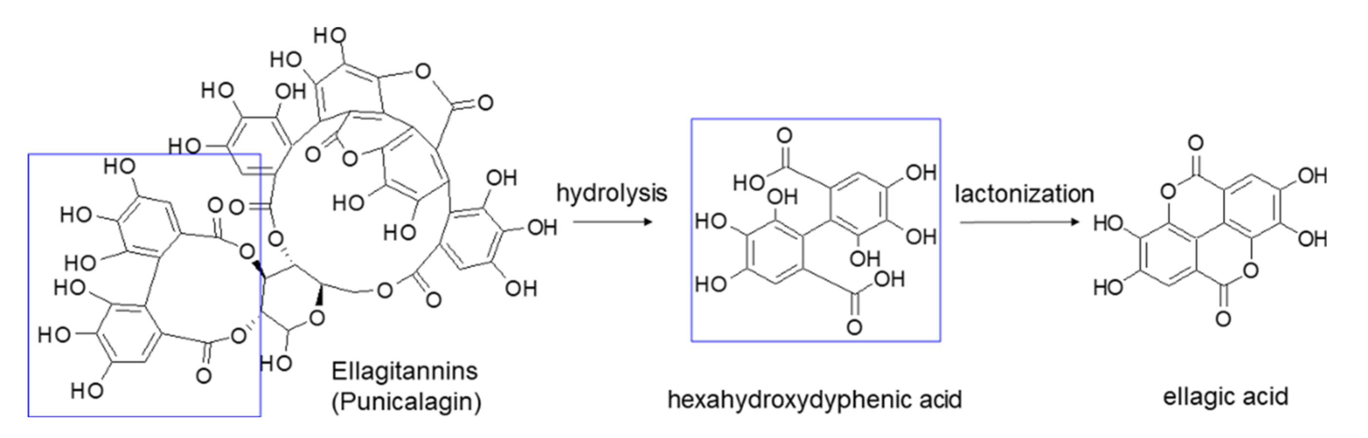
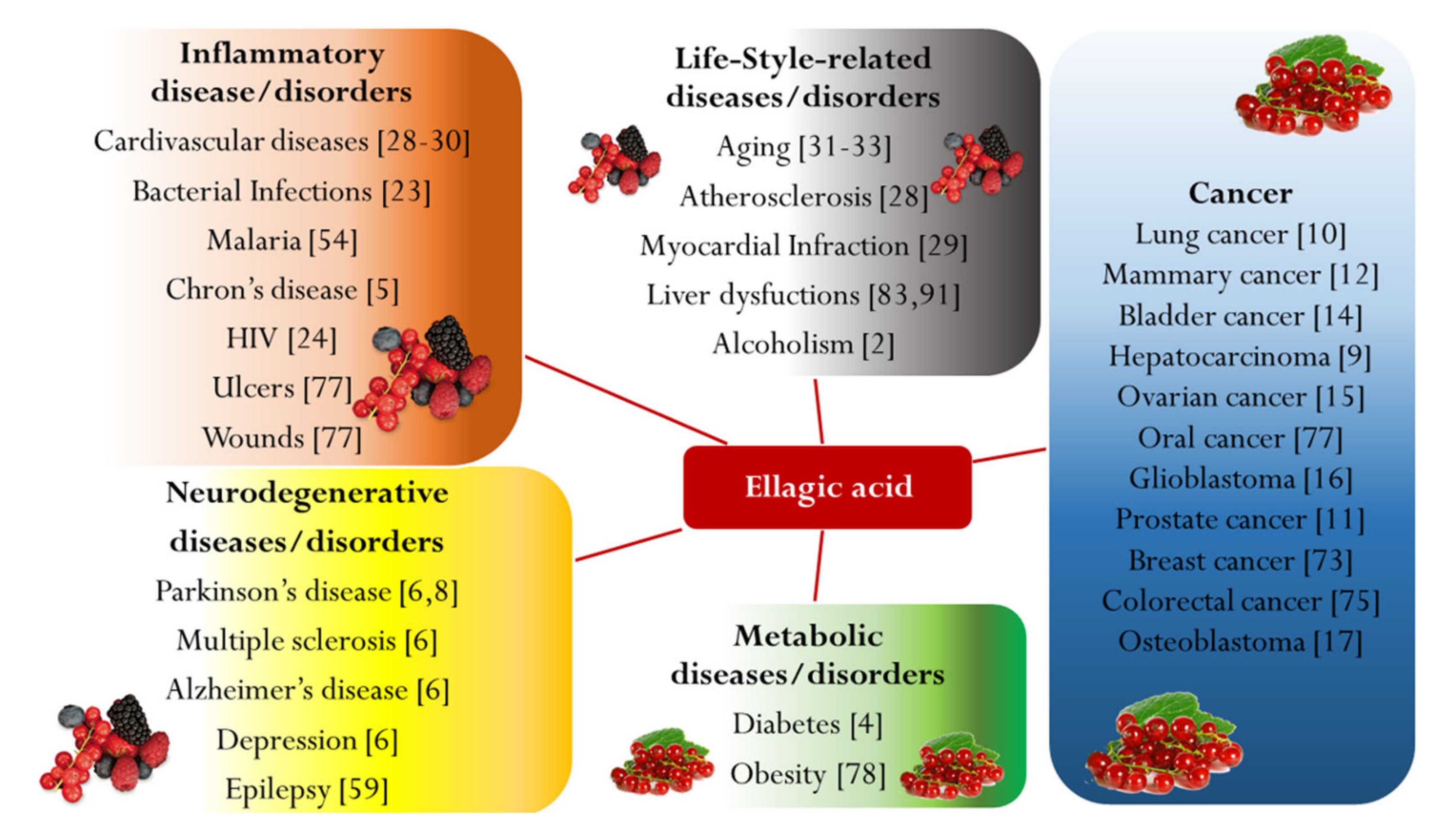
Table 1.
Solubility of ellagic acid in various vehicles.
| Vehicles | Solubility (mg/mL) | Temperature | Ref. | |
|---|---|---|---|---|
| Solvents | N-methyl-2-pyrrolidone | 25 | 37 °C | [36] |
| DMSO | 2.5 | 37 °C | [36] | |
| Pyridine | 2.0 | 37 °C | [36] | |
| Methanol | (671 ± 17) × 10−3 | 37 °C | [36] | |
| Ethanol | 1.02 ± 0.04 | 25 °C | [96] | |
| Cosolvents | PEG 200 | 4.178 | 25 °C | [94] |
| PEG 400 | 11.0 ± 0.5 | 25 °C | [96] | |
| Propylene glycol | 2.1 ± 0.1 | 25 °C | [96] | |
| Oils | Palmester 3575 1 | 0.030 | 25 °C | [94] |
| Cottonseed oil | 0.005 | 25 °C | [94] | |
| Soybean oil | 0.29 ± 0.01 | 25 °C | [95] | |
| Castor oil | 1.63 ± 0.07 | 25 °C | [96] | |
| Oleic acid | 0.29 ± 0.01 | 25 °C | [96] | |
| Ethyl oleate | 2.34 ± 0.06 | 25 °C | [96] | |
| Surfactants | Tween 20 | 1.605 | 25 °C | [94] |
| Sucrose esters | 0.115 | 25 °C | [94] | |
| Isopropyl myristate | 1.94 ± 0.07 | 25 °C | [96] | |
| Cremophor RH40 2 | 2.5 ± 0.1 | 25 °C | [96] | |
| Tween 80 | 3.5 ± 0.1 | 25 °C | [96] | |
| Lecithin | 0.085 ± 0.004 | 25 °C | [96] | |
| Poloxamer F68 | 0.036 ± 0.002 | 25 °C | [96] | |
| Aqueous Solutions | Phosphate buffer pH 7.4 | (33 ± 16) × 10−3 | 37 °C | [36] |
| Phosphate buffer pH 6.8 | (11.1 ± 0.4) × 10−3 | 25 °C | [96] | |
| Acetate buffer pH 4.5 | (6.9 ± 0.3) × 10−3 | 25 °C | [96] | |
| Distilled water | (8.2 ± 0.4) × 10−3 | 25 °C | [96] | |
| HCl 0.1 M in water | (1.03 ± 0.06) × 10−3 | 25 °C | [96] |
1 Caprylic/capric triacylglycerols 2 Polyoxyl 40 hydrogenated castor oil.
Table 2.
EA micro-sized formulations and their distinctive characteristics.
| Formulation Type | Fabrication Method | Excipients | Mean Size | Remarkable Features | Highlights | Ref. |
|---|---|---|---|---|---|---|
| Micro-sized EA | Anti-solvent precipitation | - | n.a. | Use of a syringe pump Good dispersion | In vitro and in vivo inhibition of blood parasites | [54] |
| Micro-sized EA | Supercritical anti-solvent process | Eudragit® L 100 | 3.73 µm | Co-precipitate product EA content 49% Residual NMP 148 ppm | Increased EA dissolution rate | [55] |
| Amorphous solid dispersion | Freeze drying | Maltodextrin | n.a. | Use of cloudberry extract | Higher storage stability up to 32 days Food supplement formulation | [56] |
| Amorphous solid dispersion | Spray drying | Hydroxypropyl-methyl cellulose acetate succinate | n.a. | EA solubility 280 µg/mL EA content 25%Stable supersaturated EA solution at pH 6.8 | pH-sensitive polymer Minimal release in the stomach, quite fast at pH 6.8 (35% after 0.5 h) | [57] |
| Amorphous solid dispersion | Spray drying | Pectin | 10 µm | EA solubility 63 µg/mL EA content 21% No organic solvent used | High biocompatibility Suitable to formulate antioxidant-rich functional food | [58] |
| Amorphous solid dispersion | Spray drying | Alginic acid | 670 nm | EA solubilized in basic solution Crosslinking with CaCl2 Complete release after 8 h | Highly biocompatible formulation Improved in vivo neuroprotective and anticonvulsant effect in orally treated mice | [60] |
| Polymeric microspheres | Emulsion-evaporation technique in oil phase (acetone/light liquid paraffin) | Eudragit® P-4135F | 113 µm | EA content 35% EE 81% Total release after 6 h | pH responsive release Ileocaecal targeting | [70] |
| Polymeric microspheres | Ionotropic gelation by sodium alginate | Chitosan | 4.36 µm | EE 29% Sustained release | Obesity prevention and treatment | [78] |
| Peptide microtubes | Self-assembling | bis(N-α-amido threonine)-1, 5-pentane dicarboxylate | 0.5–1 µm | EE 80% Rate release dependent on the EA deprotonation process at different pH | High biocompatibility Enhanced antibacterial activity | [83] |
| Phospholipidvesicles | Anti-solvent precipitation | Hydrogenated soy phosphatidyl-choline | 1–3 µm | EE 29% | High biocompatibility Liver protection Relative AUC increase by2.8-fold | [83] |
Table 3.
EA nano-sized formulations and their distinctive characteristics.
| Formulation Type | Fabrication Method | Excipients | Mean Size | Remarkable Features | Highlights | Ref. |
|---|---|---|---|---|---|---|
| Nano-sized EA | Anti-solvent precipitation | - | 428 nm | EA water solubility 11.67 µg/mL Lyophilized product with maltodextrin as diluent Residual NMP 405 ppm | Higher radical scavenging activity Enhanced relative AUC by 2 times | [52] |
| Polymer nanospheres | Emulsion-diffusion-evaporation | PLGA or PCL | 125 nm | EA content 62% Slow EA release (about 24% after 6 days) | Sustained release for 20 days Good stability Potential prophylaxis system Higher in situ uptake and greater in vivo nephron-protection in CyA-treated rats | [22,72] |
| Dendrimer | Self-assembling | Aminoacid-modifed hetero dendrimer | 70 nm | EA solubility 9 mg/mL EA content 53% | EA solubility increase | [58] |
| Inclusion complex | Freeze drying | HP-β-CD | n.a. | Formation of 1:2 EA:HP-β-CD complex Increased total EA solubility up to 54.40 µg/mL 60% EA released after 0.5 h | Enhanced in vivo anti-inflammatory effect | [61] |
| Inclusion complex | Freeze drying | β-CD | n.a. | Formation of 1:2 EA:β-CD complex Increased EA solubility up to 39.14 µg/mL Less than 30% EA released after 0.5 h | Enhanced in vivo anti-inflammatory effect | [62,63] |
| Inclusion complex | Overnight shaking | β-CD nanosponge | 423 nm | EA solubility up to 49.79 µg/mL EA content 69% Prolonged release | High biocompatibility Increased relative AUC by 2.2-fold | [64] |
| Inclusion complex | Stirring-ultrasonic and final freeze drying | HP-β-CD | n.a. | Optimized production process EE 84% | Antibacterial activity | [67] |
| Inclusion complex | Precipitation | Arene-Ru metalla-prisms | n.a. | Yield 92% High complex stability | Enhanced antitumor activity against A549 cells | [69] |
| Polymeric nanospheres | Double emulsion-evaporation (w/o/w) | PLGA-PEG | 175 nm | Opsonization avoided Prolonged circulation time in blood Suitable for i.v. administration | Inhibition of breast cancer cell growth More than 2-fold IC50 reduction in MCF-7 cells | [73] |
| Polymeric nanospheres | Emulsion-evaporation in aqueous phase | PLGA coated with chitosan and PEG | 255 nm | Opsonization avoided Sustained release Suitable for i.v. administration | 3-fold IC50 reduction in HepG2 and HCT 116 cells | [74] |
| Polymeric nanospheres | Emulsion-diffusion-evaporation | PCL | 193 nm | EA content 58% EE 66% Slow EA release: approx. 48% after 8 days | Long-term release 6.9-fold cytotoxicity increase against HCT 116 cells Improved relative AUC by 3.6 times | [75] |
| Polymeric nanospheres | Ionic gelation by sodium tripolyphosphate | Chitosan | 176 nm | EA content 33% Rapid release up to 8 h, then more controlled up to 48 h | More than 3-fold IC50 reduction in KB cells Faster clotting time | [76,77] |
| Polymeric nanocapsules | Anti-solvent coprecipitation | Zein | 72 nm | Shell thickness of 20 nm 326 mg EA loaded per 1 g of excipient Sustained release up to 6 days | High biocompatibility Enhanced in vivo anti-inflammatory effect Relative AUC increased by 8.7-fold | [79] |
| Adsorption complex | Overnight shaking | Functionalized graphene oxide | Sheets ranged from 20 to 120 nm | Easy EA loading by physisorption EA solubility up to 610 µg/mL 1.22 g EA loaded per 1 g of excipient 23% EA released at pH 4 38% at pH 10 after 72 h | Enhanced in vitro cytotoxicity against MCF7 and HT29 cells | [85] |
| Solid lipid nanoparticle | Hot homogenization method | Precirol® Poloxamer 407 Tween 80 | 100 nm | EE 89% EA content 36% Initial burst effect (>40%) followed by a sustained release | Enhanced antitumor activity against PC3 cells | [89] |
| Liposome | Overnight mixing | Phospholipid nanoparticles solution (10%) in form of nanospheres | n.a. | EE 60% Good EA protection in different pH buffers and metal ion containing solutions | For nutritional supplements as adjuvant therapy in cancer | [91] |
| Self-nanoemulsifying delivery systems | Preliminary EA and soy lecithin complex obtained by anti-solvent precipitation | Soy lecithin Captex® 500 Cremophor® RH40 PEG 400 Tocopherol | 106 nm | EE 95% Fast release of nearly 95% after 1 h | Improved ex vivo intestinal permeability | [93] |
| Self-nanoemulsifying delivery systems | Vortex mixing | Palmester® 3575 Tween 80 PEG 400 | 120 nm | 2.5 mg/mL EA loading into SNEDDS Fast release | Food-grade components Increased relative AUC by 6.6-fold | [94] |
| Self-nanoemulsifying delivery systems | Vortex mixing | Ethyl oleate Tween 80 PEG 400 | 45 nm | 4 mg/g EA loading into SNEDDS Adjunct of PVP 0.5% as stabilizer Less fast release | Enhanced in vitro and in vivo radical scavenging activity | [96] |
| Polymeric nanospheres | Emulsion-diffusion-evaporation | PLGA | 259 nm | Fixed formulation with CoQ10 EE of 70% for EA and of 72% for CoQ10 | Prolonged control on cholesterol levels in rats | [98] |
| Polymeric nanospheres | Self-assembling and dialysis method for drug loading | Poly (N-iso-propylacrylamide-PEG acrylate) | 200 nm | Combined formulation with paclitaxel Controlled release (only 8% after 2 h) EE 98% for EA and 92% for paclitaxel | Enhanced in vitro cytotoxicity against MCF-7 cells | [99] |
| Nanostructured lipid carrier | Hot emulsification-ultrasonication | Almond oil Compritol® ATO Phosphatidylcholine Gelucire® | 85 nm | Co-delivery of EA, fluvastatin, alpha lipoic acid Total drug released within 3 h | Enhanced in vitro cytotoxicity against PC3 cells | [100] |
| Polymeric nanoparticles | EA nanocrystallization by anti-solvent precipitation + polyelectrolyte electrostatic complexation | Lactoferrin Chondroitin sulfate | 192 nm | Inhalable combined formulation with doxorubicin | Low cost of production Easy scalability Excipients usable in food products | [102] |
© 2020 by the authors. Licensee MDPI, Basel, Switzerland. This article is an open access article distributed under the terms and conditions of the Creative Commons Attribution (CC BY) license (http://creativecommons.org/licenses/by/4.0/).
Share and Cite
MDPI and ACS Style
Zuccari, G.; Baldassari, S.; Ailuno, G.; Turrini, F.; Alfei, S.; Caviglioli, G. Formulation Strategies to Improve Oral Bioavailability of Ellagic Acid. Appl. Sci. 2020, 10, 3353. https://doi.org/10.3390/app10103353
AMA Style
Zuccari G, Baldassari S, Ailuno G, Turrini F, Alfei S, Caviglioli G. Formulation Strategies to Improve Oral Bioavailability of Ellagic Acid. Applied Sciences. 2020; 10(10):3353. https://doi.org/10.3390/app10103353
Chicago/Turabian StyleZuccari, Guendalina, Sara Baldassari, Giorgia Ailuno, Federica Turrini, Silvana Alfei, and Gabriele Caviglioli. 2020. "Formulation Strategies to Improve Oral Bioavailability of Ellagic Acid" Applied Sciences 10, no. 10: 3353. https://doi.org/10.3390/app10103353
Note that from the first issue of 2016, this journal uses article numbers instead of page numbers. See further details here.