
Poly(α-hydroxy Acids)-Based Cell Microcarriers


Department of Mining-Metallurgy Engineering and Materials Science & POLYMAT, Faculty of Engineering, University of the Basque Country (UPV/EHU), Bilbao 480130, Spain
*
Author to whom correspondence should be addressed.
Appl. Sci. 2016, 6(12), 436; https://doi.org/10.3390/app6120436
Submission received: 27 October 2016
/
Revised: 10 December 2016
/
Accepted: 12 December 2016
/
Published: 16 December 2016
(This article belongs to the Special Issue Biodegradable and Biocompatible Nanoparticles)
Abstract
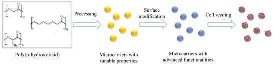
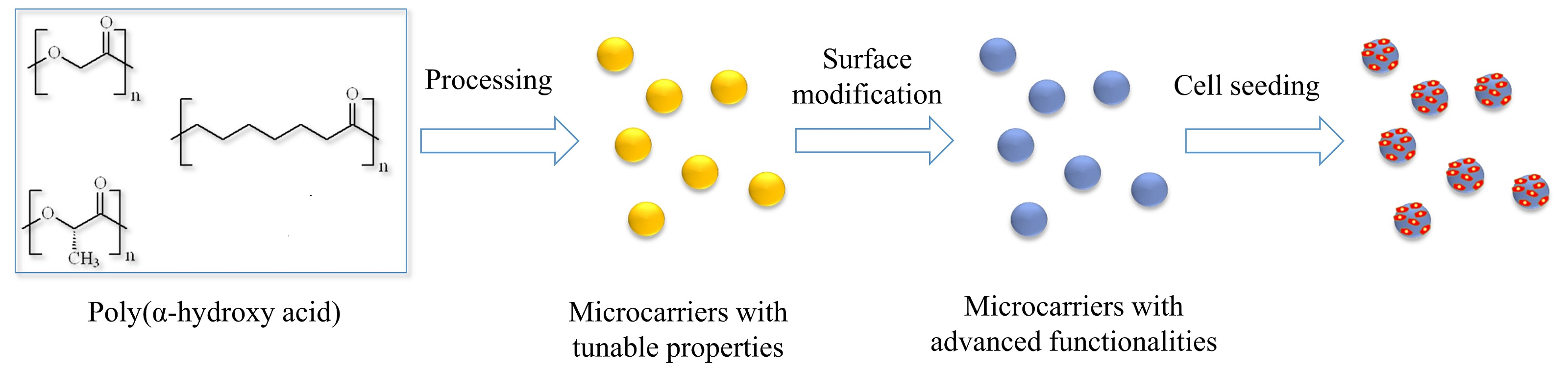
:Biodegradable poly(α-hydroxyacids) have gained increasing interest in the biomedical field for their use as cell microcarriers thanks to their biodegradability, biocompatibility, tunable mechanical properties/degradation rates and processability. The synthesis of these poly(α-hydroxyacids) can be finely controlled to yield (co)polymers of desired mechanical properties and degradation rates. On the other hand, by simple emulsion-solvent evaporation techniques, microspheres of controlled size and size distribution can be fabricated. The resulting microspheres can be further surface-modified to enhance cell adhesion and proliferation. As a result of this process, biodegradable microcarriers with advanced functionalities and surface properties that can be directly employed as injectable cell microcarriers are obtained.
1. Introduction
In 1967, van Wezel proved that microscopic particles, eventually termed “microcarriers”, could serve as a substrate for adherent cells to attach to and proliferate [1]. In this first study, dextran-based positively charged diethylaminoethyl-Sephadex (DEAE-Sephadex) microparticles were employed for the attachment and growth of several cell-lines and primary cells. In view of the results obtained, the possibility of transferring the intrinsic advantages from suspension culture to adherent cells was foreseen. Adherent cells have traditionally been expanded into monolayer systems that, due to the inherent low ratio of surface to volume, show limited scalability. Thus, the combination of microcarriers and bioreactors represent, accordingly, a promising approach to scale-up the expansion of cells to clinically relevant quantities. Moreover, several culture parameters (e.g., pH, temperature, CO2, etc.) can easily be monitored and controlled in these bioreactors, which facilitate the optimization and automation of the cell culture process [2,3]. In these systems, cells are also able to undergo “bead-to-bead” migration so sub-cultivation can be performed in the absence of proteolytic enzymes (e.g., trypsin) by simply adding more microcarriers to the culture [4].
Apart from the aforementioned advantages, microcarriers have also been successfully employed to keep the phenotype of several cells, which cannot be always achieved in a monolayer culture. For example, tenocytes expanded in a monolayer usually display a more rounded morphology, and the ratio of type III to type I collagen increases with passages. In contrast, human tenocytes cultured on Cytodex microcarriers in a spinner flask bioreactor [5] were able to proliferate over two weeks and were still able to synthesize type I collagen and decorin. When cultured in a monolayer, chondrocytes also tend to lose their phenotype, acquiring a fibroblastic-like appearance and show a downregulation in the expression of hyaline cartilage markers, aggrecan and collagen type II. However, when cultured on several commercially available microcarrier (e.g., CultiSpher, Cytodex, Hillex, etc.) chondrocytes were able to keep their rounded morphology and to secrete collagen type II after 14 days [6,7]. In another study, human adipose-derived stem cells (hADSCs) showed upregulation of pluripotent gene markers (Oct4, Sox2, Nanog, Rex1) and enhanced pro-angiogenic properties, determined by the tubular network formation ability of human umbilical vein endothelial cells (HUVECs) in the presence of hADSCs seeded on microcarriers when cultured on gelatin microspheres with respect to hADSCs cultured in a monolayer [8].
Several studies compared the performance of cells in commercially available microcarriers [9,10], and these are summarized in Table 1. Since each cell type responds differently to different microcarriers, the choice of the microcarrier will be directly determined by the particular application under consideration. Apart from the aforementioned commercially available microcarriers, research has also focused on the development of other kinds of microcarriers with improved performance and advanced functionalities [11,12,13,14,15,16,17]. For the present review, microcarriers that are made out of biodegradable and biocompatible poly(α-hydroxyacids) will be considered. These materials have attracted increasing interest for their use as microcarriers due to their biodegradability, biocompatibility, tunable mechanical properties/degradation rates and possibility of being employed directly as injectable cell microcarriers in cell-based therapies. For the regeneration of several tissues cell-based therapies have been studied, particularly for those that show limited capacity to spontaneously regenerate, for example, intervertebral discs [18], cartilage [19] and the nervous system [20], among others. The direct injection of living cells in the absence of supporting biomaterials has shown limited efficacy in clinical trials because of poor cell survival, uncontrolled differentiation, low retention/integration into the host tissue, etc. Therefore, a combination of cells with biomaterials has been proposed as a strategy to overcome the aforementioned obstacles. Among these, injectable systems such as hydrogels [21] or microcarriers have shown promising results.
The present review first considers the current strategies that have been adopted for the synthesis of biodegradable and biocompatible poly(α-hydroxyacids) with tunable mechanical properties and degradation rates. Then, the fabrication methods that yield micron-sized particles are briefly summarized. Finally, how these microparticles have been applied as cell microcarriers in biomedical applications are considered.
2. Polylactide and Its Copolyesters: Towards Tunable Mechanical Properties and Degradation Rates
Among the various biodegradable and/or bioresorbable synthetic polymers employed in the fabrication of cell microcarriers, poly(lactide) (PLA) and PLA-based copolyesters have proven to be the most attractive and useful. PLA is a high strength and high modulus thermoplastic, which can be easily processed by conventional thermoplastic techniques like injection moulding, blow moulding, thermoforming and extrusion [22]. Figure 1 illustrates the PLA cycle from its synthesis to its hydrolytic degradation that results in the formation of lactic acid.
PLAs obtained by ring-opening polymerization (ROP) are the most commonly studied examples due to the possibility of an accurate control of the chemistry and production of high molecular weight polymers, compared to the other polymerization strategy (polycondensation). Lactide, which is obtained by the polycondensation of lactic acid followed by a depolymerization into the dehydrated cyclic dimer (Step I in Figure 1) (3,6-dimethyl-1,4-dioxane-2,5-dione), can be polymerized by ring-opening into high molar polymers (Step II in Figure 1). Due to the two stereoforms of lactic acid, the corresponding optically active lactide can be found in two different versions: d,d-lactide and l,l-lactide. In addition, lactide can be formed from one d- and one l-lactic acid molecule yielding d,l-lactide (meso-lactide). PLA is degraded by hydrolytic cleavage of the backbone ester bonds, converting long polymer chains into shorter ones (Step III in Figure 1) ultimately to low molecular weight water soluble oligomers and monomers (Step IV in Figure 1) [23]. Chain scission results in the formation of carboxylic end-groups that, due to their acidic nature, will enhance the rate of further hydrolysis.
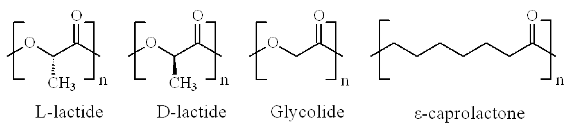
The simplest polymer in this category is the homopolymer of one stereoisomer of lactide, poly(l-lactide) (PLLA) or poly(d-lactide) (PDLA), which show a glass transition and melting temperature of about 55 °C and 175 °C, respectively [24]. It is well-known that crystalline regions in the polymer structure are highly resistant to hydrolytic degradation in regard to amorphous regions. Thus, the degradation and reabsorption rates of the homopolymers are relatively slow due to their high crystallinity [25,26,27]. In order to reduce the crystallinity of PLLA or PDLA, accelerate the degradation rate and avoid the formation of highly-resistant crystalline fragments, the copolymerization of l-lactide with other monomers has been proposed [28]. The chemical structures of the monomeric units that will be analyzed in this section are illustrated in Figure 2.
Apart from the homopolymer of one stereoisomer of lactide presented above, other polymers in this category are the copolymers of lactides of different stereoforms, with the combinations mainly affecting the polymer properties. In this sense, various studies [29,30] have reported a decrease in crystallinity fraction in poly(d,l-lactide) (PDLLA) copolymers when reducing the optical purity (approaching to the racemic mixture of l- and d-enantiomers). As the hydrolytic degradation occurs faster in amorphous regions than in crystalline regions, the reduction in the crystallinity fraction in these polymers clearly accelerate the degradation rate of PLAs. Many studies described in the literature on polylactides are aimed at the hydrolytic degradation of PDLLA 50:50, the copolymer resulting from the synthesis of a racemic mixture (50:50) of l- and d-enantiomers. Alexis et al. [31] concluded that the degradation rate of PDLLA 50:50 was almost 10 times faster than that of PLLA, with degradation rates (K) of 0.0088 and 0.0009 day−1, respectively. Other authors have also studied hydrolytic degradation of PDLLA copolymers with various l/d enantiomeric compositions. Sabbatier et al. [32] worked with two PLLAs with a slightly different d-lactide content (1.4% vs. 3.8%). Although the difference in composition between these two materials was slight, the in vitro degradation study revealed a completely different hydrolysis rate. In this sense, PLLA containing 1.4% of d-lactide suffered ~25% molecular weight loss over 90 days submerged in water, whereas PLLA containing 3.8% of d-lactide underwent a ~45% of molecular weight loss over the same time period, as determined by nuclear magnetic resonance (NMR). Saha and co-workers [33] analyzed the hydrolysis of three PLLAs with a different content of d-lactide units (0.0%, 0.2% and 1.2%). The obtained K values of these three materials were 0.00043 day−1 for PLLA containing 0% of d-lactide, 0.00080 day−1 for PLLA containing 0.2% of d-lactide and 0.00142 day−1 for PLLA containing 1.2% of d-lactide. All of these results demonstrate that the incorporation of d-lactide to PLLA (or vice versa) is a valid strategy for reducing the crystallinity of the final product, thus the degradation rate was accelerated and the formation of crystalline residues avoided.
Lactide and glycolide copolymers have been also considered as a strategy for increasing the hydrolytic degradation rate of PLAs. Polyglycolide (PGA) is a more hydrophilic polymer [34], and it is less resistant to hydrolytic degradation, probably due to the lack of a pendant voluminous methyl group that could hinder the absorption of water and the corresponding cleavage of ester bonds in PLAs. Alexis et al. [31] compared the degradation behavior of PLLA, PDLLA (50:50), poly(l-lactide-co-glycolide) (80:20) (PLGA) copolymer and poly(d,l-lactide-co-glycolide) (53:47) (PDLGA) copolymer. As previously stated, PDLLA (K = 0.0088 day−1) displayed a much faster degradation rate than PLLA (K = 0.0009 day−1) because of its completely amorphous character. The PLGA copolymer showed a K of 0.0127 day−1, demonstrating that the incorporation of glycolide units and the subsequent reduction in crystallinity greatly increases the degradation rate of PLLA. PLGA suffered microstructural rearrangements during degradation and the material shifted from completely amorphous to semi-crystalline due to the reorganization of the l-lactide chains. Finally, the PDLGA copolymer presented a K of 0.0506 day−1 due to the incorporation of glycolide units and the preservation of its amorphous nature during the degradation study. This same effect was also observed in other studies [35,36]. Lu et al. [35] worked with PDLGA porous tissue-engineering scaffolds with different compositions (85:15 vs. 50:50). Scaffolds of PDLGA 85:15 had half-degradation times (defined as the time required to reach the half of the initial molecular weight) of around 11 weeks, whereas scaffolds of PDLGA 50:50 had half-degradation times of around three weeks. Again, the incorporation of more hydrophilic glycolide units significantly increased both in vitro and in vivo degradation rates. Wu et al. [36] compared the in vitro degradation of tissue-engineering scaffolds made out of PDLLA, PDLGA 85:15 and PDLGA 75:25. As expected, PDLGA 75:25 displayed the highest degradation rate with a value of K equal to 0.153 week−1 (half-degradation time = 2.2 weeks), whereas PDLGA 85:15 and PDLLA showed a value of K equal to 0.074 (half-degradation time = 4.0 weeks) and 0.023 week−1, respectively (half-degradation time = 11.1 weeks).
In view of these results, it could be expected that increasing the glycolide content in PLGA or PDLGA copolymers would always lead to copolymers with faster degradation rates. However, it has to be considered that glycolide units are also prone to form more hydrolytically stable crystalline domains when their content in the copolymers reaches a specific threshold value. For this reason, PLGA or PDLGA copolymers with a composition of 50:50 or similar, where the crystallization capability of both lactide and glycolide units is restricted, usually display higher degradation rates than the homopolymers (PLA or PGA) and the copolymers with higher content of glycolide [37] that are crystallizable.
The (co)polymers presented so far display a very similar mechanical behavior at room and body temperatures, with high elastic modulus and rather low elongation at break values, due to their relatively high glass transition temperatures. These materials are, therefore, clearly inappropriate for numerous tissue-engineering and biomedical applications that require highly flexible biodegradable materials. In these cases, copolymerization of lactide with lower glass transition temperature polymers, such as poly(ε-caprolactone) or poly(trimethylene carbonate), could produce a solution to the above-mentioned needs. In this sense, gradual incorporation of ε-caprolactone units to PLLA or PDLLA polymers shifts the mechanical properties of the material from glassy plastic to elastomeric. Previous studies [38,39] have reported a decrease in elastic modulus from ~1700 MPa to ~12 MPa in poly(l-lactide-co-ε-caprolactone) (PLCL) copolymers with ε-caprolactone content ranging from 0% to 30%. The incorporation of ε-caprolactone also increased the elongation at break of these polymers from ~8% for pure PLLA to ~500% for PLCL with 30% of ε-caprolactone.
Regarding the hydrolytic degradation behavior of poly(d,l-lactide-co-ε-caprolactone) (PDLCL) and PLCL copolymers, the situation is much more complex than in PDLGA or PLGA copolymers, and contradictory results can be found in the literature [40,41,42]. In the case of PDLGA or PLGA copolymers, the addition of glycolide (below the limit where glycolide can also form crystalline domains) accelerates the degradation behavior of PLAs due to the incorporation of hydrolytically less resistant units and a reduction in the crystallinity of PLLA polymers. In PLCL copolymers, the incorporation of ε-caprolactone (below the limit where ε-caprolactone can also form crystalline domains) to PLAs lowers the crystallization capability of PLLA, but will, at the same time, provide more hydrolytically resistant units. Although these two effects are antagonistic, the results found in literature indicate that the reduction in crystallinity affects the degradation behavior to a larger extent [40,41,43]. For example, Garkhal et al. [40] compared the degradation behavior of three PLCLs having 90:10 (90% l-lactide, 10% ε-caprolactone), 75:25 and 50:50 compositions. PLCL 50:50 displayed a completely amorphous character, whereas PLCL 75:25 and PLCL 90:10 had a melting enthalpy (ΔHm) of 22.3 and 39.1 J·g−1, respectively, which correspond to the energy required to melt l-lactide crystals. Samples of PLCL 50:50 lost ~25% of its initial mass after six weeks submerged in phosphate buffered saline (PBS), while PLCL 75:25 lost ~13% and no mass loss was detected for PLCL 90:10. These results clearly indicate that, despite adding more hydrolytically resistant ε-caprolactone units, the reduction in crystallinity played a more important role in the degradation of the studied polymers. A summary of the degradation rates discussed above is shown in Table 2. Considering that various types of samples (i.e., films, plates, three-dimensional scaffolds, etc.) and conditions were employed for these experiments, a direct comparison between different studies cannot be made. In any case, degradation rates of different orders of magnitude have been reported for different polymer formulations, indicating the importance of the polymer composition on the degradation rate.
Apart from the composition of the copolymer, the chain microstructure also determines many important properties of the final material. Depending on the synthesis conditions (reaction temperature and time, catalyst, reaction rates of the monomers, etc.), different chain microstructures, ranging from blocky to random, can be achieved. In block copolymers, a longer average sequence chain length is obtained in comparison to random copolymers, which present a distribution that follows Bernoullian statistics. Apart from these two structures, intermediate ones can also be obtained, with block lengths between those of random and di-block copolymers. Although the impact of chain microstructure on the properties of copolymers is usually underestimated in the literature, several studies have demonstrated the importance that feature has on the mechanical properties [44], thermo-degradative behavior [45], crystalline structure [46], drug release [47] and hydrolytic degradation [41,48,49]. In general, it has been reported that copolymers with the same composition, but blockier chain microstructures undergo slower degradation than those copolymers with more randomized structures. For example, Fernández et al. [41] studied the hydrolytic degradation of two PLCL copolymers with similar composition (74% l-lactide and 26% ε-caprolactone) but different chain microstructure. The blockier copolymer, with an average sequence length of l-lactide (lLA) of 8.16, showed a degradation rate of 0.022 day−1. The PLCL with a more random distribution of sequences, with a lLA of 4.01, showed a degradation rate of 0.034 day−1.
In conclusion, it can be stated that PLA homopolymers display a slow degradation rate, with the formation of undesired crystalline residues that can remain in the body for years. However, several strategies have been considered to solve these problems. Copolymers of lactides of different stereoforms are able to reduce the crystallinity of the homopolymers, leading to faster degradation rates. The copolymerization with hydrolytically less resistant monomers, such as glycolide, will also accelerate the degradation of PLAs and avoid the formation of crystalline domains. Finally, when elastomeric behavior is required, copolymerization of lactide with lower glass transition temperature polymers, such as poly(ε-caprolactone) or poly(trimethylene carbonate), could be employed. Although ε-caprolactone is more resistant to hydrolysis than lactide, the results found in the literature indicate that the reduced crystallinity obtained in these copolymers affects the degradation behavior to a greater extent.
3. Methods for the Fabrication of Microcarriers
Microcarriers made out of biodegradable poly(α-hydroxyacids) have traditionally been fabricated via the single (oil-in-water (O/W)) or double emulsion-(water-in-oil-in-water (W1/O/W2)) solvent evaporation method. In the former, the polymer of interest is dissolved in an organic solvent (e.g., dichloromethane, trichloromethane, etc.) and subsequently added to an aqueous phase containing an emulsifier (e.g., polyvinyl alcohol, sodium dodecyl sulfate, Tween 80, etc.) to form an emulsion by mixing with a stirrer, homogenizer or sonicator (Figure 3). In the latter, a primary W1/O emulsion is first prepared and then emulsified in a W1/O/W2 emulsion. This method has been widely employed for the fabrication of drug/gene/protein micron- or nanosized delivery vehicles thanks to the possibility of incorporating water soluble entities in the internal aqueous phase (W1) [50,51].
The size of the particles developed needs to be considered carefully, since it will play a pivotal role in the application of microparticles as cell microcarriers. Thus, smaller microparticles will provide a larger surface area, and, accordingly, will provide more area for the cells to adhere to. However, very small particles (i.e., in the nano-size range) may be taken up by cells but not be useful as cell microcarriers [52]. Many studies are still being carried out to clarify the mechanism of uptake, but it has been suggested that particles below 200 nm are internalized by receptor–mediate endocytosis, whereas larger ones are internalized by phagocytosis [53]. On the other hand, the size of the microparticles will also determine the release rate and profile of the encapsulated entity (i.e., protein, drug, etc.) [50]. Several process parameters affect the size and size distribution of the resulting microcarriers, such as the concentration of the polymer in the oil phase, concentration/nature of the emulsifier, viscosity of the internal aqueous phase (W1), volume of the external aqueous phase (W2), mixing time/speed, etc. [54,55,56,57]. Of these, the concentration of the polymer in the oil phase and the mixing time/speed of the second emulsion play a major role in determining the particle size and distribution. Accordingly, when the concentration of PCL in dichloromethane was increased from 0.08 to 0.33 g·mL−1, the resulting size of the particles increased from 4.2 to 11.6 µm. Conversely, when the stirring speed of the second emulsion was increased from 6500 to 21,500 rpm, the particle size decreased from 37.9 to 7 µm [54]. Similarly, Benoit et al. [55] reported an increase in particle size from 2.12 to 5.88 µm when the concentration of PCL was increased from 0.5% to 6%. In contrast, the particle sized decreased from 4.40 to 3.26 µm when the mixing time was increased from 2 to 15 min.
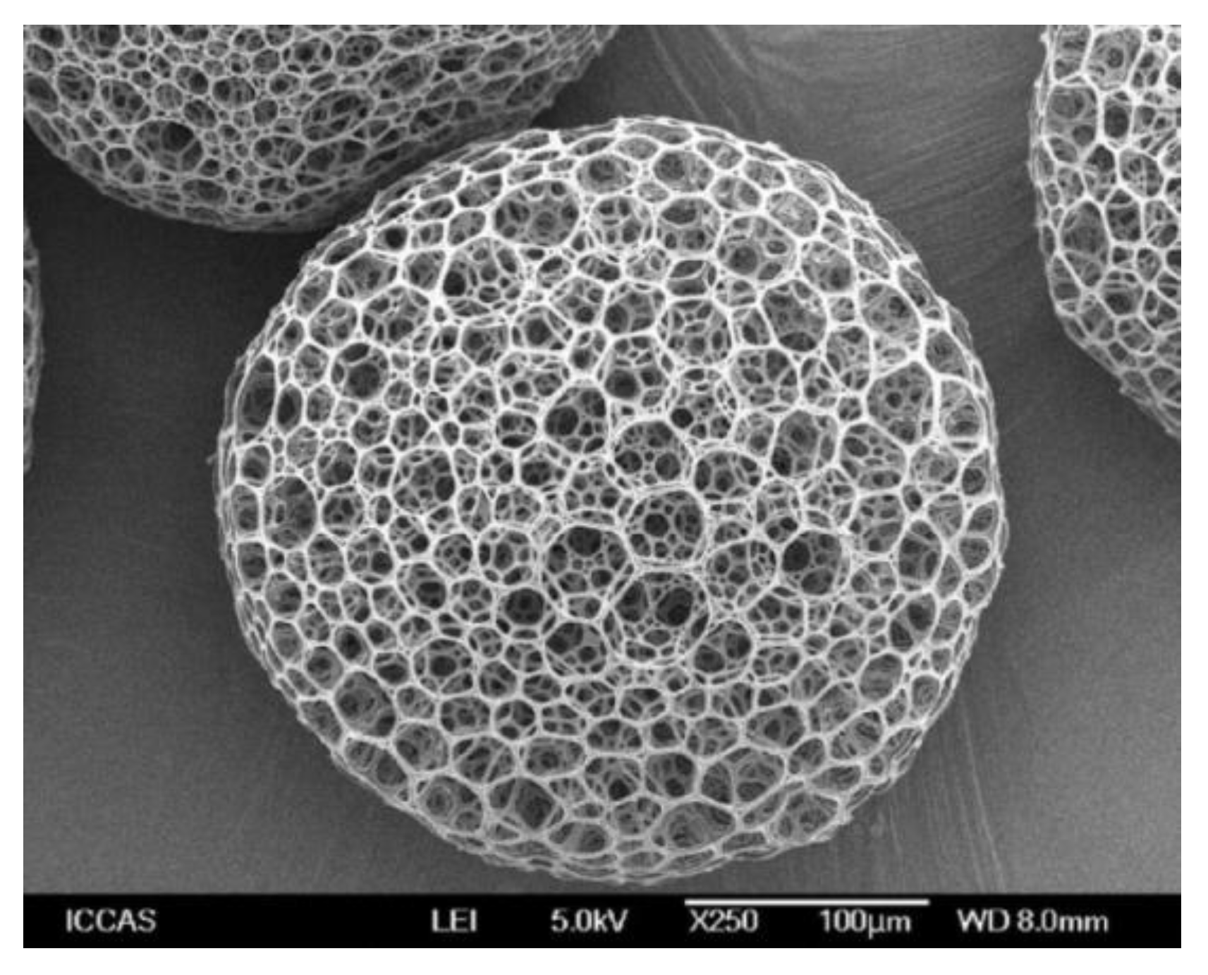
Apart from traditional solid microparticles, there is increasing interest in porous microparticles for use as microcarriers due to their larger surface area, reduced density and enhanced transport of nutrients and oxygen within the porous structure. For the fabrication of porous microcarriers, several modifications have been suggested in the emulsion–solvent evaporation method. The incorporation of ammonium bicarbonate in the internal aqueous phase (W1) yielded microparticles with high interconnected porosity because of the carbon dioxide and ammonia gas bubbles that are produced during the solvent evaporation [58,59,60,61] (Figure 4). As for solid microparticles, several process parameters (e.g., concentration of ammonium bicarbonate in the internal aqueous phase, mixing time/speed, concentration of the polymer in the oil phase, type of solvent employed in the oil phase, post-treatment with NaOH, etc.) can be modulated to tune the pore size and distribution [58,59,60,61]. Other porogens, such as camphene (which is removed by sublimation, leaving a highly porous structure) [62,63] or Pluronic F127 [64] (which is extracted from the structure by contact with water) have been also proposed as promising strategies for the fabrication of porous microparticles.
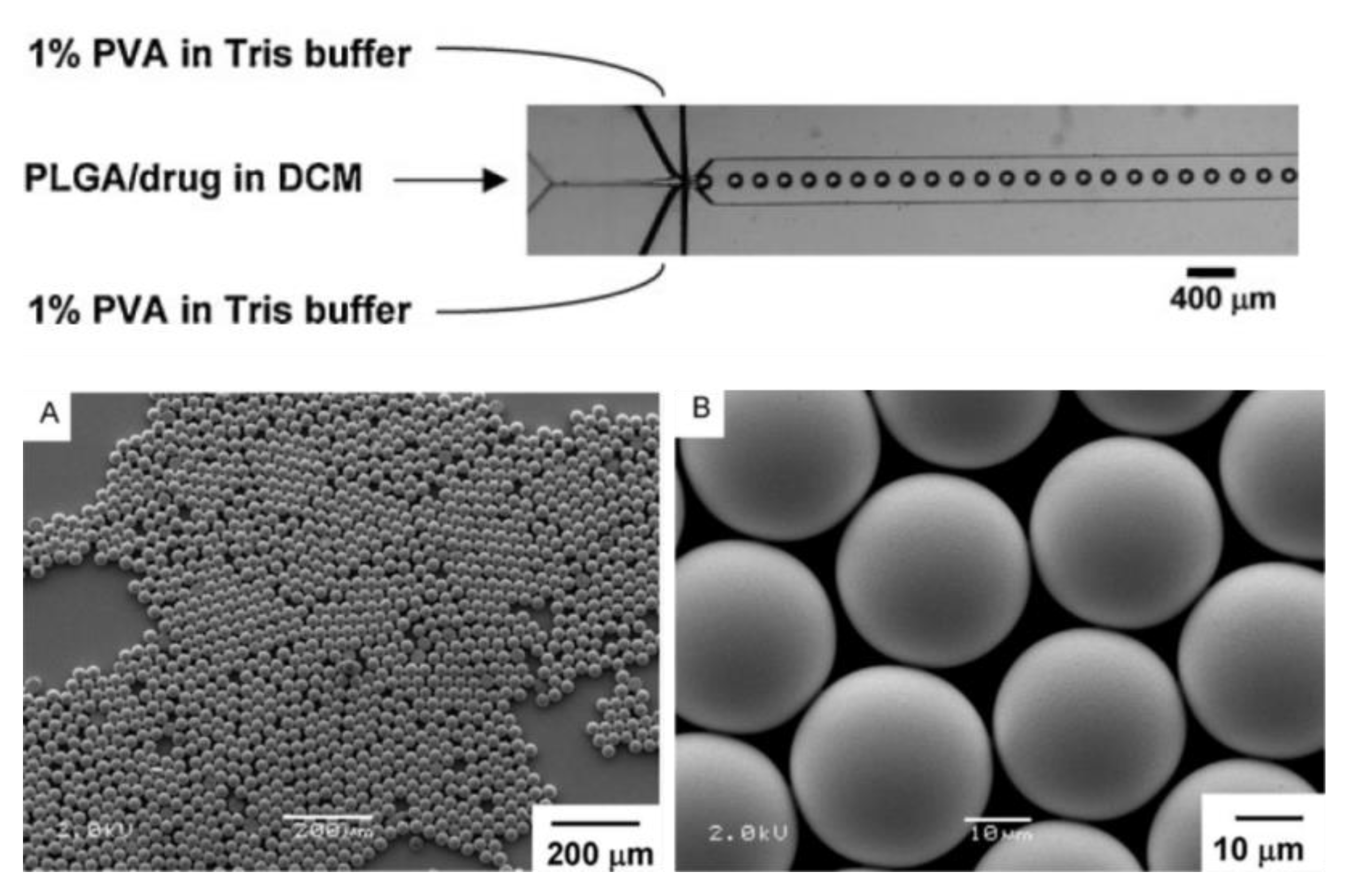
One of the main drawbacks to the emulsion–solvent evaporation described above is the wide particle size distribution that is obtained. This usually requires post-processing via separation methods (e.g., filtration, centrifugation, etc.) for the homogenization of the particle size, and the batch-to-batch variation. An elegant method to overcome this problem is the use of microfluidic devices to generate polymer droplets that further solidify into uniform-sized microparticles [65,66,67]. Apart from synthetic polymers, this method has been satisfactorily exploited for the fabrication of natural polymer-based microparticles [68]. In one of its configurations, known as the flow-focusing device, the dispersed phase (i.e., organic solvent containing the polymer of interest) is injected into the continuous phase (i.e., aqueous solutions with an emulsifier), and, consequently, droplets of uniform size are formed (Figure 5). Finally, by removing the organic solvent, solid particles with narrow particle size distribution are obtained. Changing the polymer concentration in the dispersed phase or modulating the flow-rates allow a precise control over the resulting particle size.
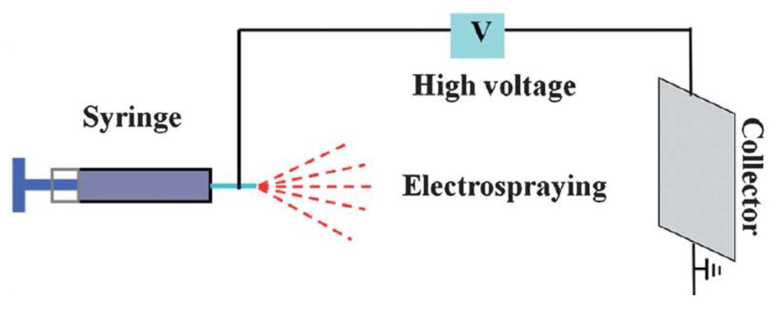
Electrospraying represents a completely different approach for the fabrication of microcarriers made out of biodegradable poly(α-hydroxyacids), and has received less attention than the aforementioned methods, probably due to the need for more complex equipment for its implementation [69,70,71]. In this technique, a polymer solution is loaded into a syringe that flows at a constant rate along a highly charged needle. Then, a high voltage source is used to inject charge of certain polarity into the polymer solution, which is subsequently accelerated in the form of nano- or micro-fiber/droplets toward a collector with opposite polarity once the electrostatic force has overcome the cohesive force of the droplet. As the solution travels, the solvent is evaporated and the particles (or fibers) are recovered from the collector (Figure 6). Polymer concentration, flow rate and applied voltage are commonly modulated to control the size of the resulting particles [70].
The atomization process is similar to electrospraying, and a liquid meniscus is formed here first. This is subsequently split by an external force (e.g., ultrasonic, hydrodynamic, electrical) into monodisperse droplets [72]. In one particular case, ethyl lactate, a green, water-miscible, biodegradable solvent, was employed to prepare polylactide microparticles via the atomization/solvent displacement process [73]. By adjusting the solution and gas flow rates as well as solution concentration, polylactide microparticles in the 60–180 µm range with reduced polydispersity were obtained. More importantly, this method avoids the use of hazardous chlorinated organic solvents and represents a promising approach for the fabrication of polymer particles that are intended for use as cell microcarriers.
In summary, even if several techniques have been considered in the literature, emulsion–solvent evaporation-based methods still remain the gold standard for the fabrication of biodegradable microcarriers.
4. The Use of Poly(α-hydroxyacids) as Cell Microcarriers
The poly(α-hydroxyacids) considered in this review have many inherent advantages for use as cell microcarriers—for example, their biodegradability, biocompatibility, tunable mechanical properties/degradation rates, processability, etc. However, due to their intrinsic hydrophobicity and lack of cell binding sites, surface modifications that promote cell adhesion and proliferation are usually necessary for their use as cell microcarriers [74]. These surface modifications, apart from determining cell adhesion and proliferation, also play an important role in the migration of cells from the microcarrier to the tissue/area of interest. Accordingly, it has been reported that functionalization of the surface of the microcarriers with proteins or peptides, via physisorption, facilitates cell migration compared to a surface modification based on a covalent coating [75].
Hong et al. [76] reported that collagen-coated PLA microspheres provided a better support for chondrocyte adhesion and viability compared to non-coated spheres. For the immobilization of collagen, microspheres were first aminolyzed via incubation in a hexanediamine/n-propanol solution at 60 °C. The amount of NH2 groups on the surface of the microcarriers increased with the incubation time. However, in order to avoid extensive degradation of the polymer matrix, aminolysis treatment was limited to 8 min. After aminolysis, NH2 groups were transferred into aldehyde groups by a treatment with glutaraldehyde, and, finally, collagen type I was covalently coupled via Schiff base formation between the amino groups from collagen and aldehyde groups from the surface of the microcarriers.
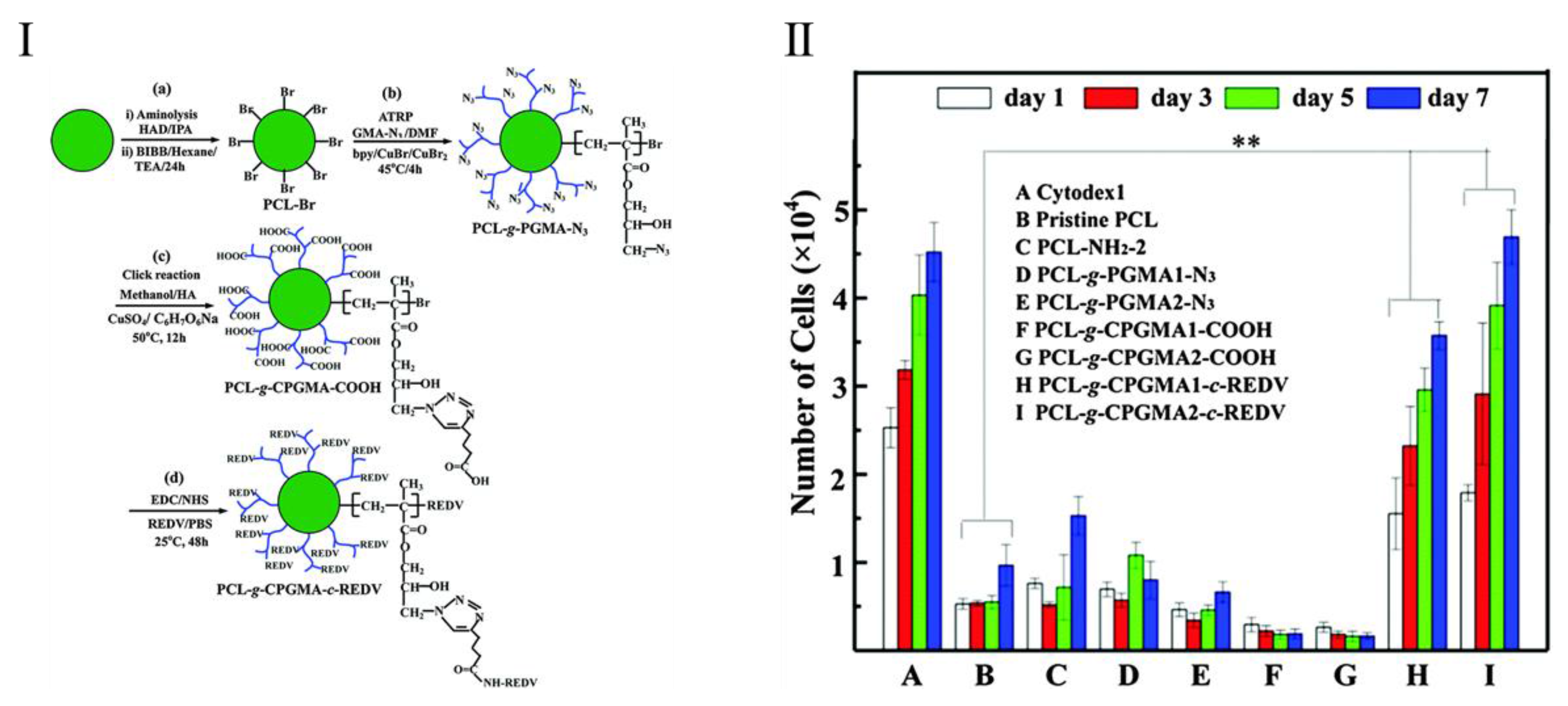
In order to avoid conformational changes associated with large biomacromolecules (i.e., proteins), short peptides can also be incorporated on the surface of microparticles to provide active domains for cell binding [77,78]. These peptides can be easily incorporated via the carbodiimide chemistry on the surface of microparticles bearing carboxylic groups [77] or previously functionalized with gelatin [78]. In these two examples, incorporation of RGD peptide clearly improved the initial adhesion and subsequent proliferation of chondrocytes. In another example [79], REDV peptide was incorporated onto the surface of PCL microspheres to enhance the adhesion and proliferation of endothelial cells (Figure 7). To achieve this, NH2 groups were first incorporated onto the surface of PCL microspheres by aminolysis using 1,6-hexanediamine solution in isopropanol at 40 °C for 60 min. Afterwards, the NH2 groups were employed to immobilize α-bromoisobutyl bromide (BIBB) via a triethylamine (TEA)-catalyzed condensation reaction. Brominated PCL surfaces then underwent a surface-initiated atom-transfer radical-polymerization (ATRP) reaction to graft azido-terminated poly(glycidyl methacrylate) (PGMA-N3) polymer brushes on the surface of the microspheres. After carboxylation of PGMA-N3 polymer brushes via the azide-alkyne click reaction, REDV was covalently immobilized by carbodiimide chemistry. Pristine PCL was found to be an unfavourable substrate for HUVEC attachment and growth. In contrast, after functionalization with REDV the adhesion and proliferation of cells was significantly improved, and similar to that obtained in Cytodex 1, which was used as a positive control.
Short peptides can also be incorporated so as to trigger the differentiation of stem cells toward specific cell lineages [80]. In one particular case, insulin-like growth factor 1 (IGF-1) was immobilized on the surface of PLGA microparticles. The microparticles were first functionalized with polydopamine, which has been employed satisfactorily to coat several nano- and micro-devices [81,82], and further incubated in IGF-1 solutions of different concentrations (1, 10 or 100 ng·mL−1) for 20 min at room temperature. Cell adhesion and osteogenic differentiation, determined by the alkaline phosphatase activity and expression of osteogenic markers (RUNX2 and OPN), were promoted by the aforementioned surface modification. Another strategy to induce the differentiation of cells seeded on the surface of biodegradable microcarriers includes the encapsulation of biological factors, which, once released, induce the differentiation of the surrounding cells [83]. For example, retinoic acid was encapsulated in PLGA microparticles that were used as microcarriers for P19 embryonal carcinoma cells. After seven days in culture, P19 cells show positive staining for a neuronal-specific nuclear protein (NeuN), demonstrating successful differentiation of P19 cells toward neurons.
In view of the promising in vitro results mentioned above, it is not surprising that scientists have gone a step further and tried to prove the feasibility of biodegradable microcarriers for in vivo applications [84,85]. For example, Kang et al. [85] seeded rabbit chondrocytes on PLGA microcarriers that were subsequently injected into a mouse. After nine weeks of implantation, a cartilaginous tissue was formed at the site of implantation. The tissue was characterized by histological and immunohistochemical analysis, confirming the presence of sulfated glycosaminogycans and collagen type II, both of which are major components of the extracellular matrix of cartilage.
5. Conclusions
Cell microcarriers have traditionally been employed to promote the expansion of adherent cells in bioreactor-based culture systems, bringing about scalability and better control over the process parameters. When biodegradable and biocompatible poly(α-hydroxyacids) are employed as cell microcarriers, the resulting systems can be further employed as injectable cell microcarriers, which can potentially be used in cell-based therapies. Advances in polymer synthesis and surface modification strategies can yield polymer microcarriers with tunable mechanical properties/degradation rates and advanced functionalities. From the perspective of materials, the possibility of finely tuning the mechanical properties, degradation rates and degradation by-products has not yet been fully exploited. Thus, only a few lactide-based (co)polymers (i.e., mainly PLLA, PLGA and PLCL) have been employed as biodegradable microcarriers. The use of new synthetic routes or different monomers may open up the possibility of designing poly(α-hydroxyacids)-based microcarriers with improved mechanical properties, and, more importantly, that degrade into harmless degradation by-products (i.e., non-crystalline residues, non-acidic by-products, etc.). Maintaining the phenotype of cells in vitro, such as mesenchymal stem cells, tenocytes, etc., still presents a challenge for the scientific community. With this in mind, it is foreseen that advanced surface modifications or the controlled release of biologically relevant entities may play a major role in the future. Finally, more in vivo studies and preclinical data will be necessary to determine the potential of poly(α-hydroxyacids)-based microcarriers in the clinical field.
Acknowledgments
The authors are thankful for funds from the Spanish Ministry of Innovation and Competitiveness MINECO (MAT2013-45559-P) and the Basque Government, Department of Education, Lingüistic politics and Culture (GIC12/161-IT-632-13).
Conflicts of Interest
The authors declare no conflict of interest.
References
- Van Wezel, A.L. Growth of cell-strains and primary cells on micro-carriers in homogeneous culture. Nature 1967, 216, 64–65. [Google Scholar] [CrossRef] [PubMed]
- Levine, D.W.; Wang, D.I.C.; Thilly, W.G. Optimization of growth surface parameters in microcarrier cell culture. Biotechnol. Bioeng. 1979, 21, 821–845. [Google Scholar] [CrossRef]
- Yuan, Y.; Kallos, M.S.; Hunter, C.; Sen, A. Improved expansion of human bone marrow-derived mesenchymal stem cells in microcarrier-based suspension culture. J. Tissue Eng. Regen. Med. 2014, 8, 210–225. [Google Scholar] [CrossRef] [PubMed]
- Melero-Martin, J.M.; Dowling, M.-A.; Smith, M.; Al-Rubeai, M. Expansion of chondroprogenitor cells on macroporous microcarriers as an alternative to conventional monolayer systems. Biomaterials 2006, 27, 2970–2979. [Google Scholar] [CrossRef] [PubMed]
- Stich, S.; Ibold, Y.; Abbas, A.; Ullah, M.; Sittinger, M.; Ringe, J.; Schulze-Tanzil, G.; Müller, C.; Kohl, B.; John, T. Continuous cultivation of human hamstring tenocytes on microcarriers in a spinner flask bioreactor system. Biotechnol. Prog. 2014, 30, 142–151. [Google Scholar] [CrossRef] [PubMed]
- Malda, J.; van Blitterswijk, C.A.; Grojec, M.; Martens, D.E.; Tramper, J.; Riesle, J. Expansion of bovine chondrocytes on microcarriers enhances redifferentiation. Tissue Eng. 2003, 9, 939–948. [Google Scholar] [CrossRef] [PubMed]
- Malda, J.; Kreijveld, E.; Temenoff, J.S.; van Blitterswijk, C.A.; Riesle, J. Expansion of human nasal chondrocytes on macroporous microcarriers enhances redifferentiation. Biomaterials 2003, 24, 5153–5161. [Google Scholar] [CrossRef]
- Kodali, A.; Lim, T.C.; Leong, D.T.; Tong, Y.W. Cell–microsphere constructs formed with human adipose-derived stem cells and gelatin microspheres promotes stemness, differentiation, and controlled pro-angiogenic potential. Macromol. Biosci. 2014, 14, 1458–1468. [Google Scholar] [CrossRef] [PubMed]
- Lecina, M.; Ting, S.; Choo, A.; Reuveny, S.; Oh, S. Scalable platform for human embryonic stem cell differentiation to cardiomyocytes in suspended microcarrier cultures. Tissue Eng. C Methods 2010, 16, 1609–1619. [Google Scholar] [CrossRef] [PubMed]
- Frauenschuh, S.; Reichmann, E.; Ibold, Y.; Goetz, P.M.; Sittinger, M.; Ringe, J. A microcarrier-based cultivation system for expansion of primary mesenchymal stem cells. Biotechnol. Prog. 2007, 23, 187–193. [Google Scholar] [CrossRef] [PubMed]
- Jin, G.-Z.; Kim, J.-H.; Park, J.-H.; Choi, S.-J.; Kim, H.-W.; Wall, I. Performance of evacuated calcium phosphate microcarriers loaded with mesenchymal stem cells within a rat calvarium defect. J. Mater. Sci. Mater. Med. 2012, 23, 1739–1748. [Google Scholar] [CrossRef] [PubMed]
- Perez, R.A.; El-Fiqi, A.; Park, J.H.; Kim, T.H.; Kim, J.H.; Kim, H.W. Therapeutic bioactive microcarriers: Co-delivery of growth factors and stem cells for bone tissue engineering. Acta Biomater. 2014, 10, 520–530. [Google Scholar] [CrossRef] [PubMed]
- Varani, J.; Dame, M.; Beals, T.F.; Wass, J.A. Growth of three established cell lines on glass microcarriers. Biotechnol. Bioeng. 1983, 25, 1359–1372. [Google Scholar] [PubMed]
- Lam, A.T.-L.; Li, J.; Chen, A.K.-L.; Reuveny, S.; Oh, S.K.-W.; Birch, W.R. Cationic surface charge combined with either vitronectin or laminin dictates the evolution of human embryonic stem cells/microcarrier aggregates and cell growth in agitated cultures. Stem Cells Dev. 2014, 23, 1688–1703. [Google Scholar] [CrossRef] [PubMed]
- Heng, B.C.; Li, J.; Chen, A.K.-L.; Reuveny, S.; Cool, S.M.; Birch, W.R.; Oh, S.K.-W. Translating human embryonic stem cells from 2-dimensional to 3-dimensional cultures in a defined medium on laminin- and vitronectin-coated surfaces. Stem Cells Dev. 2011, 21, 1701–1715. [Google Scholar] [CrossRef] [PubMed]
- Luo, B.; Loh, Q.L.; Chong Wong, M.T.; Tan, N.S.; Choong, C. Bioactivated protein-based porous microcarriers for tissue engineering applications. J. Mater. Chem. B 2014, 2, 7795–7803. [Google Scholar] [CrossRef]
- Yang, Z.; Yuan, S.; Liang, B.; Liu, Y.; Choong, C.; Pehkonen, S.O. Chitosan microsphere scaffold tethered with rgd-conjugated poly(methacrylic acid) brushes as effective carriers for the endothelial cells. Macromol. Biosci. 2014, 14, 1299–1311. [Google Scholar] [CrossRef] [PubMed]
- Pereira, D.R.; Silva-Correia, J.; Oliveira, J.M.; Reis, R.L. Hydrogels in acellular and cellular strategies for intervertebral disc regeneration. J. Tissue Eng. Regen. Med. 2013, 7, 85–98. [Google Scholar] [CrossRef] [PubMed]
- Correia, C.R.; Reis, R.L.; Mano, J.F. Multiphasic, multistructured and hierarchical strategies for cartilage regeneration. In Engineering Mineralized and Load Bearing Tissue; Bertassoni, L.E., Coelho, P.G., Eds.; Springer: Cham, Switzerland, 2015; pp. 143–160. [Google Scholar]
- Tam, R.Y.; Fuehrmann, T.; Mitrousis, N.; Shoichet, M.S. Regenerative therapies for central nervous system diseases: A biomaterials approach. Neuropsychopharmacology 2014, 39, 169–188. [Google Scholar] [CrossRef] [PubMed]
- Thomas, D.; Fontana, G.; Chen, X.; Sanz-Nogues, C.; Zeugolis, D.I.; Dockery, P.; O’Brien, T.; Pandit, A. A shape-controlled tuneable microgel platform to modulate angiogenic paracrine responses in stem cells. Biomaterials 2014, 35, 8757–8766. [Google Scholar] [CrossRef] [PubMed]
- Gupta, A.P.; Kumar, V. New emerging trends in synthetic biodegradable polymers—Polylactide: A critique. Eur. Polym. J. 2007, 43, 4053–4074. [Google Scholar] [CrossRef]
- Edlund, U.; Albertsson, A.C. Polyesters based on diacid monomers. Adv. Drug Deliv. Rev. 2003, 55, 585–609. [Google Scholar] [CrossRef]
- Garlotta, D. A literature review of poly(lactic acid). J. Polym. Environ. 2001, 9, 63–84. [Google Scholar] [CrossRef]
- Bergsma, J.E.; de Bruijn, W.C.; Rozema, F.R.; Bos, R.R.M.; Boering, G. Late degradation tissue response to poly(l-lactide) bone plates and screws. Biomaterials 1995, 16, 25–31. [Google Scholar] [CrossRef]
- Tams, J.; Rozema, F.R.; Bos, R.R.M.; Roodenburg, J.L.N.; Nikkels, P.G.J.; Vermey, A. Poly(l-lactide) bone plates and screws for internal fixation of mandibular swing osteotomies. Int. J. Oral Maxillofac. Surg. 1996, 25, 20–24. [Google Scholar] [CrossRef]
- Tsuji, H.; Ikarashi, K. In vitro hydrolysis of poly(l-lactide) crystalline residues as extended-chain crystallites. Part I: Long-term hydrolysis in phosphate-buffered solution at 37 °C. Biomaterials 2004, 25, 5449–5455. [Google Scholar] [CrossRef] [PubMed]
- Södergård, A.; Stolt, M. Properties of lactic acid based polymers and their correlation with composition. Prog. Polym. Sci. 2002, 27, 1123–1163. [Google Scholar] [CrossRef]
- Sarasua, J.-R.; Prud’homme, R.E.; Wisniewski, M.; le Borgne, A.; Spassky, N. Crystallization and melting behavior of polylactides. Macromolecules 1998, 31, 3895–3905. [Google Scholar] [CrossRef]
- Tsuji, H.; Ikada, Y. Crystallization from the melt of poly(lactide)s with different optical purities and their blends. Macromol. Chem. Phys. 1996, 197, 3483–3499. [Google Scholar] [CrossRef]
- Alexis, F.; Venkatraman, S.; Kumar Rath, S.; Gan, L.-H. Some insight into hydrolytic scission mechanisms in bioerodible polyesters. J. Appl. Polym. Sci. 2006, 102, 3111–3117. [Google Scholar] [CrossRef]
- Sabbatier, G.; le Nouën, D.; Chevallier, P.; Durand, B.; Laroche, G.; Dieval, F. Air spun poly(lactic acid) nanofiber scaffold degradation for vascular tissue engineering: A 1H NMR study. Polym. Degrad. Stab. 2012, 97, 1520–1526. [Google Scholar] [CrossRef]
- Saha, S.K.; Tsuji, H. Effects of molecular weight and small amounts of d-lactide units on hydrolytic degradation of poly(l-lactic acid)s. Polym. Degrad. Stab. 2006, 91, 1665–1673. [Google Scholar] [CrossRef]
- Yoon, J.-S.; Jung, H.-W.; Kim, M.-N.; Park, E.-S. Diffusion coefficient and equilibrium solubility of water molecules in biodegradable polymers. J. Appl. Polym. Sci. 2000, 77, 1716–1722. [Google Scholar] [CrossRef]
- Lu, L.; Peter, S.J.; Lyman, M.D.; Lai, H.-L.; Leite, S.M.; Tamada, J.A.; Vacanti, J.P.; Langer, R.; Mikos, A.G. In vitro degradation of porous poly(l-lactic acid) foams. Biomaterials 2000, 21, 1595–1605. [Google Scholar] [CrossRef]
- Wu, L.; Ding, J. In vitro degradation of three-dimensional porous poly(d,l-lactide-co-glycolide) scaffolds for tissue engineering. Biomaterials 2004, 25, 5821–5830. [Google Scholar] [CrossRef] [PubMed]
- Han, X.; Pan, J. Polymer chain scission, oligomer production and diffusion: A two-scale model for degradation of bioresorbable polyesters. Acta Biomater. 2011, 7, 538–547. [Google Scholar] [CrossRef] [PubMed]
- Fernández, J.; Etxeberria, A.; Sarasua, J.-R. Synthesis, structure and properties of poly(l-lactide-co-ε-caprolactone) statistical copolymers. J. Mech. Behav. Biomed. Mater. 2012, 9, 100–112. [Google Scholar] [CrossRef] [PubMed]
- Hiljanen-Vainio, M.; Karjalainen, T.; Seppälä, J. Biodegradable lactone copolymers. I. Characterization and mechanical behavior of ε-caprolactone and lactide copolymers. J. Appl. Polym. Sci. 1996, 59, 1281–1288. [Google Scholar] [CrossRef]
- Garkhal, K.; Verma, S.; Jonnalagadda, S.; Kumar, N. Fast degradable poly(l-lactide-co-ε-caprolactone) microspheres for tissue engineering: Synthesis, characterization, and degradation behavior. J. Polym. Sci. A Polym. Chem. 2007, 45, 2755–2764. [Google Scholar] [CrossRef]
- Fernández, J.; Larrañaga, A.; Etxeberría, A.; Sarasua, J.R. Effects of chain microstructures and derived crystallization capability on hydrolytic degradation of poly(l-lactide/ε-caprolactone) copolymers. Polym. Degrad. Stab. 2013, 98, 481–489. [Google Scholar] [CrossRef]
- Qian, H.; Bei, J.; Wang, S. Synthesis, characterization and degradation of aba block copolymer of l-lactide and ε-caprolactone. Polym. Degrad. Stab. 2000, 68, 423–429. [Google Scholar] [CrossRef]
- Grijpma, D.W.; Pennings, A.J. (Co)polymers of l-lactide, 1. Synthesis, thermal properties and hydrolytic degradation. Macromol. Chem. Phys. 1994, 195, 1633–1647. [Google Scholar] [CrossRef]
- Fernández, J.; Etxeberria, A.; Ugartemendia, J.M.; Petisco, S.; Sarasua, J.-R. Effects of chain microstructures on mechanical behavior and aging of a poly(l-lactide-co-caprolactone) biomedical thermoplastic-elastomer. J. Mech. Behav. Biomed. Mater. 2012, 12, 29–38. [Google Scholar] [CrossRef] [PubMed]
- Fernández, J.; Etxeberría, A.; Sarasua, J.R. Effects of repeat unit sequence distribution and residual catalyst on thermal degradation of poly(l-lactide/ε-caprolactone) statistical copolymers. Polym. Degrad. Stab. 2013, 98, 1293–1299. [Google Scholar] [CrossRef]
- Dobrzynski, P.; Li, S.; Kasperczyk, J.; Bero, M.; Gasc, F.; Vert, M. Structure−property relationships of copolymers obtained by ring-opening polymerization of glycolide and ε-caprolactone. Part 1. Synthesis and characterization. Biomacromolecules 2005, 6, 483–488. [Google Scholar] [CrossRef] [PubMed]
- Jelonek, K.; Kasperczyk, J.; Li, S.; Dobrzynski, P.; Jarzabek, B. Controlled poly(l-lactide-co-trimethylene carbonate) delivery system of cyclosporine a and rapamycine—The effect of copolymer chain microstructure on drug release rate. Int. J. Pharm. 2011, 414, 203–209. [Google Scholar] [CrossRef] [PubMed]
- Li, S.; Dobrzynski, P.; Kasperczyk, J.; Bero, M.; Braud, C.; Vert, M. Structure−property relationships of copolymers obtained by ring-opening polymerization of glycolide and ε-caprolactone. Part 2. Influence of composition and chain microstructure on the hydrolytic degradation. Biomacromolecules 2005, 6, 489–497. [Google Scholar] [CrossRef] [PubMed]
- Fernández, J.; Larrañaga, A.; Etxeberria, A.; Wang, W.; Sarasua, J.R. A new generation of poly(lactide/ε-caprolactone) polymeric biomaterials for application in the medical field. J. Biomed. Mater. Res. A 2014, 102, 3573–3584. [Google Scholar] [CrossRef] [PubMed]
- Mundargi, R.C.; Babu, V.R.; Rangaswamy, V.; Patel, P.; Aminabhavi, T.M. Nano/micro technologies for delivering macromolecular therapeutics using poly(d,l-lactide-co-glycolide) and its derivatives. J. Controll. Release 2008, 125, 193–209. [Google Scholar] [CrossRef] [PubMed]
- Desai, M.P.; Labhasetwar, V.; Amidon, G.L.; Levy, R.J. Gastrointestinal uptake of biodegradable microparticles: Effect of particle size. Pharm. Res. 1996, 13, 1838–1845. [Google Scholar] [CrossRef] [PubMed]
- Win, K.Y.; Feng, S.-S. Effects of particle size and surface coating on cellular uptake of polymeric nanoparticles for oral delivery of anticancer drugs. Biomaterials 2005, 26, 2713–2722. [Google Scholar] [CrossRef] [PubMed]
- Couvreur, P.; Puisieux, F. Nano- and microparticles for the delivery of polypeptides and proteins. Adv. Drug Deliv. Rev. 1993, 10, 141–162. [Google Scholar] [CrossRef]
- Ibraheem, D.; Iqbal, M.; Agusti, G.; Fessi, H.; Elaissari, A. Effects of process parameters on the colloidal properties of polycaprolactone microparticles prepared by double emulsion like process. Colloids Surf. A Physicochem. Eng. Asp. 2014, 445, 79–91. [Google Scholar] [CrossRef]
- Benoit, M.A.; Baras, B.; Gillard, J. Preparation and characterization of protein-loaded poly(ε-caprolactone) microparticles for oral vaccine delivery. Int. J. Pharm. 1999, 184, 73–84. [Google Scholar] [CrossRef]
- O’Donnell, P.B.; McGinity, J.W. Preparation of microspheres by the solvent evaporation technique. Adv. Drug Deliv. Rev. 1997, 28, 25–42. [Google Scholar] [CrossRef]
- Sahoo, S.K.; Panyam, J.; Prabha, S.; Labhasetwar, V. Residual polyvinyl alcohol associated with poly(d,l-lactide-co-glycolide) nanoparticles affects their physical properties and cellular uptake. J. Controll. Release 2002, 82, 105–114. [Google Scholar] [CrossRef]
- Yang, Y.; Bajaj, N.; Xu, P.; Ohn, K.; Tsifansky, M.D.; Yeo, Y. Development of highly porous large PLGA microparticles for pulmonary drug delivery. Biomaterials 2009, 30, 1947–1953. [Google Scholar] [CrossRef] [PubMed]
- Kim, T.K.; Yoon, J.J.; Lee, D.S.; Park, T.G. Gas foamed open porous biodegradable polymeric microspheres. Biomaterials 2006, 27, 152–159. [Google Scholar] [CrossRef] [PubMed]
- Shi, X.; Sun, L.; Jiang, J.; Zhang, X.; Ding, W.; Gan, Z. Biodegradable polymeric microcarriers with controllable porous structure for tissue engineering. Macromol. Biosci. 2009, 9, 1211–1218. [Google Scholar] [CrossRef] [PubMed]
- Fang, K.; Yang, F.; Zhang, Q.; Zhang, T.; Gu, N. Fabrication of nonporous and porous cationic PLGA microspheres. Mater. Lett. 2014, 117, 86–89. [Google Scholar] [CrossRef]
- Jin, G.-Z.; Park, J.-H.; Seo, S.-J.; Kim, H.-W. Dynamic cell culture on porous biopolymer microcarriers in a spinner flask for bone tissue engineering: A feasibility study. Biotechnol. Lett. 2014, 36, 1539–1548. [Google Scholar] [CrossRef] [PubMed]
- Hong, S.-J.; Yu, H.-S.; Kim, H.-W. Tissue engineering polymeric microcarriers with macroporous morphology and bone-bioactive surface. Macromol. Biosci. 2009, 9, 639–645. [Google Scholar] [CrossRef] [PubMed]
- Kim, H.K.; Chung, H.J.; Park, T.G. Biodegradable polymeric microspheres with “open/closed” pores for sustained release of human growth hormone. J. Controll. Release 2006, 112, 167–174. [Google Scholar] [CrossRef] [PubMed]
- Serra, C.A.; Chang, Z. Microfluidic-assisted synthesis of polymer particles. Chem. Eng. Technol. 2008, 31, 1099–1115. [Google Scholar] [CrossRef]
- Xu, Q.; Hashimoto, M.; Dang, T.T.; Hoare, T.; Kohane, D.S.; Whitesides, G.M.; Langer, R.; Anderson, D.G. Preparation of monodisperse biodegradable polymer microparticles using a microfluidic flow-focusing device for controlled drug delivery. Small 2009, 5, 1575–1581. [Google Scholar] [CrossRef] [PubMed]
- Li, J.; Lam, A.T.-L.; Toh, J.P.W.; Reuveny, S.; Oh, S.K.-W.; Birch, W.R. Fabrication of uniform-sized poly-ɛ-caprolactone microspheres and their applications in human embryonic stem cell culture. Biomed. Microdevices 2015, 17, 105. [Google Scholar] [CrossRef] [PubMed]
- Xu, J.H.; Li, S.W.; Tostado, C.; Lan, W.J.; Luo, G.S. Preparation of monodispersed chitosan microspheres and in situ encapsulation of bsa in a co-axial microfluidic device. Biomed. Microdevices 2009, 11, 243–249. [Google Scholar] [CrossRef] [PubMed]
- Paik, D.C.; Choi, S.W. Entrapment of protein using electrosprayed poly(d,l-lactide-co-glycolide) microspheres with a porous structure for sustained release. Macromol. Rapid Commun. 2014, 35, 1033–1038. [Google Scholar] [CrossRef] [PubMed]
- Bock, N.; Woodruff, M.A.; Hutmacher, D.W.; Dargaville, T.R. Electrospraying, a reproducible method for production of polymeric microspheres for biomedical applications. Polymers 2011, 3, 131. [Google Scholar] [CrossRef] [Green Version]
- Gao, J.; Wong, J.S.-P.; Hu, M.; Li, W.; Li, R.K.Y. Facile preparation of hierarchically porous polymer microspheres for superhydrophobic coating. Nanoscale 2014, 6, 1056–1063. [Google Scholar] [CrossRef] [PubMed]
- Senuma, Y.; Franceschin, S.; Hilborn, J.G.; Tissières, P.; Bisson, I.; Frey, P. Bioresorbable microspheres by spinning disk atomization as injectable cell carrier: From preparation to in vitro evaluation. Biomaterials 2000, 21, 1135–1144. [Google Scholar] [CrossRef]
- Levato, R.; Mateos-Timoneda, M.A.; Planell, J.A. Preparation of biodegradable polylactide microparticles via a biocompatible procedure. Macromol. Biosci. 2012, 12, 557–566. [Google Scholar] [CrossRef] [PubMed]
- Chun, K.W.; Yoo, H.S.; Yoon, J.J.; Park, T.G. Biodegradable PLGA microcarriers for injectable delivery of chondrocytes: Effect of surface modification on cell attachment and function. Biotechnol. Prog. 2004, 20, 1797–1801. [Google Scholar] [CrossRef] [PubMed]
- Levato, R.; Planell, J.A.; Mateos-Timoneda, M.A.; Engel, E. Role of ecm/peptide coatings on sdf-1α triggered mesenchymal stromal cell migration from microcarriers for cell therapy. Acta Biomater. 2015, 18, 59–67. [Google Scholar] [CrossRef] [PubMed]
- Hong, Y.; Gao, C.; Xie, Y.; Gong, Y.; Shen, J. Collagen-coated polylactide microspheres as chondrocyte microcarriers. Biomaterials 2005, 26, 6305–6313. [Google Scholar] [CrossRef] [PubMed]
- Chen, R.; Curran, S.J.; Curran, J.M.; Hunt, J.A. The use of poly(l-lactide) and rgd modified microspheres as cell carriers in a flow intermittency bioreactor for tissue engineering cartilage. Biomaterials 2006, 27, 4453–4460. [Google Scholar] [CrossRef] [PubMed]
- Tan, H.; Huang, D.; Lao, L.; Gao, C. RGD modified PLGA/gelatin microspheres as microcarriers for chondrocyte delivery. J. Biomed. Mater. Res. B Appl. Biomater. 2009, 91B, 228–238. [Google Scholar] [CrossRef] [PubMed]
- Yuan, S.; Xiong, G.; He, F.; Jiang, W.; Liang, B.; Pehkonen, S.; Choong, C. PCL microspheres tailored with carboxylated poly(glycidyl methacrylate)-REDV conjugates as conducive microcarriers for endothelial cell expansion. J. Mater. Chem. B 2015, 3, 8670–8683. [Google Scholar] [CrossRef]
- Gao, T.; Zhang, N.; Wang, Z.; Wang, Y.; Liu, Y.; Ito, Y.; Zhang, P. Biodegradable microcarriers of poly(lactide-co-glycolide) and nano-hydroxyapatite decorated with IGF-1 via polydopamine coating for enhancing cell proliferation and osteogenic differentiation. Macromol. Biosci. 2015, 15, 1070–1080. [Google Scholar] [CrossRef] [PubMed]
- Fernandez-Yague, M.A.; Larrañaga, A.; Gladkovskaya, O.; Stanley, A.; Tadayyon, G.; Guo, Y.; Sarasua, J.-R.; Tofail, S.A.M.; Zeugolis, D.I.; Pandit, A.; et al. Effects of polydopamine functionalization on boron nitride nanotube dispersion and cytocompatibility. Bioconjug. Chem. 2015, 26, 2025–2037. [Google Scholar] [CrossRef] [PubMed]
- Larrañaga, A.; Ramos, D.; Amestoy, H.; Zuza, E.; Sarasua, J.-R. Coating of bioactive glass particles with mussel-inspired polydopamine as a strategy to improve the thermal stability of poly(l-lactide)/bioactive glass composites. RSC Adv. 2015, 5, 65618–65626. [Google Scholar] [CrossRef]
- Newman, K.D.; McBurney, M.W. Poly(d,l lactic-co-glycolic acid) microspheres as biodegradable microcarriers for pluripotent stem cells. Biomaterials 2004, 25, 5763–5771. [Google Scholar] [CrossRef] [PubMed]
- Chung, H.J.; Park, T.G. Injectable cellular aggregates prepared from biodegradable porous microspheres for adipose tissue engineering. Tissue Eng. A 2009, 15, 1391–1400. [Google Scholar] [CrossRef] [PubMed]
- Kang, S.W.; Jeon, O.; Kim, B.S. Poly(lactic-co-glycolic acid) microspheres as an injectable scaffold for cartilage tissue engineering. Tissue Eng. 2005, 11, 438–447. [Google Scholar] [CrossRef] [PubMed]
Figure 1.
Schematic illustration of poly(lactide) PLA cycle. Production of the monomer (I), polymerization (II), degradation into shorter polymer chains (III), and, finally, into lactic acid (IV) is summarized.
Figure 1.
Schematic illustration of poly(lactide) PLA cycle. Production of the monomer (I), polymerization (II), degradation into shorter polymer chains (III), and, finally, into lactic acid (IV) is summarized.
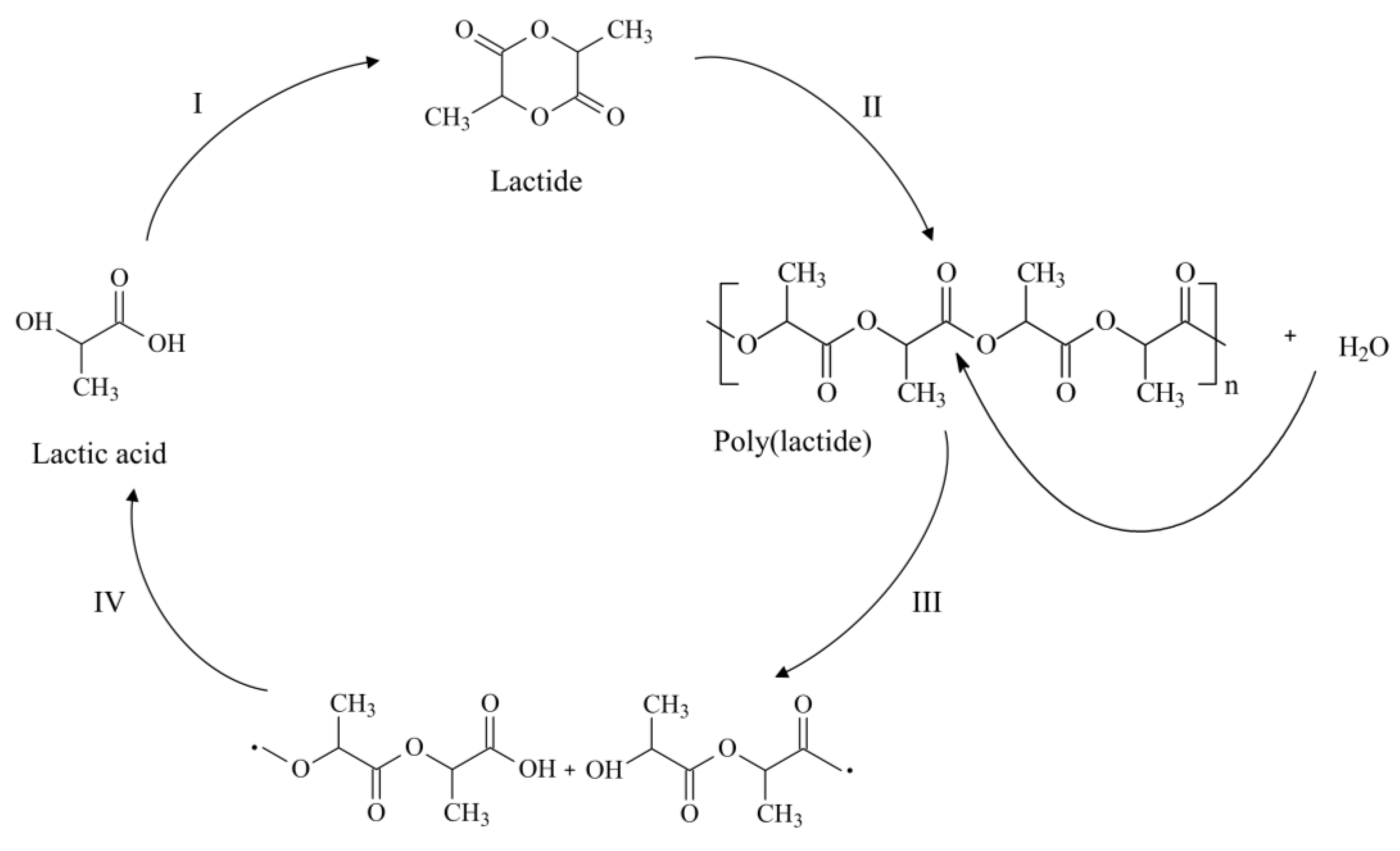
Figure 2.
Chemical structure of the monomeric units of l-lactide, d-lactide, Glycolide and ε-caprolactone.
Figure 2.
Chemical structure of the monomeric units of l-lactide, d-lactide, Glycolide and ε-caprolactone.

Figure 3.
Schematic representation of the single emulsion-solvent evaporation method.
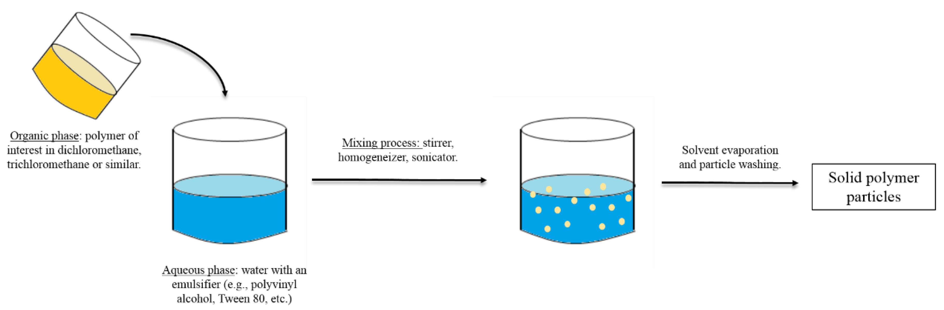
Figure 4.
Poly(d,l-lactide) (PDLLA) porous microparticles fabricated with the aid of ammonium bicarbonate and further hydrolyzed with NaOH to increase the porosity and pore-interconnectivity. Reprinted from [60] with permission from Wiley Online Library, 2009.
Figure 4.
Poly(d,l-lactide) (PDLLA) porous microparticles fabricated with the aid of ammonium bicarbonate and further hydrolyzed with NaOH to increase the porosity and pore-interconnectivity. Reprinted from [60] with permission from Wiley Online Library, 2009.

Figure 5.
Poly(l-lactide-co-glycolide) PLGA microparticles prepared via a flow-focusing device. Above, an optical micrograph of the orifice of the flow-focusing region generating droplets of the dispersed phase in the continuous phase is displayed. Below, scanning electron micrographs of the resulting particles at low (A) and high (B) magnification are represented. Reprinted from [66] with permission from Wiley Online Library, 2009.

Figure 6.
Schematic illustration of the electrospraying process. Reproduced from [71] with permission of The Royal Society of Chemistry, 2014.
Figure 6.
Schematic illustration of the electrospraying process. Reproduced from [71] with permission of The Royal Society of Chemistry, 2014.

Figure 7.
Schematic representation of the chemical reactions performed for the incorporation of REDV onto the surface of poly(ε-caprolactone) (PCL) microparticles (I). Microparticles are first aminolyzed and then the atom-transfer radical-polymerization (ATRP) initiator (α-bromoisobutyl bromide: BIBB) is incorporated (a); then, azido-terminated poly(glycidyl methacrylate) (PGMA) polymer brushes are grown via surface-initiated ATRP (b); the brushes are further carboxylated via the azide-alkyne click reaction (c); and, finally, REDV is incorporated via carbodiimide chemistry (d). The number of cells clearly increased after the incorporation of REDV in comparison to pristine PCL microparticles (II). ** indicates significant differences (p<0.01). Reproduced from Ref [79] with permission from The Royal Society of Chemistry, 2015.


| Commercial Name | Shape | Dimensions | Matrix | Surface | Surface Area (m2/g) | Manufacturer | Performance of Cells |
|---|---|---|---|---|---|---|---|
| Cytodex 1 | Spherical | ~190 µm | Cross-linked dextran | Hydrophilic diethylaminoethyl (DEAE)/Positive charge | 0.44 | GE Healthcare Life Sciences | Study by Lecina et al. [9] about the culture and differentiation toward cardiomyocites (CM) of human embryonic stem cells (hESCs):
Study by Frauenschuh et al. [10] into the expansion of bone-marrow-derived mesenchymal stem cells (MSCs) on Cytodex 1 and 3:
|
| Cytodex 3 | Spherical | ~175 µm | Cross-linked dextran | Pig skin gelatin | 0.27 | GE Healthcare Life Sciences | |
| Cytopore 1 | Spherical | ~230 µm | Cross-linked cotton cellulose | Hydrophilic DEAE/Positive charge | 1.1 | GE Healthcare Life Sciences | |
| Cultispher | Spherical | 130–380 µm | Cross-linked porcine gelatin | N/A | 1.5 | Percell Biolytica | |
| DE-53 | Cylindrical | l: 150–400 µm × d: 35–50 µm | Cellulose | Hydrophilic DEAE/Positive charge | N/A | Whatman | |
| FACT | Spherical | 125–212 µm | Polystyrene | Type I collagen | 0.036 | SoloHill | |
| Tosoh-10 | Spherical | ~10 µm | Hydroxylated methacrylate | Protamine sulphate | N/A | Tosoh | |
| Rapid Cell P | Spherical | 150–210 µm | Polymeric | N/A | 0.037 | Valeant Pharmaceuticals | |
| Hillex | Spherical | 160–200 µm | Polystyrene | N/A | 0.050 | SoloHill |
Table 2.
Some of the reported degradation rates for the poly(α-hydroxyacids) considered herein, (DL: d-lactide; LA: l-lactide; GA: Glycolide; CL: Caprolactone).
| Polymer | Composition | Degradation Rate (Day−1) | Reference |
|---|---|---|---|
| poly(l-lactide) (PLLA) | N/A | 0.0009 | [31] |
| N/A | 0.00043 | [33] | |
| poly(d,l-lactide) (PDLLA) | (DL: 0.2%) | 0.0008 | [33] |
| (DL: 1.2%) | 0.00142 | [33] | |
| (DL: 50%) | 0.0088 | [31] | |
| (DL: not reported) | 0.00386 | [36] | |
| poly(l-lactide-co-glycolide) (PLGA)/poly(d,l-lactide-co-glycolide) (PDLGA) | (LA: 80/GA: 20) | 0.0127 | [31] |
| (DL: 53/GA: 47) | 0.0506 | [31] | |
| (LA: 75/GA: 25) | 0.0219 | [36] | |
| (LA: 85/GA: 15) | 0.0106 | [36] | |
| poly(l-lactide-co-ε-caprolactone) (PLCL) | (LA: 74/CL: 26) (random) | 0.034 | [41] |
| (LA: 74/CL: 26) (blocky) | 0.022 | [41] | |
| (LA: 62/CL: 38) | 0.036 | [41] |
© 2016 by the authors; licensee MDPI, Basel, Switzerland. This article is an open access article distributed under the terms and conditions of the Creative Commons Attribution (CC-BY) license (http://creativecommons.org/licenses/by/4.0/).
Share and Cite
MDPI and ACS Style
Larrañaga, A.; Sarasua, J.-R. Poly(α-hydroxy Acids)-Based Cell Microcarriers. Appl. Sci. 2016, 6, 436. https://doi.org/10.3390/app6120436
AMA Style
Larrañaga A, Sarasua J-R. Poly(α-hydroxy Acids)-Based Cell Microcarriers. Applied Sciences. 2016; 6(12):436. https://doi.org/10.3390/app6120436
Chicago/Turabian StyleLarrañaga, Aitor, and Jose-Ramon Sarasua. 2016. "Poly(α-hydroxy Acids)-Based Cell Microcarriers" Applied Sciences 6, no. 12: 436. https://doi.org/10.3390/app6120436
Note that from the first issue of 2016, this journal uses article numbers instead of page numbers. See further details here.