Article

Cobalt (II) Complexes with Schiff Base Ligands Derived from Terephthalaldehyde and ortho-Substituted Anilines: Synthesis, Characterization and Antibacterial Activity

Department of Chemistry, North Tehran Branch, Islamic Azad University, Tehran 1651153311, Iran
*
Author to whom correspondence should be addressed.
Appl. Sci. 2018, 8(3), 385; https://doi.org/10.3390/app8030385
Submission received: 21 January 2018 / Revised: 26 February 2018 / Accepted: 4 March 2018 / Published: 6 March 2018
(This article belongs to the Special Issue Nano-systems for Antimicrobial Therapy)

Abstract

:
In this study, N-propyl-benzoguanamine-SO3H magnetic nanoparticles (MNPs) were used as a catalyst for the synthesis of new Schiff base ligands from condensation reaction of terephthalaldehyde and ortho-aniline derivatives. The bioactive ligands and their cobalt (II) complexes were characterized with nuclear magnetic resonance (1H-NMR), Fourier-transform infrared spectroscopy (FT-IR), ultraviolet-visible (UV-Visible), mass spectroscopy studies and molar conductance. The antibacterial activity of ligands and their metal complexes were screened using disc diffusion and broth dilution methods against Escherichia coli, Serratia marcescens, Pseudomonas aeruginosa (gram negative bacteria), Bacillus Subtilis and Staphylococcus aureus (gram positive bacteria). The ligands with hydroxyl group showed better biological activity when compared to other ligands. The results showed that the metal complexes have much higher antibacterial activity compare to the parent ligands. It was found that the CoL3 complex was more effective than other metal complexes used against all types of bacteria tested and it was more effective against Pseudomonas aeruginosa with diameter inhibition zone of 17 mm and minimal inhibitory concentration value of 0.15 mg/mL.

Graphical Abstract

1. Introduction

Schiff base ligands with oxygen or nitrogen donor atoms are a good class of organic compounds capable of binding to different metal ions with interesting medical and non-medical properties and very popular in the last decade [1,2]. These ligands can be easily synthesized by condensation reaction of aldehyde or ketone with a primary amine [3]. The multifarious role of transition complexes of Schiff base ligands in inorganic, metallo-organic and biochemistry have received considerable attention because of their extensive applications in a wide range of areas [4,5].
They display diverse chemical, optical and magnetic properties by modifying with different ligands [6,7,8]. It has been revealed that Schiff bases play an important role by serving as chelating ligands in the main groups and transition metal coordination chemistry; owing to their stability in different oxidative and reductive conditions [9]. The interaction of these donor ligands and metal ions gives complexes of different geometries and literature survey reveals that these complexes are potentially more biologically active compounds [10] such as anticancer, antifungal, antibacterial, antimalarial, anti-inflammatory, antiviral, and antipyretic properties [11,12,13,14]. It should be noted that metal chelation can tremendously influence the antimicrobial/bioactive behavior of the organic ligands; therefore, the synthesis of various transition metal complexes has been attempted in this field [15].
In the past few years, bacterial infection and their resistance for many antibacterial agents is a growing problem [16,17]. While there are already several classes of antibacterial agents, there has been some considerable emerging resistance in most pathogenic bacteria to these drugs [18]. For prevention of this serious medical problem, it is necessary to develop some new antibacterial agents or to expand the bioactivity of the previously used drugs [19,20]. Metal-based antibacterial compounds seem to be a promising research for designing a novel therapeutic methodology for new antibiotic drugs to control and prevent the growth of bacterial strains [21,22].
Herein, we report the synthesis of bidentate Schiff base ligands by the condensation of terephthalaldehyde with ortho-aniline derivatives in the presence of N-propyl-benzoguanamine-SO3H MNPs as a catalyst. The cobalt (II) complexes were prepared in methanol as a solvent. The synthesized compounds were characterized with several spectroscopic methods and screened for their antibacterial activity against Gram (+) and Gram (−) bacteria strains.

2. Materials and Methods

2.1. Materials

All the chemicals and solvents purchased from Merck (Darmstadt, Germany) and Sigma-Aldrich Company (St. Louis, MO, USA) and were used without further purification unless otherwise mentioned. UV-Vis (see Supplementary Materials) absorption spectra were recorded on a Cary 100 spectrophotometer (Santa Clara, CA, USA) using a 1 cm path length cell. 1H-NMR (see Supplementary Materials) spectra of ligands were collected on BRUKER 250 MHz spectrometer (Seiko, Japan) in DMSO-d6 using tetramethylsilane as internal standard. The Fourier-transform infrared spectroscopy (FTIR) spectra (see Supplementary Materials) (KBr pellets) were recorded using a Shimidzo 300 spectrometer. Melting points of compounds were obtained by an electro thermal melting point apparatus and were not corrected. Thin-Layer chromatography (TLC) was performed using n-hexane/EtOAC (1:3) as an eluent.

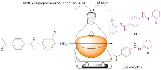
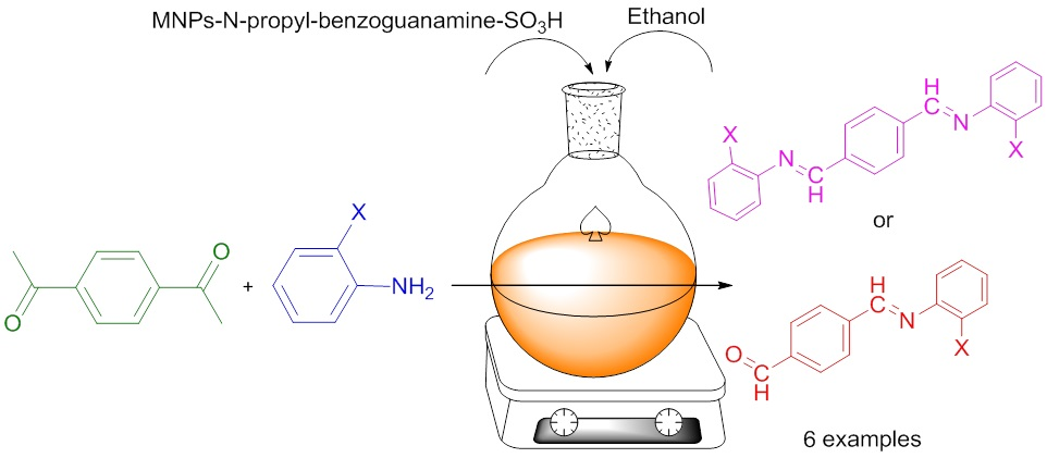
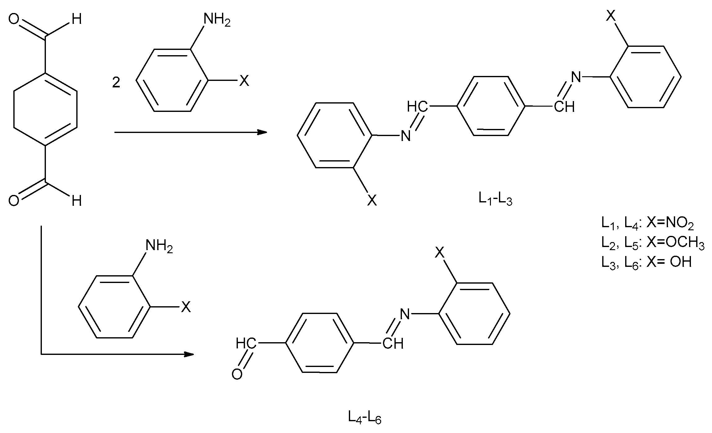
2.2. Preparation of Schiff Base Ligands

Condensation reaction of Terephthalaldehyde with o-nitroaniline, o-Anisidine and 2-aminophenol in molar ratio 1:1 and 1:2 afforded the corresponding Schiff base ligands as described below:
MNPS-N-propyl-benzoguanamine-SO3H catalyst was prepared by chemical co-precipitation according to the previous literature [23]. To a mixture of terephthalaldehyde and aniline derivative was added to N-propyl-benzoguanamine-SO3H catalyst (6 mg) in 10 mL ethanol as solvent. The reaction mixture was refluxed (100 °C) for 2–3 h. The progress of the reaction was checked with TLC. After completion of the reaction the mixture was cooled to room temperature. The catalyst was then separated by using an external magnet. The solvent was evaporated under reduced pressure and the resulting solid was obtained. The resulting was then recrystallized in ethanol.
(1,4-phenylenebis(methanylylidene))bis(2-nitroaniline) (L1): Dark Yellow solid. Yield: 84%. M.P. 208–210 °C. Selected IR data (ν, cm−1): 2924, 1630, 1449, 1348, 1012. 1H-NMR (500 MHz, DMSO-d6, δ, ppm): 10.04 (s, 2H, CH=N), 8.02–7.97 (q, 12.5 Hz, 8H, Ar-H), 7.90–7.87 (d, 7.5 Hz, 2H, Ar-H), 7.53–7.50 (d, 7.5 Hz, 2H, Ar-H). UV-Vis (DMSO): λmax (nm) = 260, 340.
(1,4-phenylenebis(methanylylidene))bis(2-methoxyaniline) (L2): Orange solid. Yield: 86%. M.P. 190–192 °C. Selected IR data (ν, cm−1): 3062, 3018, 2965, 2835, 1620, 1116. 1H-NMR (250 MHz, DMSO-d6, δ, ppm): 10.08 (s, 2H, CH=N), 8.66–8.60 (m, 2H, Ar-H), 8.14–8.02 (m, 5H, Ar-H), 7.35–6.95 (m, 8H, Ar-H), 3.80 (s, 6H, CH3). UV-Vis (DMSO): λmax (nm) = 290, 390.
(1,4-phenylenebis(methanylylidene))bis(azanylylidene))diphenol (L3): Brown solid . Yield: 64%. M.P. 295–297 °C. Selected IR data (ν, cm−1): 3412, 3100, 1614, 1059. 1H-NMR (500 MHz, DMSO-d6, δ, ppm): 9.72 (s, 2H, CH=N), 8.34–8.32 (d, 5Hz, 2H, Ar-H), 8.19–8.17 (d, 5Hz, 2H, Ar-H), 7.86–7.82 (m, 2H, Ar-H), 7.67–7.66 (d, 2.5 Hz, 1H, Ar-H), 7.48–7.42 (m, 2H, Ar-H), 7.07–7.03(m, 2, OH). UV-Vis (DMSO): λmax (nm) = 330.
4-(((2-nitrophenyl)imino)methyl)benzaldehyde (L4): Yellow solid. Yield: 68%. M.P. 203–205 °C. Selected IR data (ν, cm−1): 2948, 2900, 1702, 1619, 1568, 1356, 1073. 1H-NMR (250 MHz, CDCl3, δ, ppm): 10.99 (s, 1H, CHO), 10.06 (s, 1H, CH=N), 8.16–8.02 (m, 5H, Ar-H), 7.79–7.69 (m, 3H, Ar-H). UV-Vis (DMSO): λmax (nm) = 250, 320.
4-(((2-methoxyphenyl)imino)methyl)benzaldehyde (L5): Pale Yellow solid. Yield: 73%. M.P. 181–183 °C. Selected IR data (ν, cm−1): 3011, 2966, 2838, 1693, 1620, 1102. 1H-NMR (500 MHz, DMSO-d6, δ, ppm): 10.07 (s, 1H, CHO), 8.64 (s, 1H, CH=N), 8.03–8.01 (d, 5 Hz, 4H, Ar-H), 7.21–6.95 (m, 4H, Ar-H), 3.79 (s, 6H, CH3). UV-Vis (DMSO): λmax (nm) = 290, 370.
4-(((2-hydroxyphenyl)imino)methyl)benzaldehyde (L6): Light Green solid. Yield: 65%. M.P. 282–284 °C Selected IR data (ν, cm−1): 3431, 3147, 3046, 1703, 1652, 1028. 1H-NMR (500 MHz, DMSO-d6, δ, ppm): 10.18 (s, 1H, CHO), 9.73 (s, 1H, CH=N), 8.41–7.45 (m, 8H, Ar-H), 1.21 (s, 1H, OH). UV-Vis (DMSO): λmax (nm) = 320.

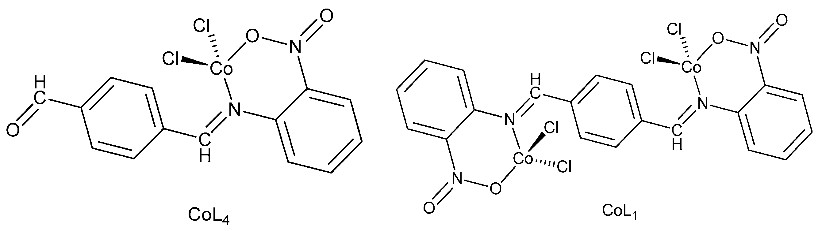
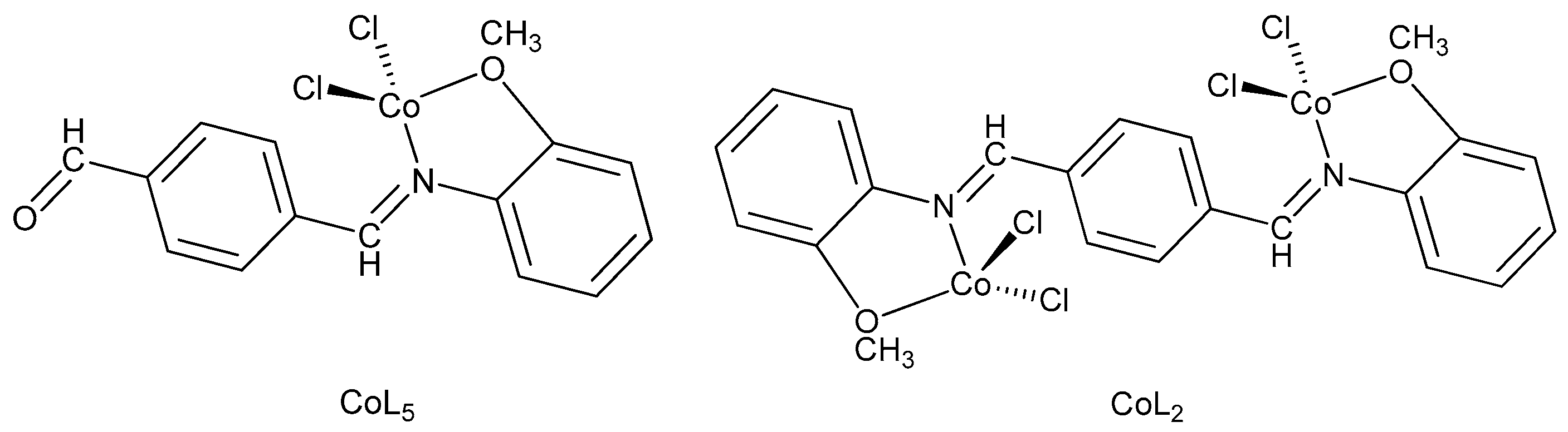
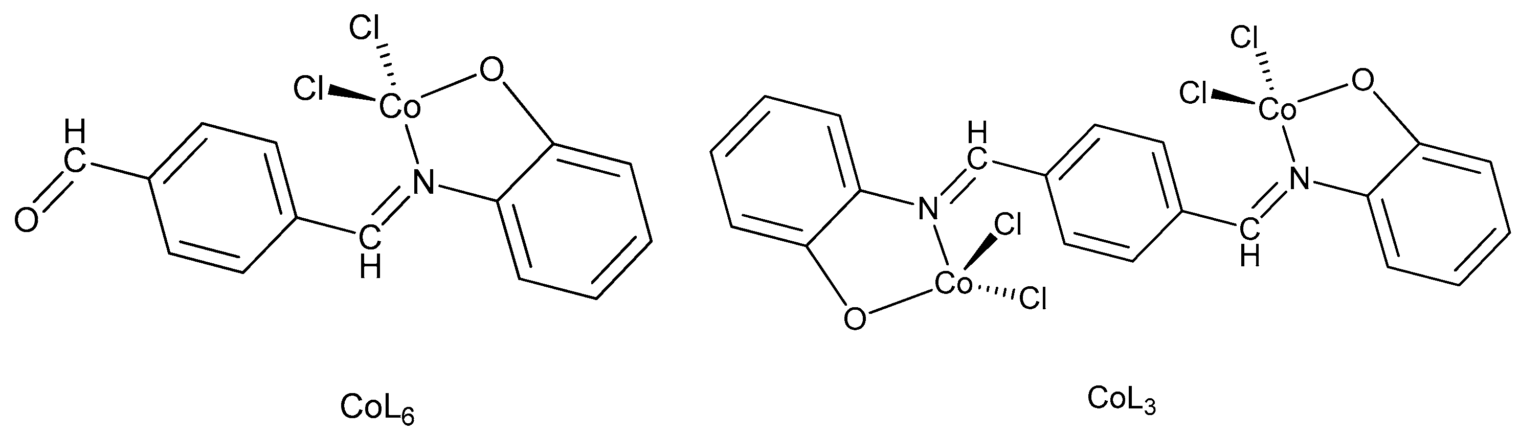
2.3. Preparation of Co (II) Complexes

All the complexes were prepared in a similar procedure. Solution of ligand in methanol (1 mmol) were mixed with CoCl2·6H2O and refluxed for 3–5 h 45 °C. The precipitate was filtered, washed with methanol and ether and then dried in a vacuum desiccator.
Complex [Co2(1,4-phenylenebis(methanylylidene))bis(2-nitroaniline)Cl4] (CoL1): Pale Green solid. Yield: 84%. M.P. 248–250 °C. Molar conductivity (Ω−1 mol−1 cm2): 22. Selected IR data (ν, cm−1): 3109, 2971, 1651, 1521, 1384, 598, 492. UV-Vis (DMSO): λmax (nm) = 300, 410, 600,690. Mass (see Supplementary Materials): [m/z]+ = 633.
Complex [Co2(1,4-phenylenebis(methanylylidene))bis(2-methoxyaniline)Cl4] (CoL2): Dark Green solid. Yield: 68%. M.P. 313–315 °C. Molar conductivity (Ω−1 mol−1 cm2): 12. Selected IR data (ν, cm−1): 3062, 3018, 1620, 1583, 1368, 511, 474. UV-Vis (DMSO): λmax (nm) = 290, 260, 610, 690, 750. Mass: [m/z]+ = 603.
Complex [Co2(1,4-phenylenebis(methanylylidene))bis(azanylylidene))diphenol)Cl4] (CoL3): Dark Brown solid. Yield: 75%. M.P. 230–232 °C. Molar conductivity (Ω−1 mol−1 cm2): 14. Selected IR data (ν, cm−1): 3100, 2841, 1650, 537, 485. UV-Vis (DMSO): λmax (nm) = 280, 370, 600, 680. Mass: [m/z]+ = 575.
Complex [Co(4-(((2-nitrophenyl)imino)methyl)benzaldehyde)Cl2] (CoL4): Green solid. Yield: 80%. M.P. 280–282 °C. Molar conductivity (Ω−1 mol−1 cm2): 8. Selected IR data (ν, cm−1): 3181, 1599, 614. UV-Vis (DMSO): λmax (nm) = 320, 600,690. Mass: [m/z]+ = 503.
Complex [Co(4-(((2-methoxyphenyl)imino)methyl)benzaldehyde)Cl2] (CoL5): Orange-Red solid. Yield: 82%. M.P. 305–307 °C. Molar conductivity (Ω−1 mol−1 cm2): 16. Selected IR data (ν, cm−1): 3100, 2900, 1619, 568, 509. UV-Vis (DMSO): λmax (nm) = 290, 370, 600, 670. Mass: [m/z]+ = 473.
Complex [Co(4-(((2-hydroxyphenyl)imino)methyl)benzaldehyde)Cl2] (CoL6): Dark Pink solid. Yield: 87%. M.P. 236–238 °C. Molar conductivity (Ω−1 mol−1 cm2): 10. Selected IR data (ν, cm−1): 2959, 1634, 649. UV-Vis (DMSO): λmax (nm) = 270, 350, 610, 670. Mass: [m/z]+ = 445.

2.4. Antibacterial Study

All the synthesized compounds were evaluated to examine their in vitro antibacterial activities against Escherichia coli (ATCC: 25922), Serratia marcescens (ATCC: 13880) and Pseudomonas aeruginosa (ATCC: 27853) as gram negative bacteria and Bacillus subtilis (ATCC: 6633), and Staphylococcus aureus (ATCC: 6838), as gram positive bacteria, by employing two methods: disk diffusion and broth dilution methods; which are recommended by the National Committee for Clinical Laboratory Standards (NCCLS) [24]. Accordingly, stock solution of each compound (2 mg/mL) was prepared by dissolving the compounds in DMSO. Prior to sensitivity testing, the bacteria strains were cultured onto Muller-Hinton agar plate and incubated for 18–24 h at 35 °C. The density of the bacteria culture required for the tests was adjusted to 0.5 McFarland (1.5 × 10⁸ CFU/mL) (CFU = Colony Forming Unit). These tests were repeated three times to ensure reliability.

2.4.1. Disc Diffusion Method

This method is based on the principles that an antibiotic-impregnated disk placed on an agar previously inoculated with the test bacterium, the pick-up moisture and the antibiotic diffused radially outward through the agar medium, yielding an antibiotic concentration gradient. For this purpose, 2 mg of the synthesized compound was dissolved in 1 mL DMSO. A bacteria culture was swabbed uniformly across lawn Hinton agar plates. Paper discs were impregnated individually with 100 μL of stock solution of the compounds. Next, the discs were placed on the inoculated agar medium and the plates incubated for 18–24 h at 35 °C. After the incubation time, antibacterial activity of each sample was determined by measuring the inhibition zone around each disc by comparing it with the standard drug (Tetracycline).

2.4.2. Broth Dilution Method

Minimal Inhibitory Concentration (MIC) value of an antibacterial agent gives a quantitative estimate of the susceptibility for each bacteria strain. MIC is defined as the lowest concentration of the antimicrobial agent which is required to inhibit the growth of the microorganism. According to this method, 1 mL of sterile Muller Hinton Broth medium were poured in tube 1–13 with two-fold dilutions of the synthesized compound (2 to 0.00195 mg/mL) and inoculated with a standardized inoculum of the bacteria (1.5 × 10⁸); then it was incubated under standardized conditions by following NCCLS guidelines. After 18–24 h of incubation at 35 °C, the MIC value was recorded as the lowest concentration of antimicrobial agent with no visible growth.

3. Results and Discussion

3.1. Characterization of Ligands

Six Schiff base Ligands (L1L6) were synthesized by condensation reaction of terephthaldehyde and o-aniline derivatives in the presence of N-propyl-benzoguanamine-SO3H MNPs under optimized condition. The magnetic nanoparticles were prepared according to the previous literature [23]. The method for the synthesis of Schiff base ligands is given in Figure 1. In order to optimize the reaction condition of starting materials, the reaction of terephthaldehyde (1 mmol) and o-nitroaniline (2 mmol) was carried out in different conditions using different solvents and catalysts with different catalyst content.
According to Table 1 the type of solvent and amount of catalyst was observed to have a significant effect on the yield of reaction using similar catalyst (N-propyl-benzoguanamine-SO3H MNPs). The highest yield was obtained up to 84%with a shorter reaction time using ethanol as solvent. In the next step, the amount of catalyst in the reaction was also examined. It is obvious from Table 1 that applying more than the specified quantity of catalyst did not have a positive effect on the yield of product and 6 mg of the catalyst represented the best yield of the reaction. It is noted from Table 1, with increasing the amount of catalyst from 3 mg to 11 mg, reaction yield was reduced 25% with a longer reaction time. The effect of p-Toluenesulfonic acid (PTSA) as catalyst was further examined which exhibited lower reaction yield with much longer reaction time.
The ligands were characterized by nuclear magnetic resonance (1H-NMR), Fourier-transform infrared spectroscopy (FT-IR) and ultraviolet-visible (UV-Visible). In the 1H-NMR spectra of the (L1L6) ligand, the singlet peaks due to the CNH (azomethine) group were observed in the range of 10.08–8.64 ppm as singlet. Regarding the 1H-NMR spectra of the compounds, it can be observed that the CNH signals of the Schiff base ligands shifted to a lower ppm (shielding) when electron donating (OH) substituent was used and it moved to a higher ppm (deshielding) when a withdrawing group (NO2) was used. Condensation of amine groups to Terephthaldehyde in all of the ligands is confirmed by the absence of the N-H protons. The signals of the methyl groups (-CH3) for L2 and L5, and OH groups for L3 and L6 ligands are observed in the range of 3.80–3.79 ppm and 1.21–1.16 ppm, respectively. In the 1H-NMR spectra of L4, L5 and L6 ligands the CHO protons are seen at 10.99, 10.07 and 10.18 ppm, respectively. The aromatic protons of the Schiff base ligands are observed in the range of 8.66–6.82 ppm. The 1H NMR spectral data of the ligands are summarized in Table 2.
The FTIR spectra of the ligands showed peaks in the range of 1652–1620 cm−1 assigned to ν(C=N). In the L4, L5 and L6 the ν(C=O) (carbonyl) stretching appeared at 1702, 1693 and 1703 cm−1, respectively. In L1 and L4 Schiff bases the bands observed in the range of 1568–1348 cm−1 are attributed to the NO2 groups, while the OH groups of L3 and L6 appeared at 3553 and 3438 cm−1, respectively.
The UV-Visible spectra of all the ligands and compounds are recorded in DMSO and the data are listed in Table 3. In the electronic spectra of L1, L2, L4 and L5 two peaks appeared which are attributed to п→п* and n→п* transitions, respectively. In L3 and L6 one intense absorption band observed in 310 and 340 nm is due to the п→п* transition. However, п→п* and n→п* absorption peaks exhibited different behaviors due to the nature of substituents. In the UV-Vis spectra of the ligands with withdrawing group (NO2) the п→п* peak shifted to a lower wavelengths (blue-shift) as compared to the ligands, whereas with electron donating group (OH) the п→п* peak observed in higher wavelengths (red-shift).

3.2. Characterization of Metal Complexes

Physical properties of all the synthesized compounds are presented in Table 4. The synthesized metal complexes were prepared in good yield (65–87%), insoluble in ethanol, methanol, chloroform and other common organic solvents but easily soluble in DMSO and DMF. Metal complexes were characterized with mass, Fourier-transform infrared (FT-IR) and Ultraviolet-visible (UV-Visible) spectroscopies. Since the cobalt is paramagnetic in nature the 1H-NMR technique was not performed. The molar conductance values of the complexes in DMSO (10−3 M solutions) were calculated at room temperature using Λ m = κ C equation; where C is the concentration of the solutions (mol/L) and κ is the measured conductivity. Measurements were performed to establish the charge of the complexes. The molar conductivity of the metal complexes lies in the range of 10–22 (Ω−1 mol−1 cm2), indicating that all the complexes were non-electrolytes.
In metal complexes with NO2 groups (CoL1 and CoL4), the NO2 group can coordinate to the metal center in various ways e.g., via the nitrogen (nitro), oxygen (nitrito), both oxygens (nitrito-O,O′) and via nitrogen and oxygen (bridging nitro) [25,26,27]. The coordination mode of this ambidentate ligand depends on the stereochemical environment around the metal ions. In the FT-IR spectra of CoL1 and CoL4 complexes the characteristic band in 492 and 614 cm−1 is observed which is due to the cobalt-oxygen stretching band; indicating the formation of nitrito isomer. The proposed structure for CoL1 and CoL4 complexes is given in Figure 2. The band due to ν(C=N) in ligands were shifted to a lower wavenumbers in complexes which indicated the involvement of azomethine group in the coordination to the cobalt center. Also the bands in the range of 1583–1592 cm−1 and 1328–1368 cm−1 are assigned to the NO2 groups. In the electronic spectra of CoL1 and CoL4 complexes the peaks in the range of 600–690 nm are associated with d-d transition; and the low intensity of these peaks indicated the symmetrical structure of these complexes. The mass spectra of these complexes were recorded at room temperature to confirm the stoichiometry of metal chelates as studied above. The molecular ion peak for the CoL1 and CoL4 complexes were observed at m/z = 633 and 573, respectively that are equal to the molecular weight of the complexes.
Figure 3 shows the proposed structure for CoL2 and CoL5 complexes. In FTIR spectra of CoL2 and CoL5 complexes containing the OMe substituent the (C-H) aliphatic functional groups appeared in the range of 2840–2900 cm−1 while the C=NH bands are observed in the range of 1619–1626 cm−1. The peaks in the range of 474–511 cm−1 are assigned to the M-O band. The electronic absorption spectrum of CoL2 and CoL5 complexes showed four peaks. The first and second peaks are due to the п→п* and n→п* transition of ligand group which shifted to higher wavelength compared to free ligand. The d-d transition bands are observed in the range 590–610 and 660–680 nm owing to 4A14B1 and 4A14B2 transitions. The mass spectra of CoL2 and CoL5 are in good agreement with the proposed structures. The mass spectra of CoL2 and CoL5 complexes showed molecular ion peak at m/z = 603 and 473, respectively, confirming their formula weight.
In the FTIR spectra of CoL3 and CoL6 complexes the stretching frequency of C=N are observed at 1650 and 1634 cm−1, respectively. The disappearance of OH groups in CoL3 and CoL6 complexes indicate the OH group of ligands has been deprotonated and coordinate to metal ions. The coordination of Schiff base ligands to metals were also proved by the υ (M-O) appearing in the range 485–649 cm−1. The electronic absorption spectra of CoL3 and CoL6 complexes are very similar to each other. In the UV-Visible spectra of these complexes the п→п* and n→п* transition of ligand group shifted to the higher wavelength upon the coordination and d-d transitions are appeared in the range 600–680 nm. In the mass spectra of CoL3 and CoL6 complexes the molecular ion peak observed at m/z = 575 and 445. The proposed structure for CoL3 and CoL6 complexes are presented in Figure 4.

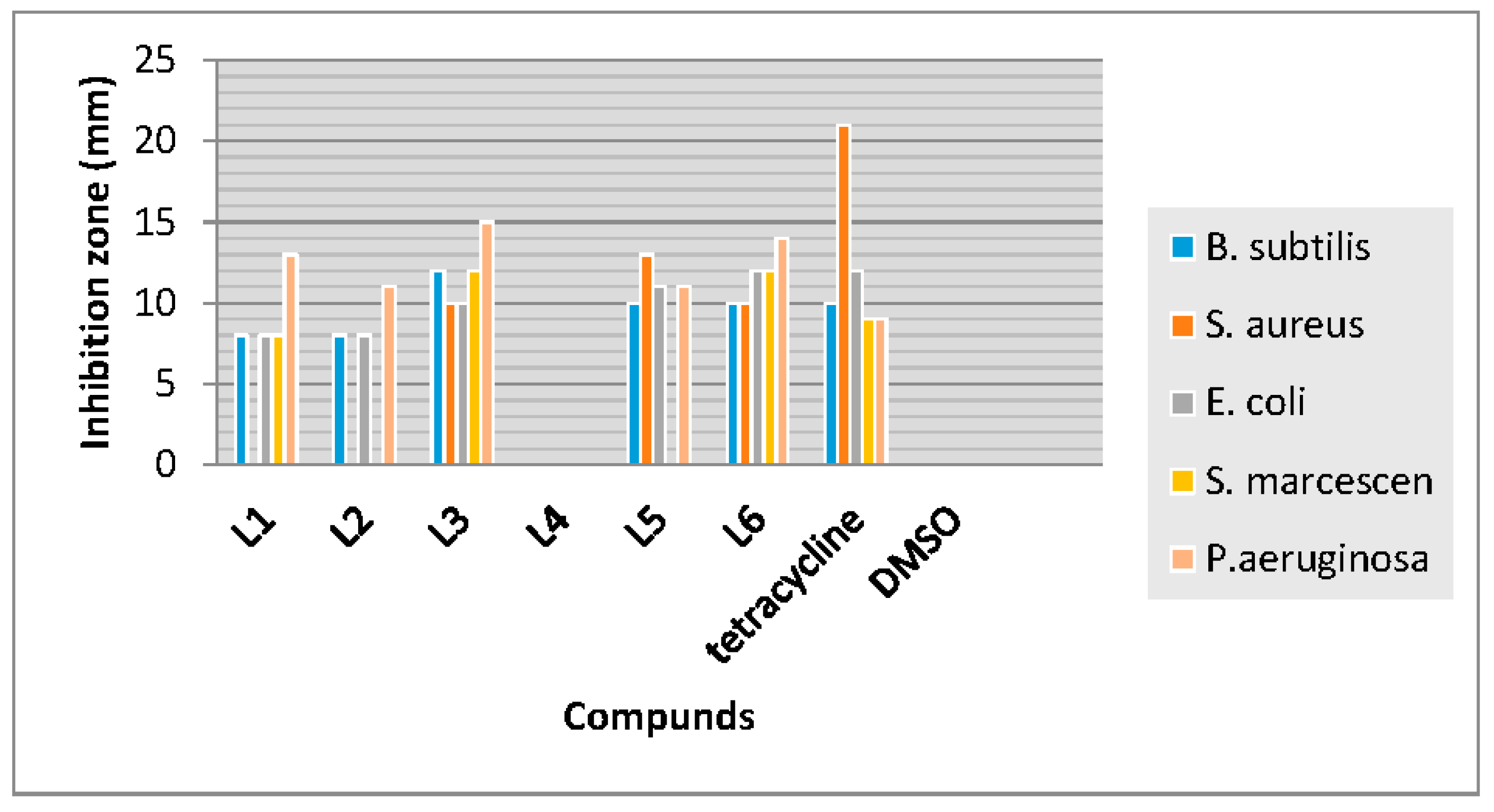
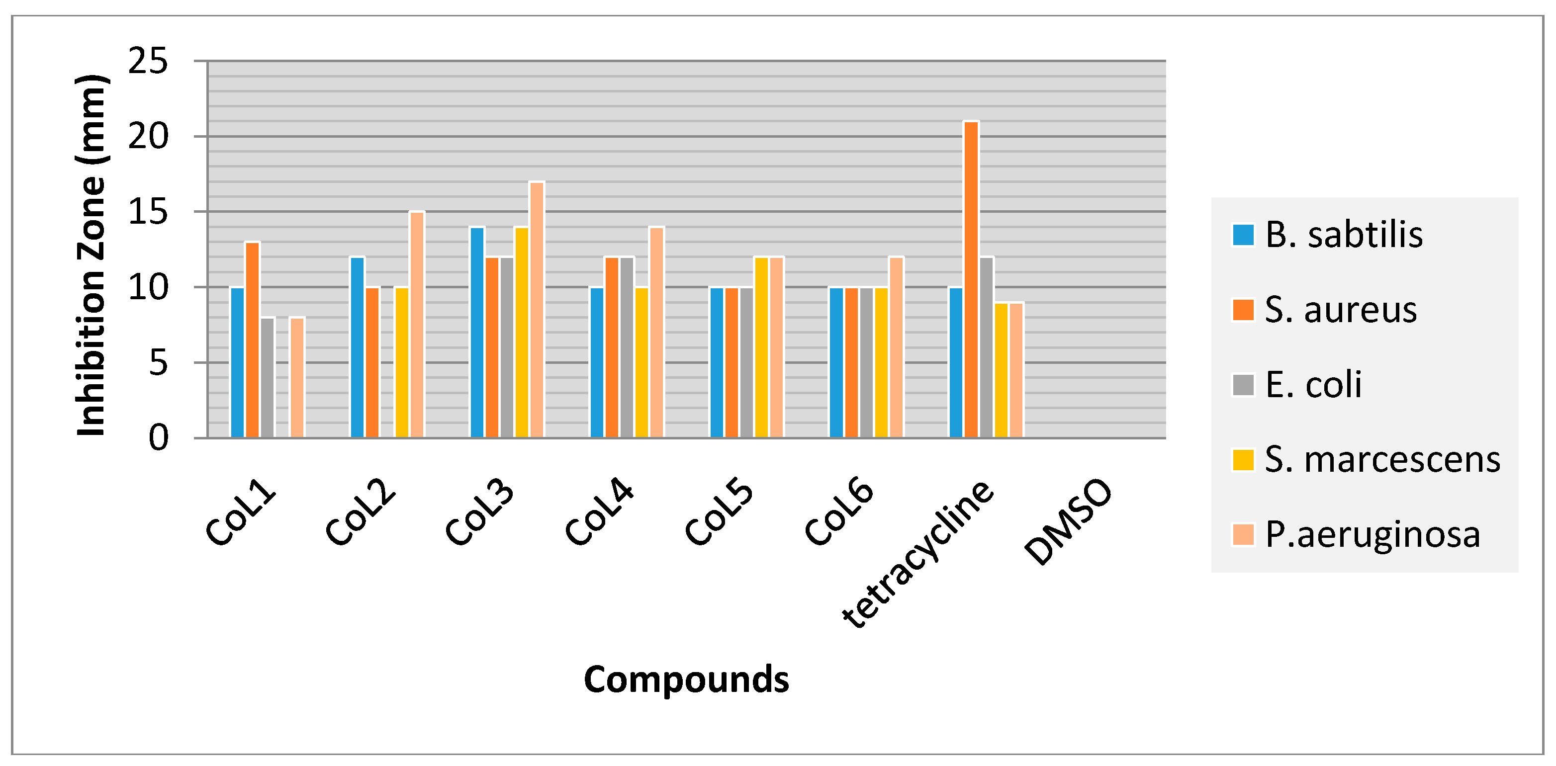
3.3. Antibacterial Activity

Antibacterial activities of the ligands, their metal complexes and standard antibiotic drug (tetracycline) were performed against gram negative bacteria (Escherichia coli, Serratia marcescens and Pseudomonas aeruginosa) and against gram positive bacteria (Bacillus Subtilis and Staphylococcus aureus) using Muller Hinton agar medium by disk diffusion and broth dilution methods are shown in Figure 5 and Figure 6, respectively.
From Figure 5 it can be observed that the L3 and L6 with OH group had relatively higher antibacterial activity compared to the other ligands tested against the bacteria strains. These ligands had better inhibitory effect against Pseudomonas aeruginosa with diameter inhibition zone of 15 and 14 mm, respectively. In contrast, the L4 with one NO2 group showed no antibacterial activity against tested bacteria strains.
As can be seen from the antibacterial activity of metal complexes in Figure 6, among all these complexes the CoL3 compound showed the higher antibacterial activity with inhibition zone of 14, 12, 12, 14 and 17 mm against B. Subtilis, S. aureus, E. coli, S. marcescen and P. aeruginosa, respectively. The Minimal Inhibitory Concentration (MIC) values of all the synthesized compounds were also recorded and results are presented in Table 5. From the data listed in Table 5, the MIC values of metal complexes were lower than that of the parent ligands. The antibacterial activities of metal complexes (CoL1 to CoL6) against P. aeruginosa were more effective than the other tested bacteria strains with MIC value in the range of 0.62 to 2.5 mg/mL. In comparison these metal complexes were less effective against E. coli.
It is observed from this study that metal chelates have a higher activity when compared to the parent ligands. Such increased activity of the metal chelates can be explained on the basis of Overtone’s concept and chelation theory [28,29,30,31]. According to Overtone’s concept of cell permeability the lipid membrane that surrounds the cell favors the passage of only lipid soluble materials due to which liposolubility is an important factor that controls antimicrobial activity. On chelation, the polarity of the metal ion is reduced to a greater extent due to the overlap of the ligand orbital and partial sharing of the positive charge of the metal ion with donor groups. Further, it increases the delocalization of p-electrons over the whole chelate ring and enhances the lipophilicity of the complex. This increased lipophilicity enhances the penetration of the complexes into lipid membranes and blocking of metal binding sites on the enzymes of the microorganism.

4. Conclusions

In this research, we successfully reported the synthesis of the Schiff base ligands (L1L6) and their Co (II) complexes from condensation of Terephthalaldehyde with ortho-anilines with high yields. The synthesis of corresponding ligands was performed under optimized condition. N-propyl-benzoguanamine-SO3H MNPs was used as a suitable catalyst in ethanol as a solvent for the synthesis of the ligands. The structures of the synthesized compounds were proposed by FTIR, 1H-NMR, UV-Vis and mass spectroscopy studies. The molar conductivity measurements showed that all the complexes were non-electrolyte. Antibacterial activities of the ligands and their metal complexes were examined against gram-positive and gram-negative bacteria strains. In general, metal complexes showed much higher antibacterial activities and better inhibitory effects than that of the ligands.

Supplementary Materials

The 1H-NMR, FTIR, UV-Vis and mass spectroscopy of synthesized compounds are available online at https://www.mdpi.com/2076-3417/8/3/385/s1.

Acknowledgments

We are grateful to the Islamic Azad University, Tehran North Branch for the financial support.

Author Contributions

N.F. conceived and designed the experiments; S.S. performed the experiments and wrote the paper; H.P. analyzed the data, supervised the experiment and contributed reagents/materials/analysis tools and M.D. and F.M. proofing the entire work. All authors read and approved the manuscript.

Conflicts of Interest

The authors declare that there is no conflict of interest.

References

  1. Liu, Y.-T.; Sheng, J.; Yin, D.-W.; Xin, H.; Yang, X.-M.; Qiao, Q.-Y.; Yang, Z.-J. Ferrocenyl chaconne-based Schiff bases and their metal complexes: Highly efficient, solvent-free synthesis, characterization, biological research. J. Organomet. Chem. 2018, 856, 27–33. [Google Scholar] [CrossRef]
  2. Lashanizadegan, M.; Shayegan, S.; Sarkheil, M. Copper(II) complex of (±)trans-1,2-cyclohexanediamine azo-linked Schiff base ligand encapsulated in nanocavity of zeolite-Y for the catalytic oxidation of olefins. J. Serbian Chem. Soc. 2016, 81, 153–162. [Google Scholar] [CrossRef]
  3. Yu, H.; Zhang, W.; Yu, Q.; Huang, F.-P.; Bian, H.-D.; Liang, H. Ni(II) Complexes with Schiff Base Ligands: Preparation, Characterization, DNA/Protein Interaction and Cytotoxicity Studies. Molecules 2017, 22, 1772. [Google Scholar] [CrossRef] [PubMed]
  4. Ganguly, R.; Sreenivasulu, B.; Vital, J. Amino acid—Containing reduced Schiff bases as the building blocks for metallasupramolecular structures. Coord. Chem. Rev. 2008, 252, 1027–1050. [Google Scholar] [CrossRef]
  5. Golcu, A.; Tumer, M.; Demirelli, H.; Wheatley, R.A. Cd and Cu complexes of polydentate Schiff base ligands: Synthesis, characterization, properties and biological activity. Inorg. Chim. Acta 2005, 153, 1785–1797. [Google Scholar] [CrossRef]
  6. Sarkar, S.; Jana, M.; Mondal, T.; Sinha, C. Ru-halide-carbonyl complexes of naphthylazoimidazoles; synthesis, spectra, electrochemistry, catalytic and electronic structure. J. Organomet. Chem. 2012, 716, 129–137. [Google Scholar] [CrossRef]
  7. Khandar, A.; Nejati, K.; Rezvani, Z. Syntesis, characterization, and study of the use of cobalt Schiff base complexes as catalysts for the oxidation of styrene by molecular oxygen. Molecules 2005, 10, 302–311. [Google Scholar] [CrossRef] [PubMed]
  8. Raman, N.; Ravichandran, S.; Thangaraja, C. Copper, Cobalt, Nickel and zink complexes of Schiff base derived from benzil-2,4-dinitrophenylhydrazone wiyh aniline. J. Chem. Sci. 2004, 116, 215–219. [Google Scholar] [CrossRef]
  9. Al Zoubi, W.; Ko, Y.G. Schiff base complexes and their versatile applications as catalysts in oxidation of organic compounds: Part I. Appl. Organomet. Chem. 2016, 31. [Google Scholar] [CrossRef]
  10. Siva, C.; Silva, D.; Modolo, L.; Alves, R.; Rwsende, M.; Martins, C.; Fatima, A. Schiff bases: A short review of their antimicrobial activities. J. Adv. Res. 2011, 2, 1–8. [Google Scholar] [CrossRef]
  11. Afradi, M.; Foroughifar, N.; Psdar, H.; Moghhanian, H. Facile green one-pot synthesis of novel thiazolo[3,2-a]pyrimidine derivatives using Fe3O4 L-argainine and their biological investigation as potent antimicrobial agents. Appl. Organomet. Chem. 2016, 1–16. [Google Scholar] [CrossRef]
  12. Anitha, C.; Sheela, C.; Tharmaraj, P.; Sumathi, S. Spectroscopic studies and biological evaluation of some transition metal complexses of azo schiff base ligand derived from (1-phenyl-2,3-dimethyl-4-aminopyrazol-5-one) and 5-((4-chlorophenyl)diazenyl)-2-hydroxybenzaldehyde. Spectrochim. Acta Part A 2002, 96, 493–900. [Google Scholar] [CrossRef] [PubMed]
  13. Raman, N.; Raja, Y.P.; Kulandaisamy, A. Synthesis and characterisation of Cu(II), Ni(II), Mn(II), Zn(II) and VO(II) Schiff base complexes derived fromo-phenylenediamine and acetoacetanilide. J. Chem. Sci. 2001, 113, 183–189. [Google Scholar] [CrossRef]
  14. Raman, N.; Thangaraja, C.; Johnsonraja, S. Synthesis, spectral characterization, redox and antimicrobial activity of Schiff base transition metal(II) complexes derived from 4-aminoantipyrine and 3-salicylideneacetylacetone. Cent. Eur. J. Chem. 2005, 3, 537–555. [Google Scholar] [CrossRef]
  15. Streater, M.; Taylor, P.D.; Hider, R.C.; Porter, J. Novel 3-hydroxyl-2(1H)-pyridinones. Synthesis, iron(III)-chelating properties and biological activity. J. Med. Chem. 1990, 33, 1749–1755. [Google Scholar] [CrossRef] [PubMed]
  16. Abdel-Latif, S.A.; Hassib, H.B.; Issa, Y.M. Studies on some salycylaldehyde schiff base derivatives and their complexes with Cr, Mn, Ni, Cu. Spectrochim. Acta Part A 2007, 67, 950–957. [Google Scholar] [CrossRef] [PubMed]
  17. Lo, K.; Cornell, H.; Nicoletti, G.; Jackson, N.; Hügel, H. Study of Fluorinated β-Nitrostyrenes as Antimicrobial Agents. Appl. Sci. 2012, 2, 114–128. [Google Scholar] [CrossRef]
  18. Tolazzal, M.; Tarafder, H.; Ali, M.; Juan, D. Complexes of a tridentate ONS Schiff base. Synthesis and biological properties. Transit. Metal Chem. 2000, 25, 456–460. [Google Scholar] [CrossRef]
  19. Khojasteh, R.; Matin, S.J. Synthesis, characterization and antimicrobial activity of some metal complexes of heptadentate schiif base ligand derived from acetylacetone. Russ. J. Appl. Chem. 2015, 88, 921–925. [Google Scholar] [CrossRef]
  20. Hou, S.H.; Jihui, J.; Huang, X.; Wang, X.; Ma, L.; Shen, W.; Kang, F.; Hiang, Z.-H. Silver Nanoparticles-Loaded Exfoliated Graphite and Its Anti-Bacterial Performance. Appl. Sci. 2017, 7, 852. [Google Scholar] [CrossRef]
  21. Lee, J.; Purushothaman, B.; Li, Z.; Kulsi, G.; Song, J.M. Synthesis, Characterization, and Antibacterial Activities of High-Valence Silver Propamidine Nanoparticles. Appl. Sci. 2017, 7, 736. [Google Scholar] [CrossRef]
  22. Wang, S.X.; Zhang, F.J.; Feng, Q.P.; Li, Y.L. Synthesis, characterization, and antibacterial activity of transition metal complexes with 5-hydroxy-7,4′-dimethoxyflavone. J. Inorg. Biochem. 1992, 46, 251–257. [Google Scholar] [CrossRef]
  23. Gholami Dehbalaei, M.; Foroughifar, N.; Pasdar, H.; Khajeh-Amiri, A. N-Propyl benzoguanamine sulfonic acid supported on magnetic Fe3O4 nanoparticles: A novel and efficient magnetically heterogeneous catalyst for the synthesis of 1,8-dioxo-decahydroacridine derivatives. New J. Chem. 2018, 42, 327–335. [Google Scholar] [CrossRef]
  24. Camus, A.; Marsich, N.; Lanfredi, A.M.M.; Ugozzoli, F.; Massera, C. Copper(II) nitrito complexes with 2,20-dipyridylamine. Crystal structures of the [(acetato)(2,20-dipyridylamine)(nitrito-O,O0)copper(II)] and [(2,20-dipyridylamine) (nitrito-O,O0)(_-nitrito-O)copper(II)]2_2(acetonitrile). Inorg. Chim. Acta 2000, 309, 1–9. [Google Scholar] [CrossRef]
  25. Mautner, F.A.; Vicente, R.; Massoud, S.S. Structure determination of nitrito- and thiocyanato-copper(II) complexes: X-ray structures of [Cu(Medpt)(ONO)(H2O)]ClO4(1), [Cu(dien)(ONO)]ClO4 (2) and [Cu2(Medpt)2(_N,S-NCS)2](ClO4)2 (3) (Medpt = 3,30-diamino-N-methyldipropylamine and dien = diethylenetriamine). Polyhedron 2006, 25, 1673–1680. [Google Scholar] [CrossRef]
  26. Eslami, A. Thermoanalytical study of linkage isomerism in coordination compounds: Part I. Reinvestigation of thermodynamic and thermokinetic of solid state interconversion of nitrito (ONO) and nitro (NO2) isomers of pentaaminecobalt(III) chloride by means of DSC. Thermochim. Acta 2004, 409, 189–193. [Google Scholar] [CrossRef]
  27. Pasdar, H.; Hedayati Saghavaz, B.; Foroughifar, N.; Davallo, M. Synthesis, Characterization and Antibacterial Activity of Novel 1,3-Diethyl-1,3-bis(4-nitrophenyl)urea and Its Metal(II) Complexes. Molecules 2017, 22, 2125. [Google Scholar] [CrossRef] [PubMed]
  28. El-Megharbel, S.M.; Adam, A.M.; Meghdad, A.S.; Refat, M.S. Synthesis and molecular structure of moxifloxacin drug with metal ions as amodel drug against some kinds of bacteria and fungi. Russ. J. Gen. Chem. 2015, 85, 2366–2371. [Google Scholar] [CrossRef]
  29. Patel, M.N.; Pansuriya, P.B.; Parmar, P.A.; Gandhi, D.S. Synthesis, characterization and thermal and biocidal aspects of drug-based metal complexes. Pharm. Chem. J. 2008, 42, 687–692. [Google Scholar] [CrossRef]
  30. Hedayati Saghavaz, B.; Pasdar, H.; Foroughifar, N. Novel dinuclear metal complexes of guanidine-pyridine hybrid ligand: Synthesis, structural characterization and biological activity. Biointerface Res. Appl. Chem. 2016, 6, 1842–1846. [Google Scholar]
  31. Khadivi, R.; Pasdar, H.; Foroughifar, N.; Davallo, M. Synthesis and structural characterization of metal complexes derived from substituted guanidine-pyridine as potential antibacterial agents. Biointerface Res. Appl. Chem. 2017, 7, 2238–2242. [Google Scholar]
Figure 1. The method for synthesis of Schiff base ligands.
Applsci 08 00385 g001
Figure 2. Proposed structure for CoL1 and CoL4 complexes.
Applsci 08 00385 g002
Figure 3. Proposed structure for CoL2 and CoL5 complexes.
Applsci 08 00385 g003
Figure 4. Proposed structure for CoL3 and CoL6 complexes.
Applsci 08 00385 g004
Figure 5. Graphical presentation of antibacterial activity of ligands against bacterial strains, measuring the inhibition zone (mm). DMSO: dimethyl sulfoxide.
Applsci 08 00385 g005
Figure 6. Graphical presentation of antibacterial activity of Co complexes against bacterial strains, measuring the inhibition zone (mm).
Applsci 08 00385 g006
Table 1. Optimization of reaction condition for synthesis of (L1L6) ligands. MNPs: magnetic nanoparticles; PTSA: p-Toluenesulfonic acid.
EntryCatalyst (mg)SolventTime (min)Yield (%)
1MNPs-N-propyl-benzoguanamine-SO3H (3)H2O30045
2MNPs-N-propyl-benzoguanamine-SO3H (3)EtOH:H2O21053
3MNPs-N-propyl-benzoguanamine-SO3H (3)EtOH18068
4MNPs-N-propyl-benzoguanamine-SO3H (6)EtOH12084
5MNPs-N-propyl-benzoguanamine-SO3H (9)EtOH21075
6MNPs-N-propyl-benzoguanamine-SO3H (11)EtOH30063
7PTSA (6)EtOH48035
Table 2. 1H NMR spectral data of the ligands.
CompoundsHCN (ppm)CHO (ppm)Ar-H (ppm)CH3 (ppm)OH (ppm)
L110.04-8.02–7.50--
L210.08-8.66–6.953.80-
L39.72-8.34–6.82-1.16
L410.0610.998.16–7.69--
L58.6410.078.03–6.953.79-
L69.7310.188.41–7.45-1.21
Table 3. Electronic spectra data for ligands and complexes.
CompoundsBand Position (nm)Assignment
L1260π→π*
350n→p*
L2300π→π*
390n→p*
L3310π→π*
L4290π→π*
320n→p*
L5290π→π*
370n→p*
L6340π→π*
CoL1310π→π*
350n→p*
6004A14B1
6904A14B2
CoL2280π→π*
410n→p*
6104A14B1
6804A14B2
CoL3280π→π*
370n→p*
6004A14B1
6804A14B2
CoL4290π→π*
350n→p*
6104A14B1
6904A14B2
CoL5260π→π*
310n→p*
5904A14B1
6604A14B2
CoL6270π→π*
390n→p*
6054A14B1
6804A14B2
Table 4. Physical properties of synthesized compounds. M. W.: molecular weight; M. P.: melting point.
CompoundsM. W. (g/mol)Yield (%)ColorMolar Conductivity (Ω−1 mol−1 cm2)M. P. (°C)
L137484Dark yellow-208–210
L234486Orange-190–192
L331664Brown-295–297
L425468Yellow-203–205
L523973Pale Yellow-181–183
L622565Light green-282–284
CoL163365Pale green22284–250
CoL260368Dark green12313–315
CoL357575Dark brown14230–232
CoL450380Green18280–28
CoL547382Orange-red16305–307
CoL644587Dark-pink10236–238
Table 5. Minimal inhibitory concentration (mg/mL) ligands and metal complexes based on broth dilution. method.
CompoundsB. subtilsS. aureusE. coliS. marcescenP. aeruginosa
L12.510552.5
L22.5102.552.5
L31.25552.50.31
L4-----
L52.51.252.552.5
L62.52.52.52.51.25
CoL11.252.52.52.52.5
CoL21.255102.51.25
CoL30.152.51.250.310.15
CoL42.51.251.251.250.62
CoL51.251.251.250.620.62
CoL61.251.252.51.250.62
Tetracycline52.5555

