Review

Roles of Sphingolipid Metabolism in Pancreatic β Cell Dysfunction Induced by Lipotoxicity

1
University Paris Diderot, Sorbonne Paris City, Unit of Functional and Adaptative Biology UMR 8251 CNRS, 75205 Paris Cedex 13, France
2
Department of Medical Biotechnology and Translational Medicine, University of Milan, LITA Segrate, Via Fratelli Cervi 93, 20090 Segrate (MI), Italy
3
School of Biology and Environmental Science and UCD Earth Institute, University College Dublin, Belfield, Dublin 4, Ireland
*
Author to whom correspondence should be addressed.
These authors contributed equally to this work.
J. Clin. Med. 2014, 3(2), 646-662; https://doi.org/10.3390/jcm3020646
Submission received: 5 March 2014 / Revised: 10 April 2014 / Accepted: 11 April 2014 / Published: 20 June 2014
(This article belongs to the Special Issue Obesity, Diabetes and Metabolic Syndrome)

Abstract

:
Pancreatic β cells secrete insulin in order to maintain glucose homeostasis. However, various environmental stresses such as obesity have been shown to induce loss of secretory responsiveness in pancreatic β cells and pancreatic β cell apoptosis which can favor the development of type 2 diabetes (T2D). Indeed, elevated levels of free fatty acids (FFAs) have been shown to induce β cell apoptosis. Importantly, the chronic adverse effects of FFAs on β cell function and viability are potentiated in the presence of hyperglycaemia, a phenomenon that has been termed gluco-lipotoxicity. The molecular mechanisms underlying the pathogenesis of gluco-lipotoxicity in pancreatic β cells are not completely understood. Recent studies have shown that sphingolipid metabolism plays a key role in gluco-lipotoxicity induced apoptosis and loss of function of pancreatic β cells. The present review focuses on how the two main sphingolipid mediators, ceramides and sphingoid base-1-phosphates, regulate the deleterious effects of gluco-lipotoxicity on pancreatic β cells. The review highlights the role of a sphingolipid biostat on the dysregulation of β cell fate and function induced by gluco-lipotoxicity, offering the possibility of new therapeutic targets to prevent the onset of T2D.

Graphical Abstract

1. Introduction

In mammals, the levels of plasma glucose are tightly regulated to maintain normo-glycemia, and chronic hypoglycemia or hyperglycemia can result in injuries to various tissues. Insulin secreted by pancreatic β cells plays a major role in the maintenance of normo-glycemia. The biological machinery of pancreatic β cells enables them to regulate the rate of insulin secretion in response to variations in plasma glucose levels. Diabetes is a disease characterized by a chronic abnormal elevation of plasma glucose concentrations. Type 1 diabetes is an autoimmune disease against pancreatic β cells whereas type two diabetes (T2D) is a multi-factorial disease due to two principal dysfunctions: (1) a loss of insulin secretion and (2) insulin resistance, which is defined by the failure of insulin to elicit anabolic effects and stimulate glucose uptake in targeted tissues.
The etiology of T2D is not well established but it is certain that loss of insulin secretion is directly linked to a loss of function and apoptosis of pancreatic β cells [1,2]. The insulin secretory capacity and reduction in β cell mass, observed during T2D, are thought to be amplified by the development of chronic hyperglycaemia, a phenomenon that has been termed “glucotoxicity” [2]. In addition to hyperglycaemia, accumulated evidence suggests that T2D is often associated with abnormalities in lipid metabolism and excessive levels of circulating lipids [3,4]. Indeed, it has been shown that 44% of diabetic patients are also obese [5], suggesting a link between obesity and T2D. Free fatty acids (FFAs) are important physiological fuels for islets, and act as supplemental nutrients able to potentiate insulin secretion in response to glucose [6,7]. However, chronically elevated levels of FFAs in circulation have been postulated to cause peripheral insulin resistance and impairment of β-cell insulin secretion, a phenomenon that has been termed “lipotoxicity” [8,9]. The molecular mechanisms underlying the pathogenesis of lipotoxicity in pancreatic β cells are not completely understood. Usually, low levels of FFAs are readily degraded by β-oxidation and are therefore non-toxic for β-cells. In contrast, chronic elevation of FFA levels raised the levels of long chain acyl-CoA (LC-CoA), which serve for complex lipid synthesis [8,10]. Interestingly, it has been proposed that a specific class of lipids, namely sphingolipids, and in particular ceramides, are important mediators of FFA-induced β cell dysfunction and apoptosis [8]. In the present review, we will illustrate the mechanisms involved in pancreatic β cell lipotoxicity with a special focus on the role of ceramide metabolism at the molecular, cellular and systems levels. In addition, we will also discuss the role of other sphingolipid metabolites, such as sphingosine-1-phosphate (S1P) in the control of pancreatic β cell lipotoxicity. A better understanding of the role of sphingolipid metabolism involved in pancreatic β cell lipotoxicity may open up the potential for identification of pharmacological targets for the prevention and/or treatment of obesity associated T2D.

2. Lipid Metabolism in Pancreatic β Cells

Lipids have many biological functions; they serve as energy reserves, signalling molecules, and as major membrane components. In pancreatic β cells, lipids are extracted from plasma lipoproteins through lipoprotein lipases secreted by pancreatic β cells [11]. Lipids are converted into long chain acyl-CoA (LC-CoA) by the Acyl-CoA synthase in the cells [12]. LC-CoA may be then exported to mitochondria through carnitine palmitoyl-CoA transferase 1 (CPT-1) before being channeled into the β-oxidation pathway in order to provide energy. Interestingly, the fate of LC-CoA in pancreatic β cells is determined by intracellular glucose levels. At low glucose concentrations, FFAs are used to produce energy through the β-oxidation pathway [13]. In contrast, at high glucose concentrations, FFA metabolism will be preferentially oriented to lipid esterification. The main metabolites able to control the lipid metabolism in response to glucose appear to be malonyl-CoA and the LC-CoA [10]. High glucose concentrations accelerate its metabolism and increase intra-cytoplasmic citrate concentration through the Krebs cycle. The resulting citrate is then converted into malonyl-CoA by the acetyl-CoA carboxylase (ACC) [14,15,16]. Normally, malonyl-CoA is used as a substrate by FFA synthase (FAS) to produce FFAs; however in pancreatic β cells, the activity of FAS is lower than the activity of ACC [17], favouring the accumulation of malonyl-CoA. Interestingly, increased malonyl-CoA levels can inhibit CPT-1 activity leading to a reduction in mitochondrial transport of LC-CoA and its subsequent β-oxidation. The consequences of cytoplasmic FFA accumulation are the production of lipids derived by various metabolic pathways, such as esterification or diglyceride synthesis [14,18], or phospholipid synthesis [19,20]. In addition to these effects, the glucose also induces the expression of genes involved in lipogenesis via the transcriptional factor SREBP1c, which activates de novo lipogenic genes [21].

3. The Phenomenon of Pancreatic β Cell Gluco-Lipotoxicity

It has been estimated by the World Health Organization that obesity is the main epidemic of the century, affecting about 1 billion people around the world [5]. Obesity has many deleterious physiological effects like cellular energy imbalances that can result in impairment of insulin secretion and the development of insulin resistance. Many hypotheses suggest a link between insulin resistance and obesity and T2D. Among these hypotheses, it has been proposed that hypoxia, fibrosis, and inflammation observed during obesity will impair adipose expansion and therefore the storage of excess FFAs. This phenomenon leads to ectopic storage of lipids [22,23,24]. The FFAs which accumulate in non-adipose tissues such as muscle and liver induce the activation of non-oxidative metabolic pathways. These pathways can lead to the production of toxic lipids. This phenomenon is called lipotoxicity [22,23,24]. It has been shown that lipotoxicity can result in insulin resistance of skeletal muscle, hepatic steatosis [25,26] and impairment of insulin secretion by β-cells [8,9]. Indeed, palmitate, one of the most abundant FFAs in plasma, has detrimental effects on β-cell function, including impairment of glucose-induced insulin release [27,28], defective insulin gene expression [29,30,31], and induction of β-cell apoptosis [32,33,34,35,36]. Importantly, the chronic adverse effects of FFAs on β cell function and viability are potentiated by hyperglycaemia, a phenomenon that has been termed “gluco-lipotoxicity” [37,38]. It has been observed that β cell apoptosis induced by palmitate is highly potentiated in the presence of elevated glucose concentrations [32,39]. As proposed above, when glucose and lipid levels are simultaneously elevated, glucose inhibits FFA oxidation and stimulates the incorporation not only of endogenous LC-CoA but also excess FFAs into the synthesis of complex lipids [8,10,39]. Interestingly, among these lipids produced, ceramides have been suggested to be important mediators of FFA-induced β-cell dysfunction and apoptosis [8,39,40].

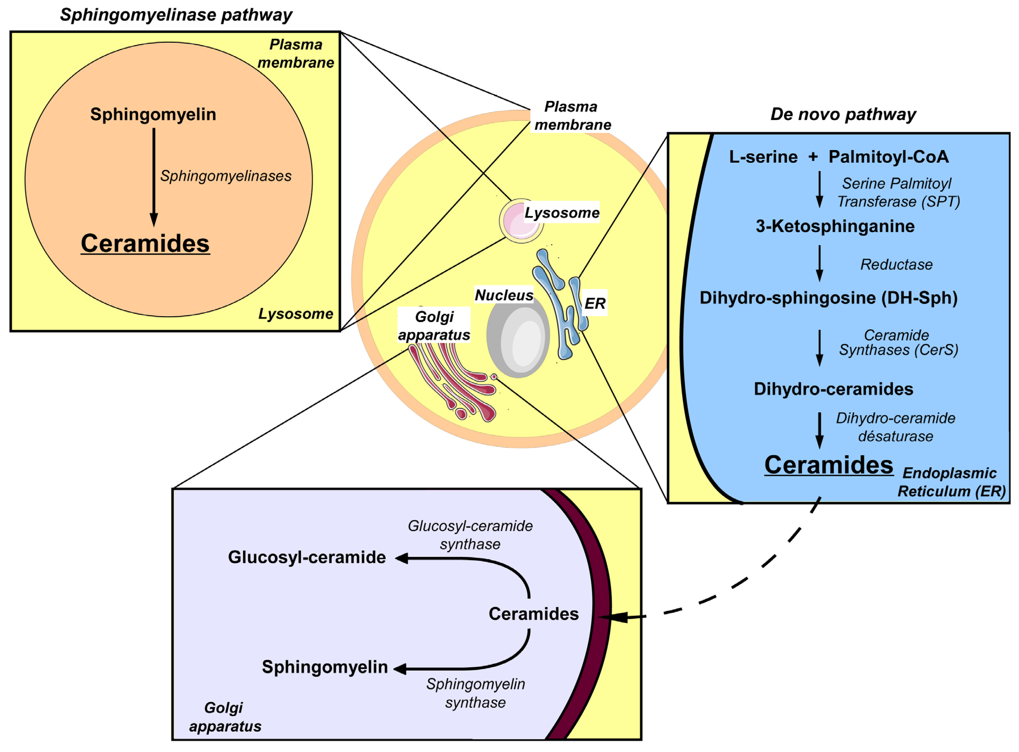
4. Sphingolipid Metabolism

It is well established that sphingolipids are structural components of cellular membranes. However, several studies have now shown that sphingolipids have important roles in the regulation of some cellular processes [41,42]. Moreover, the deregulation of these processes plays a key role in the onset of pathologies affecting cellular proliferation and apoptosis [42,43,44]. Several pathways for sphingolipid metabolism have been described [45]. One of these pathways is the de novo synthesis pathway which is initiated on the cytoplasmic face of the endoplasmic reticulum (ER) (Figure 1). The first step of this pathway is the condensation of l-serine with palmitoyl-CoA to form 3-ketosphinganine. This reaction is catalyzed by serine palmitoyl-transferase (SPT) [41]. 3-Ketosphinganine is then reduced to dihydrosphingosine (DH-Sph) by 3-ketosphinganine reductase. The resulting DH-Sph acts as a substrate for ceramide synthases (CerS) leading to the production of dihydro-ceramides. Dihydro-ceramides can be transformed into ceramides by dihydroceramide desaturases [46]. Depending on the LC-CoA attached to the DH-Sph by CerS, the dihydroceramides and the ceramides are classified into different species with varying FA chain lengths (14–32 carbons in mammals) and saturation of the carbon chain [47]. Ceramides can be transported to the Golgi apparatus [48] where they are converted into sphingomyelin by sphingomyelin synthase or into glucosyl-ceramides by glucosyl-ceramide synthase. In addition to de novo synthesis, another metabolic pathway also leads to ceramide production and this involves the degradation of sphingomyelin into ceramide by sphingomyelinases. This process takes place in the lysosomal membrane and the cytoplasmic membrane [41]. In this web of sphingolipid metabolism, ceramides are considered as central players in the metabolism of sphingolipids. Indeed, ceramides are the precursors of many lipid messengers. For example, ceramides can be metabolized into sphingosine which is in turn phosphorylated to yield sphingosine-1-phosphate (S1P) [41]. Ceramides can also be phosphorylated by ceramide kinase to form ceramide-1-phosphate [41].
Figure 1. Synthesis of sphingolipids in mammalian cells. Two main pathways to produce sphingolipids exist in mammals. The de novo pathway of synthesis starts on the cytoplasmic face of the endoplasmic reticulum (ER). The first step of this pathway is the condensation of l-serine with palmitoyl-CoA to form a 3-ketosphinganine. This reaction is catalyzed by serine palmitoyl-transferase (SPT). 3-Ketosphinganine is then reduced to form dihydrosphingosine (DH-Sph) by 3-ketopshinganine reductase. DH-Sph is a substrate of ceramide synthases (CerS) which produce dihydro-ceramide. Dihydroceramides are transformed into ceramides by dihydroceramide desaturase. Ceramide can be transported to the Golgi apparatus to be transformed into more complex sphingolipids such as sphingomyelin and glucosyl-ceramides. A second synthesis pathway is the catabolic pathway that leads to the degradation of sphingomyelin (SM) into ceramides by sphingomyelinases. This process takes place in the lysosomal and plasma membranes.
Jcm 03 00646 g001

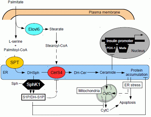
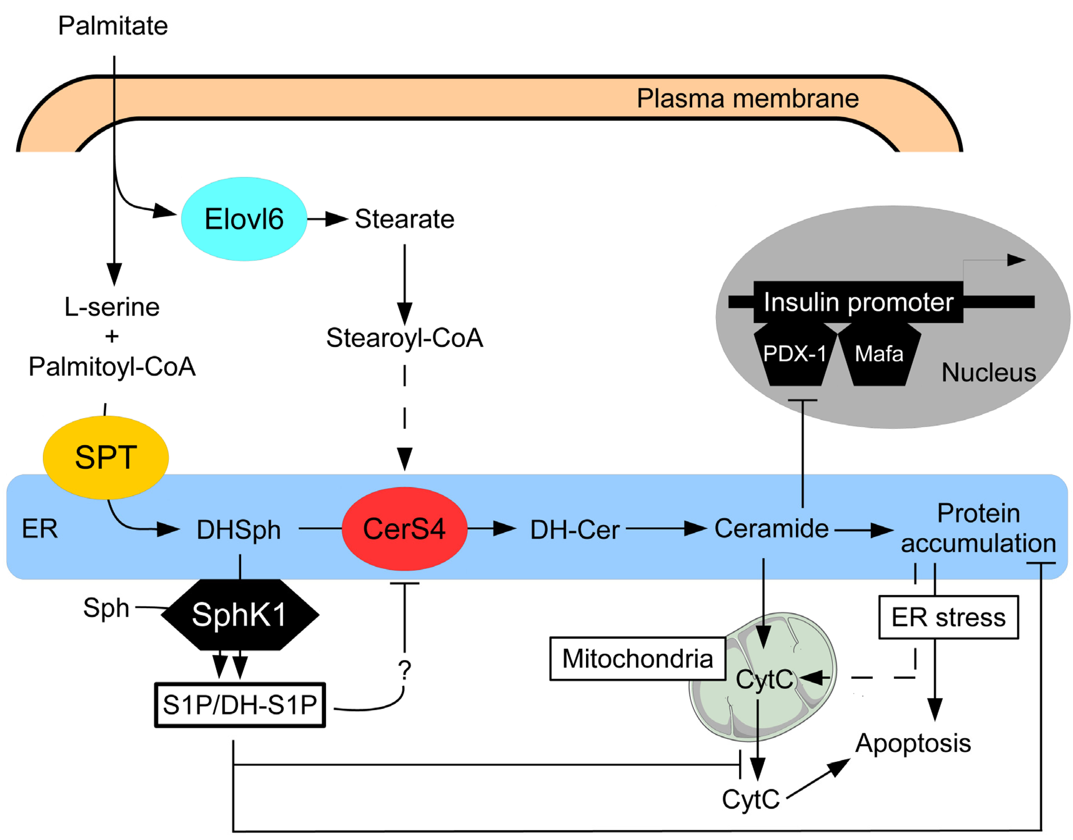
5. Ceramide and Pancreatic β Cell Gluco-Lipotoxicity

As noted above, among the lipids produced during lipotoxicity, ceramides have been implicated in the deleterious effects of gluco-lipotoxicity on pancreatic β cells and in a more general manner, on T2D and obesity. For example, patients with T2D present an increase in sphingolipid levels in plasma, muscle, adipose tissue and liver [25,49]. The accumulation of sphingolipids has been correlated with an increase in insulin resistance [50,51], loss of function and death of pancreatic β cells [8]. Additionally, sphingolipids also appear to also play a role in the complication of obesity and T2D. Interestingly, the two forms of SPT (SPT 1 and 2) have a Km for l-serine and the palmitoyl-CoA near to their physiological concentrations [52,53], and as such, are sensitive to relatively small variations in the concentrations of their substrates. Moreover, condensation of l-serine and palmitoyl-coA is the rate-limiting step in de novo synthesis of ceramides [54]. These two characteristics of SPTs suggest that excessive concentrations of palmitate and its activated form palmitoyl-CoA in a cell can induce the aberrant production of ceramides. It has been shown that diminution of pancreatic β cell mass and loss of insulin secretion are correlated to an increase in intracellular ceramide levels and SPT expression in islet of Langerhans of obese Zucker diabetic fatty (ZDF) rats [35].

5.1. Effect of Ceramide on Pancreatic β Cell Function

Previous studies have demonstrated that cellular permeable analogues of ceramide impaired insulin production in pancreatic β-cells [55]. Under gluco-lipotoxic conditions, several studies showed a correlation between de novo ceramide synthesis and defective insulin expression in islet of Langerhans [8]. In islet of Langerhans and pancreatic β cell lines, ceramide dependent inhibition of insulin expression is mediated by the dephosphorylation of protein kinases, ERK1/2 by PP2A [56,57]. Moreover, the de novo ceramide pathway inhibits the nuclear translocation of two important transcription factors, PDX-1 and Mafa, for insulin-induced gene expression [8]. However, the intracellular signaling pathways mediating these effects are mostly unknown. Previous studies have shown that glucose regulates insulin-induced gene transcription via the serine/threonine Per-Arnt-Sim domain-containing kinase (PASK) [56]. Interestingly, palmitate decrease glucose-induced expression of PASK mRNA and protein levels [56], suggesting that ceramides may function as negative regulators of PASK. It appears that in pancreatic β cells, inhibition of ceramidase, which increased intracellular levels of ceramides, also leads to a reduction in insulin expression [57]. More recently, genetic deletion of sphingomyelin synthase 1 (SMS1) has been shown to induce accumulation of ceramides in islet of Langerhans, dysfunction of mitochondria and the subsequent inhibition of insulin secretion [58]. In Yano’s study, accumulation of ceramides affected mitochondria membrane integrity and induced an excessive production of ROS which were partially responsible for the defect in insulin secretion in pancreatic β cells [58]. Pharmacological inhibition of SMS or down-regulation by siRNA of the ceramide transporter (CERT), which regulate ceramide transport between the ER and Golgi apparatus, increased dysfunction of pancreatic β cells induced by palmitate by affecting binding of transcription factors to the insulin promoter [59]. Altogether, these results suggest that ceramide accumulation, either through the induction of de novo synthesis or reduced conversion into more complex sphingolipids, is involved in loss of function in pancreatic β cells.

5.2. Effect of Ceramide on Pancreatic β Cell Survival

It has been shown that ceramide analogues can block proliferation and induce apoptosis of β cells and islets of Langerhans in culture [60,61]. De novo ceramide synthesis induced by palmitate has also been implicated in pancreatic β cell apoptosis [8,39]. Indeed, inhibition of de novo ceramide synthesis abrogates pancreatic β cell apoptosis induced by palmitate [36,39,62]. More importantly, treatment of ZDF rats with a potent inhibitor of SPT, cyclo-serine, attenuated islet of Langerhans apoptosis and prevented the appearance of hyperglycemia [35]. Ceramide accumulation induced by (gluco)-lipotoxicity induces pancreatic β cell apoptosis through different pathways. FFAs have been shown to induce accumulation of ceramides in ER of pancreatic β cells [40,63]. To date, the mechanisms used by (gluco)-lipotoxicity to block the transport of ceramides remain unknown. However, ceramide accumulation leads to ER stress which plays a key role in lipotoxic induced apoptosis [40,63,64]. Biden and co-workers showed that localized ceramide production and the associated decrease of SM levels in the ER in response to lipotoxicity is important to initiate ER stress in β cell [40,63]. Ceramide induced pancreatic β cell apoptosis is mediated by mitochondrial dysfunction and ROS production [60,61]. This result is also supported by the observation that ceramide dependent ROS production induced by gluco-lipotoxicity inhibits insulin secretion [8]. Evidence to date suggests that ceramide induce apoptosis through mitochondria injuries and subsequent production of ROS [65]. Nevertheless, the role of ROS in gluco-lipotoxicity is still unclear because in some cases, ROS have been excluded from ceramide-induced loss of function or apoptosis of pancreatic β cells [40,66]. Moreover, other hypotheses have been proposed about cellular signals involved in apoptosis of pancreatic β cell induced by ceramides. For example, it has been proposed that β cell apoptosis is induced through the activation of PP2A and the subsequent dephosphorylation of AKT [67], as seen in muscle tissues [68].

5.3. Role of Different Ceramide Species in Gluco-Lipotoxicity Induced Pancreatic β Cell Apoptosis

Ceramide species are distinguishable by the length and/or saturation of their N-acyl chains [47,69,70]. Because ceramide species have many biological roles depending on the cell type and on the inducing signals for their production [47], it is therefore important to determine changes in ceramide species in response to biological stimuli. Indeed, recent studies have reported distinct cellular functions for ceramides with specific N-acyl chain lengths [47,70]. The emergence of lipidomic analysis in the field of sphingolipid metabolism allows us to determine the effect of various treatments, not only on ceramide accumulation but also on changes in levels of ceramide species within the ceramide pool [52]. In pancreatic β cells, it appears that gluco-lipotoxicity modifies the levels of specific ceramide species, specifically C18:0, C22:0 and C24:1 [39]. In mammals, the variety of ceramide species relies on the existence of a family of enzymes, the ceramide synthases (CerS) [47,70]. Six CerS have been identified and they possessed a characteristic substrate preference for particular fatty acyl-CoA [47,70]. Interestingly, this increase in ceramide species is associated with an augmentation of CerS4 expression, which was found to play a critical role in pancreatic β cell apoptosis [39]. Collectively, the results suggest that gluco-lipotoxicity induces β cell apoptosis not only through the induction of de novo ceramide biosynthesis, but also through the formation of ceramides with specific carbon chain lengths rather than an overall increase in ceramide content. This hypothesis is supported by a recent study showing that pancreatic β cell apoptosis is potentiated when high glucose levels increased expression of FA elongase 6 (Elovl6) [71], an enzyme which catalyzes FFA elongation [72]. Importantly, the activity of Elovl-6 is mostly involved in elongation of de novo-synthesized palmitate to produce stearate, suggesting that gluco-lipotoxicity could mediate β-cell apoptosis at least through ceramide C18:0 by a concomitant up-regulation of CerS4 and Elovl-6. Recently, Prentki and co-workers have shown that gluco-lipotoxicity induced early changes in lipid partitioning in order to induce β-cell dysfunction and apoptosis [73]. Indeed, gluco-lipotoxicity induced the expression of proteins which favored FFA esterification, such as SCD-1 through FFA desaturation, and decreased the expression of enzymes involved in lipid oxidation, such as the β subunit of AMP-kinase. These results suggest that in addition to downstream lipid partitioning induced by gluco-lipotoxicity, the synthesis of specific ceramide species could also play a critical role in β cell apoptosis. However, the precise role of specific ceramide species in β cell dysfunction induced by gluco-lipotoxicity remains to be clarified. Indeed, a recent study showed that the inability of CerS2 knock-out mice to synthesize C22–C24 ceramide species did not result in defective insulin secretion [74]. Therefore, it will be important, in the future, to validate these observations in vivo by using rodent models of T2D associated to obesity, and ultimately in human islets of Langerhans in order to identify potent therapeutic targets to limit pancreatic β cell gluco-lipotoxicity.

6. Role of Sphingoid Base Phosphates in T2D

During the past decade, other sphingolipid metabolites, namely sphingoid base phosphates, have been shown to have a role in T2D. The sphingoid base phosphates are S1P and its analogue dihydroS1P, which are respectively produced by phosphorylation of Sph and DH-Sph by sphingosine kinase 1 (SphK1) and sphingosine kinase 2 (SphK2) [45]. S1P is known to induce proliferation, survival, migration and calcium mobilization in many cell types [42]. In T2D patients and rodent models of T2D, the activity of SphK1 and/or mRNA of SphK1 are increased in myocardium [75]. Interestingly, palmitate [76] or glucose [77,78], taken separately, not only increased sphingoid base phosphate levels but also SphK1 activity in different cell types such as muscle and endothelial cells. Moreover, treatment with S1P analogs protected cells from apoptosis [79,80] induced by hyperglycemia. In addition, in vivo over-expression of SphK1 gene in KK/Ay type 2 diabetic mice [81] or treatment mice under high fat diet with S1P analog [82] reduced the deleterious appearance of insulin resistance and T2D. Nevertheless, the role of the SphK1/S1P axis in T2D is still unclear. Indeed, induction of diabetes by an injection of streptozotocin leads to the activation of SphK1 associated to an inflammatory phenotype in vascular endothelium and retina [77,83]. Moreover, the SphK1/S1P axis appears to stimulate the expression of cytokine by adipocytes isolated from ob/ob mice [84] and in obese T2D patients [85].

6.1. Sphingoid Base Phosphates and Insulin Secretion

In pancreatic β cells, SphK1 and SphK2 and four of the five S1PR are expressed [85,86,87]. Expression and activity of SphK1 and SphK2 are stimulated by cytokines such as IL1β, TNFα and interferon-γ, but also by glucose and acetylcholine [77,87]. The role of S1P in insulin secretion is still unclear. Indeed, it has been shown that adding S1P to culture medium of pancreatic INS-1 β cell line and islets of Langerhans inhibits insulin secretion in response to glucose potentiated by GLP-1. The S1P1 receptor, coupled to Gi appears to be involved in this phenomenon [86]. In contrast to this study, S1P was found to stimulate secretion of insulin in islet of Langerhans [88] and in pancreatic HIT-T 15 β cell line [89,90]. Interestingly, Ozcan and colleagues showed that SphK2 plays a critical role in S1P production and insulin secretion induced by glucose [90]. To date, SphK1 has not been implicated in the control of insulin secretion. However, most of these studies have been conducted in pancreatic β cell lines or isolated normal islets. It will thus be important to investigate the in vivo role of sphingoid base phosphates by analyzing insulin secretion in available knock-out mice for SphK1 and SphK2 [91,92].

6.2. Sphingoid Base Phosphates Pancreatic β Cell Survival

In pancreatic β cells, the SphK1/S1P axis plays a critical role in survival. Indeed, adding S1P to culture medium of human or murine islets of Langerhans inhibited apoptosis induced by cytokines [93]. In this case, the anti-apoptotic effect of S1P is mediated by the S1P2 or S1P3 receptors, and the activation of the PKC pathway [93,94]. Interestingly, activation of SphK by glucose has also been implicated in β cell proliferation [76]. It was observed that gluco-lipotoxicity increased sphingoid base phosphates via up-regulation of the SphK1 pathway in pancreatic β cells [88]. Pharmacological inhibition of SphK activity drastically potentiates β cell apoptosis whereas over-expression of SphK1 partially inhibits β cell apoptosis induced by gluco-lipotoxicity. The anti-apoptotic effect of sphingoid base-1-phosphates are likely to be mediated by opposing signaling pathways to ceramide-induced apoptosis. Indeed, over-expression of SphK1 not only inhibited lipotoxicity-induced loss of mitochondrial transmembrane potential and cytochrome c release in pancreatic β cells [95] but also impaired protein trafficking between ER and Golgi which contributed to lipotoxicity-induced ER stress [88]. Another anti-apoptotic effect of sphingoid base phosphates involved the alteration of lipotoxicity-induced ceramide synthesis in pancreatic β cells [88]. While the mechanisms for this latter process remain to be established, it could potentially involve inhibition of CerS by S1P [96]. The in vivo protective role of SphK1 against pancreatic β cell apoptosis induced by gluco-lipotoxicity was supported by a recent study by Qi and co-workers who showed that while a high fat diet-fed wild-type (WT) mice developed glucose intolerance, all high fat diet-fed Sphk1 knock-out mice manifested evident diabetes, a phenomenon due to a drastic decrease in pancreatic β cell mass [95]. Nevertheless, the in vivo role of SphK1 still remains unclear since a recent study showed that under high fat diet-fed mice, SphK1 deficiency was associated with enhanced insulin signaling in adipose and muscle tissues and improved systemic insulin sensitivity and glucose tolerance [85]. Despite this recent study, the anti-apoptotic role of sphingoid base phosphates in pancreatic β cells are in agreement with the work of Boslem et al., who showed that ceramide removal from the ER, by favoring their conversion into glucosylceramides [40], is critical for β cell survival under gluco-lipotoxic conditions. Our findings suggest that the SphK1/sphingoid base phosphates axis could constitute a new therapeutic strategy for preventing pancreatic β cell death and thus the onset of T2D associated with obesity. In the future, it will be important to identify potent activators of the SphK1/sphingoid base phosphates axis in pancreatic β cells. In support of this, a recent work by Holland et al. [97] showed that adiponectin can preserve pancreatic β cell function by shifting the ceramide/S1P ratio towards higher relative levels of S1P.

7. Conclusions

Because of their inter-convertibility and opposing effects, the dynamic balance between sphingoid base phosphates and ceramides has been proposed to be an important factor that determines cell fate [42], a concept called the sphingolipid biostat. The results of recent in vitro and in some cases in vivo studies lend credence to the observation that pancreatic β cell apoptosis induced by gluco-lipotoxicity is regulated by such a sphingolipid biostat (Figure 2). The existence of this biostat offers the possibility to find new therapeutic targets in order to limit pancreatic β cells failure observed during obesity, and therefore to prevent the appearance of T2D.
Figure 2. Role of sphingolipid biostat in β-cell apoptosis during gluco-lipotoxicity. Palmitate has been shown to induce ceramide accumulation by a dual mechanism involving serine palmitoyl-transferase (SPT) and the formation of ceramides with specific N-acyl chain lengths by ceramide synthase 4 (CerS4). Fatty acid elongase 6 (Elovl-6) provides preferential substrate for CerS4 by converting palmitate into stearate. Ceramides reduces insulin expression through inhibiting the binding of pancreatic and duodenal homeobox 1 (PDX-1) and Mafa transcriptional factors to insulin promoter. Ceramide accumulation can also induce pancreatic β cell apoptosis. However, palmitate also induces sphingosine kinase 1 (SphK1) expression, which will channel the preferential metabolic flow of newly formed dihydrosphingosine (DHSph) towards its phosphorylation into dihydrosphingosine-1-phosphate (DHS1P) and increase S1P levels. Accumulation of these sphingoid base phosphates in the ER will play a protective role against palmitate-induced ceramide-dependent apoptotic β cell death. Sphingoid base phosphates inhibit ceramide synthesis by acting probably on CerS4 activity and restore protein trafficking in the endoplasmic reticulum (ER), alleviating ER stress and attenuating cytochrome C (CytC) release from mitochondria. DH-Cer: dihydro-ceramides; Sph: sphingosine.
Jcm 03 00646 g002

Acknowledgements

We thank the many members of the Magnan’s lab for their contributions to the studies that were quoted in this review. We apologize to those authors whose works were not cited due to space limitations. Julien Véret is supported by a post-doctoral fellowship from the University Paris Diderot. We also thank anonymous reviewers for comments that improved the manuscript.

Abbreviations

FFA
free fatty acid
LC-CoA
long chain acyl-CoA
S1P
shingosine-1-phosphate
CPT-1
carnitine palmitoyl-CoA transferase 1
ACC
acetyl-CoA carboxylase
FAS
fatty acid synthase
ER
endoplasmic reticulum
SPT
serine palmitoyl-transferase
DH-Sph
dihydrosphingosine
CerS
ceramide synthases
PASK
serine/threonine Per-Arnt-Sim domain-containing kinase
SMS1
sphingomyelin synthase 1
SphK
sphingosine kinase

References

  1. Klöppel, G.; Löhr, M.; Habich, K.; Oberholzer, M.; Heitz, P.U. Islet Pathology and the Pathogenesis of Type 1 and Type 2 Diabetes Mellitus Revisited. Surv. Synth. Pathol. Res. 1985, 4, 110–125. [Google Scholar]
  2. Butler, A.E.; Janson, J.; Bonner-Weir, S.; Ritzel, R.; Rizza, R.A.; Butler, P.C. Beta-cell Deficit and Increased Beta-cell Apoptosis in Humans with Type 2 Diabetes. Diabetes 2003, 52, 102–110. [Google Scholar] [CrossRef]
  3. Khan, S.R.; Ayub, N.; Nawab, S.; Shamsi, T.S. Triglyceride Profile in Dyslipidaemia of Type 2 Diabetes Mellitus. J. Coll. Physicians Surg. Pak. 2008, 18, 270–273. [Google Scholar]
  4. Carey, D.G.; Jenkins, A.B.; Campbell, L.V.; Freund, J.; Chisholm, D.J. Abdominal fat and insulin resistance in normal and overweight women: Direct measurements reveal a strong relationship in subjects at both low and high risk of NIDDM. Diabetes 1996, 45, 633–638. [Google Scholar] [CrossRef]
  5. World Health Organisation. Available online: http://www.who.int/fr/ (accessed on 26 February 2014).
  6. Crespin, S.R.; Greenough, W.B., III; Steinberg, D. Stimulation of Insulin Secretion by Infusion of Free Fatty Acids. J. Clin. Invest. 1969, 48, 1934–1943. [Google Scholar] [CrossRef]
  7. Stein, D.T.; Stevenson, B.E.; Chester, M.W.; Basit, M.; Daniels, M.B.; Turley, S.D.; McGarry, J.D. The Insulinotropic Potency of Fatty Acids Is Influenced Profoundly by Their Chain Length and Degree of Saturation. J. Clin. Invest. 1997, 100, 398–403. [Google Scholar] [CrossRef]
  8. Poitout, V.; Robertson, R.P. Glucolipotoxicity: Fuel Excess and Beta-cell Dysfunction. Endocr. Rev. 2008, 29, 351–366. [Google Scholar] [CrossRef]
  9. Boden, G.; Shulman, G.I. Free Fatty Acids in Obesity and Type 2 Diabetes: Defining Their Role in the Development of Insulin Resistance and Beta-cell Dysfunction. Eur. J. Clin. Invest. 2002, 32, 14–23. [Google Scholar] [CrossRef]
  10. Prentki, M.; Corkey, B.E. Are the Beta-cell Signaling Molecules Malonyl-CoA and Cystolic Long-chain Acyl-CoA Implicated in Multiple Tissue Defects of Obesity and NIDDM? Diabetes 1996, 45, 273–283. [Google Scholar] [CrossRef]
  11. Marshall, B.A.; Tordjman, K.; Host, H.H.; Ensor, N.J.; Kwon, G.; Marshall, C.A.; Coleman, T.; McDaniel, M.L.; Semenkovich, C.F. Relative Hypoglycemia and Hyperinsulinemia in Mice with Heterozygous Lipoprotein Lipase (LPL) Deficiency. Islet LPL Regulates Insulin Secretion. J. Biol. Chem. 1999, 274, 27426–27432. [Google Scholar] [CrossRef]
  12. Kim, K.H. Regulation of Mammalian Acetyl-coenzyme A Carboxylase. Annu. Rev. Nutr. 1997, 17, 77–99. [Google Scholar] [CrossRef]
  13. Tamarit-Rodriguez, J.; Vara, E.; Tamarit, J. Starvation-induced Secretory Changes of Insulin, Somatostatin, and Glucagon and Their Modification by 2-Bromostearate. Horm. Metab. Res. 1984, 16, 115–119. [Google Scholar] [CrossRef]
  14. Corkey, B.E.; Glennon, M.C.; Chen, K.S.; Deeney, J.T.; Matschinsky, F.M.; Prentki, M. A Role for Malonyl-CoA in Glucose-stimulated Insulin Secretion from Clonal Pancreatic Beta-cells. J. Biol. Chem. 1989, 264, 21608–21612. [Google Scholar]
  15. Liang, Y.; Matschinsky, F.M. Content of CoA-esters in Perifused Rat Islets Stimulated by Glucose and Other Fuels. Diabetes 1991, 40, 327–333. [Google Scholar] [CrossRef]
  16. Prentki, M.; Vischer, S.; Glennon, M.C.; Regazzi, R.; Deeney, J.T.; Corkey, B.E. Malonyl-CoA and Long Chain Acyl-CoA Esters as Metabolic Coupling Factors in Nutrient-induced Insulin Secretion. J. Biol. Chem. 1992, 267, 5802–5810. [Google Scholar]
  17. Brun, T.; Roche, E.; Assimacopoulos-Jeannet, F.; Corkey, B.E.; Kim, K.H.; Prentki, M. Evidence for an Anaplerotic/Malonyl-CoA Pathway in Pancreatic Beta-cell Nutrient Signaling. Diabetes 1996, 45, 190–198. [Google Scholar] [CrossRef]
  18. Wolf, B.A.; Easom, R.A.; McDaniel, M.L.; Turk, J. Diacylglycerol Synthesis De Novo from Glucose by Pancreatic Islets Isolated from Rats and Humans. J. Clin. Invest. 1990, 85, 482–490. [Google Scholar] [CrossRef]
  19. Dunlop, M.E.; Larkins, R.G. Pancreatic Islets Synthesize Phospholipids De Novo from Glucose via Acyl-dihydroxyacetone Phosphate. Biochem. Biophys. Res. Commun. 1985, 132, 467–473. [Google Scholar] [CrossRef]
  20. Farese, R.V.; DiMarco, P.E.; Barnes, D.E.; Sabir, M.A.; Larson, R.E.; Davis, J.S.; Morrison, A.D. Rapid Glucose-dependent Increases in Phosphatidic Acid and Phosphoinositides in Rat Pancreatic Islets. Endocrinology 1986, 118, 1498–1503. [Google Scholar] [CrossRef]
  21. Salt, I.P.; Johnson, G.; Ashcroft, S.J.; Hardie, D.G. AMP-Activated Protein Kinase Is Activated by Low Glucose in Cell Lines Derived from Pancreatic Beta Cells, and May Regulate Insulin Release. Biochem. J. 1998, 335, 533–539. [Google Scholar]
  22. Mittra, S.; Bansal, V.S.; Bhatnagar, P.K. From a Glucocentric to a Lipocentric Approach towards Metabolic Syndrome. Drug Discov. Today 2008, 13, 211–218. [Google Scholar] [CrossRef]
  23. Unger, R.H.; Orci, L. Diseases of Liporegulation: New Perspective on Obesity and Related Disorders. FASEB J. 2001, 15, 312–321. [Google Scholar] [CrossRef]
  24. Van Herpen, N.A.; Schrauwen-Hinderling, V.B. Lipid Accumulation in Non-adipose Tissue and Lipotoxicity. Physiol. Behav. 2008, 94, 231–241. [Google Scholar] [CrossRef]
  25. Kotronen, A.; Seppänen-Laakso, T.; Westerbacka, J.; Kiviluoto, T.; Arola, J.; Ruskeepää, A.L.; Yki-Järvinen, H.; Oresic, M. Comparison of Lipid and Fatty Acid Composition of the Liver, Subcutaneous and Intra-abdominal Adipose Tissue, and Serum. Obesity (Silver Spring) 2010, 18, 937–944. [Google Scholar] [CrossRef]
  26. Yetukuri, L.; Katajamaa, M.; Medina-Gomez, G.; Seppänen-Laakso, T.; Vidal-Puig, A.; Oresic, M. Bioinformatics Strategies for Lipidomics Analysis: Characterization of Obesity Related Hepatic Steatosis. BMC Syst. Biol. 2007, 1. [Google Scholar] [CrossRef]
  27. Sako, Y.; Grill, V.E. A 48-Hour Lipid Infusion in the Rat Time-dependently Inhibits Glucose-induced Insulin Secretion and B Cell Oxidation through a Process Likely Coupled to Fatty Acid Oxidation. Endocrinology 1990, 127, 1580–1589. [Google Scholar] [CrossRef]
  28. Zhou, Y.P.; Grill, V.E. Long-term Exposure of Rat Pancreatic Islets to Fatty Acids Inhibits Glucose-induced Insulin Secretion and Biosynthesis through a Glucose Fatty Acid Cycle. J. Clin. Invest. 1994, 93, 870–876. [Google Scholar] [CrossRef]
  29. Kelpe, C.L.; Moore, P.C.; Parazzoli, S.D.; Wicksteed, B.; Rhodes, C.J.; Poitout, V. Palmitate Inhibition of Insulin Gene Expression Is Mediated at the Transcriptional Level via Ceramide Synthesis. J. Biol. Chem. 2003, 278, 30015–30021. [Google Scholar]
  30. McGarry, J.D.; Dobbins, R.L. Fatty Acids, Lipotoxicity and Insulin Secretion. Diabetologia 1999, 42, 128–138. [Google Scholar] [CrossRef]
  31. Ritz-Laser, B.; Meda, P.; Constant, I.; Klages, N.; Charollais, A.; Morales, A.; Magnan, C.; Ktorza, A.; Philippe, J. Glucose-induced Preproinsulin Gene Expression Is Inhibited by the Free Fatty Acid Palmitate. Endocrinology 1999, 140, 4005–4014. [Google Scholar]
  32. El-Assaad, W.; Buteau, J.; Peyot, M.L.; Nolan, C.; Roduit, R.; Hardy, S.; Joly, E.; Dbaibo, G.; Rosenberg, L.; Prentki, M. Saturated Fatty Acids Synergize with Elevated Glucose to Cause Pancreatic Beta-cell Death. Endocrinology 2003, 144, 4154–4163. [Google Scholar] [CrossRef]
  33. Lupi, R.; Dotta, F.; Marselli, L.; Del Guerra, S.; Masini, M.; Santangelo, C.; Patané, G.; Boggi, U.; Piro, S.; Anello, M.; et al. Prolonged Exposure to Free Fatty Acids Has Cytostatic and Pro-apoptotic Effects on Human Pancreatic Islets: Evidence that Beta-cell Death is Caspase Mediated, Partially Dependent on Ceramide Pathway, and Bcl-2 Regulated. Diabetes 2002, 51, 1437–1442. [Google Scholar] [CrossRef]
  34. Maedler, K.; Spinas, G.A.; Dyntar, D.; Moritz, W.; Kaiser, N.; Donath, M.Y. Distinct Effects of Saturated and Monounsaturated Fatty Acids on Beta-cell Turnover and Function. Diabetes 2001, 50, 69–76. [Google Scholar] [CrossRef]
  35. Shimabukuro, M.; Higa, M.; Zhou, Y.T.; Wang, M.Y.; Newgard, C.B.; Unger, R.H. Lipoapoptosis in Beta-cells of Obese Prediabetic fa/fa Rats. Role of Serine Palmitoyltransferase Overexpression. J. Biol. Chem. 1998, 273, 32487–32490. [Google Scholar]
  36. Shimabukuro, M.; Zhou, Y.T.; Levi, M.; Unger, R.H. Fatty Acid-induced Beta Cell Apoptosis: A Link between Obesity and Diabetes. Proc. Natl. Acad. Sci. USA 1998, 95, 2498–2502. [Google Scholar] [CrossRef]
  37. Prentki, M.; Joly, E.; El-Assaad, W.; Roduit, R. Malonyl-CoA Signaling, Lipid Partitioning, and Glucolipotoxicity: Role in Beta-cell Adaptation and Failure in the Etiology of Diabetes. Diabetes 2002, 51, S405–S413. [Google Scholar] [CrossRef]
  38. Weir, G.C.; Laybutt, D.R.; Kaneto, H.; Bonner-Weir, S.; Sharma, A. Beta-cell Adaptation and Decompensation during the Progression of Diabetes. Diabetes 2001, 50, S154–S159. [Google Scholar] [CrossRef]
  39. Véret, J.; Coant, N.; Berdyshev, E.V.; Skobeleva, A.; Therville, N.; Bailbé, D.; Gorshkova, I.; Natarajan, V.; Portha, B.; Le Stunff, H. Ceramide Synthase 4 and De Novo Production of Ceramides with Specific N-Acyl Chain Lengths Are Involved in Glucolipotoxicity-induced Apoptosis of INS-1 β-Cells. Biochem. J. 2011, 438, 177–189. [Google Scholar] [CrossRef]
  40. Boslem, E.; Meikle, P.J.; Biden, T.J. Roles of ceramide and sphingolipids in pancreatic β-cell function and dysfunction. Islets 2012, 4, 177–187. [Google Scholar] [CrossRef]
  41. Hannun, Y.A.; Obeid, L.M. Principles of Bioactive Lipid Signalling: Lessons from Sphingolipids. Nat. Rev. Mol. Cell Biol. 2008, 9, 139–150. [Google Scholar] [CrossRef]
  42. Spiegel, S.; Milstien, S. Sphingosine-1-phosphate: An Enigmatic Signalling Lipid. Nat. Rev. Mol. Cell Biol. 2003, 4, 397–407. [Google Scholar] [CrossRef]
  43. Kihara, A.; Mitsutake, S.; Mizutani, Y.; Igarashi, Y. Metabolism and Biological Functions of Two Phosphorylated Sphingolipids, Sphingosine 1-Phosphate and Ceramide 1-Phosphate. Prog. Lipid Res. 2007, 46, 126–144. [Google Scholar] [CrossRef]
  44. Taha, T.A.; Hannun, Y.A.; Obeid, L.M. Sphingosine Kinase: Biochemical and Cellular Regulation and Role in Disease. J. Biochem. Mol. Biol. 2006, 39, 113–131. [Google Scholar] [CrossRef]
  45. Le Stunff, H.; Milstien, S.; Spiegel, S. Generation and Metabolism of Bioactive Sphingosine-1-Phosphate. J. Cell. Biochem. 2004, 92, 882–899. [Google Scholar] [CrossRef]
  46. Causeret, C.; Geeraert, L.; van der Hoeven, G.; Mannaerts, G.P.; van Veldhoven, P.P. Further Characterization of Rat Dihydroceramide Desaturase: Tissue Distribution, Subcellular Localization, and Substrate Specificity. Lipids 2000, 35, 1117–1125. [Google Scholar] [CrossRef]
  47. Pewzner-Jung, Y.; Ben-Dor, S.; Futerman, A.H. When Do Lasses (Longevity Assurance Genes) Become CerS (Ceramide Synthases)? Insights into the Regulation of Ceramide Synthesis. J. Biol. Chem. 2006, 281, 25001–25005. [Google Scholar] [CrossRef]
  48. Hanada, K.; Kumagai, K.; Yasuda, S.; Miura, Y.; Kawano, M.; Fukasawa, M.; Nishijima, M. Molecular Machinery for Non-vesicular Trafficking of Ceramide. Nature 2003, 426, 803–809. [Google Scholar] [CrossRef]
  49. Haus, J.M.; Kashyap, S.R.; Kasumov, T.; Zhang, R.; Kelly, K.R.; Defronzo, R.A.; Kirwan, J.P. Plasma Ceramides Are Elevated in Obese Subjects with Type 2 Diabetes and Correlate with the Severity of Insulin Resistance. Diabetes 2009, 58, 337–343. [Google Scholar]
  50. Holland, W.L.; Brozinick, J.T.; Wang, L.P.; Hawkins, E.D.; Sargent, K.M.; Liu, Y.; Narra, K.; Hoehn, K.L.; Knotts, T.A.; Siesky, A.; et al. Inhibition of Ceramide Synthesis Ameliorates Glucocorticoid-, Saturated-fat-, and Obesity-induced Insulin Resistance. Cell Metab. 2007, 5, 167–179. [Google Scholar] [CrossRef]
  51. Park, T.S.; Hu, Y.; Noh, H.L.; Drosatos, K.; Okajima, K.; Buchanan, J.; Tuinei, J.; Homma, S.; Jiang, X.C.; Abel, E.D.; Goldberg, I.J. Ceramide Is a Cardiotoxin in Lipotoxic Cardiomyopathy. J. Lipid Res. 2008, 49, 2101–2112. [Google Scholar] [CrossRef]
  52. Merrill, A.H., Jr.; Wang, E.; Mullins, R.E. Kinetics of Long-chain (Sphingoid) Base Biosynthesis in Intact LM Cells: Effects of Varying the Extracellular Concentrations of Serine and Fatty Acid Precursors of this Pathway. Biochemistry 1988, 27, 340–345. [Google Scholar] [CrossRef]
  53. Williams, R.D.; Nixon, D.W.; Merrill, A.H., Jr. Comparison of Serine Palmitoyltransferase in Morris Hepatoma 7777 and Rat Liver. Cancer Res. 1984, 44, 1918–1923. [Google Scholar]
  54. Perry, D.K. Serine Palmitoyltransferase: Role in Apoptotic De Novo Ceramide Synthesis and other Stress Responses. Biochim. Biophys. Acta 2002, 1585, 146–152. [Google Scholar] [CrossRef]
  55. Sjöholm, A. Ceramide Inhibits Pancreatic Beta-cell Insulin Production and Mitogenesis and Mimics the Actions of Interleukin-1 Beta. FEBS Lett. 1995, 367, 283–286. [Google Scholar] [CrossRef]
  56. Fontés, G.; Semache, M.; Hagman, D.K.; Tremblay, C.; Shah, R.; Rhodes, C.J.; Rutter, J.; Poitout, V. Involvement of Per-Arnt-Sim Kinase and Extracellular-regulated Kinases-1/2 in Palmitate Inhibition of Insulin Gene Expression in Pancreatic Beta-cells. Diabetes 2009, 58, 2048–2058. [Google Scholar] [CrossRef]
  57. Guo, J.; Qian, Y.; Xi, X.; Hu, X.; Zhu, J.; Han, X. Blockage of Ceramide Metabolism Exacerbates Palmitate Inhibition of Pro-insulin Gene Expression in Pancreatic Beta-cells. Mol. Cell. Biochem. 2010, 338, 283–290. [Google Scholar] [CrossRef]
  58. Yano, M.; Watanabe, K.; Yamamoto, T.; Ikeda, K.; Senokuchi, T.; Lu, M.; Kadomatsu, T.; Tsukano, H.; Ikawa, M.; Okabe, M.; et al. Mitochondrial Dysfunction and Increased Reactive Oxygen Species Impair Insulin Secretion in Sphingomyelin Synthase 1-Null Mice. J. Biol. Chem. 2011, 286, 3992–4002. [Google Scholar] [CrossRef]
  59. Guo, J.; Zhu, J.X.; Deng, X.H.; Hu, X.H.; Zhao, J.; Sun, Y.J.; Han, X. Palmitate-induced Inhibition of Insulin Gene Expression in Rat Islet β-Cells Involves the Ceramide Transport Protein. Cell. Physiol. Biochem. 2010, 26, 717–728. [Google Scholar] [CrossRef]
  60. Veluthakal, R.; Palanivel, R.; Zhao, Y.; McDonald, P.; Gruber, S.; Kowluru, A. Ceramide Induces Mitochondrial Abnormalities in Insulin-secreting INS-1 Cells: Potential Mechanisms Underlying Ceramide-mediated Metabolic Dysfunction of the Beta Cell. Apoptosis 2005, 10, 841–850. [Google Scholar] [CrossRef]
  61. Syed, I.; Szulc, Z.M.; Ogretmen, B.; Kowluru, A. l-Threo-C6-pyridinium-ceramide Bromide, a Novel Cationic Ceramide, Induces NADPH Oxidase Activation, Mitochondrial Dysfunction and Loss in Cell Viability in INS 832/13 β-Cells. Cell. Physiol. Biochem. 2012, 30, 1051–1058. [Google Scholar] [CrossRef]
  62. Maedler, K.; Oberholzer, J.; Bucher, P.; Spinas, G.A.; Donath, M.Y. Monounsaturated Fatty Acids Prevent the Deleterious Effects of Palmitate and High Glucose on Human Pancreatic Beta-cell Turnover and Function. Diabetes 2003, 52, 726–733. [Google Scholar] [CrossRef]
  63. Boslem, E.; Weir, J.M.; MacIntosh, G.; Sue, N.; Cantley, J.; Meikle, P.J.; Biden, T.J. Alteration of Endoplasmic Reticulum Lipid Rafts Contributes to Lipotoxicity in Pancreatic β-cells. J. Biol. Chem. 2013, 288, 26569–26582. [Google Scholar] [CrossRef]
  64. Eizirik, D.L.; Cardozo, A.K.; Cnop, M. The Role for Endoplasmic Reticulum Stress in Diabetes Mellitus. Endocr. Rev. 2008, 29, 42–61. [Google Scholar] [CrossRef]
  65. Kogot-Levin, A.; Saada, A. Ceramide and the Mitochondrial Respiratory Chain. Biochimie 2014. [Google Scholar] [CrossRef]
  66. Moore, P.C.; Ugas, M.A.; Hagman, D.K.; Parazzoli, S.D.; Poitout, V. Evidence against the Involvement of Oxidative Stress in Fatty Acid Inhibition of Insulin Secretion. Diabetes 2004, 53, 2610–2616. [Google Scholar] [CrossRef]
  67. Lang, F.; Ullrich, S.; Gulbins, E. Ceramide formation as a target in beta-cell survival and function. Expert Opin. Ther. Targets 2011, 15, 1061–1071. [Google Scholar] [CrossRef]
  68. Blouin, C.M.; Prado, C.; Takane, K.K.; Lasnier, F.; Garcia-Ocana, A.; Ferré, P.; Dugail, I.; Hajduch, E. Plasma Membrane Subdomain Compartmentalization Contributes to Distinct Mechanisms of Ceramide Action on Insulin Signaling. Diabetes 2010, 59, 600–610. [Google Scholar] [CrossRef]
  69. Merril, A.H., Jr. SphinGOMAP—A Web-based Biosynthetic Pathway Map of Sphingolipids and Glycosphingolipids. Glycobiology 2005, 15, 15G. [Google Scholar] [CrossRef]
  70. Mizutani, Y.; Kihara, A.; Igarashi, Y. Mammalian Lass6 and Its Related Family Members Regulate Synthesis of Specific Ceramides. Biochem. J. 2005, 390, 263–271. [Google Scholar] [CrossRef]
  71. Green, C.D.; Olson, L.K. Modulation of Palmitate-induced Endoplasmic Reticulum Stress and Apoptosis in Pancreatic β-Cells by Stearoyl-CoA Desaturase and Elovl6. Am. J. Physiol. Endocrinol. Metab. 2011, 300, E640–E649. [Google Scholar] [CrossRef]
  72. Guillou, H.; Zadravec, D.; Martin, P.G.P.; Jacobsson, A. The Key Roles of Elongases and Desaturases in Mammalian Fatty Acid Metabolism: Insights from Transgenic Mice. Prog. Lipid Res. 2010, 49, 186–199. [Google Scholar] [CrossRef]
  73. El-Assaad, W.; Joly, E.; Barbeau, A.; Sladek, R.; Buteau, J.; Maestre, I.; Pepin, E.; Zhao, S.; Iglesias, J.; Roche, E.; Prentki, M. Glucolipotoxicity alters lipid partitioning and causes mitochondrial dysfunction, cholesterol, and ceramide deposition and reactive oxygen species production in INS832/13 ss-cells. Endocrinology 2010, 151, 3061–3073. [Google Scholar] [CrossRef]
  74. Park, J.W.; Park, W.J.; Kuperman, Y.; Boura-Halfon, S.; Pewzner-Jung, Y.; Futerman, A.H. Ablation of very long acyl chain sphingolipids causes hepatic insulin resistance in mice due to altered detergent-resistant membranes. Hepatology 2013, 57, 525–532. [Google Scholar] [CrossRef]
  75. Baranowski, M.; Blachnio-Zabielska, A.; Hirnle, T.; Harasiuk, D.; Matlak, K.; Knapp, M.; Zabielski, P.; Gorski, J. Myocardium of Type 2 Diabetic and Obese Patients Is Characterized by Alterations in Sphingolipid Metabolic Enzymes but Not by Accumulation of Ceramide. J. Lipid Res. 2010, 51, 74–80. [Google Scholar] [CrossRef]
  76. Hu, W.; Bielawski, J.; Samad, F.; Merrill, A.H., Jr.; Cowart, L.A. Palmitate Increases Sphingosine-1-phosphate in C2C12 Myotubes via Upregulation of Sphingosine Kinase Message and Activity. J. Lipid Res. 2009, 50, 1852–1862. [Google Scholar] [CrossRef]
  77. Mastrandrea, L.D.; Sessanna, S.M.; Del Toro, A.; Laychock, S.G. ATP-Independent Glucose Stimulation of Sphingosine Kinase in Rat Pancreatic Islets. J. Lipid Res. 2010, 51, 2171–2180. [Google Scholar] [CrossRef]
  78. Wang, L.; Xing, X.P.; Holmes, A.; Wadham, C.; Gamble, J.R.; Vadas, M.A.; Xia, P. Activation of the Sphingosine Kinase-signaling Pathway by High Glucose Mediates the Proinflammatory Phenotype of Endothelial Cells. Circ. Res. 2005, 97, 891–899. [Google Scholar] [CrossRef]
  79. Whetzel, A.M.; Bolick, D.T.; Hedrick, C.C. Sphingosine-1-phosphate Inhibits High Glucose-mediated ERK1/2 Action in Endothelium through Induction of MAP Kinase Phosphatase-3. Am. J. Physiol. Cell Physiol. 2009, 296, C339–C345. [Google Scholar] [CrossRef]
  80. You, B.; Ren, A.; Yan, G.; Sun, J. Activation of Sphingosine Kinase-1 Mediates Inhibition of Vascular Smooth Muscle Cell Apoptosis by Hyperglycemia. Diabetes 2007, 56, 1445–1453. [Google Scholar] [CrossRef]
  81. Ma, M.M.; Chen, J.L.; Wang, G.G.; Wang, H.; Lu, Y.; Li, J.F.; Yi, J.; Yuan, Y.J.; Zhang, Q.W.; Mi, J.; Wang, L.S.; Duan, H.F.; Wu, C.T. Sphingosine Kinase 1 Participates in Insulin Signalling and Regulates Glucose Metabolism and Homeostasis in KK/Ay Diabetic Mice. Diabetologia 2007, 50, 891–900. [Google Scholar] [CrossRef]
  82. Kendall, M.R.; Hupfeld, C.J. FTY720, a Sphingosine-1-phosphate Receptor Modulator, Reverses High-fat Diet-induced Weight Gain, Insulin Resistance and Adipose Tissue Inflammation in C57BL/6 Mice. Diabetes Obes. Metab. 2008, 10, 802–805. [Google Scholar] [CrossRef]
  83. Maines, L.W.; French, K.J.; Wolpert, E.B.; Antonetti, D.A.; Smith, C.D. Pharmacologic Manipulation of Sphingosine Kinase in Retinal Endothelial Cells: Implications for Angiogenic Ocular Diseases. Invest. Ophthalmol. Vis. Sci. 2006, 47, 5022–5031. [Google Scholar] [CrossRef]
  84. Samad, F.; Hester, K.D.; Yang, G.; Hannun, Y.A.; Bielawski, J. Altered Adipose and Plasma Sphingolipid Metabolism in Obesity: A Potential Mechanism for Cardiovascular and Metabolic Risk. Diabetes 2006, 55, 2579–2587. [Google Scholar] [CrossRef]
  85. Wang, J.; Badeanlou, L.; Bielawski, J.; Ciaraldi, T.P.; Samad, F. Sphingosine Kinase 1 Regulates Adipose Proinflammatory Responses and Insulin Resistance. Am. J. Physiol. Endocrinol. Metab. 2014, 306, E756–E768. [Google Scholar] [CrossRef]
  86. Laychock, S.G.; Tian, Y.; Sessanna, S.M. Endothelial Differentiation Gene Receptors in Pancreatic Islets and INS-1 Cells. Diabetes 2003, 52, 1986–1993. [Google Scholar] [CrossRef]
  87. Mastrandrea, L.D.; Sessanna, S.M.; Laychock, S.G. Sphingosine Kinase Activity and Sphingosine-1 Phosphate Production in Rat Pancreatic Islets and INS-1 Cells: Response to Cytokines. Diabetes 2005, 54, 1429–1436. [Google Scholar] [CrossRef]
  88. Véret, J.; Coant, N.; Gorshkova, I.A.; Giussani, P.; Fradet, M.; Riccitelli, E.; Skobeleva, A.; Goya, J.; Kassis, N.; Natarajan, V.; et al. Role of Palmitate-induced Sphingoid Base-1-phosphate Biosynthesis in INS-1 β-Cell Survival. Biochim. Biophys. Acta 2013, 1831, 251–262. [Google Scholar] [CrossRef]
  89. Shimizu, H.; Okajima, F.; Kimura, T.; Ohtani, K.; Tsuchiya, T.; Takahashi, H.; Kuwabara, A.; Tomura, H.; Sato, K.; Mori, M. Sphingosine 1-Phosphate Stimulates Insulin Secretion in HIT-T 15 Cells and Mouse Islets. Endocr. J. 2000, 47, 261–269. [Google Scholar] [CrossRef]
  90. Cantrell Stanford, J.; Morris, A.J.; Sunkara, M.; Popa, G.J.; Larson, K.L.; Özcan, S. Sphingosine 1-Phosphate (S1P) Regulates Glucose-stimulated Insulin Secretion in Pancreatic Beta Cells. J. Biol. Chem. 2012, 287, 13457–13464. [Google Scholar] [CrossRef]
  91. Allende, M.L.; Sasaki, T.; Kawai, H.; Olivera, A.; Mi, Y.; van Echten-Deckert, G.; Hajdu, R.; Rosenbach, M.; Keohane, C.A.; Mandala, S.; Spiegel, S.; Proia, R.L. Mice Deficient in Sphingosine Kinase 1 Are Rendered Lymphopenic by FTY720. J. Biol. Chem. 2004, 279, 52487–52492. [Google Scholar] [CrossRef]
  92. Kharel, Y.; Lee, S.; Snyder, A.H.; Sheasley-O’neill, S.L.; Morris, M.A.; Setiady, Y.; Zhu, R.; Zigler, M.A.; Burcin, T.L.; Ley, K.; et al. Sphingosine Kinase 2 Is Required for Modulation of Lymphocyte Traffic by FTY720. J. Biol. Chem. 2005, 280, 36865–36872. [Google Scholar] [CrossRef]
  93. Laychock, S.G.; Sessanna, S.M.; Lin, M.H.; Mastrandrea, L.D. Sphingosine 1-Phosphate Affects Cytokine-induced Apoptosis in Rat Pancreatic Islet Beta-cells. Endocrinology 2006, 147, 4705–4712. [Google Scholar] [CrossRef]
  94. Imasawa, T.; Koike, K.; Ishii, I.; Chun, J.; Yatomi, Y. Blockade of Sphingosine 1-Phosphate Receptor 2 Signaling Attenuates Streptozotocin-induced Apoptosis of Pancreatic Beta-cells. Biochem. Biophys. Res. Commun. 2010, 392, 207–211. [Google Scholar] [CrossRef]
  95. Qi, Y.; Chen, J.; Lay, A.; Don, A.; Vadas, M.; Xia, P. Loss of Sphingosine Kinase 1 Predisposes to the Onset of Diabetes via Promoting Pancreatic β-Cell Death in Diet-induced Obese Mice. FASEB J. 2013, 27, 4294–4304. [Google Scholar] [CrossRef]
  96. Levy, M.; Futerman, A.H. Mammalian Ceramide Synthases. IUBMB Life 2010, 62, 347–356. [Google Scholar]
  97. Holland, W.L.; Miller, R.A.; Wang, Z.V.; Sun, K.; Barth, B.M.; Bui, H.H.; Davis, K.E.; Bikman, B.T.; Halberg, N.; Rutkowski, J.M.; et al. Receptor-mediated Activation of Ceramidase Activity Initiates the Pleiotropic Actions of Adiponectin. Nat. Med. 2011, 17, 55–63. [Google Scholar] [CrossRef]

