Bleeding after Percutaneous Transhepatic Biliary Drainage: Incidence, Causes and Treatments

 , ,
, ,
Abstract
:1. Introduction
2. Pre-Procedure Work-Up and Risk Factors
- Current antiplatelet use
- Central biliary access
- Use of 18G (vs. 21/22G) access needle
- Left-sided drain placement *, * Inconsistently found in the literature
- Multiple passes
- Non-dilated system
- Cirrhosis
- Renal insufficiency
- Advanced age
3. Clinical Evaluation and Initial Treatment
4. Management of Portal Vein and Hepatic Artery Injuries
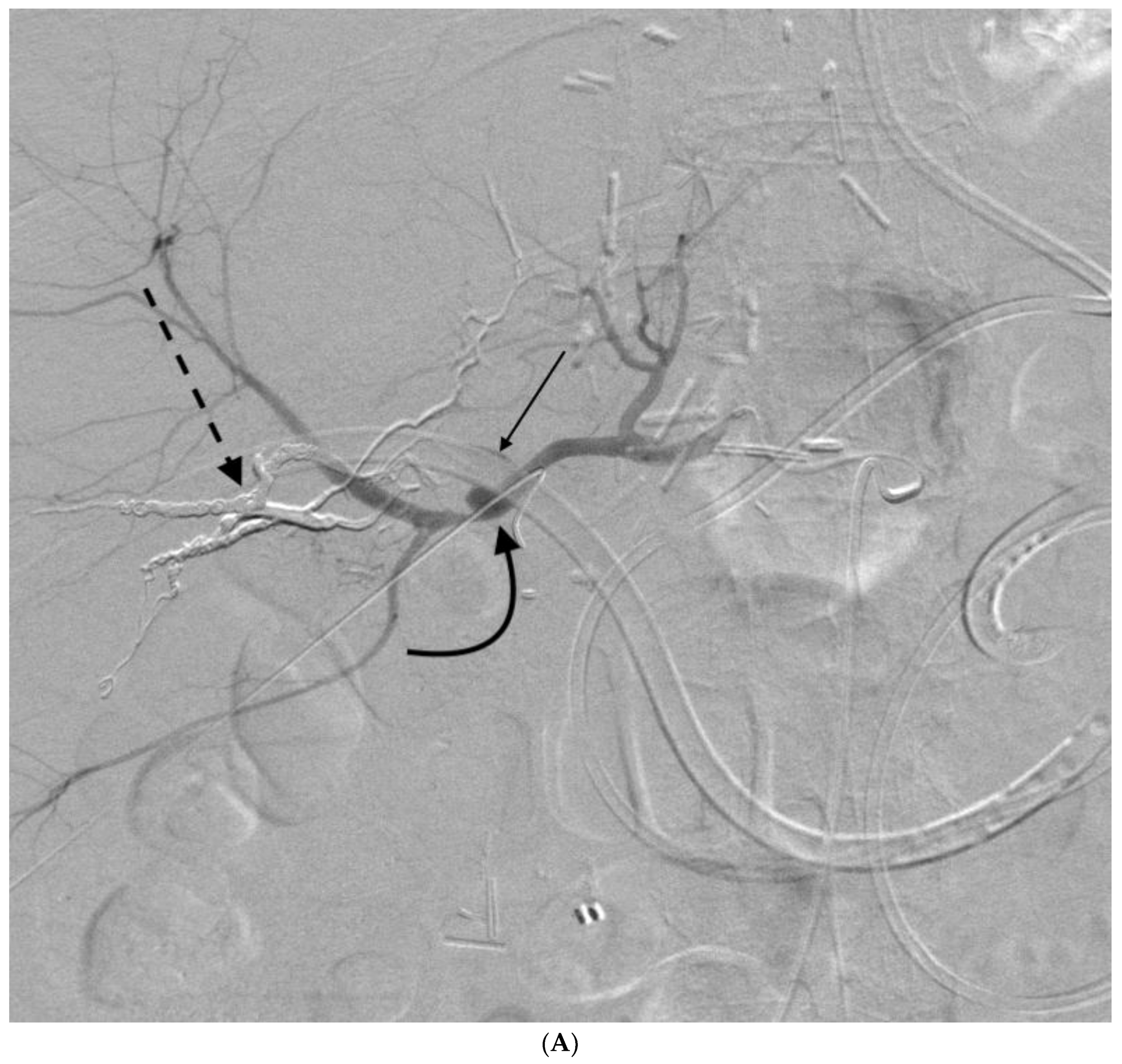
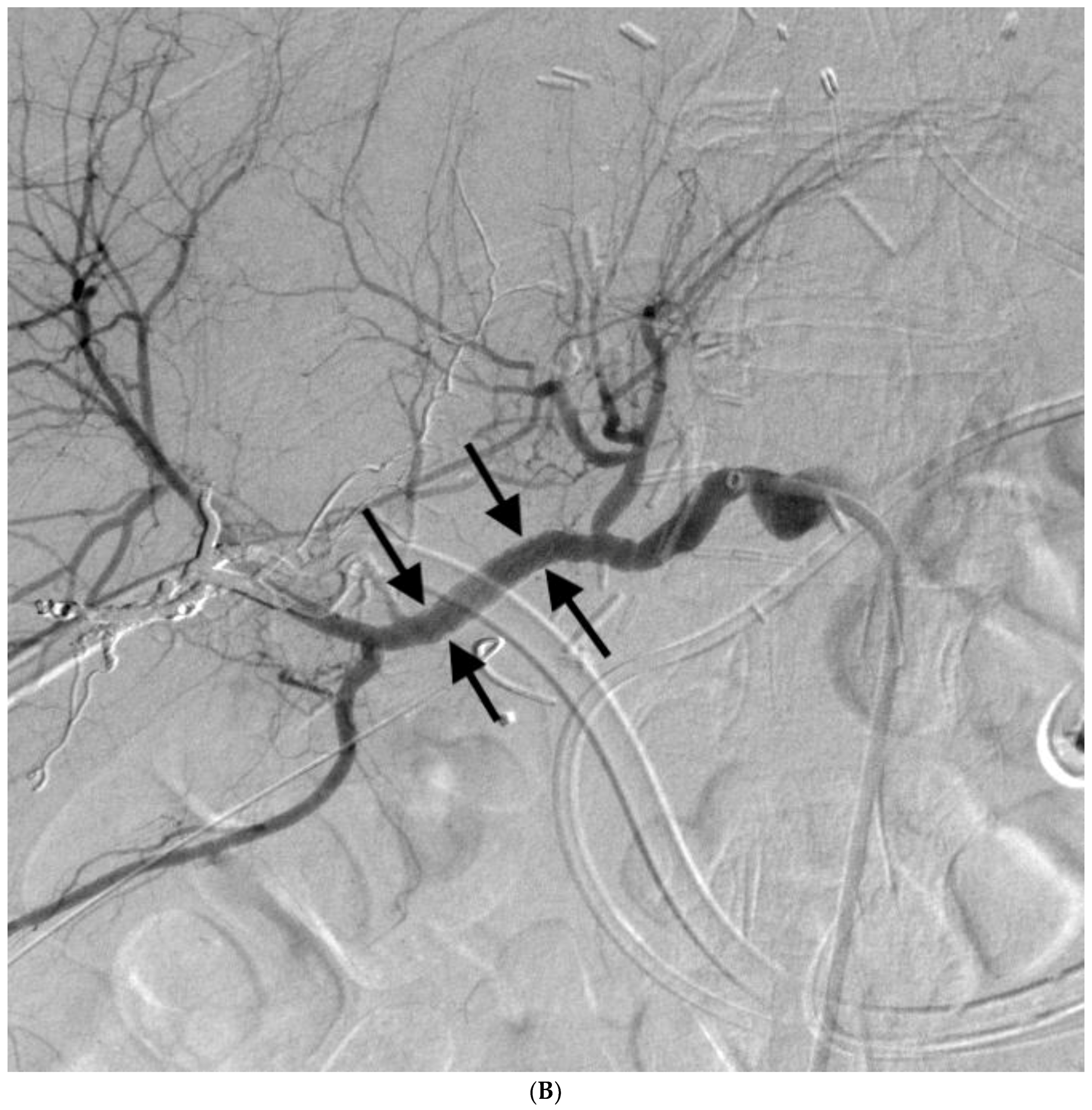
4.1. Portal Vein
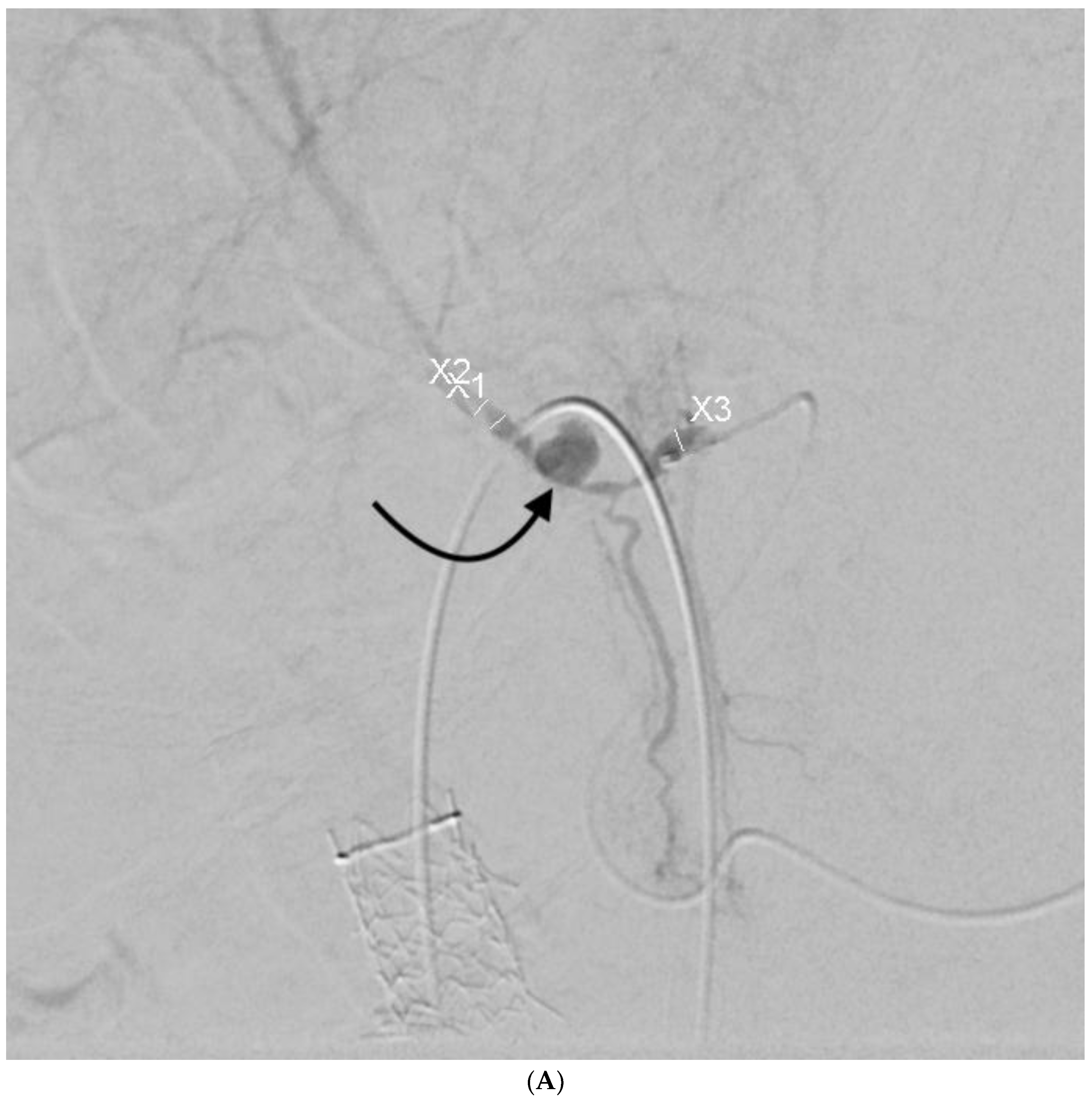
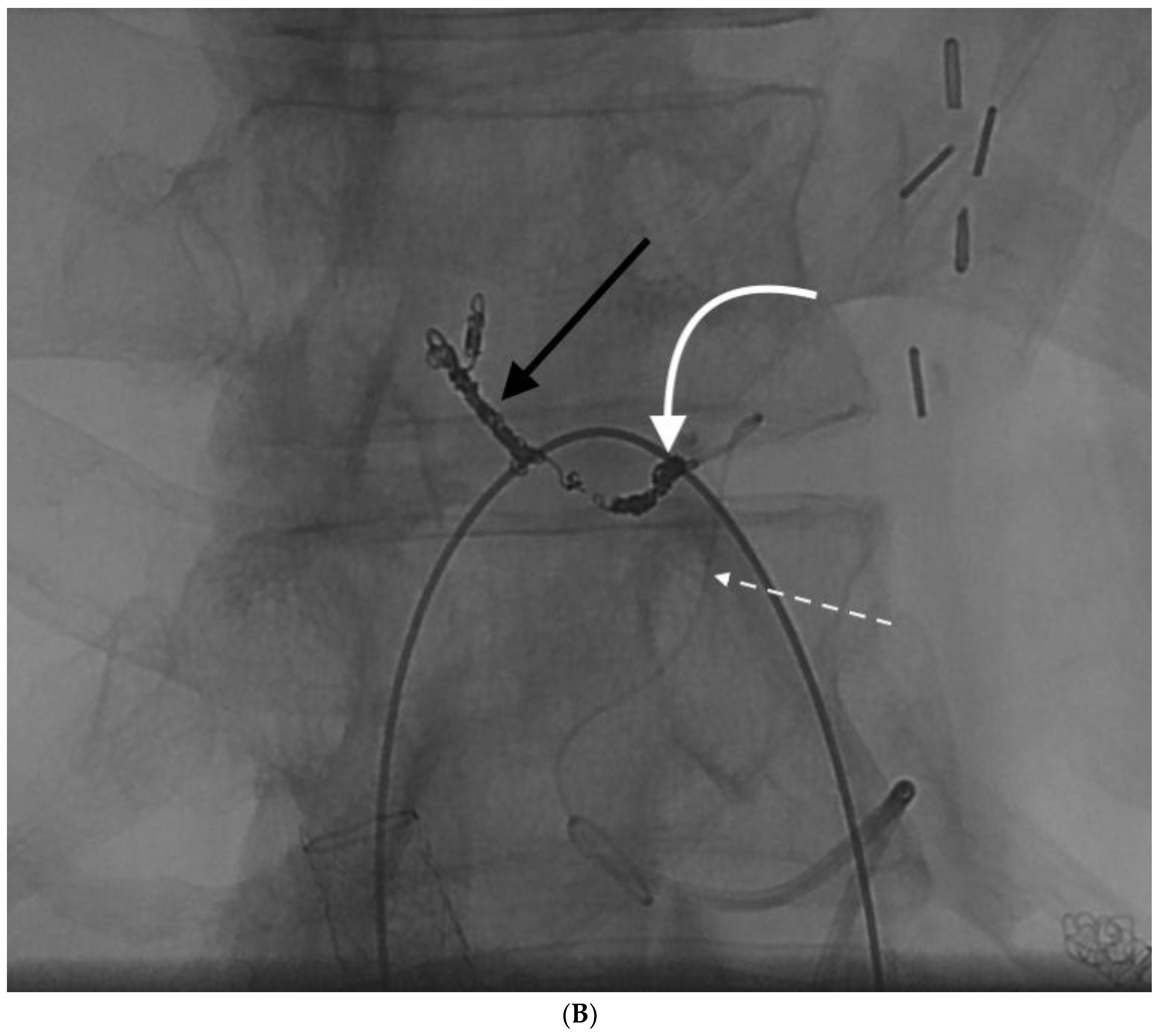
4.2. Hepatic Artery
5. Conclusions
Conflicts of Interest
References
- Zhao, X.Q.; Dong, J.H.; Jiang, K.; Huang, X.Q.; Zhang, W.Z. Comparison of percutaneous transhepatic biliary drainage and endoscopic biliary drainage in the management of malignant biliary tract obstruction: A meta-analysis. Dig. Endosc. 2015, 27, 137–145. [Google Scholar] [CrossRef] [PubMed]
- Liu, J.G.; Wu, J.; Wang, J.; Shu, G.M.; Wang, Y.J.; Lou, C.; Zhang, J.; Du, Z. Endoscopic Biliary Drainage Versus Percutaneous Transhepatic Biliary Drainage in Patients with Resectable Hilar Cholangiocarcinoma: A Systematic Review and Meta-Analysis. J. Laparoendosc. Adv. Surg. Tech. A 2018. [Google Scholar] [CrossRef] [PubMed]
- Dorcaratto, D.; Hogan, N.M.; Munoz, E.; Garces, M.; Limongelli, P.; Sabater, L.; Ortega, J. Is Percutaneous Transhepatic Biliary Drainage Better than Endoscopic Drainage in the Management of Jaundiced Patients Awaiting Pancreaticoduodenectomy? A Systematic Review and Meta-analysis. J. Vasc. Interv. Radiol. 2018. [Google Scholar] [CrossRef] [PubMed]
- Enochsson, L.; Swahn, F.; Arnelo, U.; Nilsson, M.; Lohr, M.; Persson, G. Nationwide, population-based data from 11,074 ERCP procedures from the Swedish Registry for Gallstone Surgery and ERCP. Gastrointest. Endosc. 2010, 72, 1175–1184. [Google Scholar] [CrossRef] [PubMed]
- Girotra, M.; Soota, K.; Klair, J.S.; Dang, S.M.; Aduli, F. Endoscopic management of post-liver transplant biliary complications. World J. Gastrointest. Endosc. 2015, 7, 446–459. [Google Scholar] [CrossRef] [PubMed]
- Samarasena, J.B.; Nguyen, N.T.; Lee, J.G. Endoscopic retrograde cholangiopancreatography in patients with Roux-en-Y anatomy. J. Interv. Gastroenterol. 2012, 2, 78–83. [Google Scholar] [CrossRef] [PubMed]
- Lin, L.F.; Siauw, C.P.; Ho, K.S.; Tung, J.C. ERCP in post-Billroth II gastrectomy patients: Emphasis on technique. Am. J. Gastroenterol. 1999, 94, 144–148. [Google Scholar] [CrossRef] [PubMed]
- Shimizu, H.; Itoi, T.; Sano, K. Historical development of percutaneous transhepatic biliary interventions. J. Hepatobiliary Pancreat. Sci. 2018. [Google Scholar] [CrossRef] [PubMed]
- Venkatanarasimha, N.; Damodharan, K.; Gogna, A.; Leong, S.; Too, C.W.; Patel, A.; Tay, K.H.; Tan, B.S.; Lo, R.; Irani, F. Diagnosis and Management of Complications from Percutaneous Biliary Tract Interventions. Radiographics 2017, 37, 665–680. [Google Scholar] [CrossRef] [PubMed]
- Shiau, E.L.; Liang, H.L.; Lin, Y.H.; Li, M.F.; Chiang, C.L.; Chen, M.C.; Huang, J.S.; Pan, H.B. The Complication of Hepatic Artery Injuries of 1,304 Percutaneous Transhepatic Biliary Drainage in a Single Institute. J. Vasc. Interv. Radiol. 2017, 28, 1025–1032. [Google Scholar] [CrossRef] [PubMed]
- Mueller, P.R.; van Sonnenberg, E.; Ferrucci, J.T., Jr. Percutaneous biliary drainage: Technical and catheter-related problems in 200 procedures. AJR Am. J. Roentgenol. 1982, 138, 17–23. [Google Scholar] [CrossRef] [PubMed]
- Fidelman, N.; Bloom, A.I.; Kerlan, R.K., Jr.; Laberge, J.M.; Wilson, M.W.; Ring, E.J.; Gordon, R.L. Hepatic arterial injuries after percutaneous biliary interventions in the era of laparoscopic surgery and liver transplantation: Experience with 930 patients. Radiology 2008, 247, 880–886. [Google Scholar] [CrossRef] [PubMed]
- Aung, T.H.; Too, C.W.; Kumar, N.; Damodharan, K.; Urlings, T.A.; Patel, A.; Chan, S.; Toh, L.; Gogna, A.; Irani, F.; et al. Severe Bleeding after Percutaneous Transhepatic Drainage of the Biliary System. Radiology 2016, 278, 957–958. [Google Scholar] [CrossRef] [PubMed]
- Saad, W.E.; Wallace, M.J.; Wojak, J.C.; Kundu, S.; Cardella, J.F. Quality improvement guidelines for percutaneous transhepatic cholangiography, biliary drainage, and percutaneous cholecystostomy. J. Vasc. Interv. Radiol. 2010, 21, 789–795. [Google Scholar] [CrossRef] [PubMed]
- Monden, M.; Okamura, J.; Kobayashi, N.; Shibata, N.; Horikawa, S.; Fujimoto, T.; Kosaki, G.; Kuroda, C.; Uchida, H. Hemobilia after percutaneous transhepatic biliary drainage. Arch. Surg. 1980, 115, 161–164. [Google Scholar] [CrossRef] [PubMed]
- Saad, W.E.; Davies, M.G.; Darcy, M.D. Management of bleeding after percutaneous transhepatic cholangiography or transhepatic biliary drain placement. Tech. Vasc. Interv. Radiol. 2008, 11, 60–71. [Google Scholar] [CrossRef] [PubMed]
- Shapira, Z.; Lavy, R.; Altshuler, A.; Peer, A.; Copel, L.; Halevy, A. Hemobilia as a presenting sign of hepatic artery to portal vein fistula caused by percutaneous transhepatic biliary drainage. Isr. Med. Assoc. J. 2011, 13, 64–65. [Google Scholar] [PubMed]
- Patel, I.J.; Davidson, J.C.; Nikolic, B.; Salazar, G.M.; Schwartzberg, M.S.; Walker, T.G.; Saad, W.A. Consensus guidelines for periprocedural management of coagulation status and hemostasis risk in percutaneous image-guided interventions. J. Vasc. Interv. Radiol. 2012, 23, 727–736. [Google Scholar] [CrossRef] [PubMed]
- Hamada, T.; Yasunaga, H.; Nakai, Y.; Isayama, H.; Horiguchi, H.; Fushimi, K.; Koike, K. Severe bleeding after percutaneous transhepatic drainage of the biliary system: Effect of antithrombotic agents—Analysis of 34,606 cases from a Japanese nationwide administrative database. Radiology 2015, 274, 605–613. [Google Scholar] [CrossRef] [PubMed]
- Scharf, R.E. Management of bleeding in patients using antithrombotic agents: Prediction, prevention, protection and problem-oriented intervention. Hamostaseologie 2009, 29, 388–398. [Google Scholar] [CrossRef] [PubMed]
- Kim, J.H.; Baek, C.H.; Min, J.Y.; Kim, J.S.; Kim, S.B.; Kim, H. Desmopressin improves platelet function in uremic patients taking antiplatelet agents who require emergent invasive procedures. Ann. Hematol. 2015, 94, 1457–1461. [Google Scholar] [CrossRef] [PubMed]
- Choi, S.H.; Gwon, D.I.; Ko, G.Y.; Sung, K.B.; Yoon, H.K.; Shin, J.H.; Kim, J.; Oh, J.Y.; Song, H.Y. Hepatic arterial injuries in 3110 patients following percutaneous transhepatic biliary drainage. Radiology 2011, 261, 969–975. [Google Scholar] [CrossRef] [PubMed]
- Rivera-Sanfeliz, G.M.; Assar, O.S.; LaBerge, J.M.; Wilson, M.W.; Gordon, R.L.; Ring, E.J.; Kerlan, R.K., Jr. Incidence of important hemobilia following transhepatic biliary drainage: Left-sided versus right-sided approaches. Cardiovasc. Interv. Radiol. 2004, 27, 137–139. [Google Scholar] [CrossRef]
- Weber, A.; Gaa, J.; Rosca, B.; Born, P.; Neu, B.; Schmid, R.M.; Prinz, C. Complications of percutaneous transhepatic biliary drainage in patients with dilated and nondilated intrahepatic bile ducts. Eur. J. Radiol. 2009, 72, 412–417. [Google Scholar] [CrossRef] [PubMed]
- Liu, Y.S.; Lin, C.Y.; Chuang, M.T.; Tsai, Y.S.; Wang, C.K.; Ou, M.C. Success and complications of percutaneous transhepatic biliary drainage are influenced by liver entry segment and level of catheter placement. Abdom. Radiol. (NY) 2018, 43, 713–722. [Google Scholar] [CrossRef] [PubMed]
- Born, P.; Rosch, T.; Sandschin, W.; Weiss, W. Arterial bleeding as an unusual late complication of percutaneous transhepatic biliary drainage. Endoscopy 2003, 35, 978–979. [Google Scholar] [CrossRef] [PubMed]
- Bacalbasa, N.; Brezean, I.; Anghel, C.; Barbu, I.; Pautov, M.; Balescu, I.; Brasoveanu, V. Management of a Fulminant Upper Gastrointestinal Bleeding Exteriorized Through Hemobilia Due to Arteriobiliary Fistula Between the Common Bile Duct and a Right Hepatic Artery Aneurysm—A Case Report. In Vivo 2017, 31, 983–989. [Google Scholar] [PubMed]
- Zhou, H.B. Hemobilia and other complications caused by percutaneous ultrasound-guided liver biopsy. World J. Gastroenterol. 2014, 20, 3712–3715. [Google Scholar] [CrossRef] [PubMed]
- Savader, S.J.; Trerotola, S.O.; Merine, D.S.; Venbrux, A.C.; Osterman, F.A. Hemobilia after percutaneous transhepatic biliary drainage: Treatment with transcatheter embolotherapy. J. Vasc. Interv. Radiol. 1992, 3, 345–352. [Google Scholar] [CrossRef]
- Psallidas, I.; Helm, E.J.; Maskell, N.A.; Yarmus, L.; Feller-Kopman, D.J.; Gleeson, F.V.; Rahman, N.M. Iatrogenic injury to the intercostal artery: Aetiology, diagnosis and therapeutic intervention. Thorax 2015, 70, 802–804. [Google Scholar] [CrossRef] [PubMed]
- Helm, E.J.; Rahman, N.M.; Talakoub, O.; Fox, D.L.; Gleeson, F.V. Course and variation of the intercostal artery by CT scan. Chest 2013, 143, 634–639. [Google Scholar] [CrossRef] [PubMed]
- Chemelli, A.P.; Thauerer, M.; Wiedermann, F.; Strasak, A.; Klocker, J.; Chemelli-Steingruber, I.E. Transcatheter arterial embolization for the management of iatrogenic and blunt traumatic intercostal artery injuries. J. Vasc. Surg. 2009, 49, 1505–1513. [Google Scholar] [CrossRef] [PubMed]
- Broderick, S.R. Hemothorax: Etiology, diagnosis, and management. Thorac. Surg. Clin. 2013, 23, 89–96. [Google Scholar] [CrossRef] [PubMed]
- Mortimer, A.M.; Wallis, A.; Planner, A. Multiphase multidetector CT in the diagnosis of haemobilia: A potentially catastrophic ruptured hepatic artery aneurysm complicating the treatment of a patient with locally advanced rectal cancer. Br. J. Radiol. 2011, 84, e95–e98. [Google Scholar] [CrossRef] [PubMed]
- Peynircioglu, B.; Cwikiel, W. Utility of stent-grafts in treatment of porto-biliary fistula. Cardiovasc. Interv. Radiol. 2006, 29, 1156–1159. [Google Scholar] [CrossRef] [PubMed]
- Stroz, P.; Asch, M.; Salem, S.; Sniderman, K.; Gallinger, S. Transhepatic embolization of a portohepatic venous fistula. J. Vasc. Interv. Radiol. 1996, 7, 705–707. [Google Scholar] [CrossRef]
- Chanyaputhipong, J.; Lo, R.H.; Tan, B.S.; Chow, P.K. Portobiliary fistula: Successful transcatheter treatment with embolisation coils. Singap. Med J. 2014, 55, e34–e36. [Google Scholar] [CrossRef]
- Madhusudhan, K.S.; Dash, N.R.; Afsan, A.; Gamanagatti, S.; Srivastava, D.N.; Gupta, A.K. Delayed Severe Hemobilia Due to Bilio-Venous Fistula after Percutaneous Transhepatic Biliary Drainage: Treatment with Covered Stent Placement. J. Clin. Exp. Hepatol. 2016, 6, 241–243. [Google Scholar] [CrossRef] [PubMed]
- Cappucci, M.; Zarco, F.; Orgera, G.; Lopez-Rueda, A.; Moreno, J.; Laurino, F.; Barnes, D.; Tipaldi, M.A.; Gomez, F.; Fernandez, J.M.; et al. Endovascular treatment of visceral artery aneurysms and pseudoaneurysms with stent-graft: Analysis of immediate and long-term results. Cir. Esp. 2017, 95, 283–292. [Google Scholar] [CrossRef] [PubMed]
- Lu, P.H.; Zhang, X.C.; Wang, L.F.; Chen, Z.L.; Shi, H.B. Stent graft in the treatment of pseudoaneurysms of the hepatic arteries. Vasc. Endovasc. Surg. 2013, 47, 551–554. [Google Scholar] [CrossRef] [PubMed]
- Pennington, L.; Kaufman, S.; Cameron, J.L. Intrahepatic abscess as a complication of long-term percutaneous internal biliary drainage. Surgery 1982, 91, 642–645. [Google Scholar] [PubMed]
- Saad, N.E.; Saad, W.E.; Davies, M.G.; Waldman, D.L.; Fultz, P.J.; Rubens, D.J. Pseudoaneurysms and the role of minimally invasive techniques in their management. Radiographics 2005, 25 (Suppl. 1), S173–S189. [Google Scholar] [CrossRef] [PubMed]
- Charnsangavej, C.; Chuang, V.P.; Wallace, S.; Soo, C.S.; Bowers, T. Angiographic classification of hepatic arterial collaterals. Radiology 1982, 144, 485–494. [Google Scholar] [CrossRef] [PubMed]
- Tohma, T.; Cho, A.; Okazumi, S.; Makino, H.; Shuto, K.; Mochiduki, R.; Matsubara, K.; Gunji, H.; Ochiai, T. Communicating arcade between the right and left hepatic arteries: Evaluation with CT and angiography during temporary balloon occlusion of the right or left hepatic artery. Radiology 2005, 237, 361–365. [Google Scholar] [CrossRef] [PubMed]
- Hoevels, J.; Nilsson, U. Intrahepatic vascular lesions following nonsurgical percutaneous transhepatic bile duct intubation. Gastrointest. Radiol. 1980, 5, 127–135. [Google Scholar] [CrossRef] [PubMed]
- Schneider, J.; Hapfelmeier, A.; Fremd, J.; Schenk, P.; Obermeier, A.; Burgkart, R.; Forkl, S.; Feihl, S.; Wantia, N.; Neu, B.; et al. Biliary endoprosthesis: A prospective analysis of bacterial colonization and risk factors for sludge formation. PLoS ONE 2014, 9, e110112. [Google Scholar] [CrossRef] [PubMed]
- Orons, P.D.; Sheng, R.; Zajko, A.B. Hepatic artery stenosis in liver transplant recipients: Prevalence and cholangiographic appearance of associated biliary complications. AJR Am. J. Roentgenol. 1995, 165, 1145–1149. [Google Scholar] [CrossRef] [PubMed]
- Mitchell, S.E.; Shuman, L.S.; Kaufman, S.L.; Chang, R.; Kadir, S.; Kinnison, M.L.; White, R.I., Jr. Biliary catheter drainage complicated by hemobilia: Treatment by balloon embolotherapy. Radiology 1985, 157, 645–652. [Google Scholar] [CrossRef] [PubMed]
- Guzman, E.A.; McCahill, L.E.; Rogers, F.B. Arterioportal fistulas: Introduction of a novel classification with therapeutic implications. J. Gastrointest. Surg. 2006, 10, 543–550. [Google Scholar] [CrossRef] [PubMed]
- Kirchgatterer, A.; Baldinger, C.; Lenglinger, F.; Frohler, W.; Meindl, S.; Zisch, R.; Kramar, R. Portal hypertension due to traumatic hepatic arterioportal fistula: Report of successful embolization. Eur. J. Gastroenterol. Hepatol. 1998, 10, 1057–1059. [Google Scholar] [CrossRef] [PubMed]


| Portal Vein Injury | Hepatic Artery Injury |
|---|---|
| Intermittent bleeding | Constant bleeding |
| Dark blood | Pulsatile bleeding |
| Typically hemodynamically stable | Bright red blood |
| No large drop in hematocrit | Falling hematocrit by >13% of baseline * |
| Hemodynamic instability | |
| Melena | |
© 2018 by the authors. Licensee MDPI, Basel, Switzerland. This article is an open access article distributed under the terms and conditions of the Creative Commons Attribution (CC BY) license (http://creativecommons.org/licenses/by/4.0/).
Share and Cite
Quencer, K.B.; Tadros, A.S.; Marashi, K.B.; Cizman, Z.; Reiner, E.; O’Hara, R.; Oklu, R. Bleeding after Percutaneous Transhepatic Biliary Drainage: Incidence, Causes and Treatments. J. Clin. Med. 2018, 7, 94. https://doi.org/10.3390/jcm7050094
Quencer KB, Tadros AS, Marashi KB, Cizman Z, Reiner E, O’Hara R, Oklu R. Bleeding after Percutaneous Transhepatic Biliary Drainage: Incidence, Causes and Treatments. Journal of Clinical Medicine. 2018; 7(5):94. https://doi.org/10.3390/jcm7050094
Chicago/Turabian StyleQuencer, Keith B., Anthony S. Tadros, Keyan B. Marashi, Ziga Cizman, Eric Reiner, Ryan O’Hara, and Rahmi Oklu. 2018. "Bleeding after Percutaneous Transhepatic Biliary Drainage: Incidence, Causes and Treatments" Journal of Clinical Medicine 7, no. 5: 94. https://doi.org/10.3390/jcm7050094