Strontium Ions Substitution in Brushite Crystals: The Role of Strontium Chloride

Abstract
:1. Introduction
2. Materials and Methods
2.1. Brushite Cement Synthesis
2.2. Preparation of Cylindrical Brushite Cement Samples
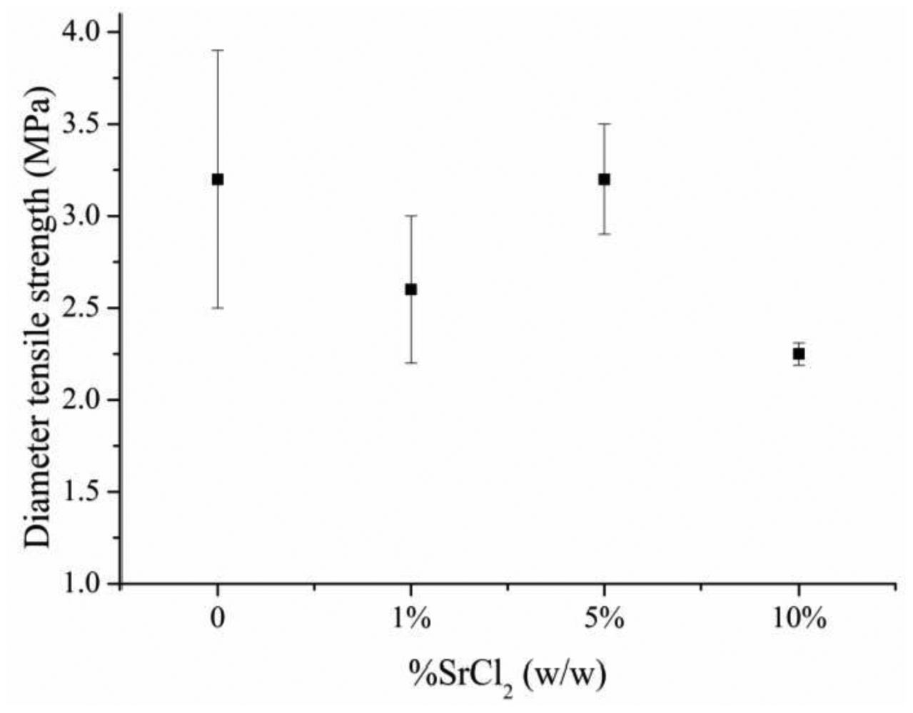
2.3. Measurement of Cement Final Setting Time and Diametral Tensile Strength
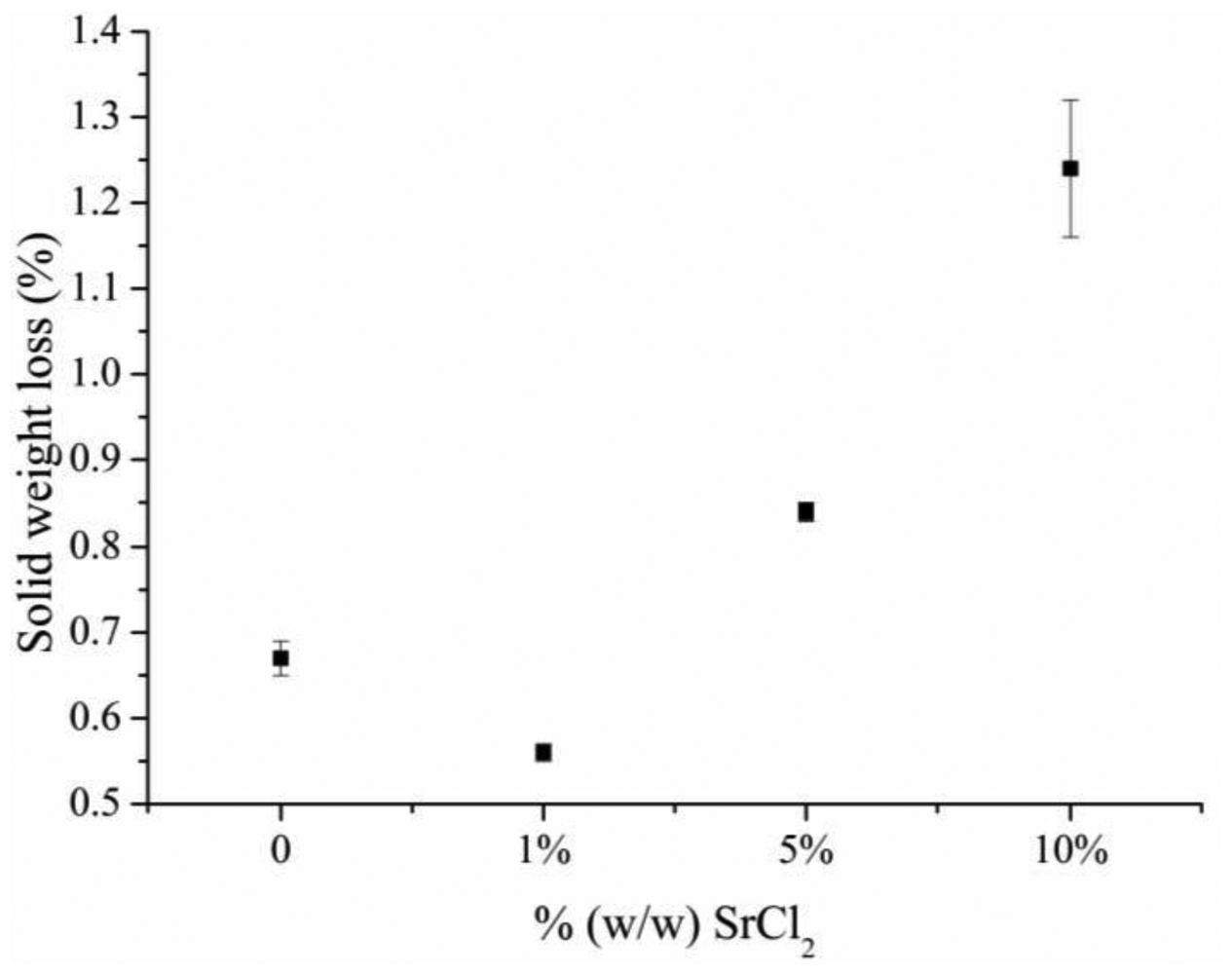
2.4. Particle Release from Cement Surface
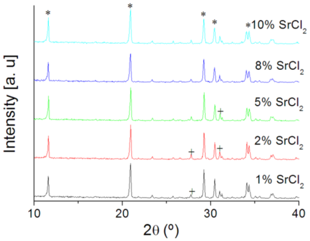
2.5. X-ray Diffraction of Brushite Cements
3. Results

| % SrCl2 (w/w) | FST (minutes) |
|---|---|
| 0 | 6.9 ± 0.3 |
| 1% | 7.1 ± 0.1 |
| 5% | 19.1 ± 0.2 |
| 10% | 45 ± 1 |


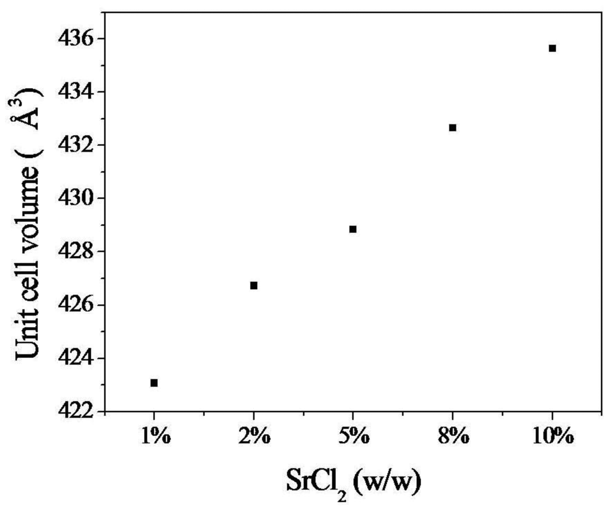
| % SrCl2 (w/w) | Cement composition | Lattice parameters (Å) | |||
|---|---|---|---|---|---|
| DCPD (%) | β-TCP (%) | a | b | c | |
| 1 | 83 | 17 | 6.365250 ± 0.002 | 15.187980 ± 0.006 | 5.812983 ± 0.003 |
| 2 | 81 | 19 | 6.367848 ± 0.002 | 15.191320 ± 0.006 | 5.814205 ± 0.002 |
| 5 | 82 | 18 | 6.369741 ± 0.001 | 15.192880 ± 0.004 | 5.815243 ± 0.002 |
| 8 | 83 | 17 | 6.376474 ± 0.004 | 15.196140 ± 0.008 | 5.818215 ± 0.003 |
| 10 | 87 | 13 | 6.375908 ± 0.002 | 15.204630 ± 0.006 | 5.818039 ± 0.003 |


4. Discussion
5. Conclusions
References
- Cummings, S.R.; Melton, L.J. Epidemiology and outcomes of osteoporotic fractures. Lancet 2002, 359, 1761–1767. [Google Scholar]
- Poole, K.E.; Compston, J.E. Osteoporosis and its management. BMJ 2006, 333, 1251–1256. [Google Scholar]
- Shorr, E.; Carter, A.C. The usefulness of strontium as an adjuvant to calcium in the remineralization of the skeleton in man. Bull. Hosp. Joint Dis. Orthop. Inst. 1952, 13, 59–66. [Google Scholar]
- McCaslin, F.E.; Janes, J.M. The effect of strontium lactate in the treatment of osteoporosis. Proc. Staff. Meet. Mayo. Clin. 1959, 34, 329–334. [Google Scholar]
- Meunier, P.J.; Roux, C.; Seeman, E.; Ortolani, S.; Badurski, J.; Spector, T.D.; Cannata, J.; Balogh, A.; Lemmel, E.M.; Pors-Nielsen, S.; Rizzoli, R.; Genant, H.K.; Reginster, J.Y. The effects of strontium ranelate on the risk of vertebral fracture in women with postmenopausal osteoporosis. New Eng. J. Med. 2004, 350, 459–468. [Google Scholar]
- Reginster, J.Y.; Seeman, E.; De Vernejoul, M.C.; Adami, S.; Compston, J.; Phenekos, C.; Devogelaer, J.P.; Diaz Curiel, M.; Sawicki, A.; Goemaere, S.; Sorensen, O.H.; Felsenberg, D.; Meunier, P.J. Strontium ranelate reduces the risk of nonvertebral fractures in postmenopausal women with osteoporosis: Treatment of Peripheral Osteoporosis (TROPOS) study. J. Clin. Endocrinol. Metab. 2005, 90, 2816–2822. [Google Scholar]
- Baron, R.; Tsouderos, Y. In vitro effects of S12911-2 on osteoclast function and bone marrow macrophage differentiation. Eur. J. Pharmacol. 2002, 450, 11–17. [Google Scholar]
- Takahashi, N.; Sasaki, T.; Tsouderos, Y.; Suda, T. S 12911-2 inhibits osteoclastic bone resorption in vitro. J. Bone Miner Res. 2003, 18, 1082–1087. [Google Scholar]
- Marie, P.J. Strontium ranelate: A physiological approach for optimizing bone formation and resorption. Bone 2006, 38, S10–S14. [Google Scholar]
- Canalis, E.; Hott, M.; Deloffre, P.; Tsouderos, Y.; Marie, P.J. The divalent strontium salts S12911 enhances bone cell replication and bone formation in vitro. Bone 1996, 18, 517–523. [Google Scholar]
- Johal, K.K.; Hill, R.G.; Brook, I.M. In vivo response of strontium and zinc based ionomeric cement implants in bone. J. Mater. Sci. Mater. Med. 2002, 13, 375–379. [Google Scholar]
- Leroux, L.; Lacout, J.L. Preparation of calcium strontium hydroxyapatites by a new route involving calcium phosphate cements. J. Mater. Res. 2001, 16, 171–176. [Google Scholar]
- Guo, D.; Xu, K.; Zhao, X.; Han, Y. Development of a strontiumcontaining hydroxyapatite bone cement. Biomaterials 2005, 26, 4073–4083. [Google Scholar]
- Rokita, E.; Hermes, C.; Nolting, H.F.; Ryczek, J. Substitution of calcium by strontium within selected calcium phosphates. J. Cryst. Growth 1993, 130, 543–552. [Google Scholar]
- Penel, G.; Leroy, N.; Van Landuyt, P.; Flautre, B.; Hardouin, P.; Lemaitre, J.; Leroy, G. Raman microspectrometry studies of bruhsite cement: In vivo evolution in a sheep model. Bone 1999, 25, 81S–84S. [Google Scholar]
- Bohner, M.; Matter, S. Brushite hydraulic cement stabilized with a magnesium salt. U.S. Patent 6,733,582, 2004.
- Alkhraisat, M.H.; Marino, F.T.; Retama, J.R.; Jerez, L.B.; Lopez-Cabarcos, E. Betatricalcium phosphate release from brushite cement surface. J. Biomed. Mater. Res. A 2008, 84, 710–717. [Google Scholar]
- Alkhraisat, M.H.; Rueda, C.; Jerez, L.B.; Tamimi, M.F.; Torres, J.; Gbureck, U.; Cabarcos, E.L. Effect of silica gel on the cohesion, properties and biological performance of brushite cement. Acta Biomater. 2010, 6, 257–265. [Google Scholar]
- Alkhraisat, M.H.; Rueda, C.; Mariño, F.T.; Torres, J.; Jerez, L.B.; Gbureck, U.; Cabarcos, E.L. The effect of hyaluronic acid on brushite cement cohesion. Acta Biomater. 2009, 5, 3150–3156. [Google Scholar]
- Christoffersen, J.; Christoffersen, M.R.; Kolthoff, N.; Barenhold, O. Effects of strontium ions on growth and dissolution of hydroxyapatite and on bone mineral detection. Bone 1997, 20, 47–54. [Google Scholar]
- Zawacki, S.J.; Koutsoukos, P.B.; Salimi, M.H.; Nancollas, G.H. The growth of calcium phosphates. Geochem. Process. Min. Surface. 1987, 32, 650–662. [Google Scholar]
© 2011 by the authors; licensee MDPI, Basel, Switzerland. This article is an open access article distributed under the terms and conditions of the Creative Commons Attribution license (http://creativecommons.org/licenses/by/3.0/).
Share and Cite
Alkhraisat, M.H.; Rueda, C.; López Cabarcos, E. Strontium Ions Substitution in Brushite Crystals: The Role of Strontium Chloride. J. Funct. Biomater. 2011, 2, 31-38. https://doi.org/10.3390/jfb2020031
Alkhraisat MH, Rueda C, López Cabarcos E. Strontium Ions Substitution in Brushite Crystals: The Role of Strontium Chloride. Journal of Functional Biomaterials. 2011; 2(2):31-38. https://doi.org/10.3390/jfb2020031
Chicago/Turabian StyleAlkhraisat, Mohammad H., Carmen Rueda, and Enrique López Cabarcos. 2011. "Strontium Ions Substitution in Brushite Crystals: The Role of Strontium Chloride" Journal of Functional Biomaterials 2, no. 2: 31-38. https://doi.org/10.3390/jfb2020031