Review

Platelet Rich Plasma: New Insights for Cutaneous Wound Healing Management

by
Deborah Chicharro-Alcántara
1,2,
Mónica Rubio-Zaragoza
1,2,*,
Elena Damiá-Giménez
1,2,
José M. Carrillo-Poveda
1,2,
Belén Cuervo-Serrato
1,2,
Pau Peláez-Gorrea
1,2 and
Joaquín J. Sopena-Juncosa
1,2
1
Bioregenerative Medicine and Applied Surgery Research Group, Animal Medicine and Surgery Department, Veterinary Faculty, Universidad Cardenal Herrera-CEU, CEU Universities, 46115 Valencia, Spain
2
García Cugat Foundation for Biomedical Research, 08006 Barcelona, Spain
*
Author to whom correspondence should be addressed.
J. Funct. Biomater. 2018, 9(1), 10; https://doi.org/10.3390/jfb9010010
Submission received: 29 December 2017 / Revised: 14 January 2018 / Accepted: 15 January 2018 / Published: 18 January 2018
(This article belongs to the Special Issue Journal of Functional Biomaterials: Feature Papers 2016)

Abstract

:
The overall increase of chronic degenerative diseases associated with ageing makes wound care a tremendous socioeconomic burden. Thus, there is a growing need to develop novel wound healing therapies to improve cutaneous wound healing. The use of regenerative therapies is becoming increasingly popular due to the low-invasive procedures needed to apply them. Platelet-rich plasma (PRP) is gaining interest due to its potential to stimulate and accelerate the wound healing process. The cytokines and growth factors forming PRP play a crucial role in the healing process. This article reviews the emerging field of skin wound regenerative therapies with particular emphasis on PRP and the role of growth factors in the wound healing process.

1. Introduction

Skin is the largest organ in vertebrates, comprising 10% of the total body mass and covers the entire surface area [1,2]. It has a crucial role in defense and survival thanks to its self-repairing and self-renewing capacity, acting as an important barrier from the outer environment to the inner environment [3]. A disturbance of the normal anatomic structure and functional integrity of the skin can be described as a wound [4]. Wound healing is a coordinated dynamic tissue repair process, which involves the interaction of multiple cell types, growth factors, cytokines, and chemokines [5,6]. If this mechanism is interrupted, chronic, non-healing wounds or excessive granulation tissue formation can appear, leading to arresting in the chronic inflammatory phase [7].
In recent years, the overall prevalence of chronic degenerative diseases associated with ageing has increased dramatically; in this way chronic wounds represent a significant biomedical burden. Millions of patients all over the world are affected by acute and chronic wounds as a result of surgeries, burns, infections, pressure ulcers, and diabetic and venous ulcers [4]. In the USA, more than 6 million people suffer from chronic wounds with an annual cost of $25 billion [8]. From the 150 million diabetes patients in the world, foot ulcerations that frequently develop into nonhealing wounds represent 15% [9]. Almost 2% of the health budgets in Europe are spent in chronic wound management [10]. The effectiveness of current chronic wound treatment is estimated at 50%, and are expensive as repetitive treatments are needed [11].
Despite significant advances in medical care and nutrition, there is a growing need to develop novel strategies to improve cutaneous wound healing. The medical field is rapidly advancing towards the development of low or non-invasive procedures and accelerated treatments that can achieve a reduced morbidity and a good functional recovery in our patients to improve their quality of life. In the last few years these simple and cost-efficient procedures have had a potential impact reducing economic costs for standard general medical treatments [12]. Regenerative medicine can be defined as an emerging interdisciplinary field in biomedical research that aims to restore, regenerate, and replace damaged tissues and cells [13].
Platelet-rich plasma (PRP) is an endogenous therapeutic technology that is gaining interest in regenerative medicine due to its potential to stimulate and accelerate tissue healing [14]. PRP is defined as an autologous biological product derived from the patient’s blood, and in which after a centrifugation process a plasma fraction is obtained with a platelet concentration higher than that in circulating blood [15]. Platelets play a crucial role in the wound healing process thanks to their hemostatic function and presence of cytokines and growth factors [16]. There are several growth factors which are known to be involved in the wound healing process, such as platelet-derived growth factor (PDGF), epidermal growth factor (EGF), fibroblast growth factor (FGF), insulin-like growth factor (IGF1, IGF2), vascular endothelial growth factor (VEGF), transforming growth factor (TGF-β), and keratinocyte growth factor (KGF) [17,18,19,20] (Figure 1).
So far, only the PDGF has been approved by the United States Food and Drug Administration (FDA) and by the European Authorities (EMEA) for clinical application in patients [21,22]. Growth factors are a group of soluble and diffusible polypeptide substances that regulate growth, differentiation, proliferation, and cellular metabolism of numerous cell types [14,23]. They promote endothelial and epithelial regeneration, stimulate angiogenesis, collagen synthesis, soft tissue healing, and hemostasis [24].
The use of growth factors to promote cutaneous wound healing has existed since the 1940s, and they can be applied in a wide range of ways, either by traditional topical or intralesional administration or by using specific scaffolds or even gene therapy [25]. Animal and human trials have reported successful PRP clinical applications for chronic skin ulcers [26,27], acute cutaneous wounds [28,29], burns [30], and plastic and cosmetic surgery [31,32].
Regardless different conventional therapeutic approaches targeting wound healing enhancement, the use of novel treatments remains still a clinical challenge. There is a continued search towards regenerative therapies to reduce health care burden. In vivo and in vitro clinical and experimental studies on cutaneous wound regenerative therapies such as PRP and stem cells are providing encouraging results. For all the above mentioned reasons, this article aims to review the regenerative skin wound healing field foccusing on platelet-rich plasma as a cost-effective therapeutic approach.

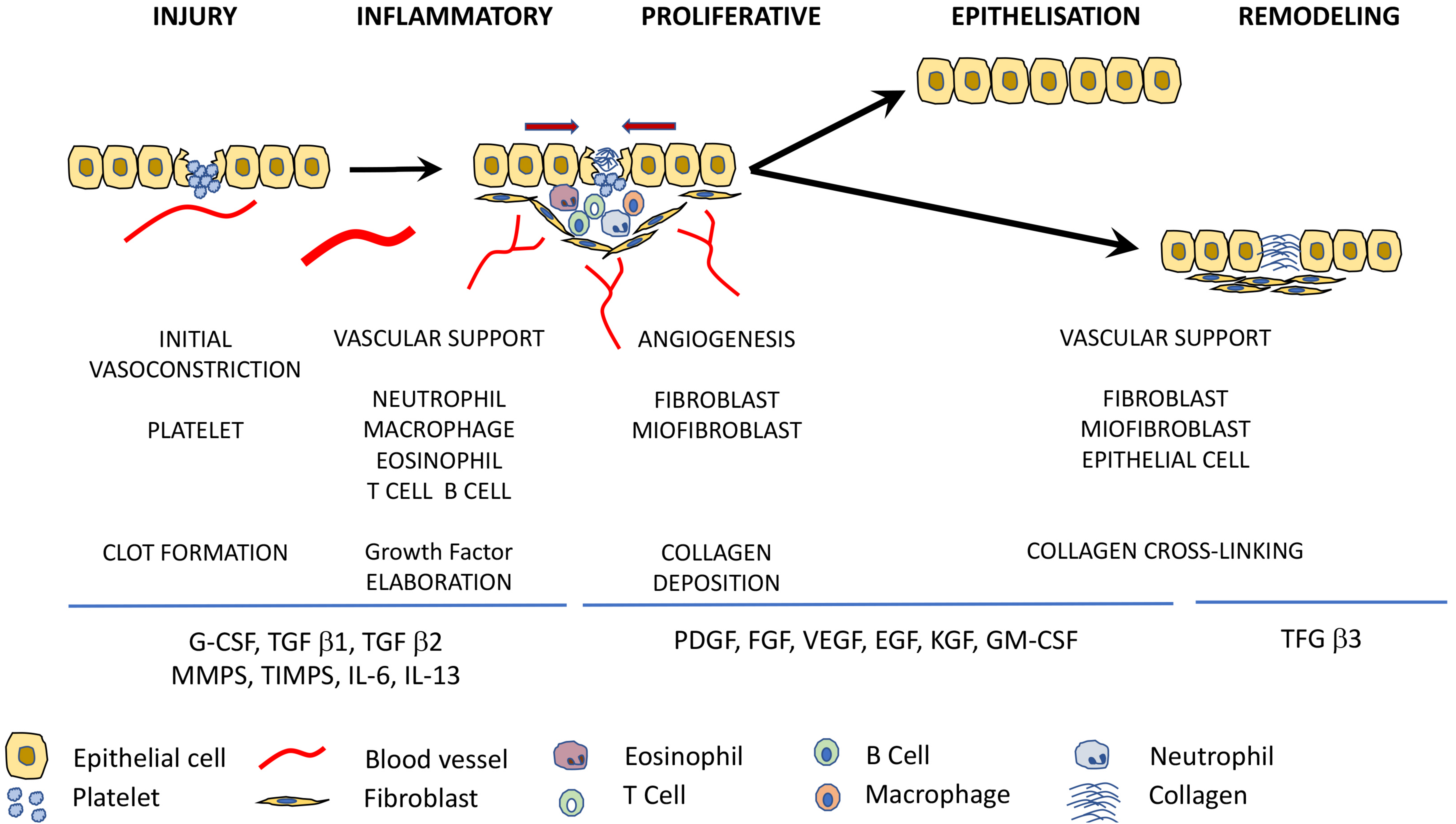
2. Stages of Wound Healing

A deep understanding of the pathophysiologic basis of wound healing has resulted in improved clinical care over the years [25]. Wound healing is one of the most complex processes in multicellular organisms, involving numerous intra- and intercellular signaling pathways in various cell types [33]. It is a biological process where physical, chemical, and cellular factors are involved with the main aim of wound repair [5].
Cutaneous wounds can either heal by repair or regeneration and there is a clear and important difference between these two types of healing. Wound repair is a physiological process that aims to obtain a tissue with inferior characteristics compared to the original one, whereas wound regeneration aims to rebuild the injured tissue with an exact copy of the original one to restore both morphology and function of the affected tissue [34,35].
The wound healing process is typically divided into three/four overlapping phases: hemostasis/inflammation, cell proliferation and remodeling, which are regulated by various cells, cytokines, and growth factors which can act directly over the responsible cells for their release, nearby cells or even distant cells [25,36,37].
Immediately after skin injury, platelets are stimulated to aggregate themselves and a fibrin clot is formed, providing hemostasis and recruiting several different cells to the wound [38,39]. Secretion of growth factors, cytokines, and other homeostatic factors for the clotting cascade also take place during this first phase [24]. The inflammatory stage is triggered by clotting and platelet degranulation. This phase is characterized by the release of serotonin, histamine and bioactive factors, which allows for the arrival of inflammatory cells into the wounded area thanks to an increase in the capillary permeability, such as neutrophils, leukocytes, and macrophages [40]. The highest concentrations of neutrophils are found from 1–2 days after injury, playing a main role in preventing bacterial infections with the help of macrophages, and they also activate keratinocytes, fibroblasts, and immune cells [38]. At the end of the inflammatory stage of wound healing macrophages develop an anti-inflammatory, profibrotic phenotype and secrete TGF-β, interleukins, and tumor necrosis factors [5]. These growth factors are going to stimulate the beginning of the proliferative stage where fibroplasia, matrix deposition, angiogénesis, and re-epithelialization processes will take place [35]. Recruitment of fibroblasts from surrounding non-injured tissue and the formation of new blood vessels thanks to the migration of endothelial cells via production of VEGF are achieved during the proliferative stage [41]. Activated fibroblasts are responsable for the secretion of an extracellular matrix containing increased levels of immature type III collagen, which will be completly different to the mature type I collagen found in normal skin and mature scars [38,42]. The highest concentration of collagen in the wound is present approximately three weeks after the initial injury [25]. Meanwhile, the fibrin plug starts to be replaced by granulation tissue from 3–5 days after injury and the wound begins to contract [39]. Finally, the last stage of the wound healing process known as remodeling or maturation takes place, a decrease in cellular content at the wounded area is observed as a result of migration and apoptosis [43]. Remodeling is a dynamic phase during which collagen forms tight cross-links to other collagen and with other protein molecules, increasing the tensile strength of the mature scar to a maximum of 80% compared to the unwounded skin [38]. Moreover, the ratio of type III to type I collagen decreases as a result of immature type III collagen conversion into mature type I collagen and this process can last up to two years [38,43].
There are several growth factors (GFs), cytokines, integrins, keratins, matrix-metaloproteinases, chemokines, and extracellular macromolecules which are going to help regulate all of these processes during wound healing [44,45]. Growth factors involved in each of the wound healing phases are shown in Table 1 [46].

Pathologic Wound Healing

There are several pathophysiological and metabolic conditions which can alter the normal wound healing process causing a delayed wound healing, and as a result chronic wounds which can persist for over six weeks [47,48].
Despite the origin of the pathologic wound healing, they are going to give rise to fibroproliferative disorders. These disorders can be classified basically in two types: hypertrophic scar and keloid formation which represent an “over-healing” response and ulcers or chronic wounds which show just the opposite an “under-healing” response [49]. Hypertrophic scars are associated with a prolonged inflammatory phase and in ulcers an increased proteolysis together with impairments in cell proliferation and migration are observed [25,50].

3. The Role of Growth Factors in the Wound Healing Process

Growth factors play an essential role in the complex process of wound healing and tissue regeneration [51]. They are signaling proteins that influence the metabolism of other cells [52]. Each GF has more than one effect on the wound healing process, and act by binding to specific receptors on cell membranes of target cells [53]. These effects include promoting chemotaxis (attracting cells into the wound), inducing cell migration and proliferation, and stimulate cells to upregulate protein production [54]. These GFs not only regulate cell migration and proliferation, but also remodel the extracellular matrix and promote angiogenesis, creating an ideal environment that favours the cutaneous wound healing process [55].
Almost every cell type in skin is involved in the production of GFs and several cells release many different types of GFs during the wound healing process [56].
Through degranulation of the alpha granules in platelets, PRP can secrete various GFs, including PDGF, VEGF, FGF, hepatocyte growth factor (HGF), and TGFβ, which have been documented to initiate wound healing process [57] (Table 2).
The main GFs which are currently known to be involved in the wound healing process are PDGF, EGF, FGF, IGF, VEGF, TGFβ, HGF, and KGF [18]. Their roles on the wound healing process are described in the following sections.

3.1. Platelet-Derived Growth Factor (PDGF)

It is one of the first factors secreted after an injury and promotes cellular reactions throughout every phase of the wound healing process [58]. PDGF stimulates many metabolic processes, such as protein and collagen synthesis, and collagenase activity and chemotaxis of fibroblasts and smooth muscle cells [59]. Additionally, it also promotes the proliferation and migration of endothelial cells, thereby exerting angiogenic effects [60] and the production of TGFβ, which initiates the synthesis of collagen [61]. Furthermore, PDGF has been shown to stimulate the production of IGF-I [59].
In addition to the fact that PDGF has a strong chemotactic effect for monocites, neutrophils and smooth muscle cells promoting mitosis of fibroblasts, endotelial, and smooth muscle cells, it also stimulates the angiogenesis process, the wound contraction, the formation of granulation tissue and wound remodeling [59].
The use of recombinant human PDGF has shown satisfactory results in the treatment of diabetic foot ulcers [62]. PDGF concentration was demonstrated to be lower in diabetic and aged mice which as a result showed a delayed injury response [63]. Similar results were obtained in a study with non-healing human ulcers [64].

3.2. Epidermal Growth Factor (EGF)

EGF is mainly secreted by platelets, although macrophages, fibroblasts, and mesenchymal stem cells can also release it, a higher concentration of the mentioned GF is found at the beginning of the healing process [61].
This GF plays a vital role in epithelial and keratinocyte cell proliferation, differentiation, growth, and migration. It is considered to play a crucial role in the wound healing process as it promotes angiogenesis [65]. It has been reported that EGF is effective in inducing epithelialization of partial thickness burns and granulation wounds [46,66]. Moreover, Kim et al. [67] observed wound healing enhancement and reduced cutaneous scaring in full thickness wounds in mice.

3.3. Fibroblast Growth Factor (FGF)

FGF contributes on re-epithelialization, angiogénesis, and granulation tissue formation [68]. Additionally, to their direct role in wound healing, it also acts indirectly, supporting the epithelialization by stimulating the release of TGF-α [69]. Together with the VEGF they are active during the proliferation phase. FGF stimulates the proliferation of fibroblasts, collagen accumulation, and accelerates the formation of granulation tissue [46]. Research studies in animal models have observed positive results, showing accelerated wound healing in a diabetic mouse model [70]. Moreover, it has been suggested that FGF influences positively in hypertrophic scar management resulting in reduced scarring [71].

3.4. Insulin Growth Factor (IGF)

The IGF is actively involved mainly in the inflammatory and proliferative phase [72]. Fibroblasts secrete IGF-1 which exert autocrine effects on themselves [58]. Low IGF concentrations have been observed in diabetic chronic wounds. Moreover, research studies carried out in diabetic rats, healthy rats, and rabbits suggest that the exogenous application of IGF-1 accelerates wound healing [73,74,75]. A stronger effect of IGF is obtained when it is used in combination with other growth factors, such as PDGF and EGF, promoting migration of keratinocytes and enhancing tissue repair [76,77].

3.5. Vascular Endothelial Growth Factor (VEGF)

During wound healing, VEGF exerts a strong paracrine effect on endothelial cells and, therefore, promotes and supports the wound angiogenesis process, becoming the main responsable growth factor to initiate the angiogenesis process in granulation tissue [63,78]. It has been demostrated that VEGF growth factor family represent key regulators of physiological and pathological vasculogenesis, angiogenesis, lymphangiogenesis, and vascular permeability [79]. To date, VEGF application has been reported in both experimental and clinical studies with successful results accelerating wound repair by increasing epithelialization, angiogenesis and granulation tissue formation in a mouse diabetic wound model [80], skin flaps in dogs [81], ischaemic ulcers [46], and diabetic ulcers in patients [82].

3.6. Transforming Growth Factor-β

TGFβ has three different isoforms: TGFβ1, -β2, -β3 which are going to carry out overlapping, but different, functions in the wound healing process [25]. The TGFβ1 isoform is present in a higher concentration during the wound healing process [44].
TGFβ3 is related to angiogenesis and TGFβ1 and -β2 are associated with scarring and fibrosis, they promote fibroblast and myofibroblast differentiation, extracellular matrix deposition, wound contraction, and scar formation [83]. TGFβ exerts a chemotactic effect for macrophages, induces the production of collagen and fibronectin and inhibits metalloproteinase activity [46]. It is important to highlight the main role TGFβ3 has in reducing deposition of collagen during the proliferative and remodelling phases, thus reducing scar formation, as shown in preclinical studies after intradermal injection of the mentioned factor in rats [84]. Moreover, low concentrations of TGFβ1 have been associated with fibroproliferative disorders of hyperthrophic scarring and keloid formation [83]. Furthermore, a different study demonstrated that TGFβ3 was necessary for re-epithelialization of excisional wounds due to the paracrine effect they exert on keratinocytes [85].

3.7. Hepatocyte Growth Factor (HGF)

This growth factor is secreted by mesenchymal cells and has a main role regulating cell growth, motility and morphogenesis in cells such as epithelial and endothelial cells. Thus, it is directly related to epithelial repair, granulation tissue formation and neovascularization [86]. Additionally, a synergistic effect between HGF and VEGF over endothelial cells has been demostrated providing a more intense angiogenic effect at the wound area [87].

3.8. Keratinocyte Growth Factor (KGF)

KGF induces the proliferation and migration of keratinocytes during the wound healing process, mainly at the remodelation phase. Enhancement of epithelial cell proliferation has also been shown by recombinant human KGF-2 and acceleration of the healing process in venous ulcers [46].

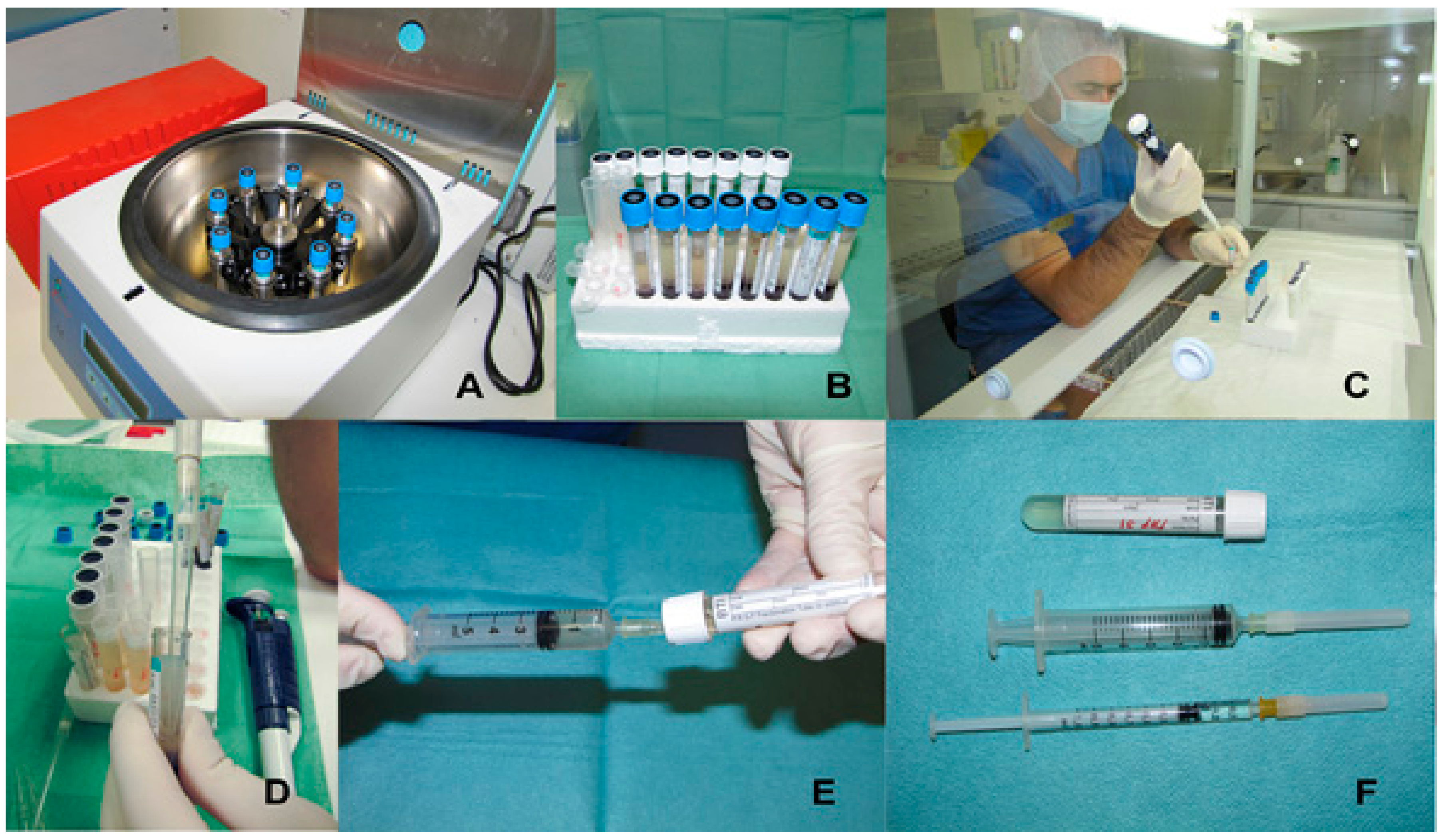
4. Platelet Rich Plasma: Preparation Methods and Therapeutic Formulations

Platelet-rich plasma can be defined as a fraction of plasma containing higher concentrations of platelets compared to whole blood and as a result an increased growth factor concentration [88]. Nowadays, PRP is widely applied in different clinical applications to promote healing in orthopaedics [89], soft tissue healing [90], nervous tissue [91], chronic skin ulcers [92], ophthalmology [93], and dentistry [94]. PRP is categorized by the Food and Drug administration (FDA) as a minimally-manipulated tissue and as an autologous blood product [95]. One of the advantages of these preparations is that they are easily obtained from the patient’s blood after a simple centrifugation process, thus, it is a safe, simple, and cost-effective product [96,97]. By controlling centrifugation parameters and the activation protocol, it is possible to control the dose of GFs and proteins that are released upon activation [98].
At the moment, there is no clear gold standard protocol for PRP generation, little characterization performed on the obtained products and lack of regulation and standardisation [96]. To characterize the main components that play a key role in tissue regeneration and formulate an adequate preparation for each specific pathophysiological situation, a well-defined simple procedure is necessary, where centrifugation conditions are fundamental to obtain a high PRP quality together with quantification and identification of platelets and lymphocytes [96,99].
In recent years there has been significant controversy regarding the nomenclature and definition of PRP [100,101]. According to Anitua and colleagues (2007), the nomenclature “platelet-rich plasma” is a vague and imprecise term. There is a large number of autologous blood preparations that differ regarding their processing protocols and preparation and, therefore, in quantitative and qualitative characteristics. Therefore, a detailed, precise, and stepwise description of the PRP preparation protocol (centrifugation conditions, platelet concentration, type of anticoagulant used, platelet activator and presence or absence of leukocytes) is required to allow a comparison in between studies [95,101]. Moreover, it is important to preserve platelet integrity and quality preventing damage or lysing of platelets to allow them to fully secrete growth factors [96].
As a result of all the above mentioned reasons, an advanced autologous PRP system was developed by the Biotechnology Institute, BTI, the Plasma Rich in Growth Factors, known as PRGF-Endoret®. It is certified by European health authorities and has been approved in Europe for the production of PRP and their application in several medical fields. PRGF is an autologous product with a moderate concentration of platelets, absence of leukocytes and provides the formation of a biological scaffold formed by fibrin for cell adhesion which will help the wound healing process [99]. The mentioned technology together with other PRP preparation methods are described in the following sections.

4.1. Preparation Methods

Within the existing methods for the preparation of PRP there is discrepancy in technical details, such as the inclusion of leukocytes or red blood cells, speed, and time of centrifugation. Although it seems that the results are similar [102].
Anitua (2004) defends the non-inclusion of leukocytes, because they can alter the function of some GFs and interfere in the anti-inflammatory action [103]. Others, such as the Harvest Technologies corporation, claims that these details are not important and that their PRP contains some red blood cells. Moreover, other authors prefer the inclusion of leukocytes for the treatment of ulcers or other surgeries, since they consider that it has an antimicrobial effect [104,105].

4.1.1. PRGF-System® (BTI Biotechnology Institute, Vitoria, Spain)

Clinically-valuable PRP contains at least one million platelets per microliter [100]. According to Anitua et al. (2004), PRGF is a more precise nomenclature to refer to this regenerative therapy and must have a platelet concentration of approximately 1.5 times the concentration in whole blood [103]. The main advantages offered by PRGF include the following: it is a biocompatible, versatile, and safe product, no leukocytes are present to prevent pro-inflammatory effects, standardized activation of platelets with calcium chloride is used to cause the release of growth factors, is a simple and fast protocol with just one centrifugation at 460× g during 8 min, has bacteriostatic properties and powerful therapeutic potential with no side-effects [99]. After single centrifugation, red blood cells are obtained at the bottom of the tube, just above white cells and finally two fractions of plasma, the upper 60% corresponds to the fraction poor in GFs, and below this the remaining 40% corresponds to PRGF [99] (Figure 2).

4.1.2. Platelet Concentrate Collection System (PCCS®Kit) (3i–Implant Innovations, Palm Beach Gardens, FL, USA)

This technique allows a platelet concentration between 1,100,000 and 2,200,000/μL, and includes leukocytes, with a concentration between 5500 and 14,800/μL. As an anticoagulant, it uses ACD-A9 and is subjected to two centrifugations: the first 3 min and 45 s at 3000 rpm and the second 13 min at 3000 rpm [106,107].

4.1.3. Gravitational Platelet Separation (GPS® System) (Biomet Merck Biomaterials, Darmstadt, Germany)

With this method a platelet concentration of 1,600,000/μL and leukocyte levels of 31,100/μL are obtained. A 60 mL syringe is used, in which 6 mL are anticoagulant (ACD-A) and 54 mL of patient’s blood. Blood undergoes centrifugation at 3200 rpm for 12 min in GPS® tubes [16,108].

4.1.4. Smart PReP® System (Harvest Technologies Corporation, Munich, Germany)

The amount of blood needed with this protocol is 52 mL in women and 48 mL in men. A double centrifugation of the blood is performed for 12 min, obtaining a PRP with a close platelet concentration to 1,250,000/μL and a leukocyte concentration of 19,261/μL [109].

4.1.5. Plateltex® (Plateltex, Bratislava, Slovakia)

The platelet concentration obtained with this method is approximately 1,600,000/μL and is carried out by means of a double centrifugation. The blood is obtained in 8.0 mL test tubes, which contain ACD-A as an anticoagulant, and are subjected to a gentle first centrifugation at 180× g for 10 min. After aspirating the supernatant plasma, a second centrifugation is performed at 1000× g for 10 min [110].
There is a wide range of PRP products obtained by different blood-spinning preparation protocols, they all aim to obtain the rich “cocktail” in GFs (Table 3). However, to succeed several challenges have to be completed, such as a complete characterization of the platelet released growth factors and proteins, evaluating individual reasons that make some products more effective than others and designing new associations with biomaterials or other regenerative therapies, such as stem cells [34,99].

4.2. Therapeutic Formulations

A very important characteristic that makes PRGF technology different from the rest of PRP products is its versatility. PRGF can be obtained in four different formulations from patient’s blood with strong therapeutic effects, depending on the coagulation and activation degree of the samples, and adapted to the clinical needs in each case. This therapeutic formulations involve: PRGF supernatant, used for conventional eye-drop and cell culture media [87], liquid PRGF commonly used in surgeries and dental implant surfaces [111], the scaffold-like PRGF (clot) formed by fibrillar and cellular components used frequently in cutaneous ulcers [112], and the elastic and dense fibrin membrane which is excellent for soft tissue injuries and to seal surgical wounds (Figure 3).

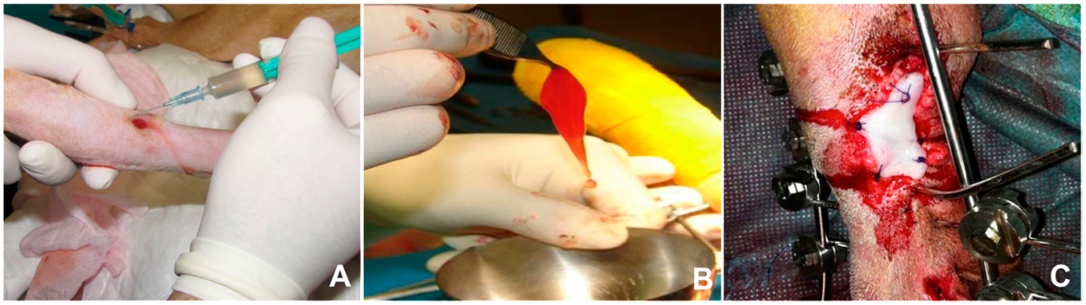
5. The Use of Platelet-Rich Plasma in Cutaneous Wound Healing

Over the last decades, the use of emerging cellular therapies, such as PRP, has gathered more attention in a wide range of diseases and settings for its potential use in the regenerative medicine field as a therapeutic agent and can have an adjunctive role in a standardized, quality treatment plan [113]. The medicine field is advancing towards the development of less-invasive and cost-effective treatments to enhance functional recovery [99]. The use of PRP has had a potential impact in reducing economic costs for standard medical treatments, although it should not be considered as a therapy that replaces certain essential conventional treatments, such as debridement of necrotized tissue, but as a complementary therapy [34,99]. A biological internal environment for tissue homeostasis restoration is created with PRP therapy by providing to the wounded area several signalling cytokines and growth factors which play a crucial role in tissue repair process by regulating inflammation, stimulating angiogénesis, and synthesis together with the remodelling of new formed tissue [113,114]. Additional advantages of PRP treatment in cutaneous wound healing includes easy methodology, cost-effective treatment, more lasting effect compared to conventional therapy, and safe treatment being at autologous product obtained from the own patient [115,116].
The use of PRP in both humans and animals is steadily increasing, and its healing properties in cutaneous wounds have been reported in many clinical and experimental studies with dogs [36,117], horses [118], humans [114,119], and other species [29,120]. Platelets play an important role in the wound healing process thanks to hemostatic functions and concentrated levels of growth factors and cytokines [121]. A higher concentration of growth factors promotes the regeneration of epithelial and endothelial cells, stimulates angiogenesis, collagen deposition, and accelerates the healing process [36]. The first clinical application of platelet-rich preparation was carried out in chronic leg ulcers and a stimulation in the formation of vascularised connective tissue was observed [122].
The main use of PRP in human clinical trials is related to chronic conditions, such as diabetic ulcers, in which the healing is impaired and are characterized by persistent inflammation due to an imbalance between pro-inflammatory and anti-inflammatory cytokines and low growth factor concentration or even due to excess reactive oxygen species [115]. In this sense, growth factors and cytokines play a crucial role in controlling oxidative damage [123]. The regenerative capacity of GFs in PRP helps shorten the recovery time for wounds and various tissue injuries in mammals [96]. In line with the aforementioned statement, Babaei et al. observed the formation of healthy granulation tissue and early complete closure of every wound in 150 patients with diabetic foot ulcers after topical application of PRP [124]. Non-healing ulcers of different etiologies were treated with subcutaneous autologous PRP injections together with topical application of PRP gel and demostrated the potential safety and efficacy of autologous PRP for chronic non-healing ulcers, appreciating a significant reduction in wound size in all treated patients with no side-effects, and additionally a reduction in pain and inflammation in the site of injury thanks to the supression of cytokine release [114]. Similar positive results were obtained in secondary wounds to necrotizing soft tissue infections after topical use of autologous PRP [125], and even in AIDS patients with chronic crural ulcers where enhanced neovascularization and reepithelialization has appreciated [126]. Furthermore, a study carried out by Man et al. [127] suggested quantitative improvements of human skin wound healing after topically treating cutaneous flaps with autologous PRP. As shown, several studies have been conducted in human medicine for the treatment of chronic wounds, showing some degree of improvement, reflected by reduction of wound area, volumen, and wound closure [128,129,130]. Using in vitro experimental approaches, Roubelakis et al. [53] studied the effect of PRP on the proliferation and migration properties of mesenchymal stem cells (MSC) and skin fibroblasts demonstrating a significant induction of the migration ability and proliferation rate of MSC and fibroblasts. In addition they also showed accelerated healing of ulcers after treatment with PRP dressings and faster neovascularisation of the affected area in real clinical patients. Results which were also obtained by other researchers in randomized, prospective, and retrospective studies. Thus, the use of platelets seems to achieve a faster healing compared to traditional methods [27,131,132]. A meta-analysis on the use of PRP in cutaneous wounds compared to control wound therapy, showed that PRP enhanced the wound healing process and ulcers improved significantly in small-hard-to-heal acute and chronic wounds and additionally PRP exert antimicrobial activity against Staphylococcus aureus and Escherichia coli [99,133]. Moreover, there are also few studies that have assessed the clinical benefit of PRP in skin ageing showing a bioregenerative action, by stimulating fibroblast proliferation, increasing anti-inflammatory factors, angiogeneic factors and proteins related to extracellular matrix remodeling [134]. In a study where patients were treated with ablative fractional carbon dioxide laser, the use of PRP showed reduced erythema and acceleration of the healing process [135]. The use of PRP can also be considered as an effective procedure for facial skin rejuvenation, showing in a clinical study an improvement in general appearance, skin firmness-sagging, and wrinkle state [136]. In addition, the role of PRP in promoting hair survival has also been demonstrated [137].
The curative properties of PRP rely on the fact that platelets in physiological conditions contain a wide variety of growth factors with a crucial healing function, that play an important role in tissue regeneration [113]. The use of PRP in animal models remain the gold-standard for testing novel regenerative therapies and these are useful for subsequent clinical application in both veterinary and human medicine. There are a number of in vivo studies in dogs and horses regarding the use of PRP in cutaneous wound therapy. Farghali et al. [45] tested the effect of perilesional subcutaneous autologous PRP infiltration in full-thickness cutaneous wounds in dogs. Regarding their clinical evaluation significant increased wound contraction and re-epithelization rate percentages were found. Moreover, a higher collagen deposition, acceleration of granulation tissue maturation, and a reduced scar formation thanks to well-organized collagen fibres was noticeable compared to control wounds. PRP stimulates type I collagen, matrix metalloproteinase I, and increases regulators of cell cycle progression to accelerate wound healing [138]. An enhanced organization of dermal collagen in PRP-treated wounds could be due to an increase in gelatinase B (MMP-9) as it seems to be crucial for the assembly of collagen fibre. In agreement with the aforementioned study, a more rapid epithelial differentiation and enhanced organization of dermal collagen was appreciated in two other clinical studies in equine wounds [118,139]. Futhermore, a recent study carried out once more in dogs studied the efficacy of intralesional injection of PRP to acute cutaneous wounds [36]. PRP-treated wounds showed both macroscopically and microscopically faster healing than control groups, with angiogenesis and upper granulation formation enhancment at day 7, in addition to a higher collagen deposition, accelerated re-epithelialization and epithelial differentiation. On the other hand, Kim et al. [140] studied the curative effect of autologous PRP with very positive results on a large skin defect in a dog.
Second intention skin healing happens when wound edges cannot be approximated as a result of several host factors such as poor blood supply, defect size, presence of infection, systemic diseases, within others, resulting in impaired wound healing [141]. A potent granulation tissue formation promoted by angiogenesis and collagen production by fibroblasts, epithelialization, and contraction is needed for closure of second intention skin defects [142]. With this purpose, Karayannopoulou et al. [143] evaluated the use of intralesional PRP on second intention wound healing of acute full-thickness skin defects in dogs and highlighted a significant increase in tissue perfusion which helps the formation of granulation tissue and wound healing by the attraction of nutrients and oxygen to the wounded area, a better collagen architecture in PRP-treated wounds was also shown. The use of locally-injected PRP in survival of skin flaps in dogs was evaluated in a research study carried out by the same authors mentioned in the previous study [81]. In both human and veterinary medicine, one of the major complications regarding skin flaps is necrosis at their distal part, mainly due to insufficient vascularity and blood supply [144]. Results from the present study showed that flap survival was significantly improved in PRP-treated flaps compared to control ones and a decreased oedema was also appreciated. PRP also improved the survival of ischemic random skin flaps in rats [145,146].
With regards to the experimental topical use of PRP in rabbits and other small research models, Ostvar et al. [28] designed a research study in full-thickness cutaneous wounds in rabbits and important results were obtained such as faster healing rates and adequate granulation tissue formation in PRP-treated wounds. In addition, PRP gel also demonstrated to enhance angiogenesis, based on a significant increase in vascular density, abundant fibroblasts, and well-organized collagen bundles were also apparent in PRP treated wounds. Similar results were observed by Lee et al. [29] in rabbit full-thickness wounds, with increased epithelialization rates and angiogénesis, together with a regression of the acute inflammatory process and granulation tissue formation. Furthermore, using the PRGF System® in an experimental study in rabbits, Molina-Miñano et al. [147] observed a significant acceleration of the reepithelialization process and significant reduction of the inflammation process in the wounded area. Moreover, the same results were obtained after PRGF infiltration in experimental tongue wounds [148]. Beneficial effects with the use of heterologous blood for the preparation of PRP have also been reported in rabbits showing no adverse effects [149].
It has been suggested that PRP plays a pivotal role in tissue expansion and skin proliferation [150], enhancing cell proliferation, collagen synthesis, and neovascularization. The combined use of PRP with other treatments to offer better results has also been reported. In line with this, Lian et al. [151] showed a synergistic effect when PRP was associated with bone marrow-derived mesenchymal stem cells (BMSCs). In the same way, Park et al. [152] described an improved wound healing in PRP + hydrogel treated wounds in mouse compared to control and individually-treated wounds, resulting in a significant shortening of the healing period and enhanced angiogenesis. Manuka honey (MH) is also known due to its inherent wound healing capacity, an in vitro study was designed to determine the response of fibroblasts, endothelial cells and macrophages when subjected to culture media supplemented with MH, PRGF, or a combination of both [153], a higher increase in cellular activity was demonstrated in the presence of PRGF + MH, with fibroblasts being the most positively-responsive cells.
An overview of some of the most relevant studies about the use of PRP for skin wound management are summarized in Table 4.

6. Future Perspectives in Wound Repair

The regenerative medicine field has been steadily increasing in recent years providing a more specific understanding of the cutaneous wound healing mechanisms. However, several issues regarding the use of PRP in several medical fields still remains unclear. It is required to establish a stepwise description of the PRP preparation protocol to obtain PRP of high quality and, therefore, a clear standarization to allow comparison between studies and enable reproductibility. Quantification and identification of platelets and lymphocytes as the rest of the composition of the starting blood sample and the final product is also desirable together with centrifugation and activation methods to obtain PRP. There is also a lack of information on long-term outcomes of cutaneous wound healing using PRP. Furthermore, controlled studies with sufficient sample sizes are needed to prove the efficacy of PRP to treat wounds.
Stem cells with their unique properties to self-regenerate and differentiate into other cells are emerging as a promising candidate for acute and chronic wound treatment. The role of stem cells in the wound healing process is being extensively researched for their healing capacity in cutaneous wound healing. Specifically, mesenchymal stem cells (MSC) are involved in every phase of the wound healing process, and they also influence on the wound’s ability to progress beyond the inflammatory phase and not regress to a chronic wound state, thus increasing its interest on chronic wound treatment [6,154].

7. Conclusions

Regenerative therapies are, nowadays, the gold-standard for accelerating wound healing in both humans and different animal species as low or non-invasive procedures are prefered. The authors opinion reveal that PRP could be a safe and cost-effective treatment for cutaneous wound healing process managing to shorten the recovery period and therefore improving life quality of our patients.

Conflicts of Interest

The authors declare no conflict of interest.

References

  1. Crovetti, G.; Martinelli, G.; Issi, M.; Barone, M.; Guizzardi, M.; Campanati, B.; Moroni, M.; Carabelli, A. Platelet gel for healing cutaneous chronic wounds. Transfus. Apher. Sci. 2004, 30, 145–151. [Google Scholar] [CrossRef] [PubMed]
  2. Theoret, C. Tissue engineering in wound repair: The three “R”s—Repair, replace, regenerate. Vet. Surg. 2009, 38, 905–913. [Google Scholar] [CrossRef] [PubMed]
  3. Hassan, W.U.; Greiser, U.; Wang, W. Role of adipose-derived stem cells in wound healing. Wound Repair Regen. 2014, 22, 313–325. [Google Scholar] [CrossRef] [PubMed]
  4. Teng, M.; Huang, Y.; Zhang, H. Application of stems cells in wound Healing—An update. Wound Repair Regen. 2014, 22, 151–160. [Google Scholar] [CrossRef] [PubMed]
  5. Singer, A.J.; Clark, R.A. Cutaneous wound healing. N. Engl. J. Med. 1999, 341, 738–746. [Google Scholar] [CrossRef] [PubMed]
  6. Maxson, S.; Lopez, E.A.; Yoo, D.; Danilkovitch-Miagkova, A.; Leroux, M.A. Concise review: Role of mesenchymal stem cells in wound repair. Stem Cells Trans. Med. 2012, 1, 142–149. [Google Scholar] [CrossRef] [PubMed]
  7. Ko, S.H.; Nauta, A.; Wong, V.; Glotzbach, J.; Gurtner, G.C.; Longaker, M.T. The role of stem cells in cutaneous wound healing: What do we really know? Plast. Reconstr. Surg. 2011, 127 (Suppl. 1), 10S–20S. [Google Scholar] [CrossRef] [PubMed]
  8. Maan, Z.N.; Januszyk, M.; Rennert, R.C.; Duscher, D.; Rodrigues, M.; Fujiwara, T.; Ho, N.; Whitmore, A.; Hu, M.S.; Longaker, M.T.; et al. Noncontact, low-frequency ultrasound therapy enhances neovascularization and wound healing in diabetic mice. Plast. Reconstr. Surg. 2014, 134, 402e–411e. [Google Scholar] [CrossRef] [PubMed]
  9. Boulton, A.; Vileikyte, L.; Ragnarson-Tennvall, G. The global burden of diabetic foot disease. Lancet 2005, 12, 1719–1724. [Google Scholar] [CrossRef]
  10. Posnett, J.; Gottrup, F.; Lundgren, H.; Saal, G. The resource impact of wounds on health-care providers in Europe. J. Wound Care 2009, 18, 154–161. [Google Scholar] [CrossRef] [PubMed]
  11. Kozlik, M.; Wojcicki, P. The use of stem cells in plastic and reconstructive surgery. Adv. Clin. Exp. Med. 2014, 23, 1011–1017. [Google Scholar] [CrossRef] [PubMed]
  12. Anitua, E.; Sanchez, M.; Nurden, A.T.; Nurden, P.; Orive, G.; Andia, I. New insights into and novel applications for platelet-rich fibrin therapies. Trends Biotechnol. 2006, 24, 227–234. [Google Scholar] [CrossRef] [PubMed]
  13. Dieckmann, C.; Renner, R.; Milkova, L.; Simon, J.C. Regenerative medicine in dermatology: Biomaterials, tissue engineering, stem cells, gene transfer and beyond. Exp. Dermatol. 2010, 19, 697–706. [Google Scholar] [CrossRef] [PubMed]
  14. Anitua, E.; Alkhraisat, M.H.; Orive, G. Perspectives and challenges in regenerative medicine using plasma rich in growth factors. J. Control. Release 2012, 157, 29–38. [Google Scholar] [CrossRef] [PubMed]
  15. Ahmad, Z.; Howard, D.; Brooks, R.A.; Wardale, J.; Henson, F.M.; Getgood, A.; Rushton, N. The role of platelet rich plasma in musculoskeletal science. JRSM Short Rep. 2012, 3, 40. [Google Scholar] [CrossRef] [PubMed]
  16. Eppley, B.L.; Woodell, J.E.; Higgins, J. Platelet quantification and growth factor analysis from platelet-rich plasma: Implications for wound healing. Plast. Reconstr. Surg. 2004, 114, 1502–1508. [Google Scholar] [CrossRef] [PubMed]
  17. Falanga, V. Wound healing and its impairment in the diabetic foot. Lancet 2005, 366, 1736–1743. [Google Scholar] [CrossRef]
  18. Grazul-Bilska, A.T.; Johnson, M.L.; Bilski, J.J.; Redmer, D.A.; Reynolds, L.P.; Abdullah, A.; Abdullah, K.M. Wound healing: The role of growth factors. Drugs Today 2003, 39, 787–800. [Google Scholar] [CrossRef] [PubMed]
  19. Koveker, G.B. Growth factors in clinical practice. Int. J. Clin. Pract. 2000, 54, 590–593. [Google Scholar] [PubMed]
  20. Arwert, E.N.; Hoste, E.; Watt, F.M. Epithelial stem cells, wound healing and cancer. Nat. Rev. Cancer 2012, 12, 170–180. [Google Scholar] [CrossRef] [PubMed]
  21. Papanas, N.; Maltezos, E. Growth factors in the treatment of diabetic foot ulcers: New technologies, any promises? Int. J. Low. Extrem. Wounds 2007, 6, 37–53. [Google Scholar] [CrossRef] [PubMed]
  22. Murphy, P.S.; Evans, G.R. Advances in wound healing: A review of current wound healing products. Plast. Surg. Int. 2012, 2012, 190436. [Google Scholar] [CrossRef] [PubMed]
  23. Canalis, E.; McCarthy, T.; Centrella, M. Growth factors and the regulation of bone remodeling. J. Clin. Investig. 1988, 81, 277–281. [Google Scholar] [CrossRef] [PubMed]
  24. Foster, T.E.; Puskas, B.L.; Mandelbaum, B.R.; Gerhardt, M.B.; Rodeo, S.A. Platelet-rich plasma: From basic science to clinical applications. Am. J. Sports Med. 2009, 37, 2259–2272. [Google Scholar] [CrossRef] [PubMed]
  25. Zielins, E.R.; Atashroo, D.A.; Maan, Z.N.; Duscher, D.; Walmsley, G.G.; Hu, M.; Senarath-Yapa, K.; McArdle, A.; Tevlin, R.; Wearda, T.; et al. Wound healing: An update. Regen. Med. 2014, 9, 817–830. [Google Scholar] [CrossRef] [PubMed]
  26. Villela, D.L.; Santos, V.L. Evidence on the use of platelet-rich plasma for diabetic ulcer: A systematic review. Growth Factors 2010, 28, 111–116. [Google Scholar] [CrossRef] [PubMed]
  27. Mazzucco, L.; Medici, D.; Serra, M.; Panizza, R.; Rivara, G.; Orecchia, S.; Libener, R.; Cattana, E.; Levis, A.; Betta, P.G.; et al. The use of autologous platelet gel to treat difficult-to-heal wounds: A pilot study. Transfusion 2004, 44, 1013–1018. [Google Scholar] [CrossRef] [PubMed]
  28. Ostvar, O.; Shadvar, S.; Yahaghi, E.; Azma, K.; Fayyaz, A.F.; Ahmadi, K.; Nowrouzian, I. Effect of platelet-rich plasma on the healing of cutaneous defects exposed to acute to chronic wounds: A clinico-histopathologic study in rabbits. Diagn. Pathol. 2015, 10, 85. [Google Scholar] [CrossRef] [PubMed]
  29. Lee, H.W.; Reddy, M.S.; Geurs, N.; Palcanis, K.G.; Lemons, J.E.; Rahemtulla, F.G.; Ho, K.J.; Chen, D.T.; Davis, C.R. Feldman, D.S. Efficacy of platelet-rich plasma on wound healing in rabbits. J. Periodontol. 2008, 79, 691–696. [Google Scholar] [CrossRef] [PubMed]
  30. Pallua, N.; Wolter, T.; Markowicz, M. Platelet-rich plasma in burns. Burns 2010, 36, 4–8. [Google Scholar] [CrossRef] [PubMed]
  31. Sommeling, C.E.; Heyneman, A.; Hoeksema, H.; Verbelen, J.; Stillaert, F.B.; Monstrey, S. The use of platelet-rich plasma in plastic surgery: A systematic review. J. Plast. Reconstr. Aesthet. Surg. 2013, 66, 301–311. [Google Scholar] [CrossRef] [PubMed]
  32. Cervelli, V.; Gentile, P.; Scioli, M.G.; Grimaldi, M.; Casciani, C.U.; Spagnoli, L.G.; Orlandi, A. Application of platelet-rich plasma in plastic surgery: Clinical and in vitro evaluation. Tissue Eng. Part C Methods 2009, 15, 625–634. [Google Scholar] [CrossRef] [PubMed]
  33. Kiritsi, D.; Nystrom, A. The role of TGFbeta in wound healing pathologies. Mech. Ageing Dev. 2017. [Google Scholar] [CrossRef] [PubMed]
  34. Borena, B.M.; Martens, A.; Broeckx, S.Y.; Meyer, E.; Chiers, K.; Duchateau, L.; Spaas, J.H. Regenerative Skin Wound Healing in Mammals: State-of-the-Art on Growth Factor and Stem Cell Based Treatments. Cell. Physiol. Biochem. 2015, 36, 1–23. [Google Scholar] [CrossRef] [PubMed] [Green Version]
  35. Gurtner, G.C.; Werner, S.; Barrandon, Y.; Longaker, M.T. Wound repair and regeneration. Nature 2008, 453, 314–321. [Google Scholar] [CrossRef] [PubMed]
  36. Jee, C.H.; Eom, N.Y.; Jang, H.M.; Jung, H.W.; Choi, E.S.; Won, J.H.; Hong, I.H.; Kang, B.T.; Jeong, D.W.; Jung, D.I. Effect of autologous platelet-rich plasma application on cutaneous wound healing in dogs. J. Vet. Sci. 2016, 17, 79–87. [Google Scholar] [CrossRef] [PubMed]
  37. Ackermann, M.R. Inflammation and healing. In Pathologic Basis of Veterinary Disease, 5th ed.; Zachary, F.J., McGavin, D.M., Eds.; Elsevier: St. Louis, MO, USA, 2011; pp. 60–88. [Google Scholar]
  38. Baum, C.L.; Arpey, C.J. Normal cutaneous wound healing: Clinical correlation with cellular and molecular events. Dermatol. Surg. 2005, 31, 674–686, discussion 86. [Google Scholar] [CrossRef] [PubMed]
  39. Pastar, I.; Stojadinovic, O.; Yin, N.C.; Ramirez, H.; Nusbaum, A.G.; Sawaya, A.; Patel, S.B.; Khalid, L.; Isseroff, R.R.; Tomic-Canic, M. Epithelialization in Wound Healing: A Comprehensive Review. Adv. Wound Care 2014, 3, 445–464. [Google Scholar] [CrossRef] [PubMed]
  40. Bennett, N.T.; Schultz, G.S. Growth factors and wound healing: Biochemical properties of growth factors and their receptors. Am. J. Surg. 1993, 165, 728–737. [Google Scholar] [CrossRef]
  41. Bao, P.; Kodra, A.; Tomic-Canic, M.; Golinko, M.S.; Ehrlich, H.P.; Brem, H. The role of vascular endothelial growth factor in wound healing. J. Surg. Res. 2009, 153, 347–358. [Google Scholar] [CrossRef] [PubMed]
  42. Duffield, J.S.; Lupher, M.; Thannickal, V.J.; Wynn, T.A. Host responses in tissue repair and fibrosis. Annu. Rev. Pathol. 2013, 8, 241–276. [Google Scholar] [CrossRef] [PubMed]
  43. Wynn, T.A.; Ramalingam, T.R. Mechanisms of fibrosis: Therapeutic translation for fibrotic disease. Nat. Med. 2012, 18, 1028–1040. [Google Scholar] [CrossRef] [PubMed]
  44. Barrientos, S.; Stojadinovic, O.; Golinko, M.S.; Brem, H.; Tomic-Canic, M. Growth factors and cytokines in wound healing. Wound Repair Regen. 2008, 16, 585–601. [Google Scholar] [CrossRef] [PubMed]
  45. Farghali, H.A.; AbdElKader, N.A.; Khattab, M.S.; AbuBakr, H.O. Evaluation of subcutaneous infiltration of autologous platelet-rich plasma on skin-wound healing in dogs. Biosci. Rep. 2017, 37. [Google Scholar] [CrossRef] [PubMed]
  46. Enoch, S.; Grey, J.E.; Harding, K.G. Recent advances and emerging treatments. Br. Med. J. 2006, 332, 962–965. [Google Scholar] [CrossRef] [PubMed]
  47. Wu, S.C.; Marston, W.; Armstrong, D.G. Wound care: The role of advanced wound-healing technologies. J. Am. Podiatr. Med. Assoc. 2010, 100, 385–394. [Google Scholar] [CrossRef] [PubMed]
  48. Markova, A.; Mostow, E.N. US skin disease assessment: Ulcer and wound care. Dermatol. Clin. 2012, 30, 107–111. [Google Scholar] [CrossRef] [PubMed]
  49. Demidova-Rice, T.N.; Wolf, L.; Deckenback, J.; Hamblin, M.R.; Herman, I.M. Human platelet-rich plasma-and extracellular matrix-derived peptides promote impaired cutaneous wound healing in vivo. PLoS ONE 2012, 7, e32146. [Google Scholar]
  50. Eming, S.A.; Krieg, T.; Davidson, J.M. Inflammation in wound repair: Molecular and cellular mechanisms. J. Investig. Dermatol. 2007, 127, 514–525. [Google Scholar] [CrossRef] [PubMed]
  51. De La Mata, J. Platelet rich plasma. A new treatment tool for the rheumatologist? Reumatol. Clin. 2013, 9, 166–171. [Google Scholar] [CrossRef] [PubMed]
  52. Fortier, L.A.; Smith, R.K. Regenerative medicine for tendinous and ligamentous injuries of sport horses. Vet. Clin. N. Am. Equine Pract. 2008, 24, 191–201. [Google Scholar] [CrossRef] [PubMed]
  53. Roubelakis, M.G.; Trohatou, O.; Roubelakis, A.; Mili, E.; Kalaitzopoulos, I.; Papazoglou, G.; Pappa, K.I.; Anagnou, N.P. Platelet-rich plasma (PRP) promotes fetal mesenchymal stem/stromal cell migration and wound healing process. Stem Cell Rev. 2014, 10, 417–428. [Google Scholar] [CrossRef] [PubMed]
  54. Cross, K.J.; Mustoe, T.A. Growth factors in wound healing. Surg. Clin. N. Am. 2003, 83, 531–545. [Google Scholar] [CrossRef]
  55. Demidova-Rice, T.N.; Hamblin, M.R.; Herman, I.M. Acute and impaired wound healing: Pathophysiology and current methods for drug delivery, part 2: Role of growth factors in normal and pathological wound healing: Therapeutic potential and methods of delivery. Adv. Skin Wound Care 2012, 25, 349–370. [Google Scholar] [CrossRef] [PubMed]
  56. Moulin, V. Growth factors in skin wound healing. Eur. J. Cell. Biol. 1995, 68, 1–7. [Google Scholar] [PubMed]
  57. Dituri, F.; Mazzocca, A.; Lupo, L.; Edling, C.E.; Azzariti, A.; Antonaci, S.; Falasca, M.; Giannelli, G. PI3K class IB controls the cell cycle checkpoint promoting cell proliferation in hepatocellular carcinoma. Int. J. Cancer 2012, 130, 2505–2513. [Google Scholar] [CrossRef] [PubMed]
  58. Kiritsy, C.P.; Lynch, A.B.; Lynch, S.E. Role of growth factors in cutaneous wound healing: A review. Crit. Rev. Oral Biol. Med. 1993, 4, 729–760. [Google Scholar] [CrossRef] [PubMed]
  59. Lynch, S.E.; Nixon, J.C.; Colvin, R.B.; Antoniades, H.N. Role of platelet-derived growth factor in wound healing: Synergistic effects with other growth factors. Proc. Natl. Acad. Sci. USA 1987, 84, 7696–7700. [Google Scholar] [CrossRef] [PubMed]
  60. Heldin, C.H.; Westermark, B. Mechanism of action and in vivo role of platelet-derived growth factor. Physiol. Rev. 1999, 79, 1283–1316. [Google Scholar] [CrossRef] [PubMed]
  61. Hoosgood, G. Wound repair and specific tissue response to injury. In Textbook of Small Animal Surgery, 3rd ed.; Slater, D., Ed.; Saunders: Philadelphia, PA, USA, 2003; pp. 66–86. [Google Scholar]
  62. Steed, D.L. Clinical evaluation of recombinant human platelet-derived growth factor for the treatment of lower extremity ulcers. Plast. Reconstr. Surg. 2006, 117, 143S–149S, discussion 150S–151S. [Google Scholar] [CrossRef] [PubMed]
  63. Werner, S.; Grose, R. Regulation of wound healing by growth factors and cytokines. Physiol. Rev. 2003, 83, 835–870. [Google Scholar] [CrossRef] [PubMed]
  64. Pierce, G.F.; Tarpley, J.E.; Tseng, J.; Bready, J.; Chang, D.; Kenney, W.C.; Rudolph, R.; Robson, M.C.; Vande Berg, J.; Reid, P.; et al. Detection of platelet-derived growth factor (PDGF)-AA in actively healing human wounds treated with recombinant PDGF-BB and absence of PDGF in chronic nonhealing wounds. J. Clin. Investig. 1995, 96, 1336–1350. [Google Scholar] [CrossRef] [PubMed]
  65. Girdler, N.M.; McGurk, M.; Aqual, S.; Prince, M. The effect of epidermal growth factor mouthwash on cytotoxic-induced oral ulceration. A phase I clinical trial. Am. J. Clin. Oncol. 1995, 18, 403–406. [Google Scholar] [CrossRef] [PubMed]
  66. Brown, G.L.; Nanney, L.B.; Griffen, J.; Cramer, A.B.; Yancey, J.M.; Curtsinger, L.J., 3rd.; Holtzin, L.; Schultz, G.S.; Jurkiewicz, M.J.; Lynch, J.B. Enhancement of wound healing by topical treatment with epidermal growth factor. N. Engl. J. Med. 1989, 321, 76–79. [Google Scholar] [CrossRef] [PubMed]
  67. Kim, Y.S.; Lew, D.H.; Tark, K.C.; Rah, D.K.; Hong, J.P. Effect of recombinant human epidermal growth factor against cutaneous scar formation in murine full-thickness wound healing. J. Korean Med. Sci. 2010, 25, 589–596. [Google Scholar] [CrossRef] [PubMed]
  68. Xie, J.; Bian, H.; Qi, S.; Xu, Y.; Tang, J.; Li, T.; Liu, X. Effects of basic fibroblast growth factor on the expression of extracellular matrix and matrix metalloproteinase-1 in wound healing. Clin. Exp. Dermatol. 2008, 33, 176–182. [Google Scholar] [CrossRef] [PubMed]
  69. Niu, J.; Chang, Z.; Peng, B.; Xia, Q.; Lu, W.; Huang, P.; Tsao, M.S.; Chiao, P.J. Keratinocyte growth factor/fibroblast growth factor-7-regulated cell migration and invasion through activation of NF-kappaB transcription factors. J. Biol. Chem. 2007, 282, 6001–6011. [Google Scholar] [CrossRef] [PubMed]
  70. Tsuboi, R.; Rifkin, D.B. Recombinant basic fibroblast growth factor stimulates wound healing in healing-impaired db/db mice. J. Exp. Med. 1990, 172, 245–251. [Google Scholar] [CrossRef] [PubMed]
  71. Shi, H.X.; Lin, C.; Lin, B.B.; Wang, Z.G.; Zhang, H.Y.; Wu, F.Z.; Cheng, Y.; Xiang, L.J.; Guo, D.J.; Luo, X.; et al. The anti-scar effects of basic fibroblast growth factor on the wound repair in vitro and in vivo. PLoS ONE 2013, 8, e59966. [Google Scholar] [CrossRef] [PubMed]
  72. Gartner, M.H.; Benson, J.D.; Caldwell, M.D. Insulin-like growth factors I and II expression in the healing wound. J. Surg. Res. 1992, 52, 389–394. [Google Scholar] [CrossRef]
  73. Tsuboi, R.; Shi, C.M.; Sato, C.; Cox, G.N.; Ogawa, H. Co-administration of insulin-like growth factor (IGF)-I and IGF-binding protein-1 stimulates wound healing in animal models. J. Investig. Dermatol. 1995, 104, 199–203. [Google Scholar] [CrossRef] [PubMed]
  74. Brown, D.L.; Kane, C.D.; Chernausek, S.D.; Greenhalgh, D.G. Differential expression and localization of insulin-like growth factors I and II in cutaneous wounds of diabetic and nondiabetic mice. Am. J. Pathol. 1997, 151, 715–724. [Google Scholar] [PubMed]
  75. Yu, D.H.; Mace, K.A.; Hansen, S.L.; Boudreau, N.; Young, D.M. Effects of decreased insulin-like growth factor-1 stimulation on hypoxia inducible factor 1-alpha protein synthesis and function during cutaneous repair in diabetic mice. Wound Repair Regen. 2007, 15, 628–635. [Google Scholar] [CrossRef] [PubMed]
  76. Haase, I.; Evans, R.; Pofahl, R.; Watt, F.M. Regulation of keratinocyte shape, migration and wound epithelialization by IGF-1- and EGF-dependent signalling pathways. J. Cell Sci. 2003, 116, 3227–3238. [Google Scholar] [CrossRef] [PubMed]
  77. Greenhalgh, D.G.; Hummel, R.P.; Albertson, S.; Breeden, M.P. Synergistic actions of platelet-derived growth factor and the insulin-like growth factors in vivo. Wound Repair Regen. 1993, 1, 69–81. [Google Scholar] [PubMed]
  78. Hsu, C.; Chang, J. Clinical implications of growth factors in flexor tendon wound healing. J. Hand Surg. 2004, 29, 551–563. [Google Scholar] [CrossRef] [PubMed]
  79. Tammela, T.; Enholm, B.; Alitalo, K.; Paavonen, K. The biology of vascular endothelial growth factors. Cardiovasc. Res. 2005, 65, 550–563. [Google Scholar] [CrossRef] [PubMed]
  80. Galiano, R.D.; Tepper, O.M.; Pelo, C.R.; Bhatt, K.A.; Callaghan, M.; Bastidas, N.; Bunting, S.; Steinmetz, H.G.; Gurtner, G.C. Topical vascular endothelial growth factor accelerates diabetic wound healing through increased angiogenesis and by mobilizing and recruiting bone marrow-derived cells. Am. J. Pathol. 2004, 164, 1935–1947. [Google Scholar] [CrossRef]
  81. Karayannopoulou, M.; Papazoglou, L.G.; Loukopoulos, P.; Kazakos, G.; Chantes, A.; Giannakas, N.; Savvas, I.; Psalla, D.; Kritsepi-Konstantinou, M.; Dionyssiou, D. Locally injected autologous platelet-rich plasma enhanced tissue perfusion and improved survival of long subdermal plexus skin flaps in dogs. Vet. Comp. Orthop. Traumatol. 2014, 27, 379–386. [Google Scholar] [CrossRef] [PubMed]
  82. Hanft, J.R.; Pollak, R.A.; Barbul, A.; van Gils, C.; Kwon, P.S.; Gray, S.M.; Lynch, C.J.; Semba, C.P.; Breen, T.J. Phase I trial on the safety of topical rhVEGF on chronic neuropathic diabetic foot ulcers. J. Wound Care 2008, 17, 30–32, 34–37. [Google Scholar] [CrossRef] [PubMed]
  83. Penn, J.W.; Grobbelaar, A.O.; Rolfe, K.J. The role of the TGF-beta family in wound healing, burns and scarring: A review. Int. J. Burns Trauma 2012, 2, 18–28. [Google Scholar] [PubMed]
  84. Shah, M.; Foreman, D.M.; Ferguson, M.W. Neutralising antibody to TGF-beta 1,2 reduces cutaneous scarring in adult rodents. J. Cell Sci. 1994, 107 Pt 5, 1137–1157. [Google Scholar] [PubMed]
  85. Le, M.; Naridze, R.; Morrison, J.; Biggs, L.C.; Rhea, L.; Schutte, B.C.; Kaartinen, V.; Dunnwald, M. Transforming growth factor Beta 3 is required for excisional wound repair in vivo. PLoS ONE 2012, 7, e48040. [Google Scholar] [CrossRef] [PubMed]
  86. Conway, K.; Price, P.; Harding, K.G.; Jiang, W.G. The molecular and clinical impact of hepatocyte growth factor, its receptor, activators, and inhibitors in wound healing. Wound Repair Regen. 2006, 14, 2–10. [Google Scholar] [CrossRef] [PubMed]
  87. Anitua, E.; Andia, I.; Sanchez, M.; Azofra, J.; del Mar Zalduendo, M.; de la Fuente, M.; Nurden, P.; Nurden, A.T. Autologous preparations rich in growth factors promote proliferation and induce VEGF and HGF production by human tendon cells in culture. J. Orthop. Res. 2005, 23, 281–286. [Google Scholar] [CrossRef] [PubMed]
  88. Borrione, P.; Gianfrancesco, A.D.; Pereira, M.T.; Pigozzi, F. Platelet-rich plasma in muscle healing. Am. J. Phys. Med. Rehabil. 2010, 89, 854–861. [Google Scholar] [CrossRef] [PubMed]
  89. Hussain, N.; Johal, H.; Bhandari, M. An evidence-based evaluation on the use of platelet rich plasma in orthopedics—A review of the literature. SICOT J. 2017, 3, 57. [Google Scholar] [CrossRef] [PubMed]
  90. De Vos, R.J.; van Veldhoven, P.L.; Moen, M.H.; Weir, A.; Tol, J.L.; Maffulli, N. Autologous growth factor injections in chronic tendinopathy: A systematic review. Br. Med. Bull. 2010, 95, 63–77. [Google Scholar] [CrossRef] [PubMed]
  91. Salarinia, R.; Sadeghnia, H.R.; Alamdari, D.H.; Hoseini, S.J.; Mafinezhad, A.; Hosseini, M. Platelet rich plasma: Effective treatment for repairing of spinal cord injury in rat. Acta Orthop. Traumatol. Turc. 2017, 51, 254–257. [Google Scholar] [CrossRef] [PubMed]
  92. Ahmed, M.; Reffat, S.A.; Hassan, A.; Eskander, F. Platelet-Rich Plasma for the Treatment of Clean Diabetic Foot Ulcers. Ann. Vasc. Surg. 2017, 38, 206–211. [Google Scholar] [CrossRef] [PubMed]
  93. Ronci, C.; Ferraro, A.S.; Lanti, A.; Missiroli, F.; Sinopoli, S.; Del Proposto, G.; Cipriani, C.; De Felici, C.; Ricci, F.; Ciotti, M.; et al. Platelet-rich plasma as treatment for persistent ocular epithelial defects. Trans. Apher. Sci. 2015, 52, 300–304. [Google Scholar] [CrossRef] [PubMed]
  94. Ghoddusi, J.; Maghsudlu, A.; Jafarzadeh, H.; Jafarian, A.; Forghani, M. Histological Evaluation of the Effect of Platelet-rich Plasma on Pulp Regeneration in Nonvital Open Apex Teeth: An Animal Study. J. Contemp. Dent. Pract. 2017, 18, 1045–1050. [Google Scholar] [CrossRef] [PubMed]
  95. Chahla, J.; Cinque, M.E.; Piuzzi, N.S.; Mannava, S.; Geeslin, A.G.; Murray, I.R.; Dornan, G.J.; Muschler, G.F.; LaPrade, R.F. A Call for Standardization in Platelet-Rich Plasma Preparation Protocols and Composition Reporting: A Systematic Review of the Clinical Orthopaedic Literature. J. Bone Jt. Surg. Am. 2017, 99, 1769–1779. [Google Scholar] [CrossRef] [PubMed]
  96. Griffeth, R.J.; Garcia-Parraga, D.; Mellado-Lopez, M.; Crespo-Picazo, J.L.; Soriano-Navarro, M.; Martinez-Romero, A.; Moreno-Manzano, V. Platelet-rich plasma and adipose-derived mesenchymal stem cells for regenerative medicine-associated treatments in bottlenose dolphins (Tursiops truncatus). PLoS ONE 2014, 9, e108439. [Google Scholar] [CrossRef] [PubMed]
  97. Bernuzzi, G.; Tardito, S.; Bussolati, O.; Adorni, D.; Cantarelli, S.; Fagnoni, F.; Rossetti, A.; Azzarone, M.; Ficarelli, E.; Caleffi, E.; et al. Platelet gel in the treatment of cutaneous ulcers: The experience of the Immunohaematology and Transfusion Centre of Parma. Blood Trans. 2010, 8, 237–247. [Google Scholar]
  98. Leitner, G.C.; Gruber, R.; Neumuller, J.; Wagner, A.; Kloimstein, P.; Hocker, P.; Kormoczi, G.F.; Buchta, C. Platelet content and growth factor release in platelet-rich plasma: A comparison of four different systems. Vox Sang. 2006, 91, 135–139. [Google Scholar] [CrossRef] [PubMed]
  99. Anitua, E.; Sanchez, M.; Orive, G.; Andia, I. The potential impact of the preparation rich in growth factors (PRGF) in different medical fields. Biomaterials 2007, 28, 4551–4560. [Google Scholar] [CrossRef] [PubMed]
  100. Marx, R.E. Platelet-rich plasma (PRP): What is PRP and what is not PRP? Implant Dent. 2001, 10, 225–228. [Google Scholar] [CrossRef] [PubMed]
  101. Anitua, E.; Sanchez, M.; Orive, G.; Andia, I. Delivering growth factors for therapeutics. Trends Pharmacol. Sci. 2008, 29, 37–41. [Google Scholar] [CrossRef] [PubMed]
  102. Weibrich, G.; Kleis, W.K.; Hitzler, W.E.; Hafner, G. Comparison of the platelet concentrate collection system with the plasma-rich-in-growth-factors kit to produce platelet-rich plasma: A technical report. Int. J. Oral Maxillofac. Implants 2005, 20, 118–123. [Google Scholar] [PubMed]
  103. Anitua, E.; Andia, I.; Ardanza, B.; Nurden, P.; Nurden, A.T. Autologous platelets as a source of proteins for healing and tissue regeneration. Thromb. Haemost. 2004, 91, 4–15. [Google Scholar] [CrossRef] [PubMed]
  104. Trowbridge, C.C.; Stammers, A.H.; Woods, E.; Yen, B.R.; Klayman, M.; Gilbert, C. Use of platelet gel and its effects on infection in cardiac surgery. J. Extra-Corpor. Technol. 2005, 37, 381–386. [Google Scholar] [PubMed]
  105. Bielecki, M.; Lebowska, D. New methods of hand mobilization after operative treatment of flexor tendon injuries. Wiad. Lek. 2007, 60, 346–351. [Google Scholar] [PubMed]
  106. Appel, T.R.; Potzsch, B.; Muller, J.; von Lindern, J.J.; Berge, S.J.; Reich, R.H. Comparison of three different preparations of platelet concentrates for growth factor enrichment. Clin. Oral Implants Res. 2002, 13, 522–528. [Google Scholar] [CrossRef] [PubMed]
  107. Weibrich, G.; Kleis, W.K. Curasan PRP kit vs. PCCS PRP system. Collection efficiency and platelet counts of two different methods for the preparation of platelet-rich plasma. Clin. Oral Implants Res. 2002, 13, 437–443. [Google Scholar] [CrossRef] [PubMed]
  108. Marlovits, S.; Mousavi, M.; Gabler, C.; Erdos, J.; Vecsei, V. A new simplified technique for producing platelet-rich plasma: A short technical note. Eur. Spine J. 2004, 13 (Suppl. 1), S102–S106. [Google Scholar] [CrossRef] [PubMed]
  109. Weibrich, G.; Kleis, W.K.; Buch, R.; Hitzler, W.E.; Hafner, G. The Harvest Smart PRePTM system versus the Friadent-Schutze platelet-rich plasma kit. Clin. Oral Implants Res. 2003, 14, 233–239. [Google Scholar] [CrossRef] [PubMed]
  110. Mazzucco, L.; Balbo, V.; Cattana, E.; Borzini, P. Platelet-rich plasma and platelet gel preparation using Plateltex. Vox Sang. 2008, 94, 202–208. [Google Scholar] [CrossRef] [PubMed]
  111. Anitua, E.A. Enhancement of osseointegration by generating a dynamic implant surface. J. Oral Implantol. 2006, 32, 72–76. [Google Scholar] [CrossRef] [PubMed]
  112. Fernandez-Barbero, J.E.; Galindo-Moreno, P.; Avila-Ortiz, G.; Caba, O.; Sanchez-Fernandez, E.; Wang, H.L. Flow cytometric and morphological characterization of platelet-rich plasma gel. Clin. Oral Implants Res. 2006, 17, 687–693. [Google Scholar] [CrossRef] [PubMed]
  113. Andia, I.; Abate, M. Platelet-rich plasma: Underlying biology and clinical correlates. Regen. Med. 2013, 8, 645–658. [Google Scholar] [CrossRef] [PubMed]
  114. Suthar, M.; Gupta, S.; Bukhari, S.; Ponemone, V. Treatment of chronic non-healing ulcers using autologous platelet rich plasma: A case series. J. Biomed. Sci. 2017, 24, 16. [Google Scholar] [CrossRef] [PubMed]
  115. Lacci, K.M.; Dardik, A. Platelet-rich plasma: Support for its use in wound healing. Yale J. Biol. Med. 2010, 83, 1–9. [Google Scholar] [PubMed]
  116. Yung, Y.L.; Fu, S.C.; Cheuk, Y.C.; Qin, L.; Ong, M.T.; Chan, K.M.; Yung, P.S. Optimisation of platelet concentrates therapy: Composition, localisation, and duration of action. Asia Pac. J. Sports Med. Arthrosc. Rehabil. Technol. 2017, 7, 27–36. [Google Scholar] [CrossRef] [PubMed]
  117. Sardari, K.; Reza, M.; Kazemi, H. Effects of platelet rich plasma (PRP) on cutaneous regeneration and wound healing in dogs treated with dexamethasone. Comp. Clin. Pathol. 2011, 20, 155–162. [Google Scholar] [CrossRef]
  118. Carter, C.A.; Jolly, D.G.; Worden, C.E., Sr.; Hendren, D.G.; Kane, C.J. Platelet-rich plasma gel promotes differentiation and regeneration during equine wound healing. Exp. Mol. Pathol. 2003, 74, 244–255. [Google Scholar] [CrossRef]
  119. Dionyssiou, D.; Demiri, E.; Foroglou, P.; Cheva, A.; Saratzis, N.; Aivazidis, C.; Karkavelas, G. The effectiveness of intralesional injection of platelet-rich plasma in accelerating the healing of chronic ulcers: An experimental and clinical study. Int. Wound J. 2013, 10, 397–406. [Google Scholar] [CrossRef] [PubMed]
  120. Kimura, A.; Ogata, H.; Yazawa, M.; Watanabe, N.; Mori, T.; Nakajima, T. The effects of platelet-rich plasma on cutaneous incisional wound healing in rats. J. Dermatol. Sci. 2005, 40, 205–208. [Google Scholar] [CrossRef] [PubMed]
  121. Brissett, A.E.; Hom, D.B. The effects of tissue sealants, platelet gels, and growth factors on wound healing. Curr. Opin. Otolaryngol. Head Neck Surg. 2003, 11, 245–250. [Google Scholar] [CrossRef] [PubMed]
  122. Krupski, W.C.; Reilly, L.M.; Perez, S.; Moss, K.M.; Crombleholme, P.A.; Rapp, J.H. A prospective randomized trial of autologous platelet-derived wound healing factors for treatment of chronic nonhealing wounds: A preliminary report. J. Vasc. Surg. 1991, 14, 526–532, discussion 532–536. [Google Scholar] [CrossRef]
  123. Behm, B.; Babilas, P.; Landthaler, M.; Schreml, S. Cytokines, chemokines and growth factors in wound healing. J. Eur. Acad. Dermatol. Venereol. 2012, 26, 812–820. [Google Scholar] [CrossRef] [PubMed]
  124. Babaei, V.; Afradi, H.; Gohardani, H.Z.; Nasseri, F.; Azarafza, M.; Teimourian, S. Management of chronic diabetic foot ulcers using platelet-rich plasma. J. Wound Care 2017, 26, 784–787. [Google Scholar] [CrossRef] [PubMed]
  125. Hersant, B.; SidAhmed-Mezi, M.; Bosc, R.; Meningaud, J.P. Autologous Platelet-Rich Plasma/Thrombin Gel Combined with Split-Thickness Skin Graft to Manage Postinfectious Skin Defects: A Randomized Controlled Study. Adv. Skin Wound Care 2017, 30, 502–508. [Google Scholar] [CrossRef] [PubMed]
  126. Cieslik-Bielecka, A.; Skowronski, R.; Jedrusik-Pawlowska, M.; Pierchala, M. The application of L-PRP in AIDS patients with crural chronic ulcers: A pilot study. Adv. Med. Sci. 2017, 63, 140–146. [Google Scholar] [CrossRef] [PubMed]
  127. Man, D.; Plosker, H.; Winland-Brown, J.E. The use of autologous platelet-rich plasma (platelet gel) and autologous platelet-poor plasma (fibrin glue) in cosmetic surgery. Plast. Reconstr. Surg. 2001, 107, 229–237, discussion 238–239. [Google Scholar] [CrossRef] [PubMed]
  128. Frykberg, R.G.; Driver, V.R.; Carman, D.; Lucero, B.; Borris-Hale, C.; Fylling, C.P.; Rappl, L.M.; Clausen, P.A. Chronic wounds treated with a physiologically relevant concentration of platelet-rich plasma gel: A prospective case series. Ostomy Wound Manag. 2010, 56, 36–44. [Google Scholar]
  129. Driver, V.R.; Hanft, J.; Fylling, C.P.; Beriou, J.M.; Autologel Diabetic Foot Ulcer Study G. A prospective, randomized, controlled trial of autologous platelet-rich plasma gel for the treatment of diabetic foot ulcers. Ostomy Wound Manag. 2006, 52, 68–70, 72, 74. [Google Scholar]
  130. Steenvoorde, P.; van Doorn, L.P.; Naves, C.; Oskam, J. Use of autologous platelet-rich fibrin on hard-to-heal wounds. J. Wound Care 2008, 17, 60–63. [Google Scholar] [CrossRef] [PubMed]
  131. Amable, P.R.; Carias, R.B.; Teixeira, M.V.; da Cruz Pacheco, I.; Correa do Amaral, R.J.; Granjeiro, J.M.; Borojevic, R. Platelet-rich plasma preparation for regenerative medicine: Optimization and quantification of cytokines and growth factors. Stem Cell Res. Ther. 2013, 4, 67. [Google Scholar] [CrossRef] [PubMed]
  132. McAleer, J.P.; Sharma, S.; Kaplan, E.M.; Persich, G. Use of autologous platelet concentrate in a nonhealing lower extremity wound. Adv. Skin Wound Care 2006, 19, 354–363. [Google Scholar] [CrossRef] [PubMed]
  133. Carter, M.J.; Fylling, C.P.; Parnell, L.K. Use of platelet rich plasma gel on wound healing: A systematic review and meta-analysis. Eplasty 2011, 11, e38. [Google Scholar] [PubMed]
  134. Kim, D.H.; Je, Y.J.; Kim, C.D.; Lee, Y.H.; Seo, Y.J.; Lee, J.H.; Lee, Y. Can Platelet-rich Plasma Be Used for Skin Rejuvenation? Evaluation of Effects of Platelet-rich Plasma on Human Dermal Fibroblast. Ann. Dermatol. 2011, 23, 424–431. [Google Scholar] [CrossRef] [PubMed]
  135. Na, J.I.; Choi, J.W.; Choi, H.R.; Jeong, J.B.; Park, K.C.; Youn, S.W.; Huh, C.H. Rapid healing and reduced erythema after ablative fractional carbon dioxide laser resurfacing combined with the application of autologous platelet-rich plasma. Dermatol. Surg. 2011, 37, 463–468. [Google Scholar] [CrossRef] [PubMed]
  136. Yuksel, E.P.; Sahin, G.; Aydin, F.; Senturk, N.; Turanli, A.Y. Evaluation of effects of platelet-rich plasma on human facial skin. J. Cosmet. Laser Ther. 2014, 16, 206–208. [Google Scholar] [CrossRef] [PubMed]
  137. Li, Z.J.; Choi, H.I.; Choi, D.K.; Sohn, K.C.; Im, M.; Seo, Y.J.; Lee, Y.H.; Lee, J.H.; Lee, Y. Autologous platelet-rich plasma: A potential therapeutic tool for promoting hair growth. Dermatol. Surg. 2012, 38, 1040–1046. [Google Scholar] [CrossRef] [PubMed]
  138. Cho, J.W.; Kim, S.A.; Lee, K.S. Platelet-rich plasma induces increased expression of G1 cell cycle regulators, type I collagen, and matrix metalloproteinase-1 in human skin fibroblasts. Int. J. Mol. Med. 2012, 29, 32–36. [Google Scholar] [PubMed]
  139. DeRossi, R.; Coelho, A.C.; Mello, G.S.; Frazilio, F.O.; Leal, C.R.; Facco, G.G.; Brum, K.B. Effects of platelet-rich plasma gel on skin healing in surgical wound in horses. Acta Cir. Bras. 2009, 24, 276–281. [Google Scholar] [CrossRef] [PubMed]
  140. Kim, J.H.; Park, C.; Park, H.M. Curative effect of autologous platelet-rich plasma on a large cutaneous lesion in a dog. Vet. Dermatol. 2009, 20, 123–126. [Google Scholar] [CrossRef] [PubMed]
  141. Schreml, S.; Szeimies, R.M.; Prantl, L.; Landthaler, M.; Babilas, P. Wound healing in the 21st century. J. Am. Acad. Dermatol. 2010, 63, 866–881. [Google Scholar] [CrossRef] [PubMed]
  142. Li, J.; Chen, J.; Kirsner, R. Pathophysiology of acute wound healing. Clin. Dermatol. 2007, 25, 9–18. [Google Scholar] [CrossRef] [PubMed]
  143. Karayannopoulou, M.; Psalla, D.; Kazakos, G.; Loukopoulos, P.; Giannakas, N.; Savvas, I.; Kritsepi-Konstantinou, M.; Chantes, A.; Papazoglou, L.G. Effect of locally injected autologous platelet-rich plasma on second intention wound healing of acute full-thickness skin defects in dogs. Vet. Comp. Orthop. Traumatol. 2015, 28, 172–178. [Google Scholar] [CrossRef] [PubMed]
  144. Zhang, F.; Waller, W.; Lineaweaver, W.C. Growth factors and flap survival. Microsurgery 2004, 24, 162–167. [Google Scholar] [CrossRef] [PubMed]
  145. Takikawa, M.; Sumi, Y.; Ishihara, M.; Kishimoto, S.; Nakamura, S.; Yanagibayashi, S.; Hattori, H.; Azuma, R.; Yamamoto, N.; Kiyosawa, T. PRP&F/P MPs improved survival of dorsal paired pedicle skin flaps in rats. J. Surg. Res. 2011, 170, e189–e196. [Google Scholar] [PubMed]
  146. Li, W.; Enomoto, M.; Ukegawa, M.; Hirai, T.; Sotome, S.; Wakabayashi, Y.; Shinomiya, K.; Okawa, A. Subcutaneous injections of platelet-rich plasma into skin flaps modulate proangiogenic gene expression and improve survival rates. Plast. Reconstr. Surg. 2012, 129, 858–866. [Google Scholar] [CrossRef] [PubMed]
  147. Molina-Minano, F.; Lopez-Jornet, P.; Camacho-Alonso, F.; Vicente-Ortega, V. The use of plasma rich in growth factors on wound healing in the skin: Experimental study in rabbits. Int. Wound J. 2009, 6, 145–148. [Google Scholar] [CrossRef] [PubMed]
  148. Lopez-Jornet, P.; Camacho-Alonso, F.; Molina-Minano, F.; Vicente-Ortega, V. Effects of plasma rich in growth factors on wound healing of the tongue. Experimental study on rabbits. Med. Oral Patol. Oral Cir. Bucal 2009, 14, e425–e428. [Google Scholar] [PubMed]
  149. Abegao, K.G.; Bracale, B.N.; Delfim, I.G.; Santos, E.S.; Laposy, C.B.; Nai, G.A.; Giuffrida, R.; Nogueira, R.M. Effects of heterologous platelet-rich plasma gel on standardized dermal wound healing in rabbits. Acta Cir. Bras. 2015, 30, 209–215. [Google Scholar] [CrossRef] [PubMed]
  150. Jinming, W.; Caiyue, L.; Baojin, W.; Antang, L.; Yingfan, Z.; Hui, W.; Lie, Z.; Hua, J. Effects of Platelet-Rich Plasma on Tissue Expansion in Rabbits. Aesthet. Plast. Surg. 2017, 41, 454–460. [Google Scholar] [CrossRef] [PubMed]
  151. Lian, Z.; Yin, X.; Li, H.; Jia, L.; He, X.; Yan, Y.; Liu, N.; Wan, K.; Li, X.; Lin, S. Synergistic effect of bone marrow-derived mesenchymal stem cells and platelet-rich plasma in streptozotocin-induced diabetic rats. Ann. Dermatol. 2014, 26, 1–10. [Google Scholar] [CrossRef] [PubMed]
  152. Park, Y.G.; Lee, I.H.; Park, E.S.; Kim, J.Y. Hydrogel and Platelet-Rich Plasma Combined Treatment to Accelerate Wound Healing in a Nude Mouse Model. Arch. Plast. Surg. 2017, 44, 194–201. [Google Scholar] [CrossRef] [PubMed]
  153. Sell, S.A.; Wolfe, P.S.; Spence, A.J.; Rodriguez, I.A.; McCool, J.M.; Petrella, R.L.; Garg, K.; Ericksen, J.J.; Bowlin, G.L. A preliminary study on the potential of manuka honey and platelet-rich plasma in wound healing. Int. J. Biomater. 2012, 2012, 313781. [Google Scholar] [CrossRef] [PubMed]
  154. Isakson, M.; de Blacam, C.; Whelan, D.; McArdle, A.; Clover, A.J. Mesenchymal Stem Cells and Cutaneous Wound Healing: Current Evidence and Future Potential. Stem Cells Int. 2015, 2015, 831095. [Google Scholar] [CrossRef] [PubMed]
Figure 1. Growth factors and cytokines involved in the wound healing process (G-CSF: Granulocyte colony stimulating factor; TGF: Transforming growth factor; MMPS: Matrix metalloproteinases; TIMPS: Tissue inhibitor of MMP; IL: Interleukin; PDGF: Platelet derived growth factor; FGF: Fibroblast growth factor; VEGF: Vascular endothelial growth factor; EGF: Epidermal growth factor; KGF: Keratinocyte growth factor; GM-CSF: Granulocyte macrophage colony stimulating factor).
Jfb 09 00010 g001
Figure 2. Methodology for PRGF® preparation. (A) After blood collection under sterile conditions in sodium citrate tubes 3.8% (PRGF® collection tube 5 mL, BTI Biotechnology Institute, Álava, Spain), tubes are centrifuged at 460× g during 8 min (PRGF® System, BTI Biotechnology Institute, Álava, Spain); (B) Blood separation into three fractions: plasma containing mostly platelets (top layer), the white blood cell layer “buffy coat” (middle layer) and red blood cells (bottom layer); (C) Pipetting procedure under a laminar flow Hood; (D,E) The Plasma Poor in Growth Factors fraction (PPGF-top part of first fraction) and Plasma Rich in Growth Factors fraction (PRGF-just above the “buffy coat”) are transferred to individual fractionation tubes with no additive (PRGF® Fractionation tubes, BTI Biotechnology Institute, Álava, Spain); (F) PRGF® activator (calcium chloride) is added to the PRGF preparation (50 μL PRGF activator® per mL of preparation) to achieve platelet degranulation and release of growth factors.
Jfb 09 00010 g002
Figure 3. Plasma Rich in Growth Factors (PRGF) therapeutic formulations. (A) Liquid PRGF in a canine cutaneous ulcer; (B) Scaffold-like PRGF mixed with spongy bone tissue; (C) PRGF fibrin membrane for traumatic wound during orthopaedic surgery.
Jfb 09 00010 g003
Table 1. Growth factors involved in stages of wound healing.
Wound Healing StagesGrowth Factors
Inflammatory phaseG-CSF, TGF-β1, TGF-β2
Proliferative phasePDGF, FGF, VEGF
EpithelialisationEGF, KGF, GM-CSF
Remodeling phaseTGF-β3
G-CSF: Granulocyte colony stimulating factor, TGF: Transforming growth factor, PDGF: Platelet derived growth factor, FGF: Fibroblast growth factor, VEGF: Vascular endothelial growth factor, EGF: Epidermal growth factor, KGF: Keratinocyte growth factor, GM-CSF: Granulocyte macrophage colony stimulating factor.
Table 2. Growth factors involved in the wound healing process and their origin.
GFsOrigin
PDGFPlatelets, macrophages, endothelial cells, keratinocytes, muscle cells
VEGFPlatelets, keratinocytes, macrophages, fibroblasts
FGFMacrophages, T lymphocytes, mast cells, endothelial cells, fibroblasts, different tissues
HGFMesenchymal cells
TGFβPlatelets, T lymphocytes, macrophages, endothelial cells, keratinocytes, fibroblasts, muscle cells
PDGF: Platelet derived growth factor, VEGF: Vascular endothelial growth factor, FGF: Fibroblast growth factor, HGF: Hepatocyte growth factor, TGF: Transforming growth factor.
Table 3. Platelet-rich plasma preparation methods and main characteristics.
MethodPlatelets (×103/mL)Leukocytes (×103/mL)Blood Volume (mL)
PRGF-System® GF-EN500≈010–20
LANDESBERG550–900N/D5
PCSS®1100–22005.5–14.854
CURASAN PRP®1000–250014.8–33.18.5
GPS® SYSTEM160031.154
SMART® PREP SYSTEM125019.252
FRIADENT-SCHÜTZE PRP®144021.78.5
PLATELTEX®1600N/DN/D
SECQUIRE PRP® SYSTEMN/DN/DN/D
ARTHREX ACP®550≈09
VIVOSTAT®N/DN/D120
FIBRINET®346N/D8
REGEN PRP®430N/D10
N/D: Non-determined.
Table 4. Overview of some of the most relevant studies about the use of PRP for skin wound management.
ReferencesModelType of WoundFunctional Effects
Babaei V. et al. 2017HumanDiabetic foot ulcersImproved healing with short recovery time
Suthar M. et al. 2017HumanChronic ulcersReduction in wound size, pain and inflammation
Cieslik-B. et al. 2017HumanChronic ulcers-AIDSEnhanced neovascularization + reepithelialization
Farghali et al. 2017DogsFull-thicknessIncreased wound contraction, reepithelialization, collagen deposition and reduced scar formation
Jee CH. et al. 2016DogsFull-thicknessIncreased angiogenesis, granulation tissue, collagen deposition and re-epithelialization
Karayannopoulou et al. 2015DogsFull-thickness-2nd intentionIncreased tissue perfusion and better collagen architecture
Karayannopoulou et al. 2014DogsSkin flapsIncreased skin flap survival and reduced oedema
Ostvar O. et al. 2015RabbitsFull-thicknessEnhanced angiogenesis and collagen deposition
Molina-Miñano et al. 2009RabbitsFull-thicknessReduced inflammation, increased granulation tissue formation and re-epithelialization

