Poly-L-lactic Acid (PLLA)-Chitosan-Collagen Electrospun Tube for Vascular Graft Application

 ,
,
Abstract
:1. Introduction
2. Results
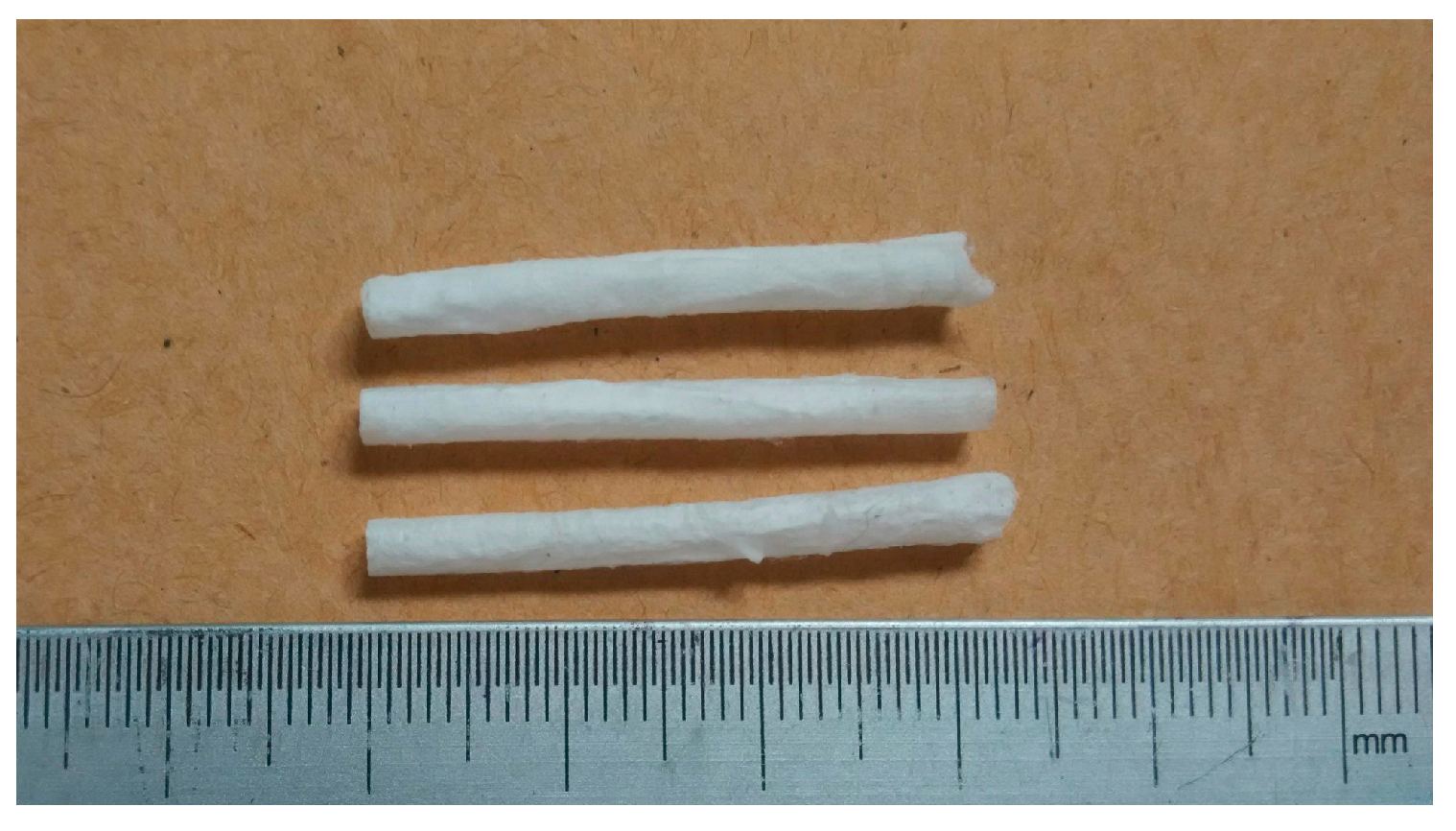
2.1. Tube
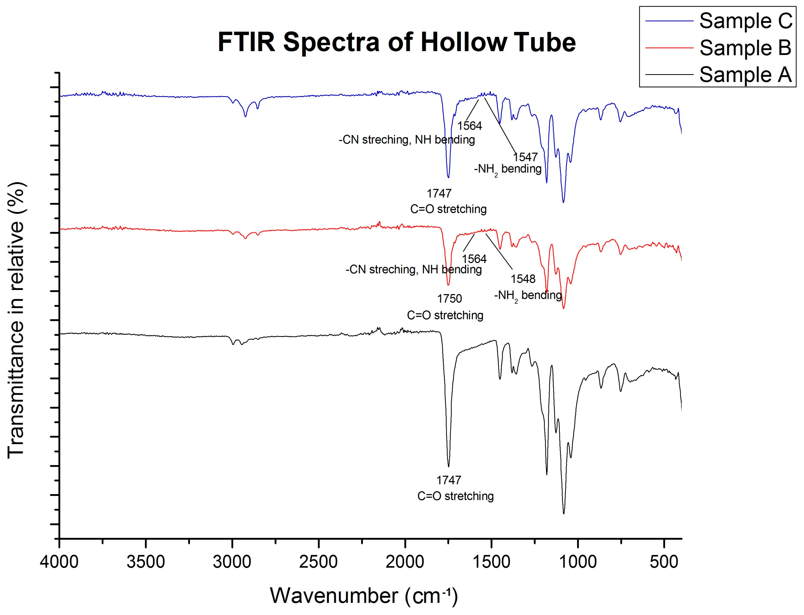
2.2. FTIR Spectra of Tubes
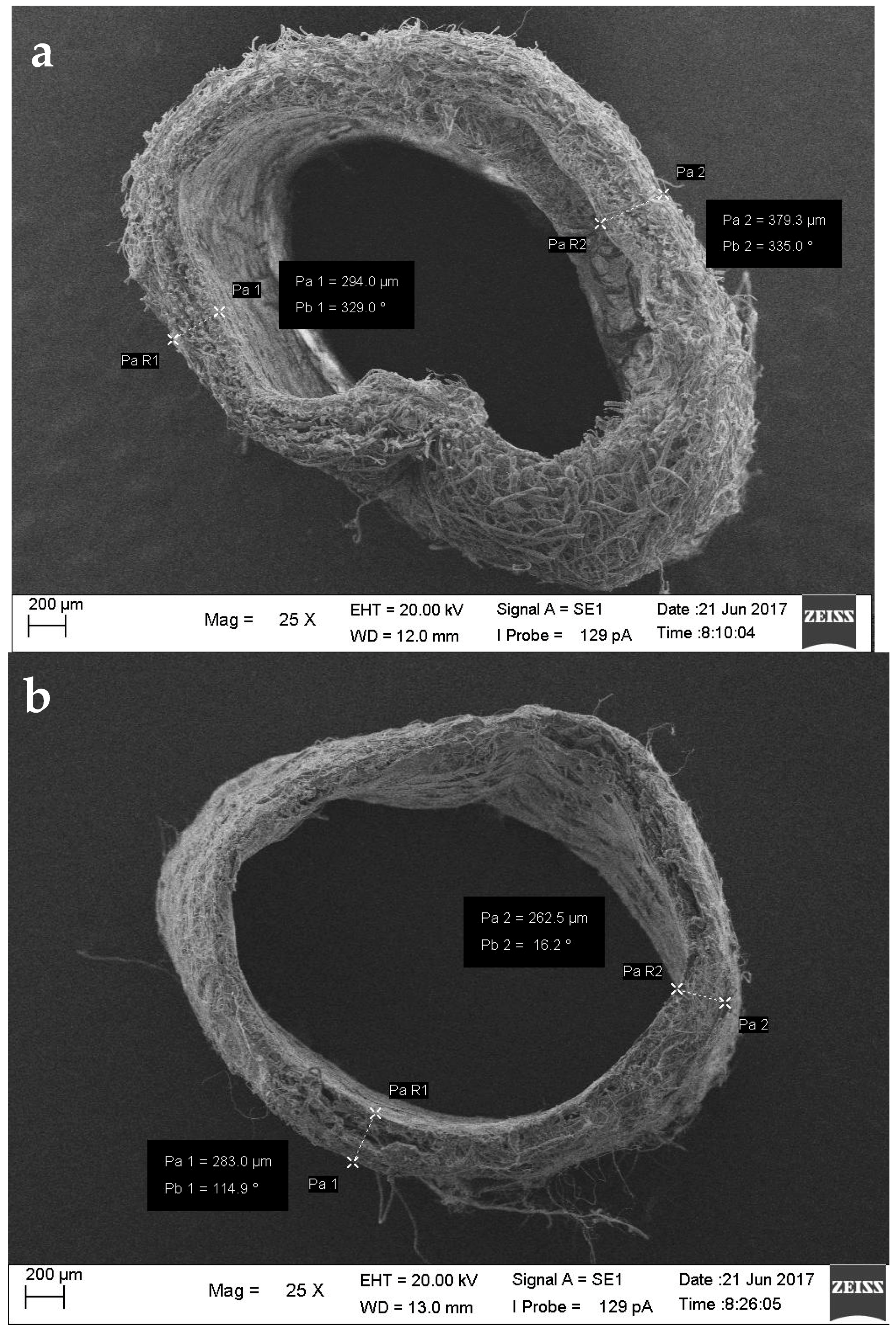
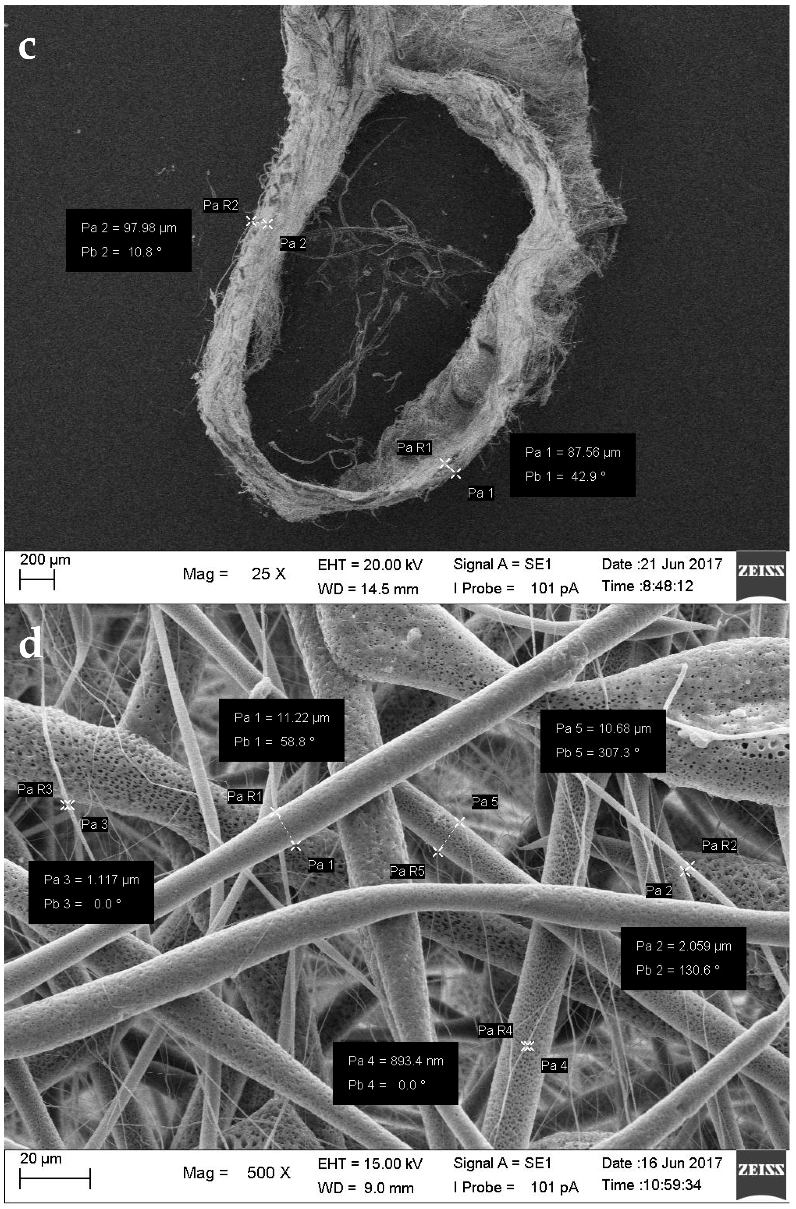
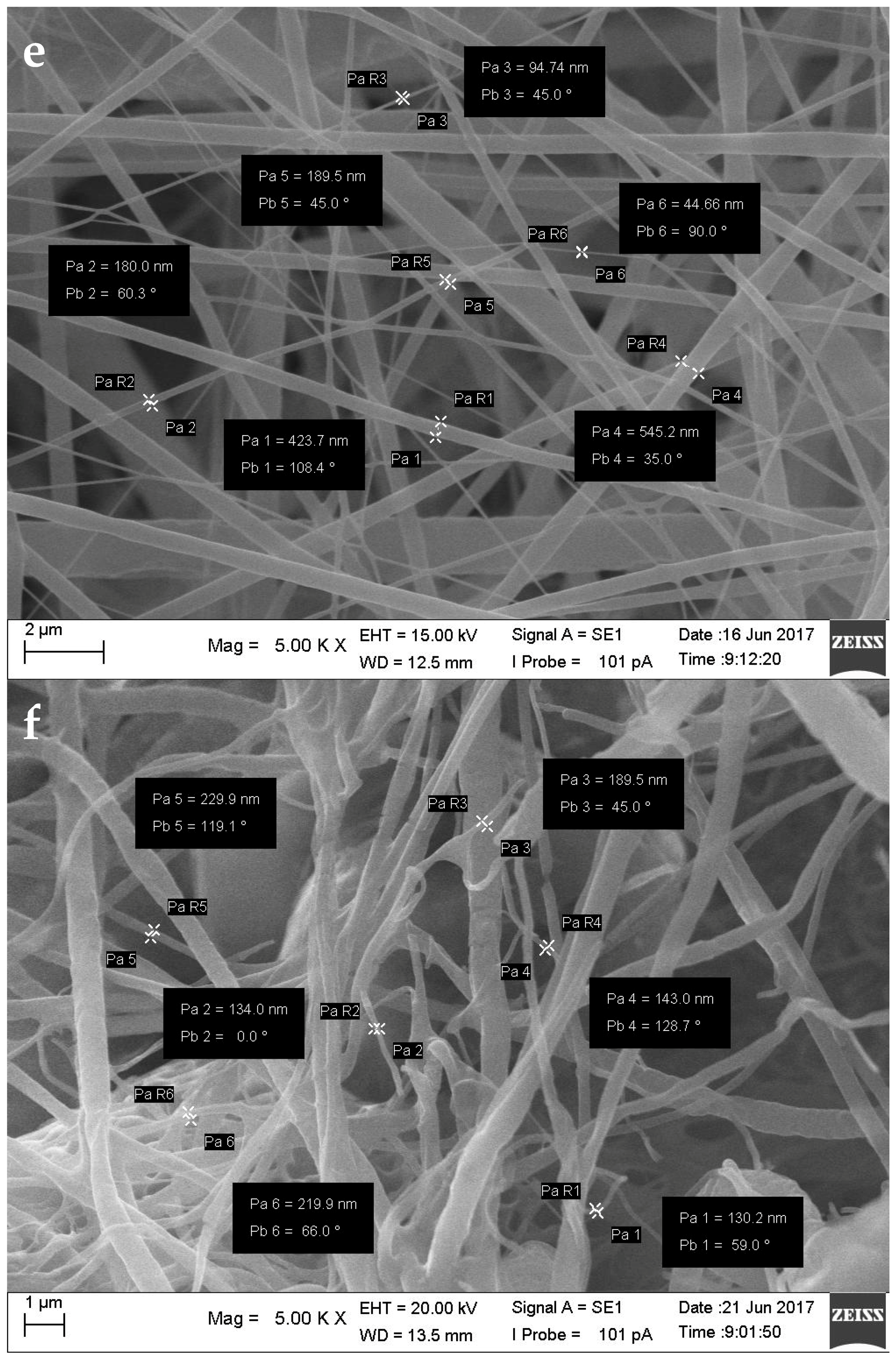
2.3. Morphology
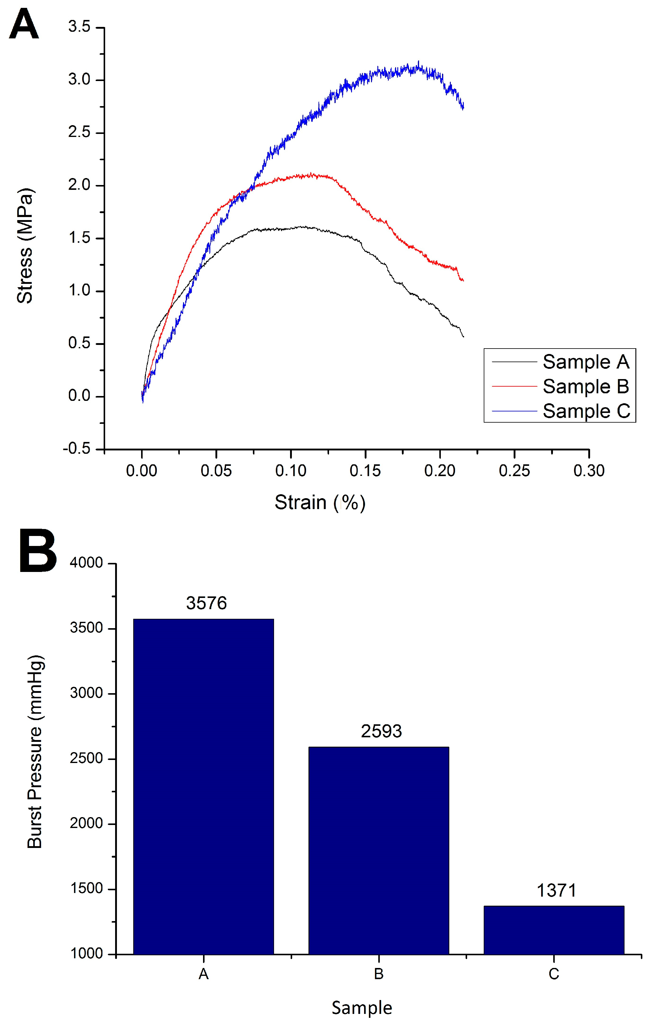
2.4. Mechanical Characteristics
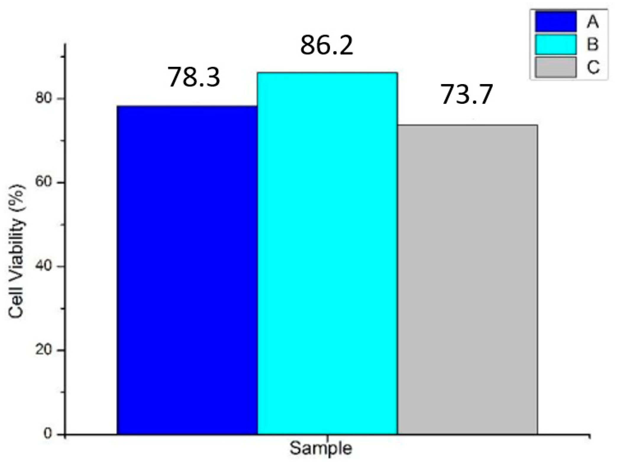
2.5. In Vitro Cell Viability
2.6. Hemolysis Assay
3. Discussion
4. Materials and Methods
4.1. Materials
4.2. Electrospinning
4.3. Evaluation
4.3.1. Fourier Transform InfraRed (FTIR)
4.3.2. Mechanical Testing
4.3.3. Morphology
4.3.4. Cell Viability Test (Cytotoxicity)
4.3.5. Hemolysis Assay
5. Conclusions
Author Contributions
Acknowledgments
Conflicts of Interest
References
- Bentzon, J.F.; Otsuka, F.; Virmani, R.; Falk, E. Mechanisms of plaque formation and rupture. Circ. Res. 2014, 114, 1852–1866. [Google Scholar] [CrossRef] [PubMed]
- Hu, J.; Sun, X.; Ma, H.; Xie, C.; Chen, Y.E.; Ma, P.X. Porous nanofibrous PLLA scaffolds for vascular tissue engineering. Biomaterials 2010, 31, 7971–7977. [Google Scholar] [CrossRef] [PubMed]
- McKenna, K.A.; Hinds, M.T.; Sarao, R.C.; Wu, P.C.; Maslen, C.L.; Glanville, R.W.; Babcock, D.; Gregory, K.W. Mechanical property characterization of electrospun recombinant human tropoelastin for vascular graft biomaterials. Acta Biomater. 2012, 8, 225–233. [Google Scholar] [CrossRef] [PubMed]
- Ravi, S.; Chaikof, E.L. Biomaterials for vascular tissue engineering. Regen. Med. 2010, 5, 107–120. [Google Scholar] [CrossRef] [PubMed]
- Puricel, S.; Arroyo, D.; Stadelmann, M. Bioresorbable Vascular Scaffolds in the Treatment of Coronary Artery Bypass Grafts. In Coronary Graft Failure: State of the Art; Tintoiu, I.C., Underwood, M.J., Cook, S.P., Kitabata, H., Abbas, A., Eds.; Springer International Publishing: Cham, Switzerland, 2016; pp. 649–653. [Google Scholar]
- Martínez, J.; Falomir, M.; Gozalbo, D. Chitin: A structural biopolysaccharide with multiple applications. eLS 2014. [Google Scholar] [CrossRef]
- Patino, M.G.; Neiders, M.E.; Andreana, S.; Noble, B.; Cohen, R.E. Collagen as an implantable material in medicine and dentistry. J. Oral Implantol. 2002, 28, 220–225. [Google Scholar] [CrossRef]
- Yin, A.; Zhang, K.; Mcclure, M.J.; Huang, C.; Wu, J.; Fang, J.; El-Newehy, M. Electrospinning collagen/chitosan/poly(L-lactic acid-co-ϵ-caprolactone) to form a vascular graft: Mechanical and biological characterization. J. Biomed. Mater. Res. A 2012, 101, 1292–1301. [Google Scholar] [PubMed]
- Coates, J. Interpretation of Infrared Spectra, A Practical Approach. In Encyclopedia of Analytical Chemistry; John Wiley & Sons, Ltd.: Chichester, UK, 2000. [Google Scholar]
- Raut, B.K.; Patil, V.N.; Cherian, G. Coronary artery dimensions in normal Indians. Indian Heart J. 2017, 69, 512–514. [Google Scholar] [CrossRef] [PubMed]
- Wilson, S.E. Vascular Access: Principles and Practice; Lippincott Williams & Wilkins: Ambler, PA, USA, 2010. [Google Scholar]
- Karimi, A.; Navidbakhsh, M.; Shojaei, A.; Faghihi, S. Measurement of the uniaxial mechanical properties of healthy and atherosclerotic human coronary arteries. Mater. Sci. Eng. C 2013, 33, 2550–2554. [Google Scholar] [CrossRef] [PubMed]
- Konig, G.; McAllister, T.; Dusserre, N.; Garrido, S.A.; Lycian, C.; Marini, A.; Fiorillo, A.; Avila, H.; Wystrychowski, W.; Zagalski, K.; et al. Mechanical properties of completely autologous human tissue engineered blood vessels compared to human saphenous vein and mammary artery. Biomaterials 2009, 30, 1542–1550. [Google Scholar] [CrossRef] [PubMed]
- Spielmann, H.; Hoffmann, S.; Liebsch, M.; Botham, P.; Fentem, J.H.; Eskes, C.; Roguet, R.; Cotovio, J.; Cole, T.; Worth, A.; et al. The ECVAM international validation study on in vitro tests for acute skin irritation: Report on the validity of the EPISKIN and EpiDerm assays and on the Skin Integrity Function Test. Altern. Lab. Anim. 2007, 35, 559–601. [Google Scholar] [PubMed]
- Xue, L.; Greisler, H.P. Biomaterials in the development and future of vascular grafts. J. Vasc. Surg. 2003, 37, 472–480. [Google Scholar] [CrossRef] [PubMed]
- Huang, Z.; Zhang, Y.; Ramakrishna, S.; Lim, C. Electrospinning and mechanical characterization of gelatin nanofibers. Polymer 2004, 45, 5361–5368. [Google Scholar] [CrossRef]
- Gauvin, R.; Guillemette, M.; Galbraith, T.; Bourget, J.M.; Larouche, D.; Marcoux, H.; Aube, D.; Hayward, C.; Auger, F.A.; Germain, L. Mechanical properties of tissue-engineered vascular constructs produced using arterial or venous cells. Tissue Eng. Part A 2011, 17, 2049–2059. [Google Scholar] [CrossRef] [PubMed]
- Prabst, K.; Engelhardt, H.; Ringgeler, S.; Hubner, H. Basic Colorimetric Proliferation Assays: MTT, WST; Resazurin. Methods Mol. Biol. 2017, 1601, 1–17. [Google Scholar] [PubMed]
- Evans, B.C.; Nelson, C.E.; Shann, S.Y.; Beavers, K.R.; Kim, A.J.; Li, H.; Nelson, H.M.; Giorgio, T.D.; Duvall, C.L. Ex vivo red blood cell hemolysis assay for the evaluation of pH-responsive endosomolytic agents for cytosolic delivery of biomacromolecular drugs. J. Visual. Exp. 2013, 73, 50166. [Google Scholar] [CrossRef] [PubMed]


| Sample | Outer Diameter (mm) and Thickness (mm) | Fiber Diameter (nm) | Fiber-to-Fiber Distance (µm) |
|---|---|---|---|
| A (PLLA) | 3.04 ± 0.02 0.39 ± 0.03 | 135.8–205.9 | 3.796–31.27 |
| B (PLLA-Collagen-Chitosan 0.5%) | 2.93 ± 0.05 0.22 ± 0.03 | 89.33–246.7 | 5.141–30.144 |
| C PLLA-Collagen-Chitosan 0.6%) | 2.87 ± 0.06 0.08 ± 0.08 | 130.2–229.9 | 4.362–30.872 |
| Sample | Tensile Strength (N/mm2) | Burst Pressure (mmHg) |
|---|---|---|
| A (Control) | 1.62 | 3576 |
| B (PLLA-Collagen-Chitosan 0.5%) | 2.13 | 2593 |
| C (PLLA-Collagen-Chitosan 0.6%) | 3.19 | 1371 |
| Sample | Average Absorbance (Hm) | Hemolysis Percentage (%) | Hemolytic Grade |
|---|---|---|---|
| A (PLLA, Control) | 0.052 | 14.63 | Hemolytic |
| B (PLLA-Collagen-Chitosan 0.5%) | 0.039 | 1.04 | Non Hemolytic |
| C (PLLA-Collagen-Chitosan 0.6%) | 0.041 | 3.14 | Slightly Hemolytic |
| Sample | Concentration (w/v %) | ||
|---|---|---|---|
| PLLA | Collagen | Chitosan | |
| A (PLLA, Control) | 10 | 0 | 0 |
| B (PLLA-Collagen-Chitosan 0.5%) | 10 | 1 | 0.5 |
| C (PLLA-Collagen-Chitosan 0.6%) | 10 | 1 | 0.6 |
© 2018 by the authors. Licensee MDPI, Basel, Switzerland. This article is an open access article distributed under the terms and conditions of the Creative Commons Attribution (CC BY) license (http://creativecommons.org/licenses/by/4.0/).
Share and Cite
Fiqrianti, I.A.; Widiyanti, P.; Manaf, M.A.; Savira, C.Y.; Cahyani, N.R.; Bella, F.R. Poly-L-lactic Acid (PLLA)-Chitosan-Collagen Electrospun Tube for Vascular Graft Application. J. Funct. Biomater. 2018, 9, 32. https://doi.org/10.3390/jfb9020032
Fiqrianti IA, Widiyanti P, Manaf MA, Savira CY, Cahyani NR, Bella FR. Poly-L-lactic Acid (PLLA)-Chitosan-Collagen Electrospun Tube for Vascular Graft Application. Journal of Functional Biomaterials. 2018; 9(2):32. https://doi.org/10.3390/jfb9020032
Chicago/Turabian StyleFiqrianti, Iffa A., Prihartini Widiyanti, Muhammad A. Manaf, Claudia Y. Savira, Nadia R. Cahyani, and Fitria R. Bella. 2018. "Poly-L-lactic Acid (PLLA)-Chitosan-Collagen Electrospun Tube for Vascular Graft Application" Journal of Functional Biomaterials 9, no. 2: 32. https://doi.org/10.3390/jfb9020032