Review

Wet-Chemical Preparation of TiO2-Based Composites with Different Morphologies and Photocatalytic Properties

Smart Materials Laboratory, Department of Applied Physics, Northwestern Polytechnical University, Xi’an 710129, China
*
Author to whom correspondence should be addressed.
Nanomaterials 2017, 7(10), 310; https://doi.org/10.3390/nano7100310
Submission received: 7 September 2017 / Revised: 30 September 2017 / Accepted: 2 October 2017 / Published: 9 October 2017
(This article belongs to the Special Issue ZnO and TiO2 Based Nanostructures)

Abstract

:
TiO2-based composites have been paid significant attention in the photocatalysis field. The size, crystallinity and nanomorphology of TiO2 materials have an important effect on the photocatalytic efficiency. The synthesis and photocatalytic activity of TiO2-based materials have been widely investigated in past decades. Based on our group’s research works on TiO2 materials, this review introduces several methods for the fabrication of TiO2, rare-earth-doped TiO2 and noble-metal-decorated TiO2 particles with different morphologies. We focused on the preparation and the formation mechanism of TiO2-based materials with unique structures including spheres, hollow spheres, porous spheres, hollow porous spheres and urchin-like spheres. The photocatalytical activity of urchin-like TiO2, noble metal nanoparticle-decorated 3D (three-dimensional) urchin-like TiO2 and bimetallic core/shell nanoparticle-decorated urchin-like hierarchical TiO2 are briefly discussed.

1. Introduction

Based on its unique chemical and physical characteristics, titanium dioxide (TiO2) has attracted much attention in many fields including paint pigments, photocatalysis, solar cells, antibacterial agents, electrical energy storage and some advanced functional materials [1,2,3]. The performance in these applications strongly depends on the microstructure, crystallinity and nanomorphology of TiO2 [1]. In particular, in the photocatalysis field, although a new type of polymeric photocatalyst—that is, graphitic carbon nitride—has been intensively investigated recently due to its huge advantages—including metal-free contents, visible light absorption ability, suitable band gap for water splitting, stability, and being environmentally benign [4,5,6,7]—TiO2 is still regarded as one of most ideal candidates for photocatalysis because of its strong oxidization, harmlessness to surroundings, chemical inactivity, good stability and low cost [1,3,8,9]. There are three important processes including photo excitation, bulk diffusion and the surface transfer of photoinduced charge carriers in photocatalysis [8]. Thus, the performance of photocatalysis depends strongly upon the charge transfer at the material surface and the light-response range of materials [1,10,11]. The processes of light harvesting and charge transfer efficiencies are affected mainly by the size, crystallinity and nanomorphology of TiO2 materials [1,8,10,11,12,13,14]. The preparation and photocatalytic properties of TiO2 with different morphologies including zero-dimensional (micro/nanospheres), one-dimensional (rods, tubes, and nanowires), two-dimensional (films, layers and sheets), and three-dimensional (porous spheres, urchin-like spheres) TiO2 structures have been widely investigated in the past decade [15,16,17,18,19,20,21]. Different ways have been developed for preparing TiO2 materials with different nanostructures. The general synthesis approaches for the fabrication of TiO2 materials include sol-gel, hydrothermal and solvothermal techniques [1]. Controlling the microscopic structures of TiO2 is still a challenge because TiO2 precursors are highly reactive.
Pure TiO2 is not a perfect photocatalyst due to the disadvantages of low photocatalytic efficiency and the narrow light-response region [8]. Doping metal ions or introducing noble metal nanoparticles onto the surface of TiO2 was demonstrated to be one of the effective ways to enhance the photocatalytic efficiency because these TiO2-based composites can combine the functions of TiO2 and metal ions or noble metals [22,23,24,25]. Furthermore, the properties of the composites can be adjusted by controlling the ingredients and the microstructures of the TiO2.
In the past two decades, our group has focused on the synthesis, electrorheological (ER) properties, luminescence properties and photocatalytic activities of TiO2-based materials [26,27,28,29,30,31,32]. A series of TiO2-based materials with different compositions, crystallinity and interior microstructures have been synthesized by different methods [33,34,35,36]. The TiO2 particles with a familiar microstructure, such as solid spheres [37], hollow spheres [29], porous spheres [28,38], hollow porous spheres [39] and urchin-like spheres [30], were synthesized and characterized in detail. In addition, some TiO2 composites with a special interior microstructure were also designed and synthesized [40,41,42,43]. According to the dielectric design, rare-earth-doped TiO2 particles were synthesized by sol-gel methods [44,45,46,47,48,49]. Inspired by the structure of biological surfaces, a kind of composite particle possessing both nano- and micro-scale structures was prepared via a hydrothermal method [50,51]. TiO2 particles with a cell-like structure were also synthesized [52]. It is noteworthy that the TiO2-based materials described above show excellent properties in different applications. For example, the rare-earth-doped TiO2, and the micro- or nano-structured composites with TiO2 have been demonstrated to show a distinct enhancement in their ER properties [44,50,51,52]. The hollow Sm3+-doped TiO2 and the monodisperse mesoporous Eu-doped TiO2 spheres have shown good luminescent performance [28,29]. The urchin-like TiO2 and urchin-like TiO2 decorated with Au, Ag, Co@Au or Co@Ag nanoparticles have shown significant improvement in photocatalytic activities [30,31,32].
Until now, there have been many review articles introducing the progress made in the field of TiO2-based materials [1,2,3,8]. Based on our group’s research work on TiO2 materials, this review is primarily concentrated on the preparation of TiO2 composites with different morphologies and the photocatalytic activities of urchin-like TiO2 composites.

2. Preparation of TiO2 and TiO2-Based Composites with Different Morphologies

2.1. Micro- and Nano-Spheres

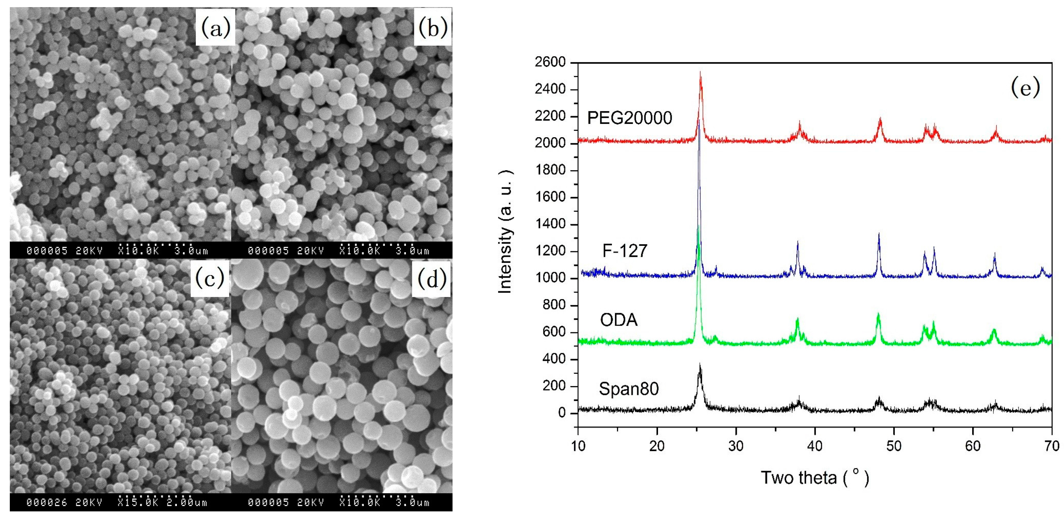
2.1.1. Solid Spheres

Spherical particles with a specific size can be used in many fields, such as photonic crystals, pigments, and so on [15]. In order to obtain monodisperse spherical TiO2 particles, many methods have been developed. However, it is still a challenge to control the morphology and size of TiO2 microspheres because of the high reactivity of precursors. Increasing the charge of the particle surface and the steric repulsion of the particles are effective methods of controlling the stability of TiO2 microspheres [53]. We have reported a simple and reproducible sol-gel method for synthesizing well-defined spherical TiO2 particles with diameters within 200–800 nm. In this method, polymers including polyethylene glycol (PEG), poly(ethylene oxide)106-poly(propylene oxide)70-poly(ethylene oxide)106 (F127) copolymer, octadecylamine (ODA), and surfactant Span-80 were used to control the size of TiO2 particles [37]. For example, quasi-monodisperse TiO2 submicron spheres were synthesized by controlling the hydrolysis of tetrabutyl titanate in ethanol containing the above polymers and small amounts of deionized water. During this process, depending on the used polymer, the transmission time from the transparent solution into white suspension changed from several seconds to minutes. As soon as the transparent solution changed into white suspension, the stirring had to be stopped and the suspension was further aged for 8 h to form quasi-monodisperse TiO2 submicron spheres. After high temperature annealing, the spheres were crystallized into the anatase phase. Figure 1 shows the quasi-monodisperse TiO2 submicron spheres with different diameters within 200–800 nm, synthesized with different polymers.

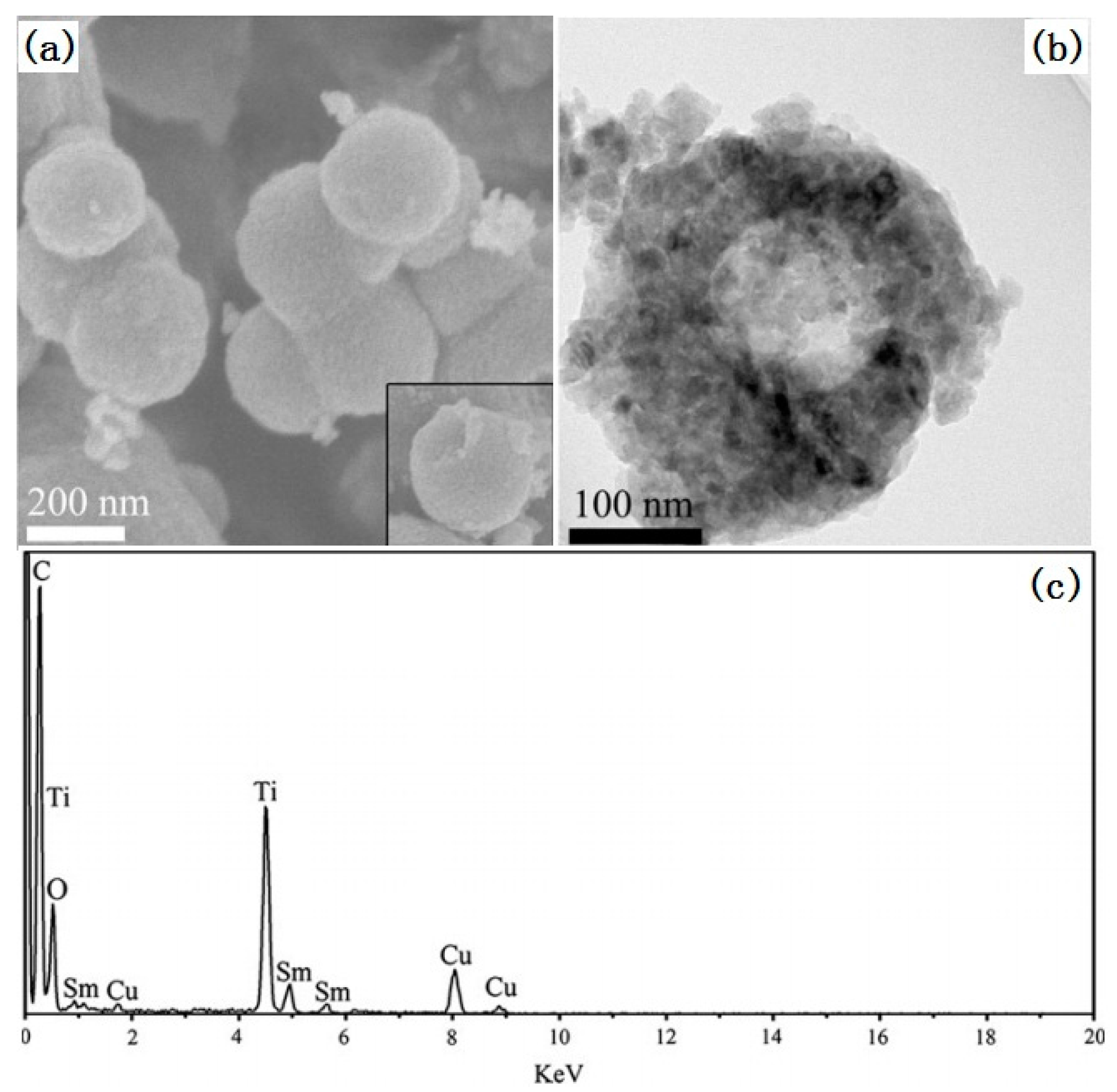
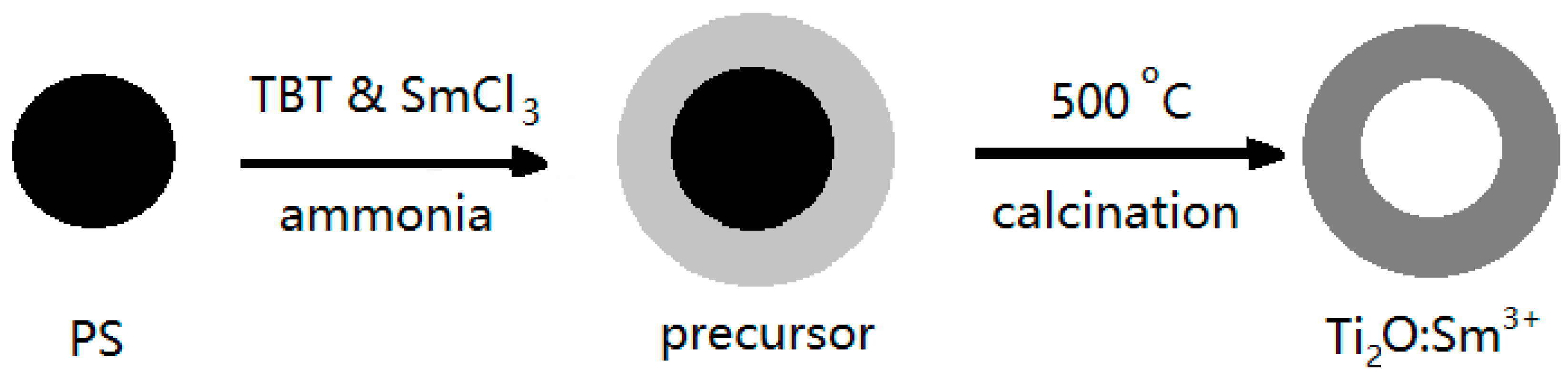
2.1.2. Hollow Spheres

Due to high specific surface area and low density, hollow structured materials have been widely used in many fields [54]. TiO2 hollow spheres with a well-defined crystal phase are highly desirable for photocatalysis use [55,56,57]. Hollow structured TiO2 can be feasibly synthesized by hard template and soft template methods. Compared to the soft template method, the hard template method is simpler, and so it is frequently used.
An et al. have used polystyrene (PS) spheres as a hard template to prepare hollow Sm3+-doped TiO2 spheres [29]. The schematic illustration of the formation mechanism is shown in Figure 2. Since the surface of PS spheres obtained by surfactant-free microemulsion polymerization is negatively charged, no additional surface modification of PS spheres is needed for the next coating of TiO2. In an ethanol/acetonitrile mixed solvent, a small amount of ammonia was used to induce the hydrolysis of tetrobutyl titanate to form the amorphous Sm3+-doped TiO2 coating layer on the surface of the PS spheres. After washing with ethanol, drying, and annealing, hollow TiO2:Sm3+ spheres, as shown in Figure 3, could be obtained.

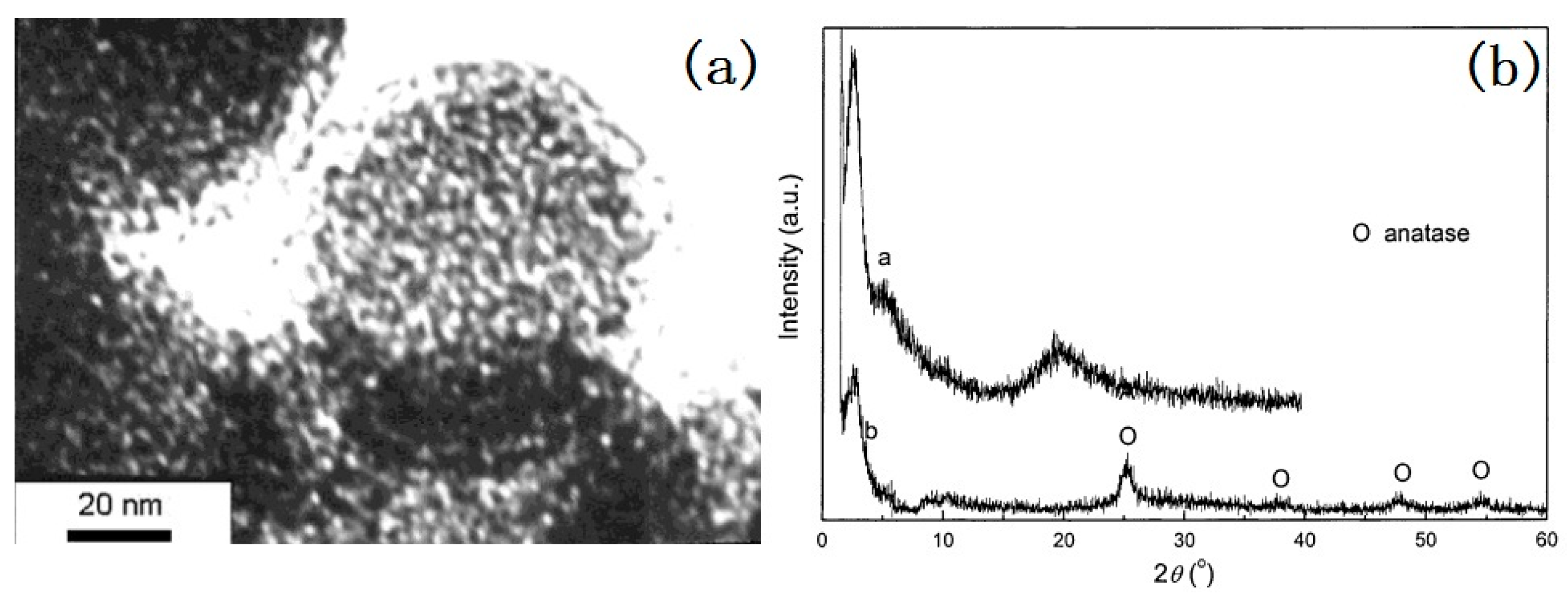
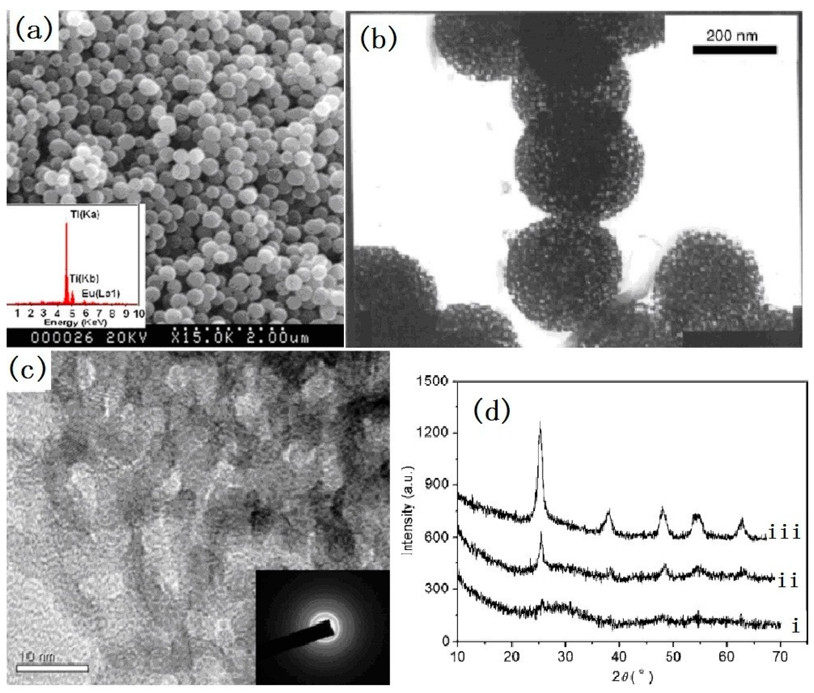
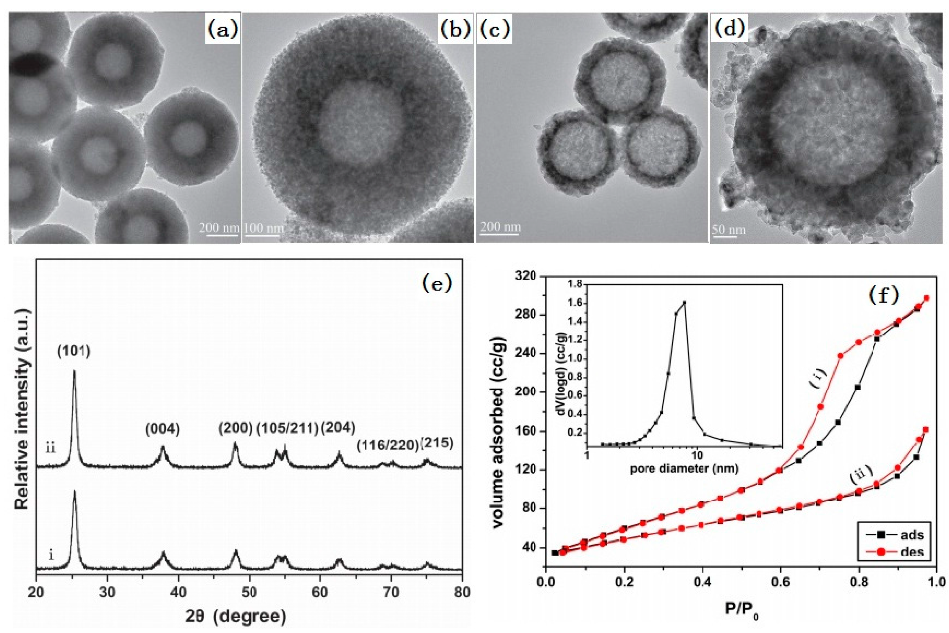
2.1.3. Porous Spheres

Due to their high surface area, porous materials are very popular for different applications including energy storage, solar cells and catalyzers [58,59,60]. Mesoporous TiO2-based materials have attracted much attention for their enhanced reactivity and light harvesting [60]. The macrochannels in mesoporous TiO2 particles have served as a light-transfer path that can introduce incident photon flux to the interior surface of the TiO2 particles [58]. A mesoporous structure gives light waves more chances to penetrate deep inside the photocatalyst and more light waves are captured. The crystallinity, pore size and composition are important for tuning the properties of mesoporous TiO2 spheres [1,8,60]. There has been intensive research concentrated on the design and preparation of porous TiO2 materials with unique porosities and tunable pore sizes [61,62,63,64,65].
We have synthesized mesoporous Ce-doped TiO2 spheres by a low-temperature hydrothermal method by using neutral dodecylamine (DDA) as a surfactant and tetrabutyl titanate as a Ti source [64]. To control the rate of hydrolysis of tetrabutyl titanate, a solvent mixture of ethanol and propanol (2:1, v/v) was used. No additional water was used to initialize the hydrolysis and condensation reaction of the tetrabutyl titanate, due to the used CeCl3·7H2O containing structured water. After the CeCl3·7H2O was dissolved, the structured water was released. DDA was able to make the solution alkaline and this made it easy to increase the rate of hydrolysis of the tetrabutyl titanate. However, the dissolution of CeCl3·7H2O also could result in a decrease of the pH value of solution. Thus, CeCl3·7H2O could service as not only as a dopant but also as an initiator and buffer. After reaction, a precipitate was formed and it was further refluxed for 2 h at 80 °C in an acid solution to get rid of the template and obtain sphere-like mesoporous Ce-doped TiO2 particles with a diameter of 100–1000 nm as shown in Figure 4. The XRD (X-ray diffraction) patterns in Figure 4 show that the TiO2 particles are semi-crystalline. The formed anatase crystalline size is very small, about 2–3 nm. In addition, from the TEM (Transmission Electron Microscopy) image, it could be found that the pore structure was worm-like, with a size of 2–3 nm. The Brunauer-Emmett-Teller (BET) surface area of the mesoporous Ce-doped TiO2 was 118 m2/g, which is much higher than the 9.6 m2/g of single-doped TiO2 obtained without a surfactant.
The mesoporous Eu-doped TiO2 spheres have also been developed by the hydrolysis of tetrabutyl titanate [28]. To increase the thickness of the pore wall, nonionic copolymer Pluronic F-127 was used as a template agent. Europium ethoxide was specially prepared as a doping agent in order to increase the rate of hydrolysis and condensation of the tetrabutyl titanate. Small amounts of water were added to initiate the reaction under stirring. As soon as the solution became slightly white after several minutes, the stirring was stopped. Meanwhile, in order to control the water content, the preparation was conducted under the protection of flowing N2. After aging for 24–48 h at 35–40 °C, the suspended particles were filtered and washed with ethanol several times. The final products were obtained after calcination at 400 °C for 4 h. The synthesized Eu-doped TiO2 particles have a spherical morphology and a mesoporous structure, with a pore size of 7–10 nm. The special surface area of the phosphor particles is 158 m2/g. The high resolution TEM images in Figure 5c show that the pore wall is semi-crystalline that many anatase nanocrystallites are dispersed in the amorphous TiO2. The XRD patterns showed in Figure 5d have indicated that no peaks corresponding to the europium compound was detected and no shift of the anatase peaks was observed after doping with Eu3+. It can be concluded that Eu3+ ions are mainly dispersed in the amorphous TiO2 region.

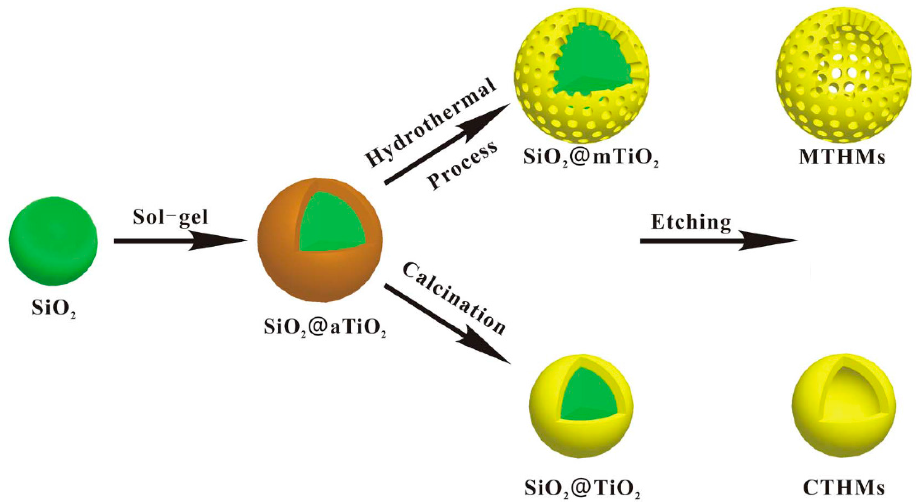
2.1.4. Hollow and Porous Spheres

Compared to single hollow spheres, hollow TiO2 spheres with a porous shell are more interesting in photocatalysis because they can provide a higher surface area and active site points, decreased diffusion resistance, and increased accessibility to reactants [66]. Several methods have been reported to synthesize hollow TiO2 spheres with a porous shell [66,67]. Among these methods, the template method is the most popular. Different sacrificial templates can be used for controlling the size and morphology of such a hollow nanostructure. The template method followed by a hydrothermal or calcination treatment is often used to synthesize hollow TiO2 spheres with a crystalline shell [39]. Figure 6 shows a typical process of preparing hollow TiO2 spheres with a crystalline shell. In this process, amorphous TiO2 was firstly coated onto the surface of SiO2 spheres by the sol-gel method in an alkaline solution. Then, the composite microspheres were subjected to a hydrothermal or calcination treatment. Meanwhile, the amorphous TiO2 was crystallized into nanocrystals and the mesoporous structure was formed by nanocrystal stacking. Finally, the SiO2 core was removed by etching in an alkaline solution. As shown in Figure 7, the sample prepared by hydrothermal treatment had a mean diameter of 620 nm with a 180 nm thick mesoporous TiO2 shell. The BET surface area was 231.1 m2/g and the pore size was 6.5 nm. However, the sample prepared by calcination had a mean diameter of 440 nm with a 90 nm thick mesoporous TiO2 shell. The BET surface area was 158.3 m2/g.

2.2. 3D Urchin-Like Hierarchical Particles

Urchin-like microspheres possess an epitaxial multilevel structure. The unique micro/nano hierarchical structure has two obvious advantages over single nano-scale or micro-scale structures when they are used as photocatalysts [54,68]. One is that urchin-like TiO2 is more efficient at absorbing incidental light because of the increase of multiple-reflection of the hierarchical microspheres [54]. The other is that urchin-like hierarchical TiO2 is easy to separate from waste water by the filtration or sedimentation methods. The template-assisted method is a familiar approach to prepare the hierarchical materials. However, it is troublesome to remove templates from products. Impurities are easily introduced into products in the process of utilization and removal of templates. The template-free method is accepted as an ideal strategy which can overcome the drawbacks. Recently, TiO2 particles with different hierarchical structures have been successfully fabricated via the template-free method [69,70,71,72,73].

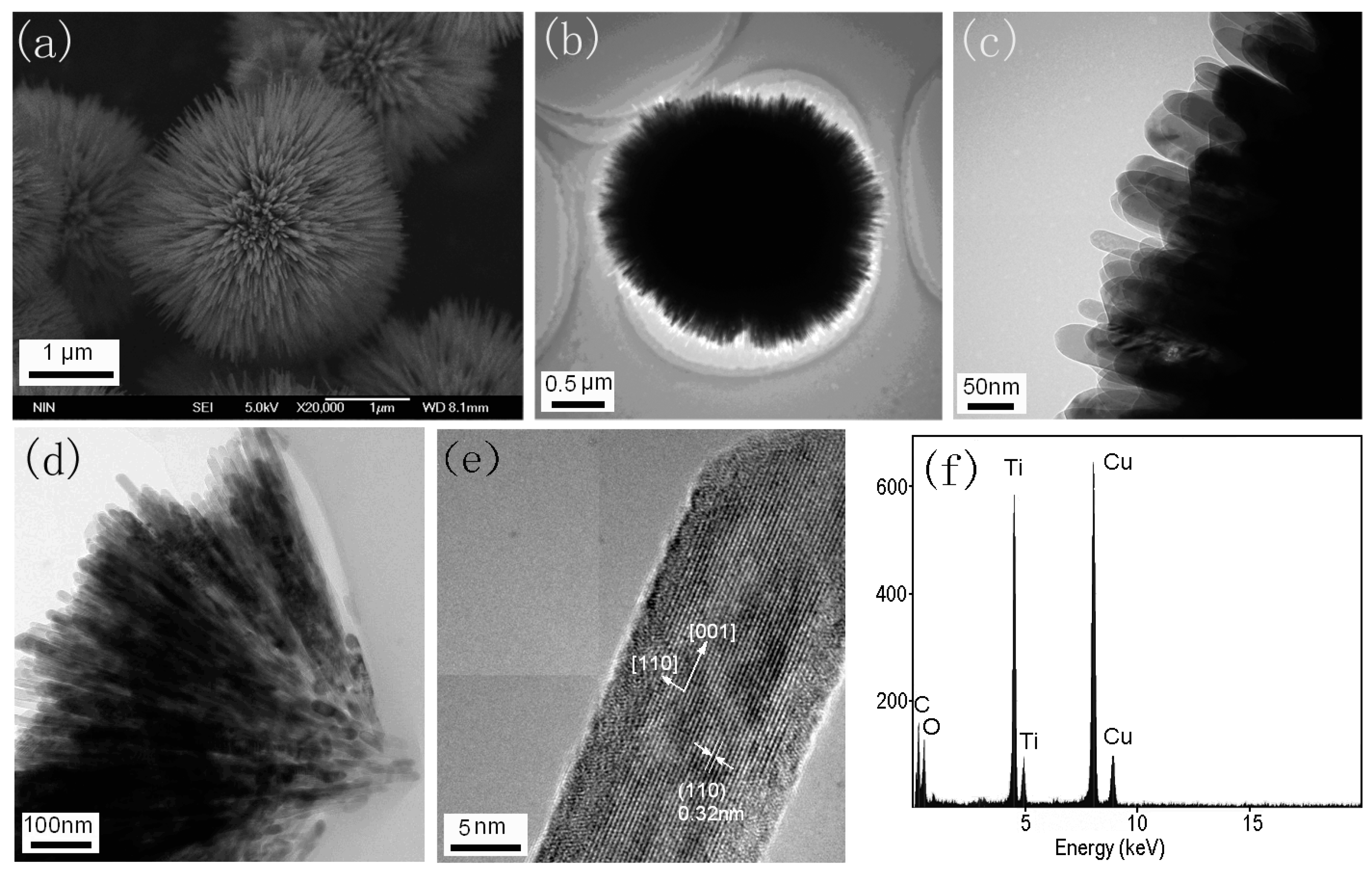
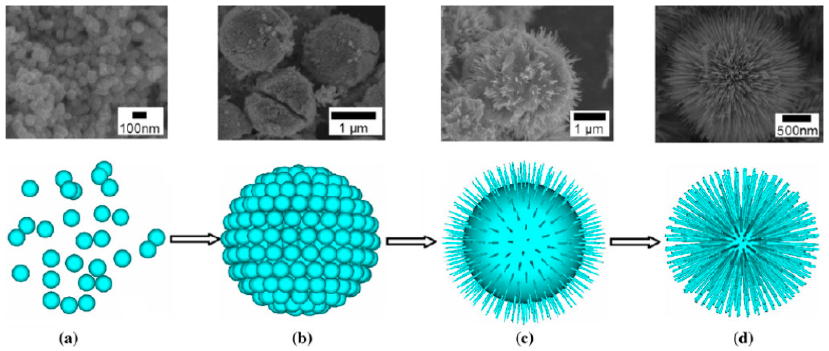
2.2.1. Urchin-Like Hierarchical TiO2 Particles

We have developed a synthesis of a kind of 3D urchin-like TiO2 microspheres via a solvothermal method without adding any surfactant or template [30]. Tetrabutyl titanate and titanium tetrachloride (TiCl4) aqueous solution were used as the reactant, and toluene was used as the solvent. The solvothermal reaction took place in a Teflon-lined autoclave at 150 °C for 24 h. Sea-urchin-like 3D hierarchical TiO2 microspheres with a uniform size of 2.5–3.0 μm were obtained (Figure 8). The 3D hierarchical microspheres were made of single crystalline rutile nanoneedles with diameters about 20–40 nm, which grew radially from the core of the microspheres. The morphology and crystal phase of the 3D hierarchical TiO2 microspheres could be influenced by some factors, such as the ratio of tetrabutyl titanate to TiCl4, the solvothermal temperature, and so on. By tracing the particle morphology change by SEM and XRD techniques, we concluded that the formation of 3D hierarchical TiO2 microspheres mainly concerned three steps, i.e., nucleation, self-assembly, dissolution and recrystallization, as depicted in Figure 9. In the nucleation stage, nanoparticles were formed. Then, the nanoparticles assembled into microspheres. Finally, the microspheres gradually changed into the urchin-like hierarchical structure by dissolution and recrystallization.

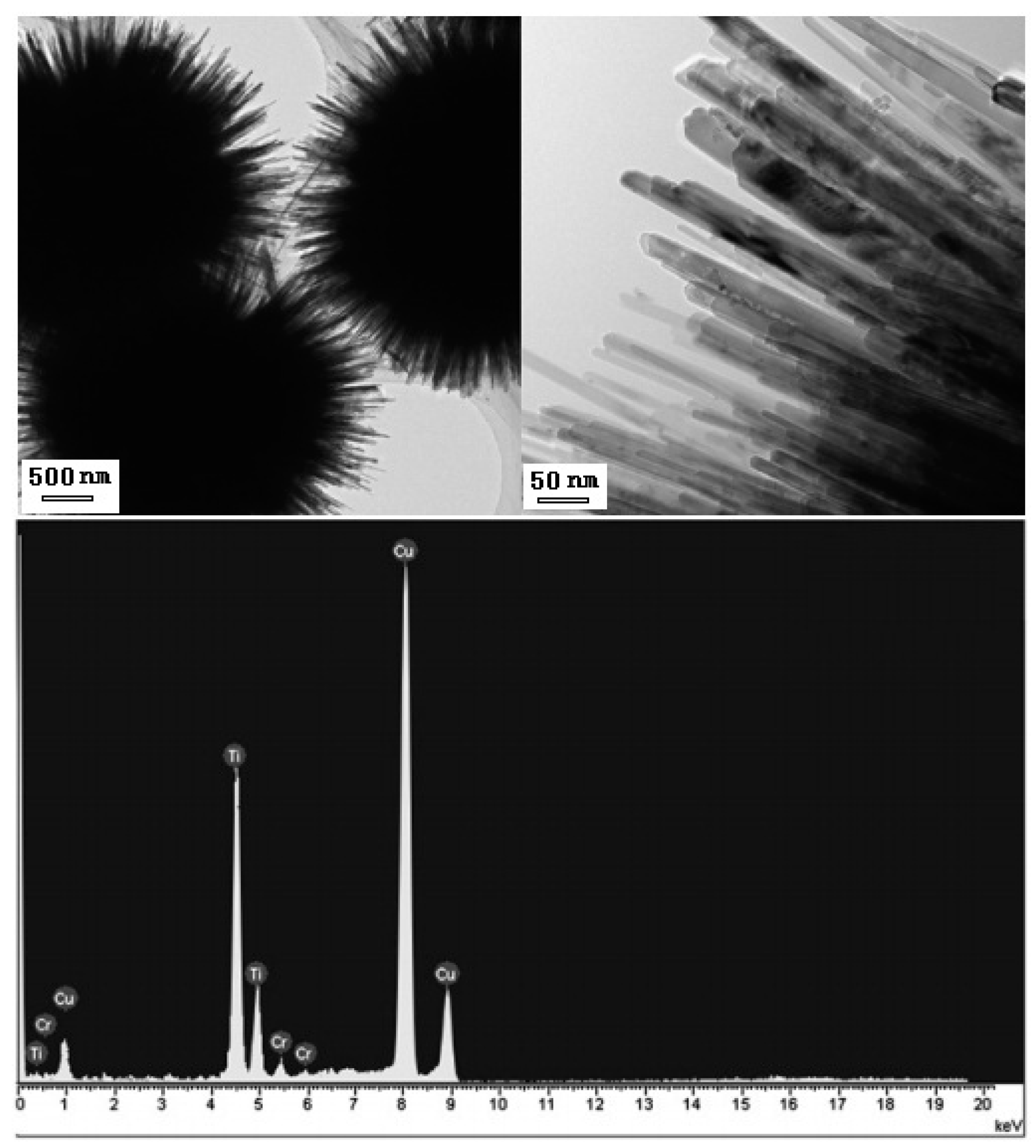
2.2.2. Cr Doped Urchin-Like Hierarchical TiO2 Particles

Urchin-like Cr-doped TiO2 particles could be also synthesized by the same solvothermal method described above in a solution of titanium tetrabutyl titanate dissolving CrCl3 [73]. The morphology of Cr-doped TiO2 particles is characterized by SEM images shown in Figure 10. The mean particle size of the hierarchical microspheres can be adjusted within 1–5 μm and the diameter of the nanorods is about 20–30 nm. The EDS results showed that the content of the Cr element in the Cr-doped TiO2 particles was ~2.9 mol %.

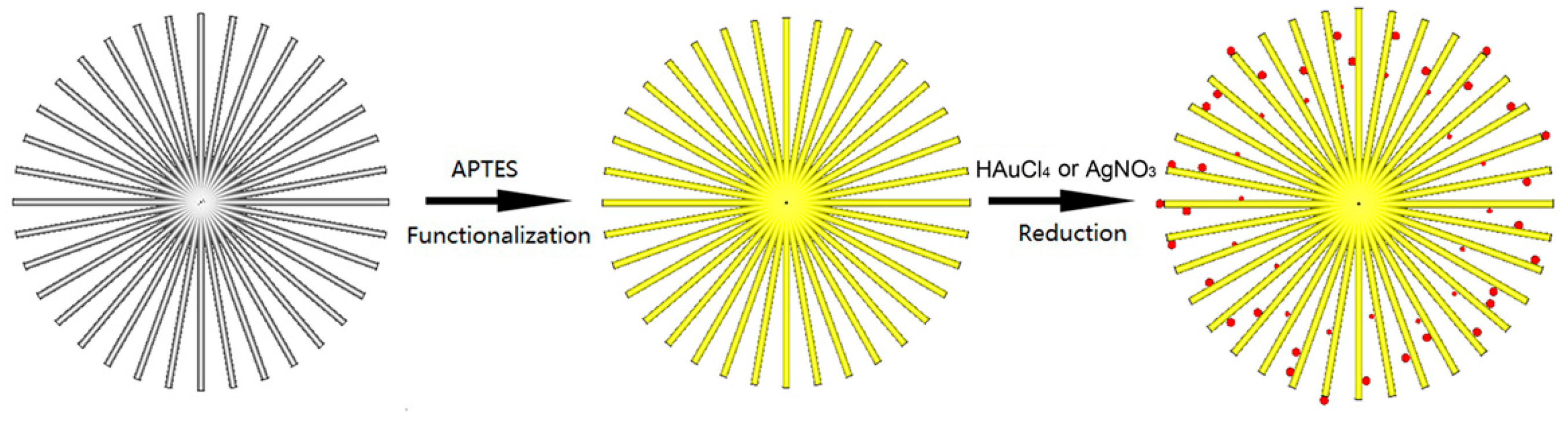
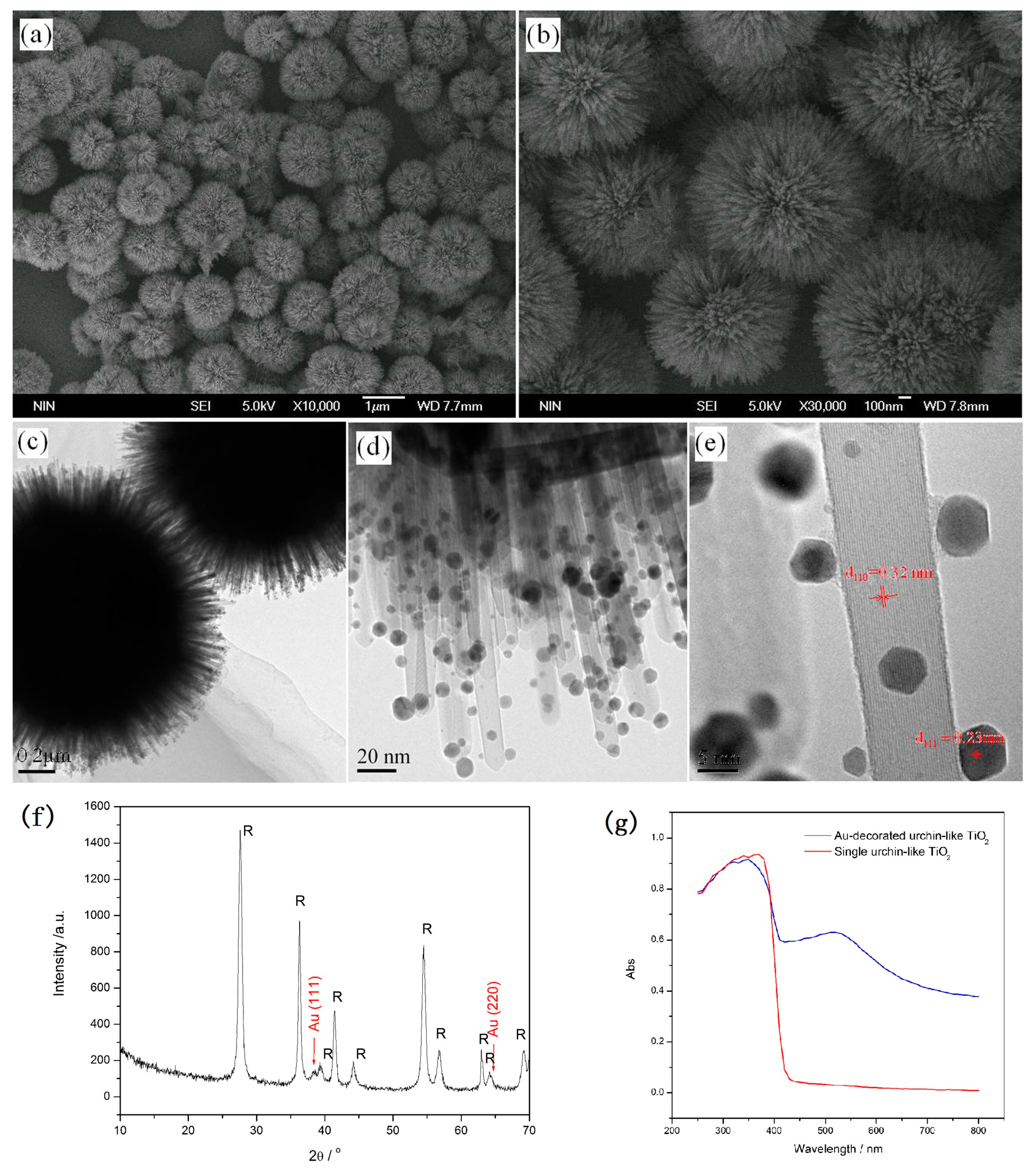
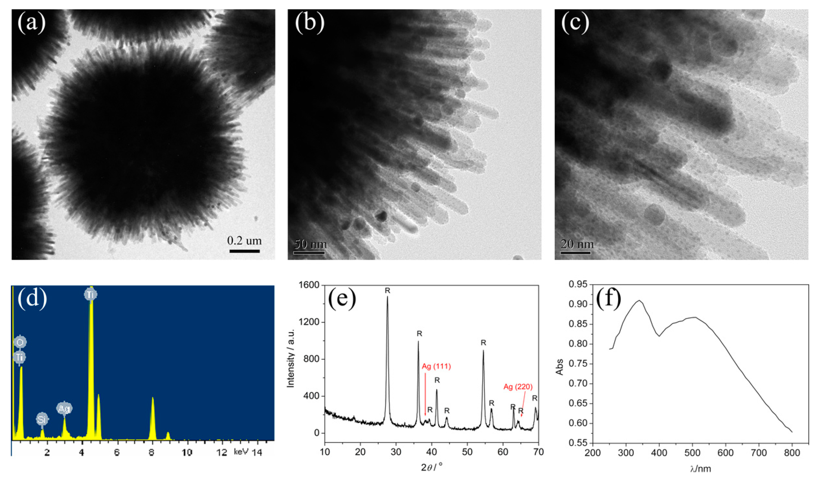
2.2.3. Noble Metal Nanoparticle-Decorated 3D Urchin-Like TiO2 Particles

Noble metal nanoparticle-decorated semiconductors are interesting for photocatalysis because of their combined properties [74]. Decorating the noble metal nanoparticles (e.g., Au, Ag and Pt) onto the surface of TiO2 is an effective method to improve the photocatalytic activity because not only light-harvesting efficiency can be enhanced due to the surface plasmon resonance of noble metal nanoparticles, but the recombination of surface radicals can also be slowed down by capturing photogenerated electrons of noble metal nanoparticles [75,76,77,78,79]. Figure 11 shows a schematic illustration of 3D urchin-like hierarchical TiO2 microspheres decorated with Au nanoparticles via a two-step wet-chemical process [31]. In the first step, the surface of the urchin-like TiO2 microspheres was modified with APTES (3-aminopropyl-triethoxysilane) that possess amidocyanogen. Then, the modified particles were decorated with Au nanoparticles in HAuCl4 aqueous solution by the reduction of NaBH4. Since the amidocyanogen could interact with Au nanoparticles by a weak covalent bond, Au nanoparticles were closely attached to the surface of the TiO2 nanostructures, as shown in Figure 12. It was seen that the Au nanoparticles with diameters of about 2–10 nm mainly adhered to the surface of the needles uniformly. Most of the Au nanoparticles possess a rhombic dodecahedra structure. The UV-Vis (ultraviolet-visible) spectra show an absorption band located at the wavelength of about 530 nm due to the surface plasmon resonance of Au nanoparticles.
The urchin-like TiO2 microspheres decorated with Ag nanoparticles could be also prepared by the similar process. As shown in Figure 13, the Ag nanoparticles with diameters of about 2 nm are decorated on the TiO2 nanoneedles homogeneously. A broad absorption band at around 500 nm, corresponding to the surface plasmon resonance of the Ag nanoparticles, appears in the UV-Vis absorption spectrum (Figure 13f).

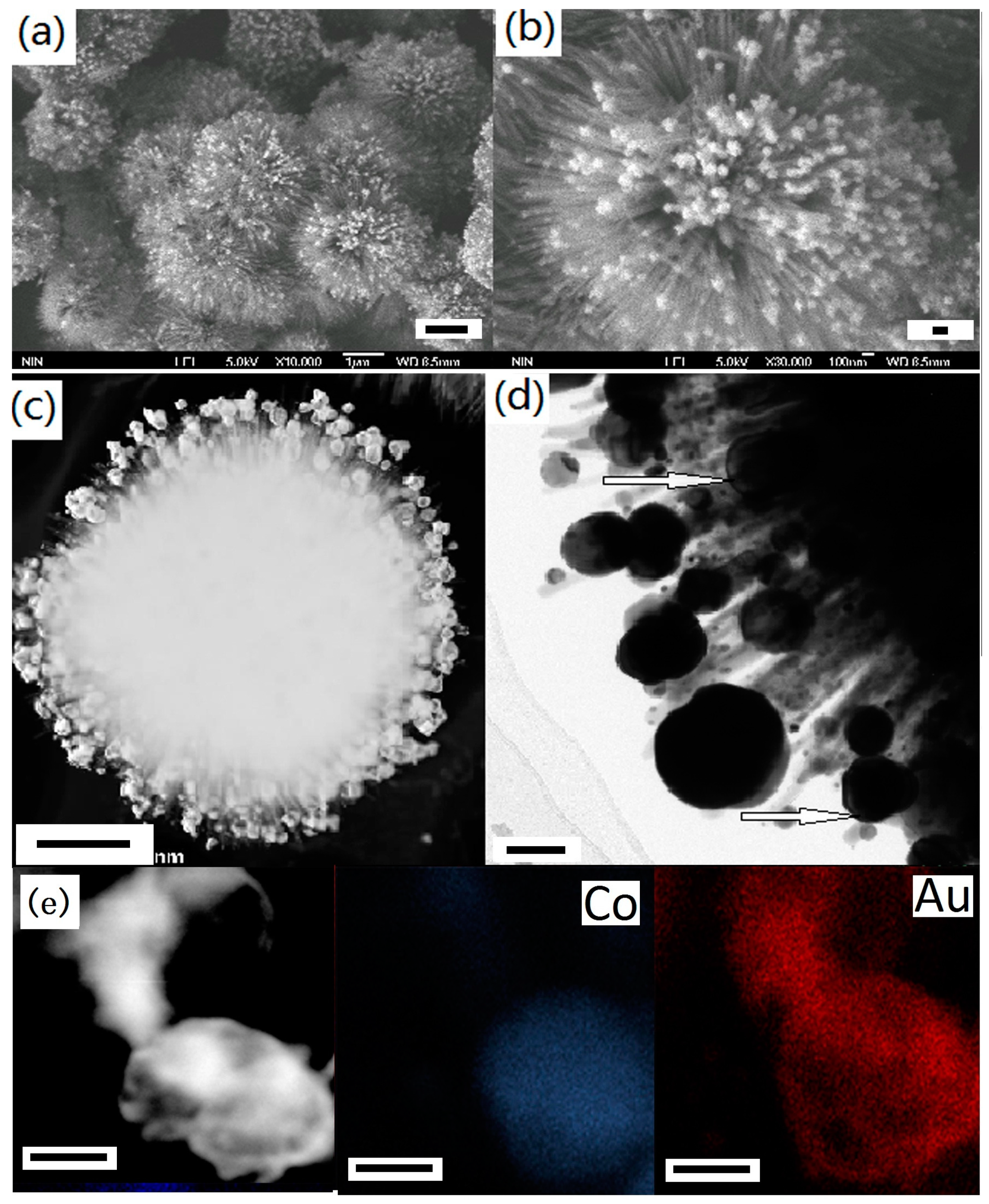
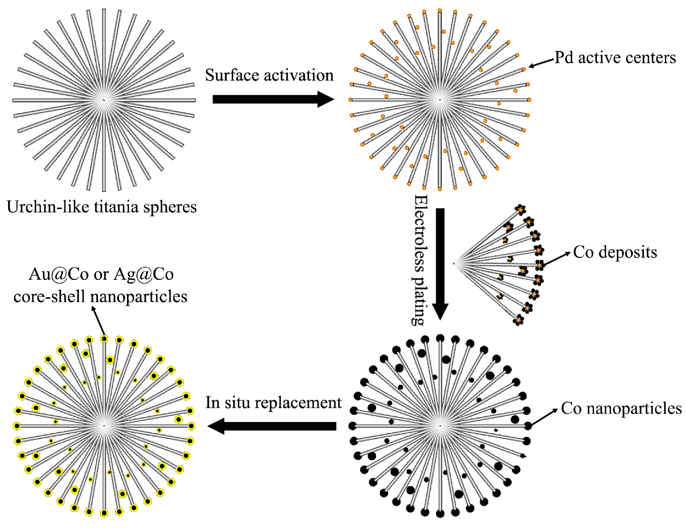
2.2.4. Core/Shell-Structured Bimetallic Nanoparticle-Decorated 3D Urchin-Like Hierarchical TiO2 Particles

Bimetallic nanostructures often show a more excellent comprehensive performance over their monometallic counterpart [80]. Especially core/shell bimetallic nanostructures with a magnetic core and a noble-metallic shell have aroused researchers’ interest [81,82,83,84,85]. The magnetic core can offer a drive force for the recycling of samples, while the noble-metal shell can improve the optical properties [85]. Figure 14 shows a typical preparation process of a kind of 3D urchin-like hierarchical TiO2 decorated with a bimetallic core/shell nanoparticle (Co@Au and Co@Ag). The preparation includes three steps, i.e., surface activation, electroless plating and in-situ replacement [32]. First, the surface of the urchin-like TiO2 microspheres was activated by implanting Pd nanodots. Then, Co nanoparticles were formed and adhered to the nanoneedle surface of the urchin-like TiO2 microspheres by electroless plating. Finally, Ag or Au were further formed and coated on the surface of the Co nanoparticles by an in-situ replacement process.
The SEM and TEM images in Figure 15 show the morphology of Co@Au/TiO2 composites. It can be seen that many core/shell nanostructured nanoparticles with diameters of 10–80 nm are attached to the surface of TiO2 nanorods. The images of the elemental mapping of core/shell nanoparticles further identify that the core is Co and the shell is Au. The size and distribution of the bimetallic particles can be adjusted by controlling the ratio of Co to TiO2 during the electroless plating process. The thickness of the Au or Ag shell could be controlled by adjusting the concentration of HAuCl4 or AgNO3 in the solution and the reaction time. Both the Co@Au/TiO2 and Co@Ag/TiO2 particles showed good response to an applied external magnetic field [32].

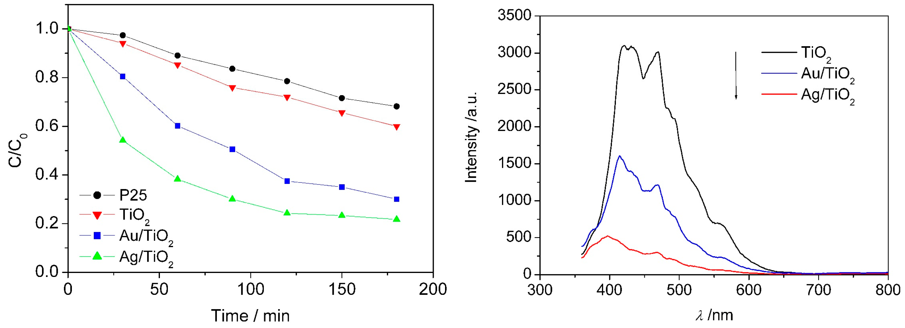
2.2.5. Photocatalytic Activity of Urchin-Like Hierarchical TiO2 and Their Composites

Although TiO2 is an ideal candidate for photocatalysis because of its strong oxidization, harmlessness to surroundings, chemical inactivity, good stability and low cost, the main weakness of TiO2 is the lack of visible light response due to the large band gap. Therefore, the question of how to increase the efficiency of visible light harvesting is an important research topic in this field [1]. Although single controlling the morphology of TiO2 materials cannot increase the efficiency of visible light harvesting, it is possible to enhance the visible light harvesting of TiO2 composites by combining ion doping or noble metal decoration with morphology control. Ion doping or noble metal decoration can induce or increase the visible light absorption of TiO2, while the absorption effect can be further enhanced by other effects from material morphology, such as decreased light scattering or increased multiple reflection, etc.
The photocatalytic efficiency of commercial P25, urchin-like TiO2 and Au or Ag-decorated urchin-like TiO2 was evaluated by degrading MB (methyl blue) under UV-Vis light irradiation. It was found that the photocatalytic degradation efficiency under the same conditions followed the order: Ag/TiO2 > Au/TiO2 > TiO2 > P25 (Figure 16). By the photoluminescence spectra, Au or Ag nanoparticles decorated on the surface of TiO2 were demonstrated to be able to effectively capture photogenerated electrons and prevent electron/hole recombining (Figure 16). In addition, the urchin-like micro/nano hierarchical structure also may increase the visible light harvesting efficiency by the multiple-reflection of nanoneedles. These should be responsible for the enhanced photocatalytic efficiency of urchin-like TiO2 microspheres after decoration with Au or Ag nanoparticles. Similarly, the light-harvesting efficiency could also be enhanced by decorating with Co@Au or Co@Ag bimetallic nanoparticles, as shown in Figure 17. As a result, the photocatalytical efficiency of urchin-like TiO2 was enhanced obviously, as shown in Figure 17, by the experiment of decolorizing methyl blue (MB) solution.

3. Summary

Based on our group’s research work, we provided a brief review of the synthesis of TiO2 with different morphologies and the photocatalytic properties of urchin-like TiO2, noble metal nanoparticle-decorated 3D urchin-like TiO2 and core/shell-structured bimetallic nanoparticle-decorated 3D urchin-like hierarchical TiO2. The examples of the fabrication of solid spheres, hollow spheres, porous spheres, and porous and hollow microspheres of anatase TiO2-based materials were introduced. The synthesis and photocatalytic efficiency of urchin-like rutile TiO2, urchin-like rutile TiO2 nanostructures decorated with Au or Ag nanoparticles and core/shell-structured bimetallic nanoparticles (Co@Au and Co@Ag) were especially introduced. The results of photocatalytic tests show that 3D urchin-like hierarchical structures have unique merits in the efficient harvesting of solar light, and decorating Au, Ag or bimetallic nanoparticles on the surface of 3D urchin-like TiO2 can promote photoinduced charge-carrier separation.

Acknowledgments

This work was supported by the National Natural Science Foundation of China (Grant Nos. 51502247 and 11674267)

Author Contributions

Liqin Xiang and Xiaopeng Zhao wrote the paper.

Conflicts of Interest

The authors declare no conflict of interest.

References

  1. Fattakhova-Rohlfing, D.; Zaleska, A.; Bein, T. Three-Dimensional Titanium Dioxide Nanomaterials. Chem. Rev. 2014, 114, 9487–9558. [Google Scholar] [CrossRef] [PubMed]
  2. Abdullah, N.; Kamarudin, S.K. Titanium dioxide in fuel cell technology: An overview. J. Power Sources 2015, 278, 109–118. [Google Scholar] [CrossRef]
  3. Chen, X.B.; Mao, S.S. Titanium dioxide nanomaterials: Synthesis, properties, modifications, and applications. Chem. Rev. 2007, 107, 2891–2959. [Google Scholar] [CrossRef] [PubMed]
  4. Zhang, G.; Lan, Z.A.; Wang, X. Conjugated Polymers: Catalysts for Photocatalytic Hydrogen Evolution. Angew. Chem. Int. Ed. 2016, 55, 15712–15727. [Google Scholar] [CrossRef] [PubMed]
  5. Kessler, F.K.; Zheng, Y.; Schwarz, D.; Merschjann, C.; Schnick, W.; Wang, X.; Bojdys, M.J. Functional carbon nitride materials-design strategies for electrochemical devices. Nat. Rev. Mater. 2017, 2, 17030. [Google Scholar] [CrossRef]
  6. Zhang, G.; Lan, Z.A.; Wang, X. Surface engineering of graphitic carbon nitride polymers with cocatalysts for photocatalytic overall water splitting. Chem. Sci. 2017, 8, 5261–5274. [Google Scholar] [CrossRef] [PubMed]
  7. Ong, W.J.; Tan, L.L.; Ng, Y.H.; Yong, S.T.; Chai, S.P. Graphitic carbon nitride (g-C3N4)-based photocatalysts for artificial photosynthesis and environmental remediation: Are we a step closer to achieving sustainability? Chem. Rev. 2016, 116, 7159–7329. [Google Scholar] [CrossRef] [PubMed]
  8. Liu, G.; Wang, L.; Yang, H.G.; Cheng, H.M.; Lu, G.Q. TiO2-based photocatalysts-crystal growth, doping and heterostructuring. J. Mater. Chem. 2010, 20, 831–843. [Google Scholar] [CrossRef]
  9. Ruokolainen, M.; Ollikainen, E.; Sikanen, T.; Kotiaho, T.; Kostiainen, R. Oxidation of tyrosine-phosphopeptides by titanium dioxide photocatalysis. J. Am. Chem. Soc. 2016, 138, 7452–7455. [Google Scholar] [CrossRef] [PubMed]
  10. Kisch, H. Semiconductor photocatalysis-mechanistic and synthetic aspects. Angew. Chem. Int. Ed. 2013, 52, 812–847. [Google Scholar] [CrossRef] [PubMed]
  11. Du, J.; Lai, X.; Yang, N.; Zhai, J.; Kisailus, D.; Su, F.; Wang, D.; Jiang, L. Hierarchically Ordered Macro-Mesoporous TiO2-Graphene Composite Films: Improved Mass Transfer, Reduced Charge Recombination, and Their Enhanced Photocatalytic Activities. ACS Nano 2011, 5, 590–596. [Google Scholar] [CrossRef] [PubMed]
  12. Li, Y.F.; Liu, Z.P. Particle size, shape and activity for photocatalysis on TiO2 anatase nanoparticles in aqueous surroundings. J. Am. Chem. Soc. 2011, 133, 15743–15752. [Google Scholar] [CrossRef] [PubMed]
  13. Meulen, T.; Mattson, A.; Oesterlund, L. A comparative study of the photocatalytic oxidation of propane on anatase, rutile, and mixed-phase anatase-rutile TiO2 nanoparticles: Role of surface intermediates. J. Catal. 2007, 25, 131–138. [Google Scholar]
  14. Kang, X.W.; Chen, S.W. Photocatalytic reduction of methylene blue by TiO2 nanotube arrays: Effects of TiO2 crystalline phase. J. Mater. Sci. 2010, 45, 2696–2702. [Google Scholar] [CrossRef]
  15. Tanaka, S.; Nogami, D.; Tsuda, N.; Miyake, Y. Synthesis of highly-monodisperse spherical TiO2 particles with diameters in the submicron range. J. Colloid Interface Sci. 2009, 334, 188–194. [Google Scholar] [CrossRef] [PubMed]
  16. Momeni, M.; Ghayeb, Y. Fabrication, characterization and photoelectrochemical performance of chromium-sensitized TiO2 nanotubes as efficient photoanodes for solar water splitting. J. Solid State Electrochem. 2016, 20, 683–689. [Google Scholar] [CrossRef]
  17. Ghicov, A.; Schmuki, P. Self-ordering electrochemistry: A review on growth and functionality of TiO2 nanotubes and other self-aligned MOx structures. Chem. Commun. 2009, 2791–2808. [Google Scholar] [CrossRef] [PubMed]
  18. Zhang, J.; Zhu, Z.; Tang, Y.; Müllen, K.; Feng, X. TiO2 Nanosheet-Mediated Construction of a Two-Dimensional TiO2/Cadmium Sulfide Heterostructure for High Hydrogen Evolution Activity. Adv. Mater. 2014, 26, 734–738. [Google Scholar] [CrossRef] [PubMed]
  19. Bartl, M.H.; Boettcher, S.W.; Frindell, K.L.; Stucky, G.D. 3-D molecular assembly of function in TiO2-based composite material systems. Acc. Chem. Res. 2005, 38, 263–271. [Google Scholar] [CrossRef] [PubMed]
  20. Zhu, T.; Li, J.; Wu, Q. Construction of TiO2 hierarchical nanostructures from nanocrystals and their photocatalytic properties. ACS Appl. Mater. Interfaces 2011, 3, 3448–3453. [Google Scholar] [CrossRef] [PubMed]
  21. Li, H.; Bian, Z.; Zhu, J.; Zhang, D.; Li, G.; Huo, Y.; Li, H.; Lu, Y. Mesoporous TiO2 spheres with tunable chamber lasmon and enhanced photocatalytic activity. J. Am. Chem. Soc. 2007, 129, 8406–8407. [Google Scholar] [CrossRef] [PubMed]
  22. Choi, W.; Termin, A.; Hoffman, M.R. The role of metal ion dopants in quantum-sized TiO2: Correlation between photoreactivity and charge carrier recombination dynamics. J. Phys. Chem. 1994, 98, 13669–13679. [Google Scholar] [CrossRef]
  23. Su, R.; Tiruvalam, R.; He, Q.; Dimitratos, N.; Kesavan, L.; Hammond, C.; Lopez-Sanchez, J.A.; Bechstein, R.; Kiely, C.J.; Hutchings, G.J.; et al. Promotion of phenol photodecomposition over TiO2 using Au, Pd, and AuPd nanoparticles. ACS Nano 2012, 6, 6284–6292. [Google Scholar] [CrossRef] [PubMed]
  24. Smirnova, N.; Vorobets, V.; Linnik, O.; Manuilov, E.; Kolbasovb, G.; Eremenkoa, A. Photoelectrochemical and photocatalytic properties of mesoporous TiO2 films modified with silver and gold nanoparticles. Surf. Interface Anal. 2010, 42, 1205–1208. [Google Scholar] [CrossRef]
  25. Arabtzis, I.M.; Stergiopoulos, T.; Andreeva, D.; Kitova, S.; Neophytides, S.G.; Falaras, P. Charaterizarion and photocatalytic activity of Au/TiO2 thin films for azo-dye degradation. J. Catal. 2003, 220, 127–135. [Google Scholar] [CrossRef]
  26. Yin, J.B.; Zhao, X.P. Preparation and Enhanced Electrorheological Activity of TiO2 Doped with Chromium Ion. Chem. Mater. 2004, 16, 321–328. [Google Scholar] [CrossRef]
  27. Zhao, X.P.; Duan, X. In situ sol-gel preparation of polysaccharide/titanium oxide hybrid colloids and their electrorheological effect. J. Colloid Interface Sci. 2002, 251, 376–380. [Google Scholar] [CrossRef] [PubMed]
  28. Yin, J.B.; Xiang, L.Q.; Zhao, X.P. Monodisperse spherical mesoporous Eu-doped TiO2 phosphor particles and the luminescence properties. Appl. Phys. Lett. 2007, 90, 113112. [Google Scholar] [CrossRef]
  29. An, G.F.; Yang, C.S.; Jin, S.H.; Chen, G.W.; Zhao, X.P. Hollow TiO2:Sm3+ spheres with enhanced photoluminescence fabricated by a facile method using polystyrene as template. J. Mater. Sci. 2013, 48, 5483–5488. [Google Scholar] [CrossRef]
  30. Xiang, L.Q.; Zhao, X.P.; Yin, J.B.; Fan, B.L. Well-organized 3-D urchin-like multilevel TiO2 microspheres with high photocatalytic activity. J. Mater. Sci. 2012, 47, 1436–1445. [Google Scholar] [CrossRef]
  31. Xiang, L.Q.; Zhao, X.P.; Shang, C.H.; Yin, J.B. Au or Ag nanoparticle-decorated 3D urchin-like TiO2 nanostructures: Synthesis, characterization, and enhanced photocatalytic activity. J. Colloid Interface Sci. 2013, 403, 22–28. [Google Scholar] [CrossRef] [PubMed]
  32. Xiang, L.Q.; Liu, S.; Yin, J.B.; Zhao, X.P. Bimetallic core/shell nanoparticle-decorated 3D urchin-like hierarchical TiO2 nanostructures with magneto-responsive and decolorization characteristics. Nanoscale Res. Lett. 2015, 10, 84. [Google Scholar] [CrossRef] [PubMed]
  33. Yin, J.B.; Zhao, X.P. Titanate nano-whisker electrorheological fluid with high suspended stability and ER activity. Nanotechnology 2006, 17, 192–196. [Google Scholar] [CrossRef]
  34. Guo, H.X.; Zhao, X.P.; Ning, G.H.; Liu, G.Q. Synthesis of Ni/Polystyrene/TiO2 multiply coated microsphere. Langmuir 2003, 19, 4884–4888. [Google Scholar] [CrossRef]
  35. Guo, H.X.; Zhao, X.P.; Guo, H.L.; Zhao, Q. Preparation of porous SiO2/Ni/TiO2 multiply microsphere responsive to electric and magnetic fields. Langmuir 2003, 19, 9799–9803. [Google Scholar] [CrossRef]
  36. Yin, J.B.; Zhao, X.P. Electrorheological fluids based on glycerol-activated TiO2 gel and silicone oil with high yield strength. J. Colloid Interface Sci. 2003, 257, 228–236. [Google Scholar] [CrossRef]
  37. Xiang, L.Q.; Yin, J.B.; Gao, W.S.; Zhao, X.P. Controllable Preparation of Quasi-monodispersed Spherical TiO2 Particles. J. Inorg. Mater. Chin. 2007, 22, 253–258. [Google Scholar]
  38. Ma, L.M.; Zheng, F.X.; Zhao, X.P. Sedimentation behaviour of hierarchical porous TiO2 microspheres electrorheological fluids. J. Intel. Mater. Syst. Struct. 2015, 26, 1936–1944. [Google Scholar] [CrossRef]
  39. Wang, J.H.; Chen, G.W.; Yin, J.B.; Luo, C.R.; Zhao, X.P. Enhanced electrorheological performance and antisedimentation property of mesoporous anatase TiO2 shell prepared by hydrothermal process. Smart Mater. Struct. 2017, 26, 035036. [Google Scholar] [CrossRef]
  40. Wang, B.X.; Zhao, X.P. Core/Shell Nanocomposite Based on the Local Polarization and Its Electrorheological Behavior. Langmuir 2005, 21, 6553–6559. [Google Scholar] [CrossRef] [PubMed]
  41. Yin, J.B.; Xia, X.; Wang, X.X.; Zhao, X.P. The electrorheological effect and dielectric properties of suspensions containing polyaniline@TiO2 nanocable-like particles. Soft Matter 2011, 7, 10978–10986. [Google Scholar] [CrossRef]
  42. Xiang, L.Q.; Zhao, X.P. Electrorheological activity of a composite of TiO2-Coated Montmorillonite. J. Mater. Chem. 2003, 13, 1529–1532. [Google Scholar] [CrossRef]
  43. Wang, B.X.; Zhao, X.P. Preparation of kaolinite/TiO2 coated nanocomposite and their electro-rheological properties. J. Mater. Chem. 2003, 13, 2248–2253. [Google Scholar] [CrossRef]
  44. Zhao, X.P.; Yin, J.B. Preparation and electrorheological characteristics of rare-earth-doped TiO2 suspensions. Chem. Mater. 2002, 14, 2258–2263. [Google Scholar] [CrossRef]
  45. Zhao, X.P.; Yin, J.B.; Xiang, L.Q.; Zhao, Q. Electrorheological fluids containing Ce-doped TiO2. J. Mater. Sci. 2002, 37, 2569–2573. [Google Scholar] [CrossRef]
  46. Zhao, X.P.; Yin, J.B.; Xiang, L.Q.; Zhao, Q. Effect of rare earth substitution on electrorheological properties of TiO2. Int. J. Mod. Phys. B 2002, 16, 2371–2377. [Google Scholar] [CrossRef]
  47. Yin, J.B.; Zhao, X.P. Temperature effect of rare earth-doped TiO2 Electrorheological fluids. J. Phys. D Appl. Phys. 2001, 34, 2063–2067. [Google Scholar] [CrossRef]
  48. Yin, J.B.; Zhao, X.P. Electrorheological effect of Cerium-Doped TiO2. Chin. Phys. Lett. 2001, 18, 1144–1146. [Google Scholar]
  49. Yin, J.B.; Zhao, X.P. Enhanced electrorheological activity of mesoporous Cr-doped TiO2 from activated pore wall and high surface area. J. Phys. Chem. B 2006, 110, 12916–12925. [Google Scholar] [CrossRef] [PubMed]
  50. Wang, B.X.; Zhao, X.P. A bionic nano-papilla particle and its super-oleophilic ability. Adv. Funct. Mater. 2005, 15, 1815–18212. [Google Scholar] [CrossRef]
  51. Xiang, L.Q.; Zhao, X.P.; Yin, J.B. Micro/nano-structured montmorillonite/TiO2 particles with high electrorheological activity. Rheol. Acta 2011, 50, 87–95. [Google Scholar] [CrossRef]
  52. Qiao, Y.P.; Zhao, X.P.; Su, Y.Y. Dielectric metamaterial particles with enhanced efficiency of mechanical/electrical energy transformation. J. Mater. Chem. 2011, 21, 394–399. [Google Scholar] [CrossRef]
  53. Eiden-Assmann, S.; Widoniak, J.; Maret, G. Synthesis and Characterization of Porous and Nonporous Monodisperse Colloidal TiO2 Particles. Chem. Mater. 2004, 16, 6–11. [Google Scholar] [CrossRef]
  54. Zhao, Y.; Jiang, L. Hollow Micro/Nanomaterials with Multilevel Interior Structures. Adv. Mater. 2009, 21, 3621–3638. [Google Scholar] [CrossRef]
  55. Réti, B.; Kiss, G.I.; Gyulavári, T.; Baan, K.; Magyari, K.; Hernadi, K. Carbon sphere templates for TiO2 hollow structures: Preparation, characterization and photocatalytic activity. Catal. Today 2017, 284, 160–168. [Google Scholar] [CrossRef]
  56. Orellana, M.; Osiglio, L.; Arnal, P.M.; Pizzio, L.R. TiO2 hollow spheres modified with tungstophosphoric acid with enhanced visible light absorption for the photodegradation of 4-chlorophenol. Photochem. Photobiol. Sci. 2017, 16, 46–52. [Google Scholar] [CrossRef] [PubMed]
  57. Liu, Y.; Lan, K.; Bagabas, A.A.; Zhang, P.; Gao, W.; Wang, J.; Sun, Z.; Fan, J.; Elzatahry, A.A.; Zhao, D. Ordered Macro/Mesoporous TiO2 Hollow Microspheres with Highly Crystalline Thin Shells for High-Efficiency Photoconversion. Small 2016, 12, 860–867. [Google Scholar] [CrossRef] [PubMed]
  58. Li, Y.; Fu, Z.Y.; Su, B.L. Hierarchically Structured Porous Materials for Energy Conversion and Storage. Adv. Funct. Mater. 2012, 22, 4634–4667. [Google Scholar] [CrossRef]
  59. Kim, Y.J.; Lee, M.H.; Kim, H.J.; Lim, G.; Choi, Y.S.; Park, N.G.; Kim, K.; Lee, W.I. Formation of Highly Efficient Dye-Sensitized Solar Cells by Hierarchical Pore Generation with Nanoporous TiO2 Spheres. Adv. Mater. 2009, 21, 3668–3673. [Google Scholar] [CrossRef]
  60. Ismail, A.A.; Bahnemann, D.W. mesoporous TiO2 photocatalysts: Preparation, characterization and reaction mechanisms. J. Mater. Chem. 2011, 21, 11686–11707. [Google Scholar] [CrossRef]
  61. Li, S.; Zheng, J.; Zhao, Y.; Liu, Y. Preparation of a three-dimensional ordered macroporous titanium dioxide material with polystyrene colloid crystal as a template. J. Appl. Polym. Sci. 2008, 107, 3903–3908. [Google Scholar] [CrossRef]
  62. Hegazy, A.; Prouzet, E. Room temperature synthesis and thermal evolution of porous nanocrystalline TiO2 anatase. Chem. Mater. 2012, 24, 245–254. [Google Scholar] [CrossRef]
  63. Zheng, X.; Lv, Y.; Kuang, Q.; Zhu, Z.; Long, X.; Yang, S. Close-Packed Colloidal SiO2 as a Nanoreactor: Generalized Synthesis of Metal Oxide Mesoporous Single Crystals and Mesocrystals. Chem. Mater. 2014, 26, 5700–5709. [Google Scholar] [CrossRef]
  64. Yin, J.B.; Zhao, X.P. Preparation and Electrorheological Activity of Mesoporous Rare-Earth-Doped TiO2. Chem. Mater. 2002, 14, 4633–4640. [Google Scholar] [CrossRef]
  65. Yin, J.B.; Zhao, X.P. Wormhole-like mesoporous Ce-doped TiO2: A new electrorheological material with high activity. J. Mater. Chem. 2003, 13, 689–695. [Google Scholar] [CrossRef]
  66. Joo, J.B.; Lee, I.; Dahl, M.; Moon, G.D.; Zaera, F.; Yin, Y. Controllable Synthesis of Mesoporous TiO2 Hollow Shells: Toward an Efficient Photocatalyst. Adv. Funct. Mater. 2013, 23, 4246–4254. [Google Scholar] [CrossRef]
  67. Bian, Z.F.; Zhu, J.; Cao, F.L.; Huo, Y.N.; Lu, Y.F.; Li, H.X. Solvothermal synthesis of well-defined TiO2 mesoporous nanotubes with enhanced photocatalytic activity. Chem. Commun. 2010, 46, 8451–8453. [Google Scholar] [CrossRef] [PubMed]
  68. Seisenbaeva, G.A.; Moloney, M.P.; Tekoriute, R.; Hardy-Dessources, A.; Nedelec, J.M.; Gun’ko, Y.K.; Kessler, V.G. Biomimetic Synthesis of Hierarchically Porous Nanostructured Metal Oxide Microparticles-Potential Scaffolds for Drug Delivery and Catalysis. Langmuir 2010, 26, 9809–9817. [Google Scholar] [CrossRef] [PubMed]
  69. Wang, C.; Yin, L.; Zhang, L.; Qi, Y.; Lun, N.; Liu, N. Large scale synthesis and gas-sensing properties of anatase TiO2 Three-dimensional hierarchical nanostructures. Langmuir 2010, 26, 12841–12848. [Google Scholar] [CrossRef] [PubMed]
  70. Rui, Y.; Wang, L.; Zhao, J.; Wang, H.; Li, Y.; Zhang, Q.; Xu, J. Template-free synthesis of hierarchical TiO2 hollow microspheres as scattering layer for dye-sensitized solar cells. Appl. Surf. Sci. 2016, 369, 170–177. [Google Scholar] [CrossRef]
  71. Zhang, D.; Li, G.; Wang, F.; Yu, J.C. Green synthesis of a self-assembled rutile mesocrystalline photocatalyst. CrystEngComm 2010, 12, 1759–1763. [Google Scholar] [CrossRef]
  72. Liu, G.; Yang, H.G.; Sun, C.; Cheng, L.; Wang, L.; Lu, G.Q.; Cheng, H.M. Titania polymorphs derived from crystalline titanium diboride. CrystEngComm 2009, 11, 2677–2682. [Google Scholar] [CrossRef]
  73. Yin, J.B.; Zhao, X.P.; Xiang, L.Q.; Xia, X.; Zhang, Z.S. Enhanced electrorheology of suspensions containing sea-urchin-like hierarchical Cr-doped TiO2 particles. Soft Matter 2009, 5, 4687–4697. [Google Scholar] [CrossRef]
  74. Vaneski, A.; Susha, A.S.; Rodríguez-Fernández, J.; Berr, M.; Jäckel, F.; Feldmann, J.; Rogach, A.L. Hybrid colloidal heterostructures of anisotropic semiconductor nanocrystals decorated with noble metals: Synthesis and function. Adv. Funct. Mater. 2011, 21, 1547–1556. [Google Scholar] [CrossRef]
  75. Kochuveedu, S.T.; Kim, D.P.; Kim, D.H. Surface lasmon induced visible light photocatalytic activity of TiO2 nanospheres decorated by Au nanoparticles with controlled configuration. J. Phys. Chem. C 2012, 116, 2500–2506. [Google Scholar] [CrossRef]
  76. Grigorieva, A.V.; Goodilin, E.A.; Derlyukova, L.E.; Anufrieva, T.A.; Tarasov, A.B.; Dobrovolskii, Y.A.; Tretyakov, Y.D. TiO2 nanotubes supported platinum catalyst in CO oxidation process. Appl. Catal. A Gen. 2009, 362, 20–25. [Google Scholar] [CrossRef]
  77. Kafizas, A.; Dunnil, C.W.; Parkin, I.P. The relationship between photocatalytic activity and Photochromic state of nanoparticulate silver surface loaded titanium dioxide thin-films. Phys. Chem. Chem. Phys. 2011, 13, 13827–13838. [Google Scholar] [CrossRef] [PubMed]
  78. Wodka, D.; Bielanska, E.; Socha, R.P.; Elzbieciak-Wodka, M.; Gurgul, J.; Nowak, P.; Warszynski, P.; Kumakiri, I. Photocatalytic activity of titanium dioxide modified by silver nanoparticles. ACS Appl. Mater. Interfaces 2010, 2, 1945–1953. [Google Scholar] [CrossRef] [PubMed]
  79. Zhang, N.; Liu, S.; Fu, X.; Xu, Y.J. Selective Pt deposition onto the face (110) of TiO2 assembled microspheres that substantially enhances the photocatalytic properties. J. Phys. Chem. C 2011, 115, 9136–9145. [Google Scholar] [CrossRef]
  80. Feng, L.L.; Wu, X.C.; Ren, L.R.; Xiang, Y.J.; He, W.W.; Zhang, K.; Zhou, W.Y.; Xie, S.S. Well-controlled synthesis of Au@Pt nanostructures by gold-nanorod-seeded growth. Chem. Eur. J. 2008, 14, 9764–9771. [Google Scholar] [CrossRef] [PubMed]
  81. Levin, C.S.; Hofmann, C.; Ali, T.A.; Kelly, A.T.; Morosan, E.; Nordlander, P.; Whitmire, K.H.; Halas, N.J. Magnetic-plasmonic core-shell nanoparticles. J. Am. Chem. Soc. 2009, 3, 1379–1388. [Google Scholar] [CrossRef] [PubMed]
  82. Chen, L.Y.; Fujita, T.; Ding, Y.; Chen, M.W. A Three-dimensional gold-decorated nanoporous copper core-shell composite for electrocatalysis and nonenzymatic biosensing. Adv. Funct. Mater. 2010, 20, 2279–2285. [Google Scholar] [CrossRef]
  83. Xuan, S.; Xiang, Y.; Wang, J.; Yu, J.C.; Leung, K.C. Preparation, Characterization, and Catalytic Activity of Core/Shell Fe3O4@Polyaniline@Au Nanocomposites. Langmuir 2009, 25, 11835–11843. [Google Scholar] [CrossRef] [PubMed]
  84. Wang, L.; Clavero, C.; Huba, Z.; Carroll, K.J.; Carpenter, E.E.; Gu, D.F.; Lukaszew, R.A. Plasmonics and Enhanced Magneto-Optics in Core-Shell Co-Ag Nanoparticles. Nano Lett. 2011, 11, 1237–1240. [Google Scholar] [CrossRef] [PubMed]
  85. Lu, Y.; Zhao, Y.; Yu, L.; Dong, L.; Shi, C.; Hu, M.J.; Xu, Y.J.; Wen, L.P.; Yu, S.H. Hydrophilic Co@Au Yolk/Shell Nanospheres: Synthesis, Assembly, and Application to Gene Delivery. Adv. Mater. 2010, 22, 1407–1411. [Google Scholar] [CrossRef] [PubMed]
Figure 1. SEM (Scanning electron microscopy) images ((a) PEG (polyethylene glycol) 20000, (b) ODA (octadecylamine), (c) F-127, (d) Span80) and the XRD (X-ray diffraction) patterns (e) of TiO2 spheres synthesized by adding different polymers [37].
Nanomaterials 07 00310 g001
Figure 2. Schematic illustration of the formation mechanism for hollow TiO2:Sm3+ spheres [29].
Nanomaterials 07 00310 g002
Figure 3. (a) SEM image, (b) TEM (Transmission Electron Microscopy) image and (c) EDS (Energy Dispersive Spectrum) of TiO2:Sm3+ hollow spheres [29].
Nanomaterials 07 00310 g003
Figure 4. TEM photograph of mesoporous-doped TiO2 and XRD patterns (a) before hydrothermal and acid treatment, (b) after hydrothermal and acid treatment of mesoporous-doped TiO2 [64].
Nanomaterials 07 00310 g004
Figure 5. (a) SEM image and EDS spectra, (b) TEM image, (c) high resolution TEM image and corresponding electron diffraction pattern of monodisperse mesoporous after 400 °C calcinations, (d) XRD patterns: (i) before calcinations, (ii) after 400 °C calcinations, and (iii) after 500 °C calcinations [28].
Nanomaterials 07 00310 g005
Figure 6. Schematic illustration of the process for the fabrication of the mesoporous TiO2 hollow microspheres [39].
Nanomaterials 07 00310 g006
Figure 7. TEM images (a,b), XRD patterns (e-i) and nitrogen adsorption-desorption isotherms (f-i) of the sample prepared by the hydrothermal process; TEM images (c,d), XRD patterns (e-ii) and nitrogen adsorption-desorption isotherms (f-ii) of the sample prepared by the calcination process [39].
Nanomaterials 07 00310 g007
Figure 8. SEM image (a), TEM images (be), and EDS (f) of the urchin-like hierarchical TiO2 [30].
Nanomaterials 07 00310 g008
Figure 9. Schematic illustration of the formation process of 3D urchin-like hierarchical TiO2: (a) nanoparticle; (b) microsphere; (c) similar urchin-like sphere; (d) urchin-like sphere [30].
Nanomaterials 07 00310 g009
Figure 10. TEM images and EDS spectra of Cr-doped urchin-like TiO2 particles [73].
Nanomaterials 07 00310 g010
Figure 11. Schematic illustration of the synthesis process of urchin-like TiO2 decorated with Au or Ag nanoparticles [31].
Nanomaterials 07 00310 g011
Figure 12. SEM images (a,b), TEM images (ce), XRD pattern (f) and UV-Vis absorption spectra (g) of Au-decorated 3D urchin-like TiO2 nanostructures [31].
Nanomaterials 07 00310 g012
Figure 13. TEM images (ac), EDS (d), XRD (e), and UV-Vis absorption spectrum (f) of Ag-decorated 3D urchin-like TiO2 nanostructures [31].
Nanomaterials 07 00310 g013
Figure 14. A schematic synthesis process of urchin-like TiO2 decorated with core/shell-structured Co@Au or Co@Ag bimetallic nanoparticles [32].
Nanomaterials 07 00310 g014
Figure 15. The SEM and TEM images of Co@Au/TiO2 composites: (a,b) SEM images, (c,d) TEM images, (e) high-resolution TEM images; (e) the local elemental mapping of Co and Au (Scale bar = 1 μm for (a); scale bar = 100 nm for (b); scale bar = 500 nm for (c); scale bar = 100 nm for (d); scale bar = 50 nm for (e)) [32].
Nanomaterials 07 00310 g015
Figure 16. Photodegradation curves of MB (methyl blue) in the presence of P25, TiO2, Au/TiO2 and Ag/TiO2 (left); Photoluminence spectra (λex = 215 nm) of pure urchin-like TiO2, Au/TiO2 and Ag/TiO2 (right) [31].
Nanomaterials 07 00310 g016
Figure 17. UV-Vis absorption spectra of TiO2, Co@Au/TiO2 and Co@Ag/TiO2 (left); UV-Vis absorption spectra of MB before and after degradation with TiO2, Co@Au/TiO2 and Co@Ag/TiO2 for 15 min at room temperature (right) [32].
Nanomaterials 07 00310 g017

