Next Issue
Volume 7, September
Previous Issue
Volume 7, July


Nanomaterials, Volume 7, Issue 8 (August 2017) – 37 articles

Cover Story (view full-size image): 
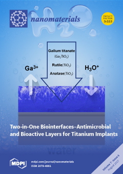
We developed novel biointerfaces for titanium implants, which simultaneously and uniquely show very high antimicrobial activity and bioactivity. Our interfaces are composed of gallium titanate (GT) accompanied by rutile and anatase. They slowly release 3.75 ppm of Ga ions for up to 7 days and formed apatite within 3 days. The antibacterial activity was shown on the GT. The multifunctional interfaces will be particularly useful for orthopaedic and dental implants since it will promote desired implant integration, while preventing infections and inhibiting bone resorption. View Paper here
  • Issues are regarded as officially published after their release is announced to the table of contents alert mailing list.
  • You may sign up for e-mail alerts to receive table of contents of newly released issues.
  • PDF is the official format for papers published in both, html and pdf forms. To view the papers in pdf format, click on the "PDF Full-text" link, and use the free Adobe Reader to open them.
Order results
Result details
Section
Select all
Export citation of selected articles as:
7592 KiB  
Article
Strong Deep-Level-Emission Photoluminescence in NiO Nanoparticles
by Ashish Chhaganlal Gandhi and Sheng Yun Wu
Nanomaterials 2017, 7(8), 231; https://doi.org/10.3390/nano7080231 - 22 Aug 2017
Cited by 130 | Viewed by 7318
Abstract
Nickel oxide is one of the highly promising semiconducting materials, but its large band gap (3.7 to 4 eV) limits its use in practical applications. Here we report the effect of nickel/oxygen vacancies and interstitial defects on the near-band-edge (NBE) and deep-level-emission (DLE) [...] Read more.
Nickel oxide is one of the highly promising semiconducting materials, but its large band gap (3.7 to 4 eV) limits its use in practical applications. Here we report the effect of nickel/oxygen vacancies and interstitial defects on the near-band-edge (NBE) and deep-level-emission (DLE) in various sizes of nickel oxide (NiO) nanoparticles. The ultraviolet (UV) emission originated from excitonic recombination corresponding near-band-edge (NBE) transition of NiO, while deep-level-emission (DLE) in the visible region due to various structural defects such as oxygen vacancies and interstitial defects. We found that the NiO nanoparticles exhibit a strong green band emission around ~2.37 eV in all samples, covering 80% integrated intensity of PL spectra. This apparently anomalous phenomenon is attributed to photogenerated holes trapped in the deep level oxygen vacancy recombining with the electrons trapped in a shallow level located just below the conducting band. Full article
(This article belongs to the Special Issue Semiconductor Nanoparticles for Electric Device Applications)
Show Figures

Graphical abstract

8616 KiB  
Article
The Effects of Silica Nanoparticles on Apoptosis and Autophagy of Glioblastoma Cell Lines
by Rafał Krętowski, Magdalena Kusaczuk, Monika Naumowicz, Joanna Kotyńska, Beata Szynaka and Marzanna Cechowska-Pasko
Nanomaterials 2017, 7(8), 230; https://doi.org/10.3390/nano7080230 - 21 Aug 2017
Cited by 60 | Viewed by 7543
Abstract
Silica nanoparticles (SiNPs) are one of the most commonly used nanomaterials in various medical applications. However, possible mechanisms of the toxicity caused by SiNPs remain unclear. The study presented here provides novel information on molecular and cellular effects of SiNPs in glioblastoma LBC3 [...] Read more.
Silica nanoparticles (SiNPs) are one of the most commonly used nanomaterials in various medical applications. However, possible mechanisms of the toxicity caused by SiNPs remain unclear. The study presented here provides novel information on molecular and cellular effects of SiNPs in glioblastoma LBC3 and LN-18 cells. It has been demonstrated that SiNPs of 7 nm, 5–15 nm and 10–20 nm induce time- and dose-dependent cytotoxicity in LBC3 and LN-18 cell lines. In contrast to glioblastoma cells, we observed only weak reduction in viability of normal skin fibroblasts treated with SiNPs. Furthermore, in LBC3 cells treated with 5–15 nm SiNPs we noticed induction of apoptosis and necrosis, while in LN-18 cells only necrosis. The 5–15 nm SiNPs were also found to cause oxidative stress, a loss in mitochondrial membrane potential, and changes in the ultrastructure of the mitochondria in LBC3 cells. Quantitative real-time PCR results showed that in LBC3 cells the mRNA levels of pro-apoptotic genes Bim, Bax, Puma, and Noxa were significantly upregulated. An increase in activity of caspase-9 in these cells was also observed. Moreover, the activation of SiNP-induced autophagy was demonstrated in LBC3 cells as shown by an increase in LC3-II/LC3-I ratio, the upregulation of Atg5 gene and an increase in AVOs-positive cells. In conclusion, this research provides novel information concerning molecular mechanisms of apoptosis and autophagy in LBC3 cells. Full article
(This article belongs to the Special Issue Frontiers in Toxicity and Functionalization of Nanomaterials)
Show Figures

Graphical abstract

2992 KiB  
Article
Facile synthesis of Mesoporouscobalt Hexacyanoferrate Nanocubes for High-Performance Supercapacitors
by Zhiyong Zhang, Jian-Gan Wang and Bingqing Wei
Nanomaterials 2017, 7(8), 228; https://doi.org/10.3390/nano7080228 - 21 Aug 2017
Cited by 14 | Viewed by 5417
Abstract
Mesoporous cobalt hexacyanoferrate nanocubes (meso–CoHCF) were prepared for the first time through a facile sacrificial template method. The CoHCF mesostructures possess a high specific surface area of 548.5 m2·g−1 and a large amount of mesopores, which enable fast mass transport [...] Read more.
Mesoporous cobalt hexacyanoferrate nanocubes (meso–CoHCF) were prepared for the first time through a facile sacrificial template method. The CoHCF mesostructures possess a high specific surface area of 548.5 m2·g−1 and a large amount of mesopores, which enable fast mass transport of electrolyte and abundant energy storage sites. When evaluated as supercapacitor materials, the meso–CoHCF materials exhibit a high specific capacitance of 285 F·g−1, good rate capability and long cycle life with capacitance retention of 92.9% after 3000 cycles in Na2SO4 aqueous electrolyte. The excellent electrochemical properties demonstrate the rational preparation of mesoporous prussian blue and its analogues for energy storage applications. Full article
(This article belongs to the Special Issue Nanomaterials Based Fuel Cells and Supercapacitors)
Show Figures

Figure 1

3812 KiB  
Article
3D Nanoporous Anodic Alumina Structures for Sustained Drug Release
by Maria Porta-i-Batalla, Elisabet Xifré-Pérez, Chris Eckstein, Josep Ferré-Borrull and Lluis F. Marsal
Nanomaterials 2017, 7(8), 227; https://doi.org/10.3390/nano7080227 - 21 Aug 2017
Cited by 25 | Viewed by 4623
Abstract
The use of nanoporous anodic alumina (NAA) for the development of drug delivery systems has gained much attention in recent years. The release of drugs loaded inside NAA pores is complex and depends on the morphology of the pores. In this study, NAA, [...] Read more.
The use of nanoporous anodic alumina (NAA) for the development of drug delivery systems has gained much attention in recent years. The release of drugs loaded inside NAA pores is complex and depends on the morphology of the pores. In this study, NAA, with different three-dimensional (3D) pore structures (cylindrical pores with several pore diameters, multilayered nanofunnels, and multilayered inverted funnels) were fabricated, and their respective drug delivery rates were studied and modeled using doxorubicin as a model drug. The obtained results reveal optimal modeling of all 3D pore structures, differentiating two drug release stages. Thus, an initial short-term and a sustained long-term release were successfully modeled by the Higuchi and the Korsmeyer–Peppas equations, respectively. This study demonstrates the influence of pore geometries on drug release rates, and further presents a sustained long-term drug release that exceeds 60 days without an undesired initial burst. Full article
(This article belongs to the Special Issue Electrochemically Engineering of Nanoporous Materials)
Show Figures

Graphical abstract

5252 KiB  
Article
Two-in-One Biointerfaces—Antimicrobial and Bioactive Nanoporous Gallium Titanate Layers for Titanium Implants
by Seiji Yamaguchi, Shekhar Nath, Yoko Sugawara, Kamini Divakarla, Theerthankar Das, Jim Manos, Wojciech Chrzanowski, Tomiharu Matsushita and Tadashi Kokubo
Nanomaterials 2017, 7(8), 229; https://doi.org/10.3390/nano7080229 - 20 Aug 2017
Cited by 45 | Viewed by 6060
Abstract
The inhibitory effect of gallium (Ga) ions on bone resorption and their superior microbial activity are attractive and sought-after features for the vast majority of implantable devices, in particular for implants used for hard tissue. In our work, for the first time, Ga [...] Read more.
The inhibitory effect of gallium (Ga) ions on bone resorption and their superior microbial activity are attractive and sought-after features for the vast majority of implantable devices, in particular for implants used for hard tissue. In our work, for the first time, Ga ions were successfully incorporated into the surface of titanium metal (Ti) by simple and cost-effective chemical and heat treatments. Ti samples were initially treated in NaOH solution to produce a nanostructured sodium hydrogen titanate layer approximately 1 μm thick. When the metal was subsequently soaked in a mixed solution of CaCl2 and GaCl3, its Na ions were replaced with Ca and Ga ions in a Ga/Ca ratio range of 0.09 to 2.33. 8.0% of the Ga ions were incorporated into the metal surface when the metal was soaked in a single solution of GaCl3 after the NaOH treatment. The metal was then heat-treated at 600 °C to form Ga-containing calcium titanate (Ga–CT) or gallium titanate (GT), anatase and rutile on its surface. The metal with Ga–CT formed bone-like apatite in a simulated body fluid (SBF) within 3 days, but released only 0.23 ppm of the Ga ions in a phosphate-buffered saline (PBS) over a period of 14 days. In contrast, Ti with GT did not form apatite in SBF, but released 2.96 ppm of Ga ions in PBS. Subsequent soaking in hot water at 80 °C dramatically enhanced apatite formation of the metal by increasing the release of Ga ions up to 3.75 ppm. The treated metal exhibited very high antibacterial activity against multidrug resistant Acinetobacter baumannii (MRAB12). Unlike other antimicrobial coating on titanium implants, Ga–CT and GT interfaces were shown to have a unique combination of antimicrobial and bioactive properties. Such dual activity is essential for the next generation of orthopaedic and dental implants. The goal of combining both functions without inducing cytotoxicity is a major advance and has far reaching translational perspectives. This unique dual-function biointerfaces will inhibit bone resorption and show antimicrobial activity through the release of Ga ions, while tight bonding to the bone will be achieved through the apatite formed on the surface. Full article
(This article belongs to the Special Issue Frontiers in Toxicity and Functionalization of Nanomaterials)
Show Figures

Graphical abstract

22510 KiB  
Article
Wettability Investigations and Wet Transfer Enhancement of Large-Area CVD-Graphene on Aluminum Nitride
by Marius Knapp, René Hoffmann, Volker Cimalla and Oliver Ambacher
Nanomaterials 2017, 7(8), 226; https://doi.org/10.3390/nano7080226 - 18 Aug 2017
Cited by 8 | Viewed by 7898
Abstract
The two-dimensional and virtually massless character of graphene attracts great interest for radio frequency devices, such as surface and bulk acoustic wave resonators. Due to its good electric conductivity, graphene might be an alternative as a virtually massless electrode by improving resonator performance [...] Read more.
The two-dimensional and virtually massless character of graphene attracts great interest for radio frequency devices, such as surface and bulk acoustic wave resonators. Due to its good electric conductivity, graphene might be an alternative as a virtually massless electrode by improving resonator performance regarding mass-loading effects. We report on an optimization of the commonly used wet transfer technique for large-area graphene, grown via chemical vapor deposition, onto aluminum nitride (AlN), which is mainly used as an active, piezoelectric material for acoustic devices. Today, graphene wet transfer is well-engineered for silicon dioxide (SiO2). Investigations on AlN substrates reveal highly different surface properties compared to SiO2 regarding wettability, which strongly influences the quality of transferred graphene monolayers. Both physical and chemical effects of a plasma treatment of AlN surfaces change wettability and avoid large-scale cracks in the transferred graphene sheet during desiccation. Spatially-resolved Raman spectroscopy reveals a strong strain and doping dependence on AlN plasma pretreatments correlating with the electrical conductivity of graphene. In our work, we achieved transferred crack-free large-area (40 × 40 mm2) graphene monolayers with sheet resistances down to 350 Ω/sq. These achievements make graphene more powerful as an eco-friendly and cheaper replacement for conventional electrode materials used in radio frequency resonator devices. Full article
(This article belongs to the Special Issue Graphene and Nanotube Based Devices)
Show Figures

Graphical abstract

3140 KiB  
Article
Tuning Properties of Iron Oxide Nanoparticles in Aqueous Synthesis without Ligands to Improve MRI Relaxivity and SAR
by Debora Bonvin, Duncan T. L. Alexander, Angel Millán, Rafael Piñol, Beatriz Sanz, Gerardo F. Goya, Abelardo Martínez, Jessica A. M. Bastiaansen, Matthias Stuber, Kurt J. Schenk, Heinrich Hofmann and Marijana Mionić Ebersold
Nanomaterials 2017, 7(8), 225; https://doi.org/10.3390/nano7080225 - 18 Aug 2017
Cited by 29 | Viewed by 4564
Abstract
Aqueous synthesis without ligands of iron oxide nanoparticles (IONPs) with exceptional properties still remains an open issue, because of the challenge to control simultaneously numerous properties of the IONPs in these rigorous settings. To solve this, it is necessary to correlate the synthesis [...] Read more.
Aqueous synthesis without ligands of iron oxide nanoparticles (IONPs) with exceptional properties still remains an open issue, because of the challenge to control simultaneously numerous properties of the IONPs in these rigorous settings. To solve this, it is necessary to correlate the synthesis process with their properties, but this correlation is until now not well understood. Here, we study and correlate the structure, crystallinity, morphology, as well as magnetic, relaxometric and heating properties of IONPs obtained for different durations of the hydrothermal treatment that correspond to the different growth stages of IONPs upon initial co-precipitation in aqueous environment without ligands. We find that their properties were different for IONPs with comparable diameters. Specifically, by controlling the growth of IONPs from primary to secondary particles firstly by colloidal and then also by magnetic interactions, we control their crystallinity from monocrystalline to polycrystalline IONPs, respectively. Surface energy minimization in the aqueous environment along with low temperature treatment is used to favor nearly defect-free IONPs featuring superior properties, such as high saturation magnetization, magnetic volume, surface crystallinity, the transversal magnetic resonance imaging (MRI) relaxivity (up to r2 = 1189 mM−1·s−1 and r2/r1 = 195) and specific absorption rate, SAR (up to 1225.1 W·gFe−1). Full article
(This article belongs to the Special Issue Magnetic Nanoparticles in Biological Applications)
Show Figures

Graphical abstract

6276 KiB  
Article
Sintering Inhibition of Silver Nanoparticle Films via AgCl Nanocrystal Formation
by Thomas Öhlund, Magnus Hummelgård and Håkan Olin
Nanomaterials 2017, 7(8), 224; https://doi.org/10.3390/nano7080224 - 17 Aug 2017
Cited by 1 | Viewed by 6739
Abstract
Electrically conductive films are key components in most printed and flexible electronics applications. For the solution processing of conductive films, inks containing silver nanoparticles (AgNPs) remain important because of their relatively easy processing and generally low resistivity after a sintering procedure. Because the [...] Read more.
Electrically conductive films are key components in most printed and flexible electronics applications. For the solution processing of conductive films, inks containing silver nanoparticles (AgNPs) remain important because of their relatively easy processing and generally low resistivity after a sintering procedure. Because the commonly used, moderate sintering temperatures of 150–300 °C are still too high for most low-cost flexible substrates, expanding the knowledge of surface-ink interactions that affect the sintering temperature is desirable. It is known that chloride ions can assist the sintering of AgNP films by displacing capping agents on the surfaces of AgNPs. However, very little is known about other possible Cl-AgNP interactions that affect the resistivity and no interaction having the opposite effect (sintering inhibition) has been identified before. Here we identify such a Cl-AgNP interaction giving sintering inhibition and find that the mechanism involves the formation of AgCl nanocrystals within the AgNP film. The AgCl formation was observed after inkjet-printing of AgNP inks with polyvinylpyrrolidone (PVP) as the capping agent onto papers with quick-absorbing coatings containing 0.3 wt % KCl. Our findings show that chloride can have opposite roles during sintering, either assisting or inhibiting the sintering depending on the prevalence of AgCl formation. The prevalence of AgCl formation depends on the absorption properties and the capping agent. Full article
(This article belongs to the Special Issue Multifunctional Metallic Nanomaterials)
Show Figures

Graphical abstract

8696 KiB  
Article
Time-Resolved Study of Nanomorphology and Nanomechanic Change of Early-Stage Mineralized Electrospun Poly(lactic acid) Fiber by Scanning Electron Microscopy, Raman Spectroscopy and Atomic Force Microscopy
by Mengmeng Wang, Yin Cai, Bo Zhao and Peizhi Zhu
Nanomaterials 2017, 7(8), 223; https://doi.org/10.3390/nano7080223 - 17 Aug 2017
Cited by 4 | Viewed by 4802
Abstract
In this study, scanning electron microscopy (SEM), Raman spectroscopy and high-resolution atomic force microscopy (AFM) were used to reveal the early-stage change of nanomorphology and nanomechanical properties of poly(lactic acid) (PLA) fibers in a time-resolved manner during the mineralization process. Electrospun PLA nanofibers [...] Read more.
In this study, scanning electron microscopy (SEM), Raman spectroscopy and high-resolution atomic force microscopy (AFM) were used to reveal the early-stage change of nanomorphology and nanomechanical properties of poly(lactic acid) (PLA) fibers in a time-resolved manner during the mineralization process. Electrospun PLA nanofibers were soaked in simulated body fluid (SBF) for different periods of time (0, 1, 3, 5, 7 and 21 days) at 10 °C, much lower than the conventional 37 °C, to simulate the slow biomineralization process. Time-resolved Raman spectroscopy analysis can confirm that apatites were deposited on PLA nanofibers after 21 days of mineralization. However, there is no significant signal change among several Raman spectra before 21 days. SEM images can reveal the mineral deposit on PLA nanofibers during the process of mineralization. In this work, for the first time, time-resolved AFM was used to monitor early-stage nanomorphology and nanomechanical changes of PLA nanofibers. The Surface Roughness and Young’s Modulus of the PLA nanofiber quantitatively increased with the time of mineralization. The electrospun PLA nanofibers with delicate porous structure could mimic the extracellular matrix (ECM) and serve as a model to study the early-stage mineralization. Tested by the mode of PLA nanofibers, we demonstrated that AFM technique could be developed as a potential diagnostic tool to monitor the early onset of pathologic mineralization of soft tissues. Full article
Show Figures

Figure 1

2881 KiB  
Article
Polyaspartamide-Based Nanoparticles Loaded with Fluticasone Propionate and the In Vitro Evaluation towards Cigarette Smoke Effects
by Emanuela Fabiola Craparo, Maria Ferraro, Elisabetta Pace, Maria Luisa Bondì, Gaetano Giammona and Gennara Cavallaro
Nanomaterials 2017, 7(8), 222; https://doi.org/10.3390/nano7080222 - 13 Aug 2017
Cited by 8 | Viewed by 3755
Abstract
This paper describes the evaluation of polymeric nanoparticles (NPs) as a potential carrier for lung administration of fluticasone propionate (FP). The chosen polymeric material to produce NPs was a copolymer based on α,β-poly(N-2-hydroxyethyl)-d,l-aspartamide (PHEA) whose backbone was [...] Read more.
This paper describes the evaluation of polymeric nanoparticles (NPs) as a potential carrier for lung administration of fluticasone propionate (FP). The chosen polymeric material to produce NPs was a copolymer based on α,β-poly(N-2-hydroxyethyl)-d,l-aspartamide (PHEA) whose backbone was derivatised with different molecules, such as poly(lactic acid) (PLA) and polyethylenglycol (PEG). The chosen method to produce NPs from PHEA-PLA-PEG2000 was the method based on high-pressure homogenization and subsequent solvent evaporation by adding Pluronic F68 during the process and trehalose before lyophilisation. Obtained colloidal FP-loaded NPs showed a slightly negative surface charge and nanometric dimensions that are maintained after storage for one year at −20 °C and 5 °C. The FP loading was about 2.9 wt % and the drug was slowly released in simulated lung fluid. Moreover, the obtained NPs, containing the drug or not, were biocompatible and did not induce cell necrosis and cell apoptosis on bronchial epithelial cells (16-HBE). Further in vitro testing on cigarette smoke extract (CSE)-stimulated 16-HBE revealed that FP-loaded NPs were able to reduce the survivin expression, while either free FP or empty NPs were not able to significantly reduce this effect. Full article
Show Figures

Figure 1

2750 KiB  
Article
Broadband Ultra-Deep Sub-Diffraction-Limit Optical Focusing by Metallic Graded-Index (MGRIN) Lenses
by Yechuan Zhu, Weizheng Yuan, Hao Sun and Yiting Yu
Nanomaterials 2017, 7(8), 221; https://doi.org/10.3390/nano7080221 - 12 Aug 2017
Cited by 12 | Viewed by 4235
Abstract
The development of techniques for efficiently confining energy in the visible and infrared spectral regions to the deep subwavelength spatial scale with dimensions as small as a few nanometers would have great significance for scientific research and engineering practices. Such an ability to [...] Read more.
The development of techniques for efficiently confining energy in the visible and infrared spectral regions to the deep subwavelength spatial scale with dimensions as small as a few nanometers would have great significance for scientific research and engineering practices. Such an ability to manipulate light is impossible for conventional dielectric lenses due to the diffraction limit. Here, we propose a metallic graded-index (MGRIN) lens formed by an array of coupled metallic waveguides with identical nanoscale widths embedded by index-varying dielectrics to enable the optical nanofocusing. The focusing mechanism of the MGRIN lens is theoretically investigated based on Hamiltonian optics, which are verified by the finite-difference time-domain (FDTD) method. Numerical results reveal that an ultra-deep subwavelength focus of 8 nm (λ/500) with a long focal depth (1.93λ) and enhanced field intensity can be achieved. Moreover, the nanofocusing capability of the MGRIN lens without redesigning the structure can be well kept when the incident wavelength changes over a broad range from visible to infrared. Our design of optical nanofocusing shows great potential for use in nano-optics and nanotechnology. Full article
(This article belongs to the Special Issue Multifunctional Metallic Nanomaterials)
Show Figures

Figure 1

2591 KiB  
Article
Preparation of Magnetic Nanoparticles via a Chemically Induced Transition: Role of Treating Solution’s Temperature
by Ting Zhang, Xiangshen Meng, Zhenghong He, Yueqiang Lin, Xiaodong Liu, Decai Li, Jian Li and Xiaoyan Qiu
Nanomaterials 2017, 7(8), 220; https://doi.org/10.3390/nano7080220 - 12 Aug 2017
Cited by 12 | Viewed by 3964
Abstract
Using FeOOH/Mg(OH)2 as precursor and FeCl2 as the treating solution, we prepared γ-Fe2O3 based nanoparticles. The FeCl2 treating solution catalyzes the chemical reactions, dismutation and oxygenation, leading to the formation of products FeCl3 and Fe [...] Read more.
Using FeOOH/Mg(OH)2 as precursor and FeCl2 as the treating solution, we prepared γ-Fe2O3 based nanoparticles. The FeCl2 treating solution catalyzes the chemical reactions, dismutation and oxygenation, leading to the formation of products FeCl3 and Fe2O3, respectively. The treating solution (FeCl2) accelerates dehydration of the FeOOH compound in the precursor and transforms it into the initial seed crystallite γ-Fe2O3. Fe2O3 grows epitaxially on the initial seed crystallite γ-Fe2O3. The epitaxial layer has a magnetically silent surface, which does not have any magnetization contribution toward the breaking of crystal symmetry. FeCl3 would be absorbed to form the FeCl3·6H2O surface layer outside the particles to form γ-Fe2O3/FeCl3·6H2O nanoparticles. When the treating solution’s temperature is below 70 °C, the dehydration reaction of FeOOH is incomplete and the as-prepared samples are a mixture of both FeOOH and γ-Fe2O3/FeCl3·6H2O nanoparticles. As the treating solution’s temperature increases from 70 to 90 °C, the contents of both FeCl3·6H2O and the epitaxial Fe2O3 increased in totality. Full article
Show Figures

Figure 1

2014 KiB  
Article
Response of Dermal Fibroblasts to Biochemical and Physical Cues in Aligned Polycaprolactone/Silk Fibroin Nanofiber Scaffolds for Application in Tendon Tissue Engineering
by Chih-Hao Chen, Shih-Hsien Chen, Chang-Yi Kuo, Meng-Lun Li and Jyh-Ping Chen
Nanomaterials 2017, 7(8), 219; https://doi.org/10.3390/nano7080219 - 11 Aug 2017
Cited by 53 | Viewed by 6036
Abstract
Silk fibroin (SF) and fiber alignment were introduced into polycaprolactone (PCL)-based electrospun nanofibers as chemical and physical cues for tendon tissue engineering applications. The physicochemical properties of random PCL (RP) nanofibers, random PCL/SF (RPSF) nanofibers and aligned PCL/SF (APSF) nanofibers were characterized for [...] Read more.
Silk fibroin (SF) and fiber alignment were introduced into polycaprolactone (PCL)-based electrospun nanofibers as chemical and physical cues for tendon tissue engineering applications. The physicochemical properties of random PCL (RP) nanofibers, random PCL/SF (RPSF) nanofibers and aligned PCL/SF (APSF) nanofibers were characterized for fiber orientation and SF blending effects. An in vitro cell culture with rabbit dermal fibroblasts (RDFBs) on nanofibers indicated that SF promotes cell proliferation to a higher extent than fiber alignment. Cells aligned in the direction of fiber axes could be confirmed through scanning electron microscopy (SEM) observation and cytoskeleton staining. The quantitative real-time polymerase chain reaction (qRT-PCR) experiments indicated up-regulated gene expression of tendon marker proteins (type I collagen (Col I), fibronectin and biglycan) on APSF nanofibers and tendon reconstruction was confirmed from Col III gene expression. Animal experiments with Achilles tendon defect repairs in rabbits were carried out with RPSF and APSF scaffolds. The beneficial effects of fiber alignment were verified from histological and immunohistochemical staining, where cell migration and extracellular matrix protein deposition tend to stretch in a parallel direction along the axial direction of APSF nanofibers with enhanced Col I and tenascin C production. Biomechanical testing indicated the tensile stiffness and maximum load of cell-seeded APSF scaffolds were 60.2 and 81.3% of normal tendon values, respectively, which are significantly higher than cell-seeded RPSF or acellular APSF and RPSF scaffolds. These results suggest that APSF nanofiber scaffolds combined with RDFBs have the potential to repair the gap defects of Achilles tendons in vivo and to effectively restore the function and structure of tendons. Full article
(This article belongs to the Special Issue Nanofibrous Scaffolds for Biomedical Application)
Show Figures

Graphical abstract

5766 KiB  
Article
Performance of Electropun Polyacrylonitrile Nanofibrous Phases, Shown for the Separation of Water-Soluble Food Dyes via UTLC-Vis-ESI-MS
by Pimolpun Niamlang, Pitt Supaphol and Gertrud E. Morlock
Nanomaterials 2017, 7(8), 218; https://doi.org/10.3390/nano7080218 - 10 Aug 2017
Cited by 9 | Viewed by 3884
Abstract
Research in the miniaturization of planar chromatography led to various approaches in manufacturing ultrathin-layer chromatography (UTLC) layers of reduced thickness (<50 µm) along with smaller instrumentation, as targeted in Office Chromatography. This novel concept merges 3D print & media technologies with miniaturized planar [...] Read more.
Research in the miniaturization of planar chromatography led to various approaches in manufacturing ultrathin-layer chromatography (UTLC) layers of reduced thickness (<50 µm) along with smaller instrumentation, as targeted in Office Chromatography. This novel concept merges 3D print & media technologies with miniaturized planar chromatography to realize an all-in-one instrument, in which all steps of UTLC are automated and integrated in the same tiny device. In this context, the development of electrospun polyacrylonitrile (PAN) nanofiber phases was investigated as well as its performance. A nanofibrous stationary phase with fiber diameters of 150–225 nm and a thickness of ca. 25 µm was manufactured. Mixtures of water-soluble food dyes were printed on it using a modified office printer, and successfully separated to illustrate the capabilities of such UTLC media. The separation took 8 min for 30 mm and was faster (up to a factor of 2) than on particulate layers. The mean hRF values ranging from 25 to 90 for the five food dyes were well spread over the migration distance, with an overall reproducibility of 7% (mean %RSD over 5 different plates for 5 dyes). The individual mean plate numbers over 5 plates ranged between 8286 and 22,885 (mean of 11,722 over all 5 dyes). The single mean resolutions RS were between 1.7 and 6.5 (for the 5 food dyes over 5 plates), with highly satisfying reproducibilities (0.3 as mean deviation of RS). Using videodensitometry, different amounts separated in parallel led to reliable linear calibrations for each dye (sdv of 3.1–9.1% for peak heights and 2.4–9.3% for peak areas). Coupling to mass spectrometry via an elution head-based interface was successfully demonstrated for such ultrathin layers, showing several advantages such as a reduced cleaning process and a minimum zone distance. All these results underline the potential of electrospun nanofibrous phases to succeed as affordable stationary phase for quantitative UTLC. Full article
(This article belongs to the Special Issue Nanomaterials for Mass Spectrometry Applications)
Show Figures

Graphical abstract

6097 KiB  
Article
Preparation and Characterization of ZnO Nanoparticles Supported on Amorphous SiO2
by Ying Chen, Hao Ding and Sijia Sun
Nanomaterials 2017, 7(8), 217; https://doi.org/10.3390/nano7080217 - 10 Aug 2017
Cited by 61 | Viewed by 6799
Abstract
In order to reduce the primary particle size of zinc oxide (ZnO) and eliminate the agglomeration phenomenon to form a monodisperse state, Zn2+ was loaded on the surface of amorphous silica (SiO2) by the hydrogen bond association between hydroxyl groups [...] Read more.
In order to reduce the primary particle size of zinc oxide (ZnO) and eliminate the agglomeration phenomenon to form a monodisperse state, Zn2+ was loaded on the surface of amorphous silica (SiO2) by the hydrogen bond association between hydroxyl groups in the hydrothermal process. After calcining the precursors, dehydration condensation among hydroxyl groups occurred and ZnO nanoparticles supported on amorphous SiO2 (ZnO–SiO2) were prepared. Furthermore, the SEM and TEM observations showed that ZnO nanoparticles with a particle size of 3–8 nm were uniformly and dispersedly loaded on the surface of amorphous SiO2. Compared with pure ZnO, ZnO–SiO2 showed a much better antibacterial performance in the minimum inhibitory concentration (MIC) test and the antibacterial properties of the paint adding ZnO–SiO2 composite. Full article
(This article belongs to the Special Issue ZnO and TiO2 Based Nanostructures)
Show Figures

Figure 1

3083 KiB  
Communication
Toxicity and T2-Weighted Magnetic Resonance Imaging Potentials of Holmium Oxide Nanoparticles
by Timur Sh. Atabaev, Yong Cheol Shin, Su-Jin Song, Dong-Wook Han and Nguyen Hoa Hong
Nanomaterials 2017, 7(8), 216; https://doi.org/10.3390/nano7080216 - 07 Aug 2017
Cited by 25 | Viewed by 4909
Abstract
In recent years, paramagnetic nanoparticles (NPs) have been widely used for magnetic resonance imaging (MRI). This paper reports the fabrication and toxicity evaluation of polyethylene glycol (PEG)-functionalized holmium oxide (Ho2O3) NPs for potential T2-weighted MRI applications. Various [...] Read more.
In recent years, paramagnetic nanoparticles (NPs) have been widely used for magnetic resonance imaging (MRI). This paper reports the fabrication and toxicity evaluation of polyethylene glycol (PEG)-functionalized holmium oxide (Ho2O3) NPs for potential T2-weighted MRI applications. Various characterization techniques were used to examine the morphology, structure and chemical properties of the prepared PEG–Ho2O3 NPs. MRI relaxivity measurements revealed that PEG–Ho2O3 NPs could generate a strong negative contrast in T2-weighted MRI. The pilot cytotoxicity experiments showed that the prepared PEG–Ho2O3 NPs are biocompatible at concentrations less than 16 μg/mL. Overall, the prepared PEG–Ho2O3 NPs have potential applications for T2-weighted MRI imaging. Full article
(This article belongs to the Special Issue Frontiers in Toxicity and Functionalization of Nanomaterials)
Show Figures

Graphical abstract

6633 KiB  
Article
Application of Metal-Organic Framework Nano-MIL-100(Fe) for Sustainable Release of Doxycycline and Tetracycline
by Seyed Dariush Taherzade, Janet Soleimannejad and Aliakbar Tarlani
Nanomaterials 2017, 7(8), 215; https://doi.org/10.3390/nano7080215 - 06 Aug 2017
Cited by 44 | Viewed by 8143
Abstract
Nanostructures of MIL-100 were synthesized and used as a drug delivery platform for two members of the Tetracycline family. Doxycycline monohydrate (DOX) and Tetracycline hydrochloride (TC) were loaded separately on nano-MIL-100 (nanoparticles of drug@carrier were abbreviated as DOX@MIL-100 and TC@MIL-100). Characterizations were carried [...] Read more.
Nanostructures of MIL-100 were synthesized and used as a drug delivery platform for two members of the Tetracycline family. Doxycycline monohydrate (DOX) and Tetracycline hydrochloride (TC) were loaded separately on nano-MIL-100 (nanoparticles of drug@carrier were abbreviated as DOX@MIL-100 and TC@MIL-100). Characterizations were carried out using FT-IR, XRD, BET, DLS, and SEM. The FT-IR spectra revealed that the drugs were loaded into the framework of the carrier. The XRD patterns of DOX@MIL-100 and TC@MIL-100 indicated that no free DOX or TC were present. It could be concluded that the drugs are well dispersed into the pores of nano-MIL-100. The microporosity of the carrier was confirmed by BJH data. BET analysis showed a reduction in the free surface for both DOX@MIL-100 and TC@MIL-100. The release of TC and DOX was investigated, and it was revealed that MIL-100 mediated the drug solubility in water, which in turn resulted in a decrease in the release rate of TC (accelerating in DOX case) without lowering the total amount of released drug. After 48 h, 96 percent of the TC was sustain released, which is an unprecedented amount in comparison with other methods. Full article
(This article belongs to the Special Issue Nanoparticles in Metal-Organic Frameworks)
Show Figures

Figure 1

2262 KiB  
Article
Structural Engineering of Metal-Mesh Structure Applicable for Transparent Electrodes Fabricated by Self-Formable Cracked Template
by Yeong-gyu Kim, Young Jun Tak, Sung Pyo Park, Hee Jun Kim and Hyun Jae Kim
Nanomaterials 2017, 7(8), 214; https://doi.org/10.3390/nano7080214 - 05 Aug 2017
Cited by 16 | Viewed by 5980
Abstract
Flexible and transparent conducting electrodes are essential for future electronic devices. In this study, we successfully fabricated a highly-interconnected metal-mesh structure (MMS) using a self-formable cracked template. The template—fabricated from colloidal silica—can be easily formed and removed, presenting a simple and cost-effective way [...] Read more.
Flexible and transparent conducting electrodes are essential for future electronic devices. In this study, we successfully fabricated a highly-interconnected metal-mesh structure (MMS) using a self-formable cracked template. The template—fabricated from colloidal silica—can be easily formed and removed, presenting a simple and cost-effective way to construct a randomly and uniformly networked MMS. The structure of the MMS can be controlled by varying the spin-coating speed during the coating of the template solution or by stacking of metal-mesh layers. Through these techniques, the optical transparency and sheet resistance of the MMS can be designed for a specific purpose. A double-layered Al MMS showed high optical transparency (~80%) in the visible region, low sheet resistance (~20 Ω/sq), and good flexibility under bending test compared with a single-layered MMS, because of its highly-interconnected wire structure. Additionally, we identified the applicability of the MMS in the case of practical devices by applying it to electrodes of thin-film transistors (TFTs). The TFTs with MMS electrodes showed comparable electrical characteristics to those with conventional film-type electrodes. The cracked template can be used for the fabrication of a mesh structure consisting of any material, so it can be used for not only transparent electrodes, but also various applications such as solar cells, sensors, etc. Full article
Show Figures

Graphical abstract

3756 KiB  
Article
Nanocomposites Based on PCL and Halloysite Nanotubes Filled with Lysozyme: Effect of Draw Ratio on the Physical Properties and Release Analysis
by Valeria Bugatti, Gianluca Viscusi, Carlo Naddeo and Giuliana Gorrasi
Nanomaterials 2017, 7(8), 213; https://doi.org/10.3390/nano7080213 - 04 Aug 2017
Cited by 24 | Viewed by 4590
Abstract
Halloysite nanotubes (HNTs) were loaded with lsozyme, as antimicrobial molecule, at a HNTs/lysozyme ratio of 1:1. Such a nano-hybrid was incorporated into a poly (ε-caprolactone) (PCL) matrix at 10 wt % and films were obtained. The nano-composites were submitted to a [...] Read more.
Halloysite nanotubes (HNTs) were loaded with lsozyme, as antimicrobial molecule, at a HNTs/lysozyme ratio of 1:1. Such a nano-hybrid was incorporated into a poly (ε-caprolactone) (PCL) matrix at 10 wt % and films were obtained. The nano-composites were submitted to a cold drawn process at three different draw ratios, λ = 3, 4, and 5, where λ is l(final length)/l0(initial length). Morphology, physical, and barrier properties of the starting nanocomposite and drawn samples were studied, and correlated to the release of the lysozyme molecule. It was demonstrated that with a simple mechanical treatment it is possible to obtain controlled release systems for specific active packaging requirements. Full article
(This article belongs to the Special Issue Polymer Nanocomposites)
Show Figures

Graphical abstract

4576 KiB  
Article
Effects of Metal Micro and Nano-Particles on hASCs: An In Vitro Model
by Silvia Palombella, Cristina Pirrone, Federica Rossi, Ilaria Armenia, Mario Cherubino, Luigi Valdatta, Mario Raspanti, Giovanni Bernardini and Rosalba Gornati
Nanomaterials 2017, 7(8), 212; https://doi.org/10.3390/nano7080212 - 03 Aug 2017
Cited by 16 | Viewed by 3736
Abstract
As the knowledge about the interferences of nanomaterials on human staminal cells are scarce and contradictory, we undertook a comparative multidisciplinary study based on the size effect of zero-valent iron, cobalt, and nickel microparticles (MPs) and nanoparticles (NPs) using human adipose stem cells [...] Read more.
As the knowledge about the interferences of nanomaterials on human staminal cells are scarce and contradictory, we undertook a comparative multidisciplinary study based on the size effect of zero-valent iron, cobalt, and nickel microparticles (MPs) and nanoparticles (NPs) using human adipose stem cells (hASCs) as a model, and evaluating cytotoxicity, morphology, cellular uptake, and gene expression. Our results suggested that the medium did not influence the cell sensitivity but, surprisingly, the iron microparticles (FeMPs) resulted in being toxic. These data were supported by modifications in mRNA expression of some genes implicated in the inflammatory response. Microscopic analysis confirmed that NPs, mainly internalized by endocytosis, persist in the vesicles without any apparent cell damage. Conversely, MPs are not internalized, and the effects on hASCs have to be ascribed to the release of ions in the culture medium, or to the reduced oxygen and nutrient exchange efficiency due to the presence of MP agglomerating around the cells. Notwithstanding the results depicting a heterogeneous scene that does not allow drawing a general conclusion, this work reiterates the importance of comparative investigations on MPs, NPs, and corresponding ions, and the need to continue the thorough verification of NP and MP innocuousness to ensure unaffected stem cell physiology and differentiation. Full article
Show Figures

Graphical abstract

7400 KiB  
Article
Synthesis of Distinct Iron Oxide Nanomaterial Shapes Using Lyotropic Liquid Crystal Solvents
by Seyyed Muhammad Salili, Matthew Worden, Ahlam Nemati, Donald W. Miller and Torsten Hegmann
Nanomaterials 2017, 7(8), 211; https://doi.org/10.3390/nano7080211 - 02 Aug 2017
Cited by 6 | Viewed by 5048
Abstract
A room temperature reduction-hydrolysis of Fe(III) precursors such as FeCl3 or Fe(acac)3 in various lyotropic liquid crystal phases (lamellar, hexagonal columnar, or micellar) formed by a range of ionic or neutral surfactants in H2O is shown to be an [...] Read more.
A room temperature reduction-hydrolysis of Fe(III) precursors such as FeCl3 or Fe(acac)3 in various lyotropic liquid crystal phases (lamellar, hexagonal columnar, or micellar) formed by a range of ionic or neutral surfactants in H2O is shown to be an effective and mild approach for the preparation of iron oxide (IO) nanomaterials with several morphologies (shapes and dimensions), such as extended thin nanosheets with lateral dimensions of several hundred nanometers as well as smaller nanoflakes and nanodiscs in the tens of nanometers size regime. We will discuss the role of the used surfactants and lyotropic liquid crystal phases as well as the shape and size differences depending upon when and how the resulting nanomaterials were isolated from the reaction mixture. The presented synthetic methodology using lyotropic liquid crystal solvents should be widely applicable to several other transition metal oxides for which the described reduction-hydrolysis reaction sequence is a suitable pathway to obtain nanoscale particles. Full article
(This article belongs to the Special Issue Nanomaterials in Liquid Crystals)
Show Figures

Graphical abstract

4871 KiB  
Article
Charge Transfer Effect on Raman and Surface Enhanced Raman Spectroscopy of Furfural Molecules
by Fu Wan, Haiyang Shi, Weigen Chen, Zhaoliang Gu, Lingling Du, Pinyi Wang, Jianxin Wang and Yingzhou Huang
Nanomaterials 2017, 7(8), 210; https://doi.org/10.3390/nano7080210 - 02 Aug 2017
Cited by 30 | Viewed by 6208
Abstract
The detection of furfural in transformer oil through surface enhanced Raman spectroscopy (SERS) is one of the most promising online monitoring techniques in the process of transformer aging. In this work, the Raman of individual furfural molecules and SERS of furfural-Mx (M [...] Read more.
The detection of furfural in transformer oil through surface enhanced Raman spectroscopy (SERS) is one of the most promising online monitoring techniques in the process of transformer aging. In this work, the Raman of individual furfural molecules and SERS of furfural-Mx (M = Ag, Au, Cu) complexes are investigated through density functional theory (DFT). In the Raman spectrum of individual furfural molecules, the vibration mode of each Raman peak is figured out, and the deviation from experimental data is analyzed by surface charge distribution. In the SERS of furfural-Mx complexes, the influence of atom number and species on SERS chemical enhancement factors (EFs) are studied, and are further analyzed by charge transfer effect. Our studies strengthen the understanding of charge transfer effect in the SERS of furfural molecules, which is important in the online monitoring of the transformer aging process through SERS. Full article
(This article belongs to the Special Issue Nanomaterials for SERS Applications)
Show Figures

Graphical abstract

3240 KiB  
Article
3D-Hydrogel Based Polymeric Nanoreactors for Silver Nano-Antimicrobial Composites Generation
by Albanelly Soto-Quintero, Ángel Romo-Uribe, Víctor H. Bermúdez-Morales, Isabel Quijada-Garrido and Nekane Guarrotxena
Nanomaterials 2017, 7(8), 209; https://doi.org/10.3390/nano7080209 - 01 Aug 2017
Cited by 10 | Viewed by 5870
Abstract
This study underscores the development of Ag hydrogel nanocomposites, as smart substrates for antibacterial uses, via innovative in situ reactive and reduction pathways. To this end, two different synthetic strategies were used. Firstly thiol-acrylate (PSA) based hydrogels were attained via thiol-ene and radical [...] Read more.
This study underscores the development of Ag hydrogel nanocomposites, as smart substrates for antibacterial uses, via innovative in situ reactive and reduction pathways. To this end, two different synthetic strategies were used. Firstly thiol-acrylate (PSA) based hydrogels were attained via thiol-ene and radical polymerization of polyethylene glycol (PEG) and polycaprolactone (PCL). As a second approach, polyurethane (PU) based hydrogels were achieved by condensation polymerization from diisocyanates and PCL and PEG diols. In fact, these syntheses rendered active three-dimensional (3D) hydrogel matrices which were used as nanoreactors for in situ reduction of AgNO3 to silver nanoparticles. A redox chemistry of stannous catalyst in PU hydrogel yielded spherical AgNPs formation, even at 4 °C in the absence of external reductant; and an appropriate thiol-functionalized polymeric network promoted spherical AgNPs well dispersed through PSA hydrogel network, after heating up the swollen hydrogel at 103 °C in the presence of citrate-reductant. Optical and swelling behaviors of both series of hydrogel nanocomposites were investigated as key factors involved in their antimicrobial efficacy over time. Lastly, in vitro antibacterial activity of Ag loaded hydrogels exposed to Pseudomona aeruginosa and Escherichia coli strains indicated a noticeable sustained inhibitory effect, especially for Ag–PU hydrogel nanocomposites with bacterial inhibition growth capabilities up to 120 h cultivation. Full article
(This article belongs to the Special Issue Polymer Nanocomposites)
Show Figures

Graphical abstract

2341 KiB  
Article
Chirality on Amorphous High-Tg Polymeric Nanofilms: Optical Activity Amplification by Thermal Annealing
by Tiziana Benelli, Massimiliano Lanzi, Laura Mazzocchetti and Loris Giorgini
Nanomaterials 2017, 7(8), 208; https://doi.org/10.3390/nano7080208 - 01 Aug 2017
Cited by 3 | Viewed by 4108
Abstract
The chiroptical properties of amorphous chiral polymers functionalized with conjugated trans-azoaromatic chromophore linked to the backbone through a chiral cyclic pyrrolidine moiety of one single configuration at the solid state, as thin films, were investigated. For the first time nanometric thin films [...] Read more.
The chiroptical properties of amorphous chiral polymers functionalized with conjugated trans-azoaromatic chromophore linked to the backbone through a chiral cyclic pyrrolidine moiety of one single configuration at the solid state, as thin films, were investigated. For the first time nanometric thin films of amorphous polymers (not liquid crystals) showed a remarkable chiral amplification upon thermal treatment at a temperature close to their Tg. The side-chain azobenzene chromophores rearrangement driven by the enhanced chain mobility seems to favor the formation of nanodomains of conformationally ordered macromolecular chains with one prevailing helical handedness whose optical activity depends on the configuration of the intrinsic chirality of the monomeric units and which as a result are stable at room temperature for a long time. Full article
(This article belongs to the Special Issue Frontiers in Chiral Nanomaterials)
Show Figures

Graphical abstract

7160 KiB  
Article
Effects of PLA Film Incorporated with ZnO Nanoparticle on the Quality Attributes of Fresh-Cut Apple
by Wenhui Li, Lin Li, Yun Cao, Tianqing Lan, Haiyan Chen and Yuyue Qin
Nanomaterials 2017, 7(8), 207; https://doi.org/10.3390/nano7080207 - 31 Jul 2017
Cited by 88 | Viewed by 5845
Abstract
A novel nanopackaging film was synthesized by incorporating ZnO nanoparticles into a poly-lactic acid (PLA) matrix, and its effect on the quality of fresh-cut apple during the period of preservation was investigated at 4 ± 1 °C for 14 days. Six wt % [...] Read more.
A novel nanopackaging film was synthesized by incorporating ZnO nanoparticles into a poly-lactic acid (PLA) matrix, and its effect on the quality of fresh-cut apple during the period of preservation was investigated at 4 ± 1 °C for 14 days. Six wt % cinnamaldehyde was added into the nano-blend film. Scanning electron microscope (SEM) analysis showed a rougher cross-section of the nano-blend films and an X-ray diffraction (XRD) was carried out to determine the structure of the ZnO nanoparticles. Compared to the pure PLA film, the nano-blend film had a higher water vapor permeability (WVP) and lower oxygen permeability. With the increase of the nanoparticles (NPs) in the PLA, the elongation at break (ε) and elastic modulus (EM) increased, while tensile strength (TS) decreased. Thermogravimetric analysis (TGA) presented a relatively good thermostability. Most importantly, the physical and biochemical properties of the fresh-cut apple were also measured, such as weight loss, firmness, polyphenol oxidase (PPO), total phenolic content, browning index (BI), sensory quality, and microbiological level. The results indicated that nano-blend packaging films had the highest weight loss at the end of storage compared to the pure PLA film; however, nanopackaging provided a better retention of firmness, total phenolic countent, color, and sensory quality. It also had a remarkable inhibition on the growth of microorganisms. Therefore, Nano-ZnO active packaging could be used to improve the shelf-life of fresh-cut produce. Full article
(This article belongs to the Special Issue Nanomaterials in Food Safety)
Show Figures

Figure 1

2359 KiB  
Article
N-Doped TiO2-Coated Ceramic Membrane for Carbamazepine Degradation in Different Water Qualities
by Enbal Luster, Dror Avisar, Inna Horovitz, Luca Lozzi, Mark A. Baker, Rossana Grilli and Hadas Mamane
Nanomaterials 2017, 7(8), 206; https://doi.org/10.3390/nano7080206 - 31 Jul 2017
Cited by 29 | Viewed by 4642
Abstract
The photocatalytic degradation of the model pollutant carbamazepine (CBZ) was investigated under simulated solar irradiation with an N-doped TiO2-coated Al2O3 photocatalytic membrane, using different water types. The photocatalytic membrane combines photocatalysis and membrane filtration in a single step. [...] Read more.
The photocatalytic degradation of the model pollutant carbamazepine (CBZ) was investigated under simulated solar irradiation with an N-doped TiO2-coated Al2O3 photocatalytic membrane, using different water types. The photocatalytic membrane combines photocatalysis and membrane filtration in a single step. The impact of each individual constituent such as acidity, alkalinity, dissolved organic matter (DOM), divalent cations (Mg2+ and Ca2+), and Cl on the degradation of CBZ was examined. CBZ in water was efficiently degraded by an N-doped TiO2-coated Al2O3 membrane. However, elements added to the water, which simulate the constituents of natural water, had an impact on the CBZ degradation. Water alkalinity inhibited CBZ degradation mostly due to increase in pH while radical scavenging by carbonate was more dominant at higher values (>200 mg/L as CaCO3). A negative effect of Ca2+ addition on photocatalytic degradation was found only in combination with phosphate buffer, probably caused by deposition of CaHPO4 or CaHPO4·2H2O on the catalyst surface. The presence of Cl and Mg2+ ions had no effect on CBZ degradation. DOM significantly inhibited CBZ degradation for all tested background organic compounds. The photocatalytic activity of N-doped TiO2-coated Al2O3 membranes gradually decreased after continuous use; however, it was successfully regenerated by 0.1% HCl chemical cleaning. Nevertheless, dissolution of metals like Al and Ti should be monitored following acid cleaning. Full article
(This article belongs to the Special Issue Nanomaterials for Water Treatment)
Show Figures

Figure 1

6082 KiB  
Article
CeO2 Nanorods Embedded in Ni(OH)2 Matrix for the Non-Enzymatic Detection of Glucose
by Yongjian Li, Panpan Guan, Fucheng Yu, Wei Li and Xiaoling Xie
Nanomaterials 2017, 7(8), 205; https://doi.org/10.3390/nano7080205 - 31 Jul 2017
Cited by 14 | Viewed by 4601
Abstract
The electrode based on cerium oxide (CeO2) nanorods embedded in nickel hydroxide (Ni(OH)2) matrix were prepared and used for detecting glucose non-enzymatically. The materials were characterized by X-ray diffraction, transmission electron microscopy (TEM), and so on. The results indicate [...] Read more.
The electrode based on cerium oxide (CeO2) nanorods embedded in nickel hydroxide (Ni(OH)2) matrix were prepared and used for detecting glucose non-enzymatically. The materials were characterized by X-ray diffraction, transmission electron microscopy (TEM), and so on. The results indicate that the response of CeO2/Ni(OH)2 nanocomposite are significantly improved due to the synergetic effect between CeO2 and Ni(OH)2. The optimum CeO2/Ni(OH)2 nanocomposite electrode exhibits a detection range from 2 μM to 6.62 mM, a sensitivity of 594 μA mM−1 cm−2, an estimated detection limit of 1.13 μM, and a response time less than 5 s. In addition, this biosensor also shows good selectivity, long term stability, and accurate measurement in juice on sale. Full article
Show Figures

Figure 1

5640 KiB  
Article
Nanoporous Structure Formation on the Surface of InSb by Ion Beam Irradiation
by Takashi Miyaji and Noriko Nitta
Nanomaterials 2017, 7(8), 204; https://doi.org/10.3390/nano7080204 - 30 Jul 2017
Cited by 8 | Viewed by 4257
Abstract
Nanoporous structures have a great potential for application in electronic and photonic materials, including field effect transistors, photonic crystals, and quantum dots. The control of size and shape is important for such applications. In this study, nanoporous structure formation on the indium antimonide [...] Read more.
Nanoporous structures have a great potential for application in electronic and photonic materials, including field effect transistors, photonic crystals, and quantum dots. The control of size and shape is important for such applications. In this study, nanoporous structure formation on the indium antimonide (InSb) surface was investigated using controlled focused ion beam irradiation. Upon increasing the ion dose, the structures grew larger, and the shapes changed from voids to pillars. The structures also became larger when the ion flux (high-dose) and accelerating voltage were increased. The structure grew obliquely on the substrate by following the ion beam irradiation of 45°. The shapes of the structures formed by superimposed ion beam irradiation were affected by primary irradiation conditions. The nanostructural features on the InSb surface were easy to control by changing the ion beam conditions. Full article
Show Figures

Figure 1

4530 KiB  
Article
Spectroscopic Characterization and Nanosafety of Ag-Modified Antibacterial Leather and Leatherette
by Maria Chiara Sportelli, Rosaria Anna Picca, Federica Paladini, Annarosa Mangone, Lorena Carla Giannossa, Cinzia Di Franco, Anna Lucia Gallo, Antonio Valentini, Alessandro Sannino, Mauro Pollini and Nicola Cioffi
Nanomaterials 2017, 7(8), 203; https://doi.org/10.3390/nano7080203 - 29 Jul 2017
Cited by 20 | Viewed by 4421
Abstract
The development of antibacterial coatings is of great interest from both industry and the consumer’s point of view. In this study, we characterized tanned leather and polyurethane leatherette, typically employed in the automotive and footwear industries, which were modified by photo-deposition of antibacterial [...] Read more.
The development of antibacterial coatings is of great interest from both industry and the consumer’s point of view. In this study, we characterized tanned leather and polyurethane leatherette, typically employed in the automotive and footwear industries, which were modified by photo-deposition of antibacterial silver nanoparticles (AgNPs). Material surface chemical composition was investigated in detail by X-ray photoelectron spectroscopy (XPS). The material’s antibacterial capability was checked against Escherichia coli and Staphylococcus aureus, as representative microorganisms in cross transmissions. Due to the presence of silver in a nanostructured form, nanosafety issues were considered, as well. Ionic release in contact media, as well as whole nanoparticle release from treated materials, were quantitatively evaluated, thus providing specific information on potential product nanotoxicity, which was further investigated through cytocompatibility MTT (3-(4,5-dimethylthiazol-2-yl)-2,5-diphenyltetrazolium bromide) assays, also after surface abrasion of the materials. The proved negligible nanoparticle release, as well as the controlled release of antibacterial ions, shed light on the materials’ potentialities, in terms of both high activity and safety. Full article
(This article belongs to the Special Issue Antimicrobial Nanomaterials and Nanotechnology)
Show Figures

Figure 1

3365 KiB  
Article
Versatility of Pyridoxal Phosphate as a Coating of Iron Oxide Nanoparticles
by Debora Bonvin, Ulrich J. Aschauer, Jessica A. M. Bastiaansen, Matthias Stuber, Heinrich Hofmann and Marijana Mionić Ebersold
Nanomaterials 2017, 7(8), 202; https://doi.org/10.3390/nano7080202 - 29 Jul 2017
Cited by 15 | Viewed by 4367
Abstract
Pyridoxal 5′-phosphate (PLP) is the most important cofactor of vitamin B6-dependent enzymes, which catalyses a wide range of essential body functions (e.g., metabolism) that could be exploited to specifically target highly metabolic cells, such as tumour metastatic cells. However, the use [...] Read more.
Pyridoxal 5′-phosphate (PLP) is the most important cofactor of vitamin B6-dependent enzymes, which catalyses a wide range of essential body functions (e.g., metabolism) that could be exploited to specifically target highly metabolic cells, such as tumour metastatic cells. However, the use of PLP as a simultaneous coating and targeting molecule, which at once provides colloidal stability and specific biological effects has not been exploited so far. Therefore, in this work iron oxide nanoparticles (IONPs) were coated by PLP at two different pH values to tune PLP bonding (e.g., orientation) at the IONP surface. The surface study, as well as calculations, confirmed different PLP bonding to the IONP surface at these two pH values. Moreover, the obtained PLP-IONPs showed different zeta potential, hydrodynamic radius and agglomeration state, and consequently different uptake by two metastatic-prostate-cancer cell lines (LnCaP and PC3). In LnCaP cells, PLP modified the morphology of IONP-containing intracellular vesicles, while in PC3 cells PLP impacted the amount of IONPs taken up by cells. Moreover, PLP-IONPs displayed high magnetic resonance imaging (MRI) r2 relaxivity and were not toxic for the two studied cell lines, rendering PLP promising for biomedical applications. We here report the use of PLP simultaneously as a coating and targeting molecule, directly bound to the IONP surface, with the additional high potential for MRI detection. Full article
Show Figures

Graphical abstract

Previous Issue
Next Issue
Back to TopTop