Polymeric Micelle of A3B-Type Lactosome as a Vehicle for Targeting Meningeal Dissemination
by

Kensuke Kurihara
1, 
Motoki Ueda
1,
Isao Hara
2,
Eiichi Ozeki
2,
Kaori Togashi
1 and
Shunsaku Kimura
3,* 1
Clinical Division of Diagnostic Radiology, Kyoto University Hospital 54 Shogoin Kawara-cho, Sakyo-ku, Kyoto 606-8507, Japan
2
Technology Research Laboratory, Shimadzu Corporation, Kyoto 619-0237, Japan
3
Department of Material Chemistry, Graduate School of Engineering, Kyoto University Kyoto-Daigaku-Katsura, Nishikyo-ku, Kyoto 615-8510, Japan
*
Author to whom correspondence should be addressed.
Nanomaterials 2018, 8(2), 79; https://doi.org/10.3390/nano8020079
Submission received: 30 November 2017
/
Revised: 22 December 2017
/
Accepted: 8 January 2018
/
Published: 31 January 2018
(This article belongs to the Special Issue Nanomaterials for Imaging, Diagnosis or Therapy)
Abstract
:Polymeric micelle of the A3B-type lactosome comprising (poly(sarcosine))3-b-poly(l-lactic acid) was labeled with 111In. The 111In-labeled A3B-type lactosome was administered to the model mice bearing meningeal dissemination and bone metastasis at mandible. With single-photon emission computed tomography (SPECT) imaging, the meningeal dissemination was identified successfully by 111In-labeled A3B-type lactosome, which was superior to 201TlCl in regard of the imaging contrast. The 111In-labeled A3B-type lactosome was also potential in imaging selectively of bone metastasis at mandible, whilst a nonspecific imaging of the whole bone was obtained by the SPECT imaging using 99mTc-HMDP. The polymeric micelle of the A3B-type lactosome was therefore found to be effective as a vehicle of 111In to be targeted to meningeal dissemination and bone metastasis.
1. Introduction
Meningeal metastasis, which develops in approximately 5–10% of cancer patients, is a fatal complication of lung cancer, breast cancer, and melanoma [1]. Melanoma of the skin, for example, will amount to 87,000 of the estimated new cancer cases in the United States, 2017 [2], and 15% of the patients with melanoma suffer from central nervous system (CNS) metastasis with a dismal prognosis [3,4]. Craniospinal Gd-MRI represents the gold standard for a neuroimaging diagnosis of leptomeningeal metastases, because fluid-attenuated inversion recovery or T2-weighted imaging is not so potential [5]. Single-photon emission computed tomography (SPECT) using thallous (Tl+) ion is another diagnostic method for CNS metastasis, because tumor cells uptake Tl+ ion, which has a similar size to K+, through the Na+-K+ ATPase [6]. These diagnostic methods, however, cannot directly associate with therapeutic treatment of meningeal metastasis for which there is currently no cure. On the other hand, the concept of theranostics [7,8], in which a vehicle, such as antibody and nanoparticles, employed for delivery of diagnostic and therapeutic agents to the target sites, has been drawing much attention. One typical example is Zevalin®, which uses a monoclonal antibody, Ibritumomab, as a delivery vehicle to non-Hodgkin lymphoma cells. 111In-labeled Ibritumomab clarifies in vivo disposition of this antibody, which determines the dose of 90Y-labled Ibritumomab for therapy [9].
Nanoparticles are another candidate for cancer medicine as a vehicle, which is based on the inherent property of nanoparticles to accumulate in solid tumors in accordance with the enhanced permeability and retention (EPR) effect [10]. There are, however, two major concerns on nanoparticles for theranostics: one is the low delivery efficiencies to tumor sites [11] and the other is the accelerated blood clearance (ABC) phenomenon, in which nanoparticles can become immunogenic after frequent doses [12,13], and efforts for nanoparticles to escape from the ABC phenomenon have been paid. The ABC phenomenon was observed with cynomolgus monkeys and minipigs especially at a low dosage [14,15]. It is therefore considered that the ABC phenomenon would be observed in humans as well. Our group has reported that polymeric micelle of A3B-type lactosome composed of (poly(sarcosine)3)-b-poly(l-lactic acid) could overcome these problems owing to its small diameter of less than 30 nm and a high surface density of hydrophilic chains [16,17,18,19,20]. A3B-type lactosome is therefore examined here on the delivery ability to meningeal dissemination. A mouse model of melanoma metastases in brain was constructed by intra-cardiac injection of B16F10 cells resulting in exclusive ventricular and leptomeningeal spread [21,22]. With this model, bone metastasis was also observed. A3B-type lactosome was prepared with mixing DOTA-poly(d-lactic acid) (DOTA-PDLA, DOTA is one of the commonly used chelator for a number of isotopes, including 111In, 177Lu, 86/90Y, 225Ac, and 44/47Sc [23]) to be labeled with 111In. SPECT imaging of B16F10 meningeal dissemination by 111In-labled A3B-type lactosome was compared with that by 201TlCl. The ability of bone metastasis imaging by A3B-type lactosome was also evaluated and compared with SPECT imaging by 99mTc-HMDP.
2. Results
2.1. Preparation of 111In-Lactosome
111In-lactosome was prepared by mixing with lyophilized DOTA-lactosome and 111InCl3 at 90 °C for five minutes, and was purified by PD-10 column chromatography (Figure 1). With this column, free Indium ions were strongly bound to the resin to leave In-chelated lactosome alone in the eluent. The resulting yield of 111In-lactosome was 79.6%.
2.2. Melanoma Brain Metastases
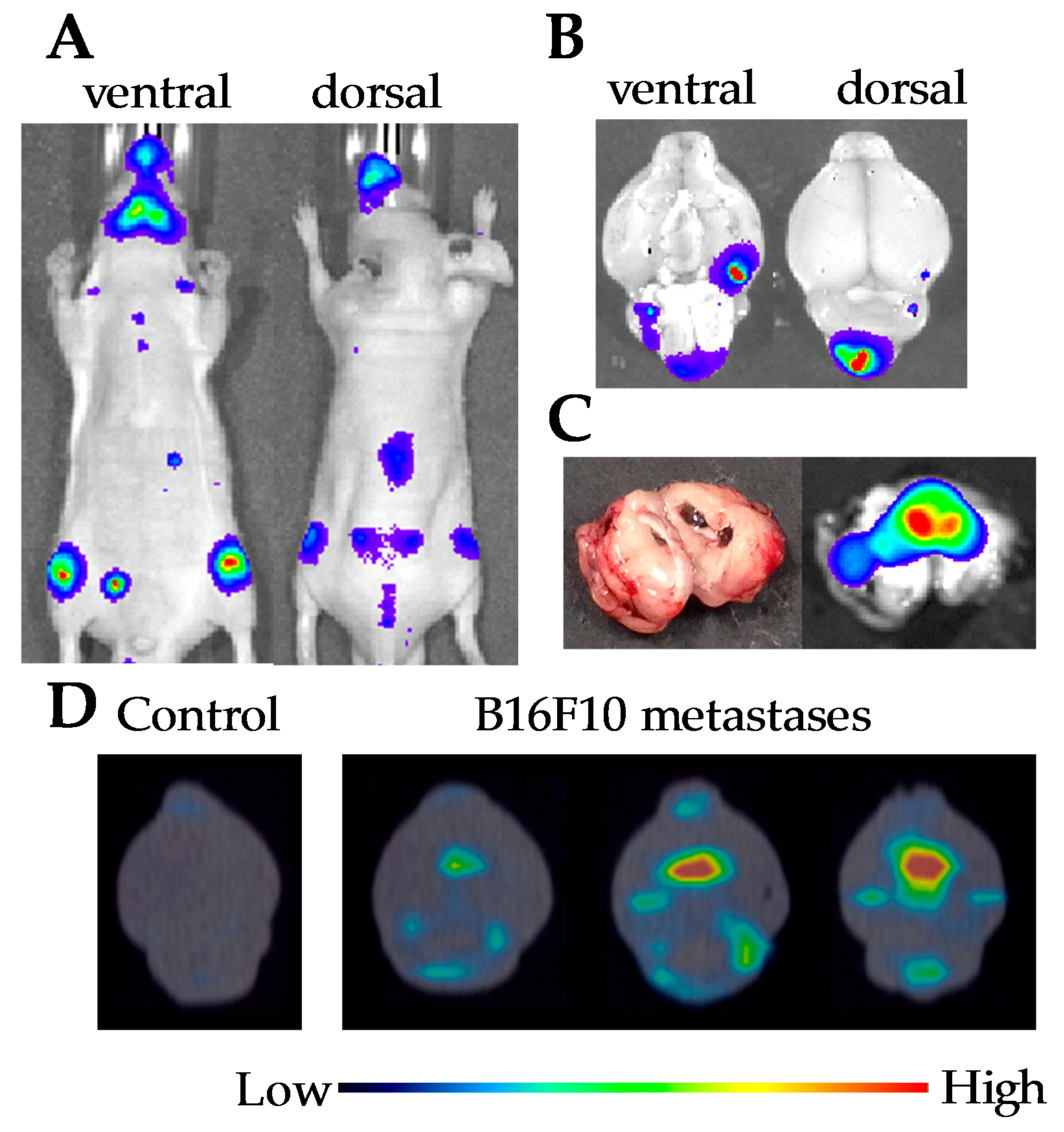
B16F10-luc2 cells (1 × 10⁵ in 0.1 mL of PBS) were injected from the left cardiac ventricle of five-week-old mice. Melanoma cells were migrated around the jaw and the knee joint at 11 days after implantation (Figure 2A). Brain metastases were confirmed by measuring of luminescence from isolated brain at 14 days after implantation (Figure 2B). Luminescent measurement of brain cross-section of a similar model mouse revealed that melanoma cells were migrated not only on the surface of brain but also in cerebral ventricle (Figure 2C). Accumulation of 111In-lactosome to the brain metastases were confirmed by SPECT/CT imaging of isolated brain at 14 days after implantation (Figure 2D).
2.3. Comparison of 111In-Lactosome and 201TlCl for SPECT/CT Imaging
Since the SPECT imaging makes it possible to collect simultaneously the data of multiple radiation sources owing to measuring the different energy radiation with choosing proper filters, different radiation species can be compared regarding their imaging abilities using an identical mouse. Comparison of 111In-lactosome and 201TlCl for SPECT/CT imaging of the brain metastases were carried out using an identical mouse. No specific accumulation of radioactivity was observed in the brain for control mice having no injection of B16F10 cells (Figure 3A). On the contrary, both of 111In-lactosome and 201TlCl accumulated successfully in the cerebral ventricle of the melanoma brain metastases (Figure 3A). On the other hand, 99mTc-HMDP failed to image the melanoma brain metastases (Figure 3B).
Metastases to the facial bone were also generated upon intracardiac injection of B16F10-luc2 cells. 111In-lactosome clearly imaged the metastasis at mandible as well (Figure 3). In the case of 201TlCl, however, accumulation of radioactivity in the muscle around the neck was also observed, which makes it difficult to recognize the bone metastasis at mandible by Tl ion (Figure 3A). 99mTc-HMDP accumulated a broad range of bones in head and neck (Figure 3B), and has difficulty in selective imaging of bone metastasis.
2.4. In Vivo Disposition in Brain
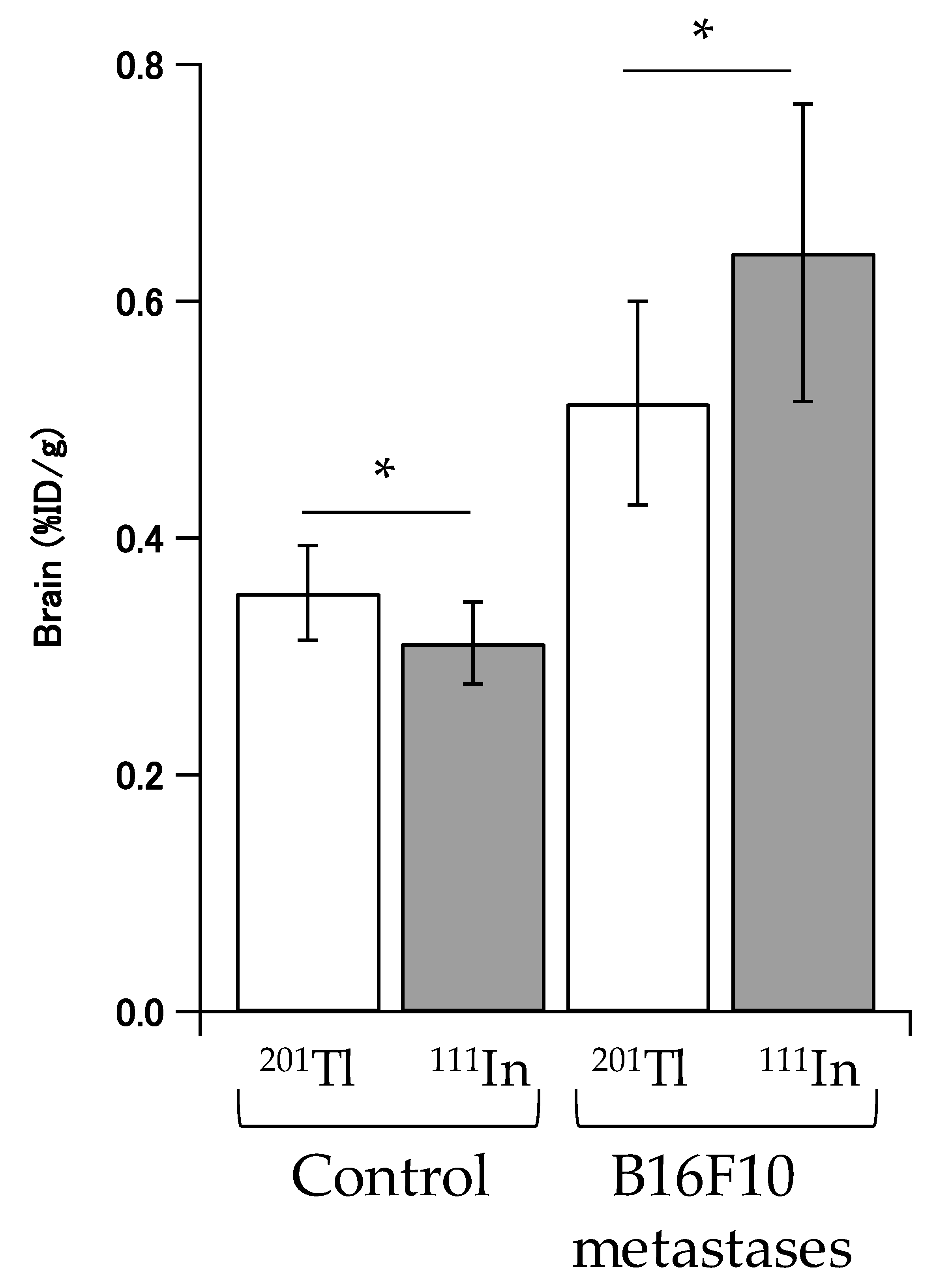
Radioactivities of 111In-lactosome and 201TlCl of the brains were measured by gamma counter using isolated brain to eliminate effects from bone metastases and muscle around the brain. Accumulation of radioactivities of 111In-lactosome and 201TlCl in brains of control mice were 0.311 ± 0.035 %ID/g and 0.354 ± 0.040 %ID/g, respectively. In contrast, accumulation of radioactivities of 111In-lactosome and 201TlCl in the brain metastases were 0.641 ± 0.126 %ID/g and 0.514 ± 0.086 %ID/g, respectively (Figure 4).
3. Discussion
Brain metastases in the leptomeningeal and cerebral ventricle were observed when the B16F10 melanoma cells were injected in the left cardiac ventricle [22]. It was known that 111In-lactosome accumulated in solid tumors by the EPR effect [24]. SPECT/CT images of brain metastases mice showed that 111In-lactosome accumulated in the metastases of the leptomeningeal and cerebral ventricle. However, in the mechanism for the selective accumulation of 111In-lactosome in the brain metastases, it is unclear whether 111In-lactosome could accumulate in the brain metastases through the spinal fluid.
SPECT imaging using 201TlCl has an intrinsic limitation because of its background uptake by muscle [25]. 99mTc-HMDP also failed to accumulate in brain metastases. On the contrary, 111In-lactosome accumulated selectively in both brain and bone metastases. In comparison with 201TlCl, 111In-lactosome accumulation in the healthy brain was extremely lowered. Assuming that the accumulation in the healthy brain was not affected by implantation of B16F10 cells, accumulation gains of 111In-lactosome and 201TlCl in the brain metastases compared with the healthy brain were estimated by the accumulation difference between them, and found to increase by 0.33 %ID/g and 0.16 %ID/g, respectively (Figure 4). Since the accumulation amount of 111In-lactosome in the brain metastases was doubled in comparison with that of 201TlCl, the imaging ability of 111In-lactosome is accordingly superior to 201TlCl.
111In-lactosome is composed of an A3B-type amphiphilic polydepsipeptide (A and B represent hydrophilic and hydrophobic blocks, respectively) of ((sarcosine)42)3-b-(l-lactic acid)30. The molecular design of the A3B-type amphiphilic polydepsipeptide is explained as follows. The A3B-type amphiphilic polydepsipeptide generated polymeric micelles of the A3B-type lactosome with a high surface density of the hydrophilic poly(sarcosine) chains resulting in escape from recognition by immune system [19]. That is, the A3B-type lactosome shows unchanged in vivo dispositions upon frequent doses, whilst the AB-type lactosome with a low surface density failed and the accelerated blood clearance was observed at second dose [18]. The evasion ability of the A3B-type lactosome from the immune system was studied with varying the hydrophilic poly(sarcosine) chain length, and the optimum chain length was found to exist between 30 to 50 mer [26]. The chain length of the hydrophobic block of poly(l-lactic acid) was also chosen to be 30 mer because the chain length allows to take a helical conformation leading to a good molecular association between the hydrophobic blocks [27]. All the points of these molecular designs place an emphasis on raising the evade ability of the nanoparticle from the immune system, and they also lead to effective accumulations in solid tumors. Recently, we are observing the delivery pathway of the A3B-type amphiphilic polydepsipeptide to lymph node in healthy mice. The pathway to meningeal dissemination therefore may be via spinal fluid which is now under investigation.
Taken together, 111In-lactosome has a high potential for selective imaging of brain and bone metastases, and therefore is applicable as a diagnostic agent. Further, 111In-lactosome distributes to every site of meningeal dissemination. With replacement of 111In with 90Y, the obtainable 90Y carrying lactosome will be applicable for therapeutic agent for meningeal dissemination, for which there is currently no cure. For this objective, the accumulation of 90Y carrying lactosome in bone marrow should be low, which is now under investigation.
4. Materials and Methods
4.1. Polymer Synthesis
DOTA-PDLA (PDLA represents poly(d-lactic acid); number-average molecular weight (Mn which was estimated by NMR) = 2.2 × 10³ (30 mer) was synthesized by condensation of NH2-PDLA and tri-tert-butyl 1,4,7,10-tetraazacyclododecane-1,4,7,10-tetraacetate as described previously [24]. Tri-tert-butyl groups were deprotected with trifluoroacetic acid. The A3B-type amphiphilic block polydepsipeptide of (poly(sarcosine))3-block-poly(l-lactic acid) (P(Sar42))3-PLLA30; Mn values of poly(sarcosine) and poly(l-lactic acid) blocks were 3.0 × 10³ (42 mer) and 2.2 × 10³ (30 mer), respectively) was similarly synthesized as described previously [26,28].
4.2. Animal and Cell Line
Mouse melanoma cell line (B16F10-luc2) was purchased from Caliper Life Sciences (Hopkinton, MA, USA). B16F10-luc2 cells were cultured in DMEM medium supplemented with 10% FBS, 1% GlutaMAX™ (Invitrogen, Carlsbad, CA, USA), 2.5 μg/mL Plasmocin™ Prophylactic (Nacalai Tesque, Kyoto, Japan), 100 U/mL penicillin, and 100 μg/mL streptomycin. Cells were incubated in a 5% CO2-humidified incubator at 37 °C. Pathogen-free male athymic BALB/c nude mice were purchased from the Japan SLC (Shizuoka, Japan). The brain metastasis model was established by injecting tumor cells (1 × 10⁵ in 0.1 mL of PBS) into the left cardiac ventricle of 5-week-old mice anesthetized by intraperitoneal injection of pentobarbital sodium. All the animal experiments were approved by the Animal Research Committee of Kyoto University.
4.3. Preparation of 111In-Lactosome
Four milligrams of (P(Sar42))3-PLLA30 and 2 mol% of DOTA-PDLA were dissolved in 2 mL of chloroform. After the polymers were completely dissolved, distill chloroform off under reduced pressure using a rotary evaporator (RE300, Yamato Scientific, Tokyo, Japan). Then 2 mL ultra-pure water was added into the dried polymer film. The self-assembled polymer micelles were obtained by sonication of polymer solution for 2 min at 85 °C. The self-assembled polymeric micelle composed of (P(Sar42))3-PLLA30 and DOTA-PDLA (DOTA-lactosome) was lyophilized and stored until use. 0.1 M sodium acetate was added into 111InCl3 solution (Nihon Medi-Physics, Tokyo, Japan) to adjust pH at 4.7 before labeling of 111In to DOTA. 111In labeled DOTA-PDLA containing A3B-type lactosome (111In-lactosome) was prepared by mixing 1 mg of lyophilized DOTA-lactosome and 0.7 mL of 111InCl3 solution at pH 4.7 and heated at 90 °C for 5 min. 111In-lactosome was purified by PD-10 column chromatography (GE Helthcare, Little Chalfont, UK) using saline to remove 111In ions that did not incorporate in DOTA (Figure 1). 111In-lactosome was concentrated by centrifugal concentrator (Amicon Ultra 50 kDa, Merck Millipore, Burlington, MA, USA) if necessary. Characterizations of the polymeric micelles were reported before [19,26,28], and they were as follows: hydrodynamic diameter by DLS: ca. 25 nm, critical association concentration: ca. 8 × 10⁻⁷ M.
4.4. Imaging of Melanoma Metastasis
The B16F10 implanted mice were used for single-photon emission computed tomography/computed tomography (SPECT/CT) imaging at 13 days after cell injection. Luminescence images of whole body of mouse and isolated brain were obtained with mice at 11 days and 14 days after intracardiac cell injection, respectively. The SPECT/CT images for isolated brains were taken at 24 h after intravenous injection of 111In-lactosome (12.8–13.7 MBq/body). An iodinated contrast agent (Iomeron350, Eisai, Tokyo, Japan) for CT was also used 437.5 mg of iodine per kilogram of body weight. The images were taken by FX3000 (Gamma Medica-Ideas, Inc., Northridge, CA, USA) for SPECT/CT and IVIS SPECTRUM (PerkinElmer, Waltham, MA, USA) for luminescent imaging. Simultaneous dual isotope SPECT/CT images were obtained using 111In-lactosome and 201TlCl (Nihon Medi-Physics) for brain metastasis and 111In-lactosome and 99mTc-HMDP (Nihon Medi-Physics) for born metastasis. The energy windows of SPECT for 111In, 201Tl, and 99mTc were 150–192, 67–74, and 133–148 keV, respectively. The SPECT/CT images for craniocervical region were taken at 24 h, 20 min, and 4 h after injections of 111In-lactosome, 201TlCl and 99mTc-HMDP, respectively. For the simultaneous dual isotope SPECT/CT imaging of 111In-lactosome and 201TlCl, 111In of 25.8 MBq and 201Tl of 23.3 MBq were injected to B16F10 metastases mice, and 111In of 26.0 MBq and 201Tl of 23.0 MBq to control mice. In the case of 111In-lactosome and 99mTc-HMDP, 111In of 19.2 MBq and 99mTc of 10.3 MBq were injected to B16F10 metastases mice, and 111In of 19.0 MBq and 99mTc of 10.5 MBq to control mice. The acquisition time was 45 min for all SPECT imaging. The mouse was anesthetized with isoflurane during the SPECT/CT imaging.
4.5. In Vivo Disposition in Brain
The B16F10 implanted mice were used for in vivo disposition in brain at 13 days after cell injection. The in vivo dispositions of 111In-lactosome and 201TlCl were determined in metastatic melanoma bearing and intact mice (n = 10). 111In-lactosome and 201TlCl of more than 0.5 MBq was injected into the identical mouse via tail vein. 111In-lactosome and 201TlCl were injected at 24 h and 20 min before the resection of brain, respectively. Radioactivities of 111In and 201Tl were detected in a gamma counter (COBRA II, Packard Instrument, Meriden, CT, USA) with energy window of 220–270 keV and 63–77 keV, respectively. The distribution of radioactivity measured and calculated for the percentage of injected dose of radioactivity per gram of tissue (%ID/g). Differences were considered statistically significant when p values were less than 0.05.
4.6. Statistical Analysis
All results are expressed as mean ± SD. Differences between groups were assessed by the t test for independent samples. p values < 0.05 were considered statistically significant.
4.7. Ethics
All of our in vivo animal experiments were approved by the Animal Research Committee of Kyoto University. Animals were treated humanely.
5. Conclusions
111In-labeled A3B-type lactosome accumulated selectively in the brain metastases of the leptomeningeal and the cerebral ventricle and in bone metastasis. Since 111In-labeled A3B-type lactosome distributed negligibly into healthy brain, bone and muscle, the SPECT imaging contrast for metastasis in the head and neck was highly potential by 111In-labeled A3B-type lactosome compared with 201TlCl and 99mTc-HMDP SPECT imaging.
Acknowledgments
This study is a part of the Innovative Techno-Hub for Integrated Medical Bio-Imaging of the Project for Developing Innovation Systems, from the Ministry of Education, Culture, Sports, Science and Technology (MEXT).
Author Contributions
K.K., M.U. and I.H. performed all practical work described in this article. E.O., K.T. and S.K. designed the project and let it. All authors contributed equally to the manuscript.
Conflicts of Interest
The authors declare no conflict of interest.
References
- Le Rhun, E.; Galanis, E. Leptomeningeal metastases of solid cancer. Curr. Opin. Neurol. 2016, 29, 797–805. [Google Scholar] [CrossRef] [PubMed]
- Siegel, R.L.; Miller, K.D.; Jemal, A. Cancer Statistics, 2017. CA Cancer J. Clin. 2017, 67, 7–30. [Google Scholar] [CrossRef] [PubMed]
- Balch, C.M.; Soong, S.J.; Gershenwald, J.E.; Thompson, J.F.; Reintgen, D.S.; Cascinelli, N.; Urist, M.; McMasters, K.M.; Ross, M.I.; Kirkwood, J.M.; et al. Prognostic factors analysis of 17,600 melanoma patients: Validation of the American joint committee on cancer melanoma staging system. J. Clin. Oncol. 2001, 19, 3622–3634. [Google Scholar] [CrossRef] [PubMed]
- Schouten, L.J.; Rutten, J.; Huveneers, H.A.M.; Twijnstra, A. Incidence of brain metastases in a cohort of patients with carcinoma of the breast, colon, kidney, and lung and melanoma. Cancer 2002, 94, 2698–2705. [Google Scholar] [CrossRef] [PubMed]
- Hatzoglou, V.; Karimi, S.; Diamond, E.L.; Lis, E.; Krol, G.; Holodny, A.I.; Young, R.J. Nonenhancing Leptomeningeal Metastases: Imaging characteristics and potential causative factors. Neurohospitalist 2016, 6, 24–28. [Google Scholar] [CrossRef] [PubMed]
- Nose, A.; Otsuka, H.; Nose, H.; Otomi, Y.; Terazawa, K.; Harada, M. Visual and semi-quantitative assessment of brain tumors using (201)Tl-SPECT. J. Med. Investig. 2013, 60, 121–126. [Google Scholar] [CrossRef]
- Wang, L.N.; Wang, Y.B.; Li, Z.J. Nanoparticle-based tumor theranostics with molecular imaging. Curr. Pharm. Biotechnol. 2013, 14, 683–692. [Google Scholar] [CrossRef] [PubMed]
- Lin, Q.Y.; Jin, C.S.; Huang, H.; Ding, L.L.; Zhang, Z.H.; Chen, J.; Zheng, G. Nanoparticle-enabled, image-guided treatment planning of target specific RNAi therapeutics in an orthotopic prostate cancer model. Small 2014, 10, 3072–3082. [Google Scholar] [CrossRef] [PubMed]
- Uhl, P.; Fricker, G.; Haberkorn, U.; Mier, W. Radionuclides in drug development. Drug Discov. Today 2015, 20, 198–208. [Google Scholar] [CrossRef] [PubMed]
- Maeda, H. Tumor-selective delivery of macromolecular drugs via the EPR effect: Background and future prospects. Bioconjug. Chem. 2010, 21, 797–802. [Google Scholar] [CrossRef] [PubMed]
- Wilhelm, S.; Tavares, A.J.; Dai, Q.; Ohta, S.; Audet, J.; Dvorak, H.F.; Chan, W.C.W. Analysis of nanoparticle delivery to tumours. Nat. Rev. Mater. 2016, 1. [Google Scholar] [CrossRef]
- Dams, E.T.M.; Laverman, P.; Oyen, W.J.G.; Storm, G.; Scherphof, G.L.; Van der Meer, J.W.M.; Corstens, F.H.M.; Boerman, O.C. Accelerated blood clearance and altered biodistribution of repeated injections of sterically stabilized liposomes. J. Pharmacol. Exp. Ther. 2000, 292, 1071–1079. [Google Scholar] [PubMed]
- Ishida, T.; Harada, M.; Wang, X.Y.; Ichihara, M.; Irimura, K.; Kiwada, H. Accelerated blood clearance of PEGylated liposomes following preceding liposome injection: Effects of lipid dose and PEG surface-density and chain length of the first-dose liposomes. J. Control. Release 2005, 105, 305–317. [Google Scholar] [CrossRef] [PubMed]
- Suzuki, T.; Ichihara, M.; Hyodo, K.; Yamamoto, E.; Ishida, T.; Kiwada, H.; Ishihara, H.; Kikuchi, H. Accelerated blood clearance of PEGylated liposomes containing doxorubicin upon repeated administration to dogs. Int. J. Pharm. 2012, 436, 636–643. [Google Scholar] [CrossRef] [PubMed]
- Suzuki, T.; Ichihara, M.; Hyodo, K.; Yamamoto, E.; Ishida, T.; Kiwada, H.; Kikuchi, H.; Ishihara, H. Influence of dose and animal species on accelerated blood clearance of PEGylated liposomal doxorubicin. Int. J. Pharm. 2014, 476, 205–212. [Google Scholar] [CrossRef] [PubMed]
- Hara, E.; Makino, A.; Kurihara, K.; Sugai, M.; Shimizu, A.; Hara, I.; Ozeki, E.; Kimura, S. Evasion from accelerated blood clearance of nanocarrier named as “Lactosome” induced by excessive administration of Lactosome. Biochim. Biophys. Acta 2013, 1830, 4046–4052. [Google Scholar] [CrossRef] [PubMed]
- Hara, E.; Makino, A.; Kurihara, K.; Ueda, M.; Hara, I.; Kawabe, T.; Yamamoto, F.; Ozeki, E.; Togashi, K.; Kimura, S. Radionuclide therapy using nanoparticle of I-131-Lactosome in combination with percutaneous ethanol injection therapy. J. Nanopart. Res. 2013, 15. [Google Scholar] [CrossRef]
- Hara, E.; Makino, A.; Kurihara, K.; Yamamoto, F.; Ozeki, E.; Kimura, S. Pharmacokinetic change of nanoparticulate formulation “Lactosome” on multiple administrations. Int. Immunopharmacol. 2012, 14, 261–266. [Google Scholar] [CrossRef] [PubMed]
- Hara, E.; Ueda, M.; Akira, M.; Hara, I.; Ozeki, E.; Kimura, S. Factors influencing in vivo disposition of polymeric micelles on multiple administrations. ACS Med. Chem. Lett. 2014, 5, 873–877. [Google Scholar] [CrossRef] [PubMed]
- Hara, E.; Ueda, M.; Kim, C.J.; Makino, A.; Hara, I.; Ozeki, E.; Kimura, S. Suppressive immune response of poly(sarcosine) chains in peptide-nanosheets in contrast to polymeric micelles. J. Pept. Sci. 2014, 20, 570–577. [Google Scholar] [CrossRef] [PubMed]
- Conley, F.K. Development of a metastatic brain-tumor model in mice. Cancer Res. 1979, 39, 1001–1007. [Google Scholar] [PubMed]
- Morsi, A.; Gaziel-Sovran, A.; Cruz-Munoz, W.; Kerbel, R.S.; Golfinos, J.G.; Hernando, E.; Wadghiri, Y.Z. Development and characterization of a clinically relevant mouse model of melanoma brain metastasis. Pigment Cell Melanoma Res. 2013, 26, 743–745. [Google Scholar] [CrossRef] [PubMed]
- Price, E.W.; Orvig, C. Matching chelators to radiometals for radiopharmaceuticals. Chem. Soc. Rev. 2014, 43, 260–290. [Google Scholar] [CrossRef] [PubMed]
- Kurihara, K.; Ueda, M.; Hara, I.; Hara, E.; Sano, K.; Makino, A.; Ozeki, E.; Yamamoto, F.; Saji, H.; Togashi, K.; et al. Inflammation-induced synergetic enhancement of nanoparticle treatments with DOXIL (R) and Y-90-Lactosome for orthotopic mammary tumor. J. Nanopart. Res. 2016, 18, 1–11. [Google Scholar] [CrossRef]
- Mullins, L.J.; Moore, R.D. The movemnet of thallium ions in muscle. J. Gen. Physiol. 1960, 43, 759–773. [Google Scholar] [CrossRef] [PubMed]
- Kurihara, K.; Ueda, M.; Hara, I.; Ozeki, E.; Togashi, K.; Kimura, S. Control of in vivo disposition and immunogenicity of polymeric micelles by adjusting poly(sarcosine) chain lengths on surface. J. Nanopart. Res. 2017, 19, 242. [Google Scholar] [CrossRef]
- Makino, A.; Yamahara, R.; Ozeki, E.; Kimura, S. Preparation of Novel Polymer Assemblies, “Lactosome”, Composed of Poly(l-lactic acid) and Poly(sarcosine). Chem. Lett. 2007, 36, 1220–1221. [Google Scholar] [CrossRef]
- Makino, A.; Hara, E.; Hara, I.; Ozeki, E.; Kimura, S. Size control of core-shell-type polymeric micelle with a nanometer precision. Langmuir 2014, 30, 669–674. [Google Scholar] [CrossRef] [PubMed]
Figure 1.
Schematic illustration of the labelling method of 111In to DOTA-lactosome and the structure of 111In-lactosome.
Figure 1.
Schematic illustration of the labelling method of 111In to DOTA-lactosome and the structure of 111In-lactosome.
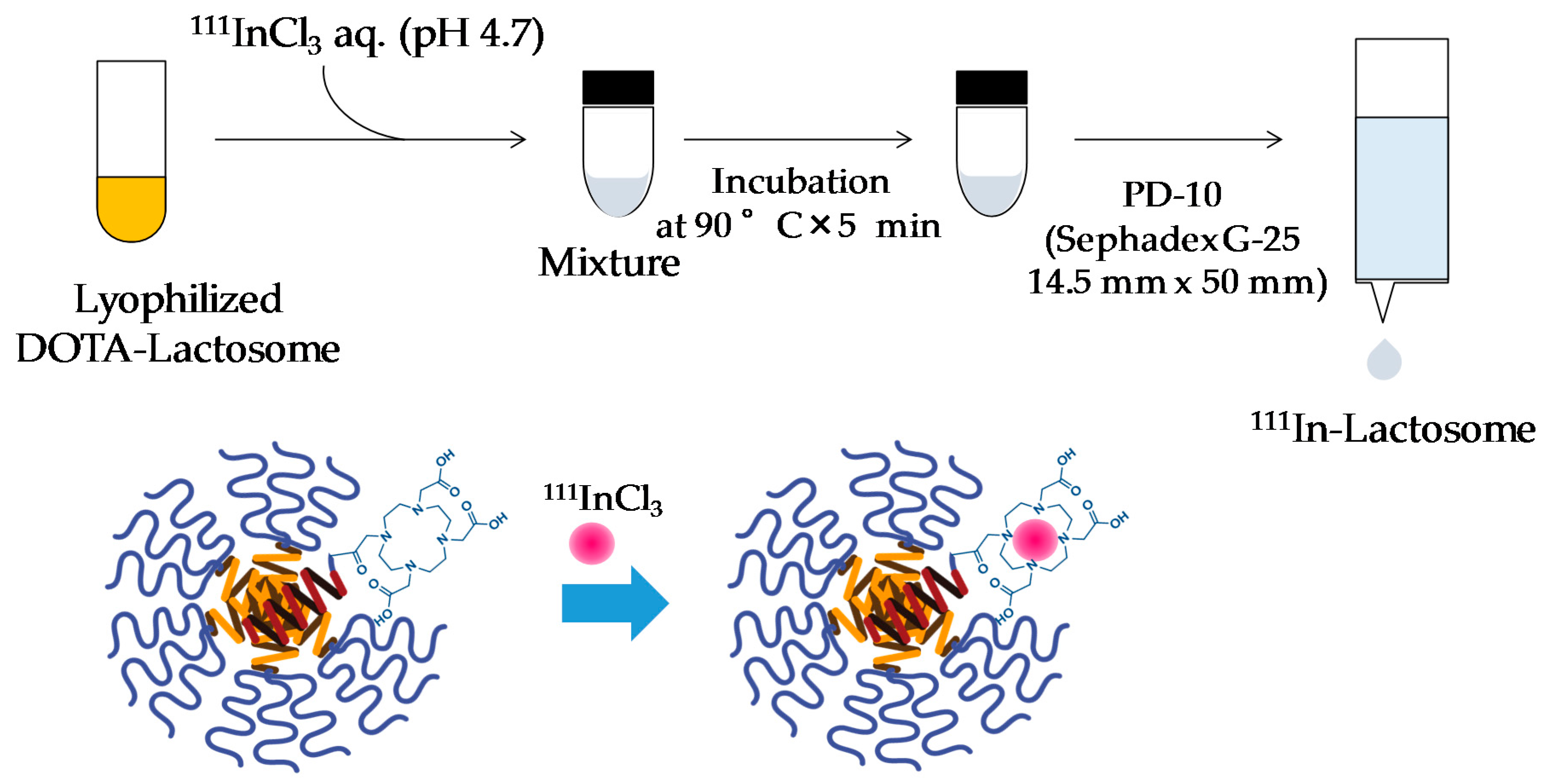
Figure 2.
Luminescence and single-photon emission computed tomography (SPECT)/CT images of the B16F10 implanted mice. (A) Luminescence images for ventral and dorsal side of the mouse at 11 days after implantation; (B) Luminescence images for isolated brain of the mouse at 14 days after implantation; (C) Luminescence and photo images for brain cross-section isolated from the B16F10 implanted mouse at 14 days after cell injection; (D) SPECT/CT images using 111In-lactosome for isolated brains of the mice at 14 days after implantation.

Figure 3.
Dual isotope SPECT/CT images of melanoma metastasis mice after injections of 111In-lactosome and 201TlCl (A); and 111In-Llctosome and 99mTc-HMDP (B). The transverse (left), sagittal (middle), and coronal (right) views centered on the cerebral ventricle are shown.
Figure 3.
Dual isotope SPECT/CT images of melanoma metastasis mice after injections of 111In-lactosome and 201TlCl (A); and 111In-Llctosome and 99mTc-HMDP (B). The transverse (left), sagittal (middle), and coronal (right) views centered on the cerebral ventricle are shown.
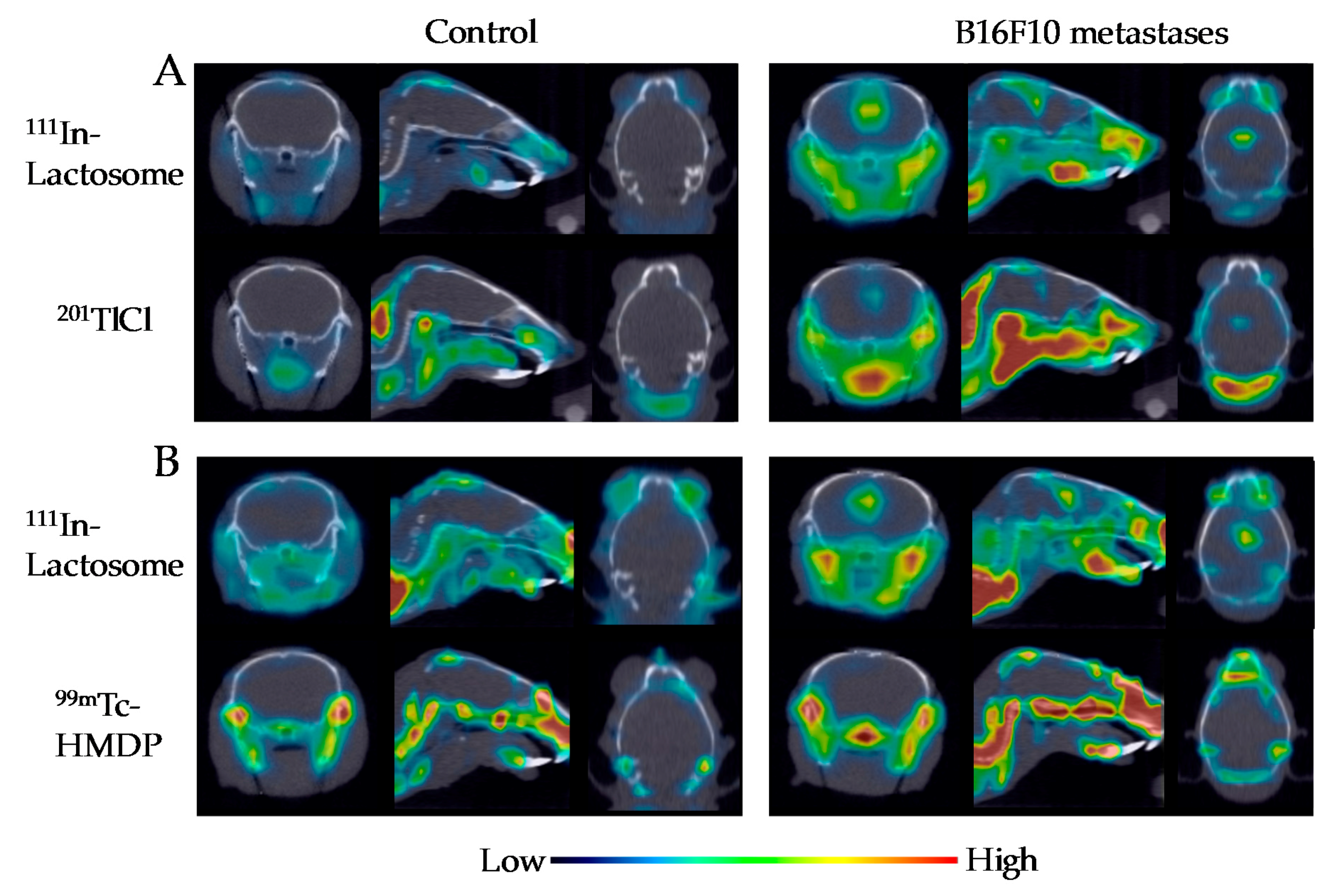
Figure 4.
Brain accumulation of 111In-lactosome (111In) and 201TlCl (201Tl) for intact and B16F10 metastasis mice (* p < 0.05). The sample number (n) = 10.
Figure 4.
Brain accumulation of 111In-lactosome (111In) and 201TlCl (201Tl) for intact and B16F10 metastasis mice (* p < 0.05). The sample number (n) = 10.

© 2018 by the authors. Licensee MDPI, Basel, Switzerland. This article is an open access article distributed under the terms and conditions of the Creative Commons Attribution (CC BY) license (http://creativecommons.org/licenses/by/4.0/).
Share and Cite
MDPI and ACS Style
Kurihara, K.; Ueda, M.; Hara, I.; Ozeki, E.; Togashi, K.; Kimura, S. Polymeric Micelle of A3B-Type Lactosome as a Vehicle for Targeting Meningeal Dissemination. Nanomaterials 2018, 8, 79. https://doi.org/10.3390/nano8020079
AMA Style
Kurihara K, Ueda M, Hara I, Ozeki E, Togashi K, Kimura S. Polymeric Micelle of A3B-Type Lactosome as a Vehicle for Targeting Meningeal Dissemination. Nanomaterials. 2018; 8(2):79. https://doi.org/10.3390/nano8020079
Chicago/Turabian StyleKurihara, Kensuke, Motoki Ueda, Isao Hara, Eiichi Ozeki, Kaori Togashi, and Shunsaku Kimura. 2018. "Polymeric Micelle of A3B-Type Lactosome as a Vehicle for Targeting Meningeal Dissemination" Nanomaterials 8, no. 2: 79. https://doi.org/10.3390/nano8020079
Note that from the first issue of 2016, this journal uses article numbers instead of page numbers. See further details here.