Next Issue
Volume 7, December
Previous Issue
Volume 7, June


Biosensors, Volume 7, Issue 3 (September 2017) – 17 articles

Cover Story (view full-size image): 
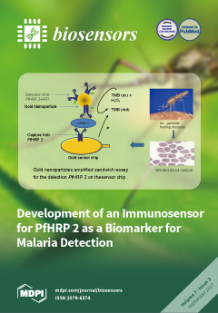
Malaria is a disease transmitted by the adult female Anopheles mosquito and is caused by the Apicomplexan Plasmodium parasite. Four species of Plasmodium are responsible for human malaria, but P. falciparum malaria is known to be the most lethal. Malaria, is transmitted easily and can claim the lives of some of its victims, with expectant mothers and children under the age of five being the most vulnerable to the disease. To date, different tests have been developed for the detection of malaria biomarkers, but they all have limitations which include being expensive, time consuming or lacking sensitivity and specificity. Hence, the development of biosensors for malaria detection can help to overcome some of the known limitations in malaria diagnosis. View this paper
  • Issues are regarded as officially published after their release is announced to the table of contents alert mailing list.
  • You may sign up for e-mail alerts to receive table of contents of newly released issues.
  • PDF is the official format for papers published in both, html and pdf forms. To view the papers in pdf format, click on the "PDF Full-text" link, and use the free Adobe Reader to open them.
Order results
Result details
Section
Select all
Export citation of selected articles as:
2344 KiB  
Article
Use of Wavelet Transform to Detect Compensated and Decompensated Stages in the Congestive Heart Failure Patient
by Pratibha Sharma, Kimberly Newman, Carlin S. Long, A. J. Gasiewski and Frank Barnes
Biosensors 2017, 7(3), 40; https://doi.org/10.3390/bios7030040 - 20 Sep 2017
Cited by 4 | Viewed by 6132
Abstract
This research work is aimed at improving health care, reducing cost, and the occurrence of emergency hospitalization in patients with Congestive Heart Failure (CHF) by analyzing heart and lung sounds to distinguish between the compensated and decompensated states. Compensated state defines stable state [...] Read more.
This research work is aimed at improving health care, reducing cost, and the occurrence of emergency hospitalization in patients with Congestive Heart Failure (CHF) by analyzing heart and lung sounds to distinguish between the compensated and decompensated states. Compensated state defines stable state of the patient but with lack of retention of fluids in lungs, whereas decompensated state leads to unstable state of the patient with lots of fluid retention in the lungs, where the patient needs medication. Acoustic signals from the heart and the lung were analyzed using wavelet transforms to measure changes in the CHF patient’s status from the decompensated to compensated and vice versa. Measurements were taken on CHF patients diagnosed to be in compensated and decompensated states by using a digital stethoscope and electrocardiogram (ECG) in order to monitor their progress in the management of their disease. Analysis of acoustic signals of the heart due to the opening and closing of heart valves as well as the acoustic signals of the lungs due to respiration and the ECG signals are presented. Fourier, short-time Fourier, and wavelet transforms are evaluated to determine the best method to detect shifts in the status of a CHF patient. The power spectra obtained through the Fourier transform produced results that differentiate the signals from healthy people and CHF patients, while the short-time Fourier transform (STFT) technique did not provide the desired results. The most promising results were obtained by using wavelet analysis. Wavelet transforms provide better resolution, in time, for higher frequencies, and a better resolution, in frequency, for lower frequencies. Full article
(This article belongs to the Special Issue Latest Wearable Biosensors)
Show Figures

Figure 1

1905 KiB  
Article
Substituting Sodium Hydrosulfite with Sodium Metabisulfite Improves Long-Term Stability of a Distributable Paper-Based Test Kit for Point-of-Care Screening for Sickle Cell Anemia
by Kian Torabian, Dalia Lezzar, Nathaniel Z. Piety, Alex George and Sergey S. Shevkoplyas
Biosensors 2017, 7(3), 39; https://doi.org/10.3390/bios7030039 - 20 Sep 2017
Cited by 9 | Viewed by 10861
Abstract
Sickle cell anemia (SCA) is a genetic blood disorder that is particularly lethal in early childhood. Universal newborn screening programs and subsequent early treatment are known to drastically reduce under-five SCA mortality. However, in resource-limited settings, cost and infrastructure constraints limit the effectiveness [...] Read more.
Sickle cell anemia (SCA) is a genetic blood disorder that is particularly lethal in early childhood. Universal newborn screening programs and subsequent early treatment are known to drastically reduce under-five SCA mortality. However, in resource-limited settings, cost and infrastructure constraints limit the effectiveness of laboratory-based SCA screening programs. To address this limitation our laboratory previously developed a low-cost, equipment-free, point-of-care, paper-based SCA test. Here, we improved the stability and performance of the test by replacing sodium hydrosulfite (HS), a key reducing agent in the hemoglobin solubility buffer which is not stable in aqueous solutions, with sodium metabisulfite (MS). The MS formulation of the test was compared to the HS formulation in a laboratory setting by inexperienced users (n = 3), to determine visual limit of detection (LOD), readout time, diagnostic accuracy, intra- and inter-observer agreement, and shelf life. The MS test was found to have a 10% sickle hemoglobin LOD, 21-min readout time, 97.3% sensitivity and 99.5% specificity for SCA, almost perfect intra- and inter-observer agreement, at least 24 weeks of shelf stability at room temperature, and could be packaged into a self-contained, distributable test kits comprised of off-the-shelf disposable components and food-grade reagents with a total cost of only $0.21 (USD). Full article
(This article belongs to the Special Issue Point-of-Care Diagnostics)
Show Figures

Graphical abstract

3121 KiB  
Article
Mediatorless Impedance Studies with Titanium Dioxide Conjugated Gold Nanoparticles for Hydrogen Peroxide Detection
by Nur Hamidah Abdul Halim, Yook Heng Lee, Radha Swathe Priya Malon Marugan and Uda Hashim
Biosensors 2017, 7(3), 38; https://doi.org/10.3390/bios7030038 - 18 Sep 2017
Cited by 19 | Viewed by 6620
Abstract
An impedimetric-based biosensor constructed using gold nanoparticles (AuNP) entrapped within titanium dioxide (TiO2) particles for hydrogen peroxide (H2O2) detection is the main feature of this research. The matrix of the biosensor employed the surface of TiO2 [...] Read more.
An impedimetric-based biosensor constructed using gold nanoparticles (AuNP) entrapped within titanium dioxide (TiO2) particles for hydrogen peroxide (H2O2) detection is the main feature of this research. The matrix of the biosensor employed the surface of TiO2, which was previously modified with an amine terminal group using 3-Aminopropyltriethoxysilane (APTS) at a low temperature to create a ready to immobilise surface for the biosensor application. Hemoglobin (Hb), which exhibits peroxidase-like activity, was used as the bioreceptor in the biosensor to detect H2O2 in solution. The analysis was carried out using an alternative impedance method, in which the biosensor exhibited a wide linear range response between 1 × 10⁻⁴ M and 1.5 × 10⁻² M and a limit of detection (LOD) of 1 × 10⁻⁵ M without a redox mediator. Full article
Show Figures

Figure 1

3714 KiB  
Article
Silicon Integrated Dual-Mode Interferometer with Differential Outputs
by Niklas Hoppe, Pascal Scheck, Rami Sweidan, Philipp Diersing, Lotte Rathgeber, Wolfgang Vogel, Benjamin Riegger, Alexander Southan and Manfred Berroth
Biosensors 2017, 7(3), 37; https://doi.org/10.3390/bios7030037 - 14 Sep 2017
Cited by 8 | Viewed by 6773
Abstract
The dual-mode interferometer (DMI) is an attractive alternative to Mach-Zehnder interferometers for sensor purposes, achieving sensitivities to refractive index changes close to state-of-the-art. Modern designs on silicon-on-insulator (SOI) platforms offer thermally stable and compact devices with insertion losses of less than 1 dB [...] Read more.
The dual-mode interferometer (DMI) is an attractive alternative to Mach-Zehnder interferometers for sensor purposes, achieving sensitivities to refractive index changes close to state-of-the-art. Modern designs on silicon-on-insulator (SOI) platforms offer thermally stable and compact devices with insertion losses of less than 1 dB and high extinction ratios. Compact arrays of multiple DMIs in parallel are easy to fabricate due to the simple structure of the DMI. In this work, the principle of operation of an integrated DMI with differential outputs is presented which allows the unambiguous phase shift detection with a single wavelength measurement, rather than using a wavelength sweep and evaluating the optical output power spectrum. Fluctuating optical input power or varying attenuation due to different analyte concentrations can be compensated by observing the sum of the optical powers at the differential outputs. DMIs with two differential single-mode outputs are fabricated in a 250 nm SOI platform, and corresponding measurements are shown to explain the principle of operation in detail. A comparison of DMIs with the conventional Mach-Zehnder interferometer using the same technology concludes this work. Full article
(This article belongs to the Special Issue Biophotonic Sensors and Applications)
Show Figures

Figure 1

3009 KiB  
Article
A Rationally Designed Reversible ‘Turn-Off’ Sensor for Glutathione
by Sabrina Heng, Xiaozhou Zhang, Jinxin Pei and Andrew D. Abell
Biosensors 2017, 7(3), 36; https://doi.org/10.3390/bios7030036 - 06 Sep 2017
Cited by 12 | Viewed by 7592
Abstract
γ-Glutamyl-cysteinyl-glycine (GSH) plays a critical role in maintaining redox homeostasis in biological systems and a decrease in its cellular levels is associated with diseases. Existing fluorescence-based chemosensors for GSH acts as irreversible reaction-based probes that exhibit a maximum fluorescence (‘turn-on’) once the reaction [...] Read more.
γ-Glutamyl-cysteinyl-glycine (GSH) plays a critical role in maintaining redox homeostasis in biological systems and a decrease in its cellular levels is associated with diseases. Existing fluorescence-based chemosensors for GSH acts as irreversible reaction-based probes that exhibit a maximum fluorescence (‘turn-on’) once the reaction is complete, regardless of the actual concentration of GSH. A reversible, reaction-based ‘turn-off’ probe (1) is reported here to sense the decreasing levels of GSH, a situation known to occur at the onset of various diseases. The more fluorescent merocyanine (MC) isomer of 1 exists in aqueous solution and this reacts with GSH to induce formation of the ring-closed spiropyran (SP) isomer, with a measurable decrease in absorbance and fluorescence (‘turn-off’). Sensor 1 has good aqueous solubility and shows an excellent selectivity for GSH over other biologically relevant metal ions and aminothiol analytes. The sensor permeates HEK 293 cells and an increase in fluorescence is observed on adding buthionine sulfoximine, an inhibitor of GSH synthesis. Full article
(This article belongs to the Special Issue Nanomaterials Based Optical Biosensors)
Show Figures

Graphical abstract

1405 KiB  
Article
A Novel Impedance Biosensor for Measurement of Trans-Epithelial Resistance in Cells Cultured on Nanofiber Scaffolds
by Robert A. Schramm, Matthew H. Koslow, Deirdre A. Nelson, Melinda Larsen and James Castracane
Biosensors 2017, 7(3), 35; https://doi.org/10.3390/bios7030035 - 31 Aug 2017
Cited by 5 | Viewed by 7255
Abstract
Nanofibrous scaffolds provide high surface area for cell attachment, and resemble the structure of the collagen fibers which naturally occur in the basement membrane and extracellular matrix. A label free and non-destructive method of assessing the interaction of cell tissue and scaffolds aids [...] Read more.
Nanofibrous scaffolds provide high surface area for cell attachment, and resemble the structure of the collagen fibers which naturally occur in the basement membrane and extracellular matrix. A label free and non-destructive method of assessing the interaction of cell tissue and scaffolds aids in the ability to discern the effective quality and magnitude of any scaffold modifications. Impedance cell spectroscopy is a biosensing method that employs a functional approach to assessing the cell monolayer. The electrical impedance barrier function of a cell monolayer represents the level of restriction to diffusion of charged species between all adjacent cells across an entire contiguous cellular monolayer. The impedance signals from many individual paracellular pathways contribute to the bulk measurement of the whole monolayer barrier function. However, the scaffold substrate must be entirely porous in order to be used with electrochemical cell impedance spectroscopy (ECIS) and cells must be closely situated to the electrodes. For purposes of evaluating cell-scaffold constructs for tissue engineering, non-invasive evaluation of cell properties while seeded on scaffolds is critical. A Transwell-type assay makes a measurement across a semi-permeable membrane, using electrodes placed on opposing sides of the membrane immersed in fluid. It was found that by suspending a nanofiber scaffold across a Transwell aperture, it is possible to integrate a fully functional nanofiber tissue scaffold with the ECIS Transwell apparatus. Salivary epithelial cells were grown on the nanofiber scaffolds and tight junction formation was evaluated using ECIS measurements in parallel with immunostaining and confocal imaging. The trans-epithelial resistance increased coordinate with cell coverage, culminating with a cell monolayer, at which point the tight junction proteins assemble and strengthen, reaching the peak signal. These studies demonstrate that ECIS can be used to evaluate tight junction formation in cells grown on nanofiber scaffolds and on effects of scaffold conditions on cells, thus providing useful biological feedback to inform superior scaffold designs. Full article
Show Figures

Figure 1

2445 KiB  
Article
Thermal Response Analysis of Phospholipid Bilayers Using Ellipsometric Techniques
by Carmen M. González-Henríquez, Vanessa A. Villegas-Opazo, Dallits H. Sagredo-Oyarce, Mauricio A. Sarabia-Vallejos and Claudio A. Terraza
Biosensors 2017, 7(3), 34; https://doi.org/10.3390/bios7030034 - 18 Aug 2017
Cited by 8 | Viewed by 7004
Abstract
Biomimetic planar artificial membranes have been widely studied due to their multiple applications in several research fields. Their humectation and thermal response are crucial for reaching stability; these characteristics are related to the molecular organization inside the bilayer, which is affected by the [...] Read more.
Biomimetic planar artificial membranes have been widely studied due to their multiple applications in several research fields. Their humectation and thermal response are crucial for reaching stability; these characteristics are related to the molecular organization inside the bilayer, which is affected by the aliphatic chain length, saturations, and molecule polarity, among others. Bilayer stability becomes a fundamental factor when technological devices are developed—like biosensors—based on those systems. Thermal studies were performed for different types of phosphatidylcholine (PC) molecules: two pure PC bilayers and four binary PC mixtures. These analyses were carried out through the detection of slight changes in their optical and structural parameters via Ellipsometry and Surface Plasmon Resonance (SPR) techniques. Phospholipid bilayers were prepared by Langmuir-Blodgett technique and deposited over a hydrophilic silicon wafer. Their molecular inclination degree, mobility, and stability of the different phases were detected and analyzed through bilayer thickness changes and their optical phase-amplitude response. Results show that certain binary lipid mixtures—with differences in its aliphatic chain length—present a co-existence of two thermal responses due to non-ideal mixing. Full article
Show Figures

Figure 1

3227 KiB  
Article
Bloch Surface Waves Biosensors for High Sensitivity Detection of Soluble ERBB2 in a Complex Biological Environment
by Alberto Sinibaldi, Camilla Sampaoli, Norbert Danz, Peter Munzert, Frank Sonntag, Fabio Centola, Agostino Occhicone, Elisa Tremante, Patrizio Giacomini and Francesco Michelotti
Biosensors 2017, 7(3), 33; https://doi.org/10.3390/bios7030033 - 17 Aug 2017
Cited by 11 | Viewed by 6608
Abstract
We report on the use of one-dimensional photonic crystals to detect clinically relevant concentrations of the cancer biomarker ERBB2 in cell lysates. Overexpression of the ERBB2 protein is associated with aggressive breast cancer subtypes. To detect soluble ERBB2, we developed an optical set-up [...] Read more.
We report on the use of one-dimensional photonic crystals to detect clinically relevant concentrations of the cancer biomarker ERBB2 in cell lysates. Overexpression of the ERBB2 protein is associated with aggressive breast cancer subtypes. To detect soluble ERBB2, we developed an optical set-up which operates in both label-free and fluorescence modes. The detection approach makes use of a sandwich assay, in which the one-dimensional photonic crystals sustaining Bloch surface waves are modified with monoclonal antibodies, in order to guarantee high specificity during the biological recognition. We present the results of exemplary protein G based label-free assays in complex biological matrices, reaching an estimated limit of detection of 0.5 ng/mL. On-chip and chip-to-chip variability of the results is addressed too, providing repeatability rates. Moreover, results on fluorescence operation demonstrate the capability to perform high sensitive cancer biomarker assays reaching a resolution of 0.6 ng/mL, without protein G assistance. The resolution obtained in both modes meets international guidelines and recommendations (15 ng/mL) for ERBB2 quantification assays, providing an alternative tool to phenotype and diagnose molecular cancer subtypes. Full article
(This article belongs to the Special Issue Biosensors for the Detection of Biomarkers)
Show Figures

Figure 1

2789 KiB  
Article
A Novel Surface Plasmon Resonance Biosensor for the Rapid Detection of Botulinum Neurotoxins
by Kruti Patel, Shmuel Halevi, Paul Melman, John Schwartz, Shuowei Cai and Bal Ram Singh
Biosensors 2017, 7(3), 32; https://doi.org/10.3390/bios7030032 - 07 Aug 2017
Cited by 20 | Viewed by 9245
Abstract
Botulinum neurotoxins (BoNTs) are Category A agents on the NIAID (National Institute of Allergy and Infectious Diseases) priority pathogen list owing to their extreme toxicity and the relative ease of production. These deadly toxins, in minute quantities (estimated human i.v. lethal dose LD [...] Read more.
Botulinum neurotoxins (BoNTs) are Category A agents on the NIAID (National Institute of Allergy and Infectious Diseases) priority pathogen list owing to their extreme toxicity and the relative ease of production. These deadly toxins, in minute quantities (estimated human i.v. lethal dose LD50 of 1–2 ng/kg body weight), cause fatal flaccid paralysis by blocking neurotransmitter release. The current gold standard detection method, the mouse-bioassay, often takes days to confirm botulism. Furthermore, there are no effective antidotes known to reverse the symptoms of botulism, and as a result, patients with severe botulism often require meticulous care during the prolonged paralytic illness. To combat potential bio-terrorism incidents of botulinum neurotoxins, their rapid detection is paramount. Surface plasmon resonance (SPR) is a very sensitive technique to examine bio-molecular interactions. The label-free, real-time analysis, with high sensitivity and low sample consumption makes this technology particularly suitable for detection of the toxin. In this study, we demonstrated the feasibility in an assay with a newly designed SPR instrument for the rapid detection of botulinum neurotoxins. The LOD (limit of detection) of the Newton Photonics (NP) SPR based assay is 6.76 pg/mL for Botulinum Neurotoxin type A Light Chain (BoNT/A LC). We established that the detection sensitivity of the system is comparable to the traditional mouse LD50 bioassay in BoNT/A using this SPR technology. Full article
(This article belongs to the Special Issue Point-of-Care Diagnostics)
Show Figures

Figure 1

2946 KiB  
Article
An Affordable Microsphere-Based Device for Visual Assessment of Water Quality
by Azra Rajwani, Brendon Restall, Nathan J. Muller, Scott Roebuck and Stephanie M. Willerth
Biosensors 2017, 7(3), 31; https://doi.org/10.3390/bios7030031 - 05 Aug 2017
Cited by 1 | Viewed by 5494
Abstract
This work developed a prototype of an affordable, long-term water quality detection device that provides a visual readout upon detecting bacterial contamination. This device prototype consists of: (1) enzyme-releasing microspheres that lyse bacteria present in a sample, (2) microspheres that release probes that [...] Read more.
This work developed a prototype of an affordable, long-term water quality detection device that provides a visual readout upon detecting bacterial contamination. This device prototype consists of: (1) enzyme-releasing microspheres that lyse bacteria present in a sample, (2) microspheres that release probes that bind the DNA of the lysed bacteria, and (3) a detector region consisting of gold nanoparticles. The probes bind bacterial DNA, forming complexes. These complexes induce aggregation of the gold nanoparticles located in the detector region. The nanoparticle aggregation process causes a red to blue color change, providing a visual indicator of contamination being detected. Our group fabricated and characterized microspheres made of poly (ε-caprolactone) that released lysozyme (an enzyme that degrades bacterial cell walls) and hairpin DNA probes that bind to regions of the Escherichia coli genome over a 28-day time course. The released lysozyme retained its ability to lyse bacteria. We then showed that combining these components with gold nanoparticles followed by exposure to an E. coli-contaminated water sample (concentrations tested—106 and 108 cells/mL) resulted in a dramatic red to blue color change. Overall, this device represents a novel low-cost system for long term detection of bacteria in a water supply and other applications. Full article
(This article belongs to the Special Issue Nanomaterials Based Optical Biosensors)
Show Figures

Graphical abstract

2124 KiB  
Review
Point-of-Care-Testing in Acute Stroke Management: An Unmet Need Ripe for Technological Harvest
by Dorin Harpaz, Evgeni Eltzov, Raymond C.S. Seet, Robert S. Marks and Alfred I.Y. Tok
Biosensors 2017, 7(3), 30; https://doi.org/10.3390/bios7030030 - 03 Aug 2017
Cited by 49 | Viewed by 15397
Abstract
Stroke, the second highest leading cause of death, is caused by an abrupt interruption of blood to the brain. Supply of blood needs to be promptly restored to salvage brain tissues from irreversible neuronal death. Existing assessment of stroke patients is based largely [...] Read more.
Stroke, the second highest leading cause of death, is caused by an abrupt interruption of blood to the brain. Supply of blood needs to be promptly restored to salvage brain tissues from irreversible neuronal death. Existing assessment of stroke patients is based largely on detailed clinical evaluation that is complemented by neuroimaging methods. However, emerging data point to the potential use of blood-derived biomarkers in aiding clinical decision-making especially in the diagnosis of ischemic stroke, triaging patients for acute reperfusion therapies, and in informing stroke mechanisms and prognosis. The demand for newer techniques to deliver individualized information on-site for incorporation into a time-sensitive work-flow has become greater. In this review, we examine the roles of a portable and easy to use point-of-care-test (POCT) in shortening the time-to-treatment, classifying stroke subtypes and improving patient’s outcome. We first examine the conventional stroke management workflow, then highlight situations where a bedside biomarker assessment might aid clinical decision-making. A novel stroke POCT approach is presented, which combines the use of quantitative and multiplex POCT platforms for the detection of specific stroke biomarkers, as well as data-mining tools to drive analytical processes. Further work is needed in the development of POCTs to fulfill an unmet need in acute stroke management. Full article
(This article belongs to the Special Issue Point-of-Care Diagnostics)
Show Figures

Figure 1

1766 KiB  
Article
In Vitro Quantified Determination of β-Amyloid 42 Peptides, a Biomarker of Neuro-Degenerative Disorders, in PBS and Human Serum Using a Simple, Cost-Effective Thin Gold Film Biosensor
by Yifan Dai, Alireza Molazemhosseini and Chung Chiun Liu
Biosensors 2017, 7(3), 29; https://doi.org/10.3390/bios7030029 - 20 Jul 2017
Cited by 19 | Viewed by 7141
Abstract
A simple in vitro biosensor for the detection of β-amyloid 42 in phosphate-buffered saline (PBS) and undiluted human serum was fabricated and tested based on our platform sensor technology. The bio-recognition mechanism of this biosensor was based on the effect of the interaction [...] Read more.
A simple in vitro biosensor for the detection of β-amyloid 42 in phosphate-buffered saline (PBS) and undiluted human serum was fabricated and tested based on our platform sensor technology. The bio-recognition mechanism of this biosensor was based on the effect of the interaction between antibody and antigen of β-amyloid 42 to the redox couple probe of K4Fe(CN)6 and K3Fe(CN)6. Differential pulse voltammetry (DPV) served as the transduction mechanism measuring the current output derived from the redox coupling reaction. The biosensor was a three-electrode electrochemical system, and the working and counter electrodes were 50 nm thin gold film deposited by a sputtering technique. The reference electrode was a thick-film printed Ag/AgCl electrode. Laser ablation technique was used to define the size and structure of the biosensor. Cost-effective roll-to-roll manufacturing process was employed in the fabrication of the biosensor, making it simple and relatively inexpensive. Self-assembled monolayers (SAM) of 3-Mercaptopropionic acid (MPA) was employed to covalently immobilize the thiol group on the gold working electrode. A carbodiimide conjugation approach using N-(3-dimethylaminopropyl)-N′-ethylcarbodiimide hydrochloride (EDC) and N–hydroxysuccinimide (NHS) was undertaken for cross-linking antibody of β-amyloid 42 to the carboxylic groups on one end of the MPA. The antibody concentration of β-amyloid 42 used was 18.75 µg/mL. The concentration range of β-amyloid 42 in this study was from 0.0675 µg/mL to 0.5 µg/mL for both PBS and undiluted human serum. DPV measurements showed excellent response in this antigen concentration range. Interference study of this biosensor was carried out in the presence of Tau protein antigen. Excellent specificity of this β-amyloid 42 biosensor was demonstrated without interference from other species, such as T-tau protein. Full article
(This article belongs to the Special Issue Biosensors for the Detection of Biomarkers)
Show Figures

Figure 1

3237 KiB  
Article
Development of an Immunosensor for PfHRP 2 as a Biomarker for Malaria Detection
by Aver Hemben, Jon Ashley and Ibtisam E. Tothill
Biosensors 2017, 7(3), 28; https://doi.org/10.3390/bios7030028 - 18 Jul 2017
Cited by 28 | Viewed by 9608
Abstract
Plasmodium falciparum histidine-rich protein 2 (PfHRP 2) was selected in this work as the biomarker for the detection and diagnosis of malaria. An enzyme-linked immunosorbent assay (ELISA) was first developed to evaluate the immunoreagent’s suitability for the sensor’s development. A gold-based [...] Read more.
Plasmodium falciparum histidine-rich protein 2 (PfHRP 2) was selected in this work as the biomarker for the detection and diagnosis of malaria. An enzyme-linked immunosorbent assay (ELISA) was first developed to evaluate the immunoreagent’s suitability for the sensor’s development. A gold-based sensor with an integrated counter and an Ag/AgCl reference electrode was first selected and characterised and then used to develop the immunosensor for PfHRP 2, which enables a low cost, easy to use, and sensitive biosensor for malaria diagnosis. The sensor was applied to immobilise the anti-PfHRP 2 monoclonal antibody as the capture receptor. A sandwich ELISA assay format was constructed using horseradish peroxidase (HRP) as the enzyme label, and the electrochemical signal was generated using a 3, 3′, 5, 5′tetramethyl-benzidine dihydrochloride (TMB)/H2O2 system. The performance of the assay and the sensor were optimised and characterised, achieving a PfHRP 2 limit of detection (LOD) of 2.14 ng·mL−1 in buffer samples and 2.95 ng∙mL−1 in 100% spiked serum samples. The assay signal was then amplified using gold nanoparticles conjugated detection antibody-enzyme and a detection limit of 36 pg∙mL−1 was achieved in buffer samples and 40 pg∙mL−1 in serum samples. This sensor format is ideal for malaria detection and on-site analysis as a point-of-care device (POC) in resource-limited settings where the implementation of malaria diagnostics is essential in control and elimination efforts. Full article
(This article belongs to the Special Issue Biosensors for the Detection of Biomarkers)
Show Figures

Graphical abstract

17236 KiB  
Article
A Label-Free and Ultrasensitive Immunosensor for Detection of Human Chorionic Gonadotrophin Based on Graphene FETs
by Kamrul Islam, Ahmed Suhail and Genhua Pan
Biosensors 2017, 7(3), 27; https://doi.org/10.3390/bios7030027 - 12 Jul 2017
Cited by 10 | Viewed by 8990
Abstract
We report on a label-free immunosensor based on graphene field effect transistors (G-FETs) for the ultrasensitive detection of Human Chorionic Gonadotrophin (hCG), as an indicator of pregnancy and related disorders, such as actopic pregnancy, choriocarcinoma and orchic teratoma. Pyrene based bioactive ester was [...] Read more.
We report on a label-free immunosensor based on graphene field effect transistors (G-FETs) for the ultrasensitive detection of Human Chorionic Gonadotrophin (hCG), as an indicator of pregnancy and related disorders, such as actopic pregnancy, choriocarcinoma and orchic teratoma. Pyrene based bioactive ester was non-covalently anchored onto the graphene channel in order to retain the sp2 lattice. The G-FET transfer characteristics showed repeatable and reliable responses in all surface modifying steps using a direct current (DC) readout system. The hCG concentration gradient showed a detection limit of ~1 pg·mL−1. The proposed method facilitates the cost-effective and viable production of graphene point-of-care devices for clinical diagnosis. Full article
(This article belongs to the Special Issue Biosensors for the Detection of Biomarkers)
Show Figures

Figure 1

2180 KiB  
Article
Lipid Bilayer Membrane in a Silicon Based Micron Sized Cavity Accessed by Atomic Force Microscopy and Electrochemical Impedance Spectroscopy
by Muhammad Shuja Khan, Noura Sayed Dosoky, Darayas Patel, Jeffrey Weimer and John Dalton Williams
Biosensors 2017, 7(3), 26; https://doi.org/10.3390/bios7030026 - 05 Jul 2017
Cited by 18 | Viewed by 7488
Abstract
Supported lipid bilayers (SLBs) are widely used in biophysical research to probe the functionality of biological membranes and to provide diagnoses in high throughput drug screening. Formation of SLBs at below phase transition temperature (Tm) has applications in nano-medicine research where [...] Read more.
Supported lipid bilayers (SLBs) are widely used in biophysical research to probe the functionality of biological membranes and to provide diagnoses in high throughput drug screening. Formation of SLBs at below phase transition temperature (Tm) has applications in nano-medicine research where low temperature profiles are required. Herein, we report the successful production of SLBs at above—as well as below—the Tm of the lipids in an anisotropically etched, silicon-based micro-cavity. The Si-based cavity walls exhibit controlled temperature which assist in the quick and stable formation of lipid bilayer membranes. Fusion of large unilamellar vesicles was monitored in real time in an aqueous environment inside the Si cavity using atomic force microscopy (AFM), and the lateral organization of the lipid molecules was characterized until the formation of the SLBs. The stability of SLBs produced was also characterized by recording the electrical resistance and the capacitance using electrochemical impedance spectroscopy (EIS). Analysis was done in the frequency regime of 10−2–105 Hz at a signal voltage of 100 mV and giga-ohm sealed impedance was obtained continuously over four days. Finally, the cantilever tip in AFM was utilized to estimate the bilayer thickness and to calculate the rupture force at the interface of the tip and the SLB. We anticipate that a silicon-based, micron-sized cavity has the potential to produce highly-stable SLBs below their Tm. The membranes inside the Si cavity could last for several days and allow robust characterization using AFM or EIS. This could be an excellent platform for nanomedicine experiments that require low operating temperatures. Full article
(This article belongs to the Special Issue Nanomaterials Based Optical Biosensors)
Show Figures

Graphical abstract

1359 KiB  
Review
Detection of Lipid and Amphiphilic Biomarkers for Disease Diagnostics
by Jessica Z. Kubicek-Sutherland, Dung M. Vu, Heather M. Mendez, Shailja Jakhar and Harshini Mukundan
Biosensors 2017, 7(3), 25; https://doi.org/10.3390/bios7030025 - 04 Jul 2017
Cited by 32 | Viewed by 14702
Abstract
Rapid diagnosis is crucial to effectively treating any disease. Biological markers, or biomarkers, have been widely used to diagnose a variety of infectious and non-infectious diseases. The detection of biomarkers in patient samples can also provide valuable information regarding progression and prognosis. Interestingly, [...] Read more.
Rapid diagnosis is crucial to effectively treating any disease. Biological markers, or biomarkers, have been widely used to diagnose a variety of infectious and non-infectious diseases. The detection of biomarkers in patient samples can also provide valuable information regarding progression and prognosis. Interestingly, many such biomarkers are composed of lipids, and are amphiphilic in biochemistry, which leads them to be often sequestered by host carriers. Such sequestration enhances the difficulty of developing sensitive and accurate sensors for these targets. Many of the physiologically relevant molecules involved in pathogenesis and disease are indeed amphiphilic. This chemical property is likely essential for their biological function, but also makes them challenging to detect and quantify in vitro. In order to understand pathogenesis and disease progression while developing effective diagnostics, it is important to account for the biochemistry of lipid and amphiphilic biomarkers when creating novel techniques for the quantitative measurement of these targets. Here, we review techniques and methods used to detect lipid and amphiphilic biomarkers associated with disease, as well as their feasibility for use as diagnostic targets, highlighting the significance of their biochemical properties in the design and execution of laboratory and diagnostic strategies. The biochemistry of biological molecules is clearly relevant to their physiological function, and calling out the need for consideration of this feature in their study, and use as vaccine, diagnostic and therapeutic targets is the overarching motivation for this review. Full article
(This article belongs to the Special Issue Biosensors for the Detection of Biomarkers)
Show Figures

Figure 1

2602 KiB  
Communication
Optical Tracking and Digital Quantification of Beating Behavior in Bioengineered Human Cardiac Organoids
by Mahesh Devarasetty, Steven Forsythe, Thomas D. Shupe, Shay Soker, Colin E. Bishop, Anthony Atala and Aleksander Skardal
Biosensors 2017, 7(3), 24; https://doi.org/10.3390/bios7030024 - 23 Jun 2017
Cited by 31 | Viewed by 8972
Abstract
Organoid and organ-on-a-chip technologies are rapidly advancing towards deployment for drug and toxicology screening applications. Liver and cardiac toxicities account for the majority of drug candidate failures in human trials. Liver toxicity generally produces liver cell death, while cardiac toxicity causes adverse changes [...] Read more.
Organoid and organ-on-a-chip technologies are rapidly advancing towards deployment for drug and toxicology screening applications. Liver and cardiac toxicities account for the majority of drug candidate failures in human trials. Liver toxicity generally produces liver cell death, while cardiac toxicity causes adverse changes in heart beat kinetics. In traditional 2D cultures, beating kinetics can be measured by electrode arrays, but in some 3D constructs, quantifying beating kinetics can be more challenging. For example, real time measurements of calcium flux or contractile forces are possible, yet rather complex. In this communication article, we demonstrate a simple sensing system based on software code that optically analyzes video capture files of beating cardiac organoids, translates these files in representations of moving pixels, and quantifies pixel movement activity over time to generate beat kinetic plots. We demonstrate this system using bioengineered cardiac organoids under baseline and drug conditions. This technology offers a non-invasive, low-cost, and incredibly simple method for tracking and quantifying beating behavior in cardiac organoids and organ-on-a-chip systems for drug and toxicology screening. Full article
Show Figures

Figure 1

Previous Issue
Next Issue
Back to TopTop