Next Issue
Volume 9, March
Previous Issue
Volume 8, September


Biosensors, Volume 8, Issue 4 (December 2018) – 46 articles

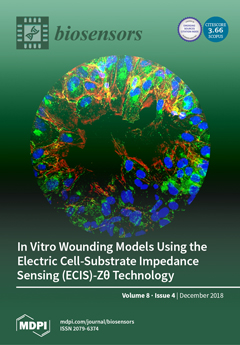
Cover Story (view full-size image): These images show differences in the wounding patterns of brain microvascular endothelial cells between 8W1E and 8W10E+ ECIS arrays. Nuclei are stained with DAPI (blue), and cells are stained for connexin43 (green) and actin (red). On 8W1E, only cell debris is left at 2 hours (green). On 8W10E+, cells remain, and cell migration occurs as the actin filaments protrude towards the center of the wounding site. View this paper.
  • Issues are regarded as officially published after their release is announced to the table of contents alert mailing list.
  • You may sign up for e-mail alerts to receive table of contents of newly released issues.
  • PDF is the official format for papers published in both, html and pdf forms. To view the papers in pdf format, click on the "PDF Full-text" link, and use the free Adobe Reader to open them.
Order results
Result details
Section
Select all
Export citation of selected articles as:
18 pages, 2228 KiB  
Review
Recent Progress in Surface Plasmon Resonance Biosensors (2016 to Mid-2018)
by Ewa Gorodkiewicz and Zenon Lukaszewski
Biosensors 2018, 8(4), 132; https://doi.org/10.3390/bios8040132 - 16 Dec 2018
Cited by 47 | Viewed by 6811
Abstract
More than 50 papers on surface plasmon resonance biosensors, published between 2016 and mid-2018, are reviewed. Papers concerning the determination of large particles such as vesicles, exosomes, cancer cells, living cells, stem cells, and microRNA are excluded, as these are covered by a [...] Read more.
More than 50 papers on surface plasmon resonance biosensors, published between 2016 and mid-2018, are reviewed. Papers concerning the determination of large particles such as vesicles, exosomes, cancer cells, living cells, stem cells, and microRNA are excluded, as these are covered by a very recent review. The reviewed papers are categorized into five groups, depending on the degree of maturity of the reported solution; ranging from simple marker detection to clinical application of a previously developed biosensor. Instrumental solutions and details of biosensor construction are analyzed, including the chips, receptors, and linkers used, as well as calibration strategies. Biosensors with a sandwich structure containing different nanoparticles are considered separately, as are SPR (Surface Plasmon Resonance) applications for investigating the interactions of biomolecules. An analysis is also made of the markers determined using the biosensors. In conclusion, there is shown to be a growing number of SPR applications in the solution of real clinical problems. Full article
(This article belongs to the Special Issue Surface Plasmon Resonance-Based Biosensors)
Show Figures

Figure 1

16 pages, 2236 KiB  
Article
Label-Free SERS Discrimination and In Situ Analysis of Life Cycle in Escherichia coli and Staphylococcus epidermidis
by Niccolò Paccotti, Francesco Boschetto, Satoshi Horiguchi, Elia Marin, Alessandro Chiadò, Chiara Novara, Francesco Geobaldo, Fabrizio Giorgis and Giuseppe Pezzotti
Biosensors 2018, 8(4), 131; https://doi.org/10.3390/bios8040131 - 15 Dec 2018
Cited by 15 | Viewed by 5856
Abstract
Surface enhanced Raman spectroscopy (SERS) has been proven suitable for identifying and characterizing different bacterial species, and to fully understand the chemically driven metabolic variations that occur during their evolution. In this study, SERS was exploited to identify the cellular composition of Gram-positive [...] Read more.
Surface enhanced Raman spectroscopy (SERS) has been proven suitable for identifying and characterizing different bacterial species, and to fully understand the chemically driven metabolic variations that occur during their evolution. In this study, SERS was exploited to identify the cellular composition of Gram-positive and Gram-negative bacteria by using mesoporous silicon-based substrates decorated with silver nanoparticles. The main differences between the investigated bacterial strains reside in the structure of the cell walls and plasmatic membranes, as well as their biofilm matrix, as clearly noticed in the corresponding SERS spectrum. A complete characterization of the spectra was provided in order to understand the contribution of each vibrational signal collected from the bacterial culture at different times, allowing the analysis of the bacterial populations after 12, 24, and 48 h. The results show clear features in terms of vibrational bands in line with the bacterial growth curve, including an increasing intensity of the signals during the first 24 h and their subsequent decrease in the late stationary phase after 48 h of culture. The evolution of the bacterial culture was also confirmed by fluorescence microscope images. Full article
(This article belongs to the Special Issue Applications of Raman Techniques in Biosensing)
Show Figures

Figure 1

17 pages, 1835 KiB  
Article
Rapid Antibody Selection Using Surface Plasmon Resonance for High-Speed and Sensitive Hazelnut Lateral Flow Prototypes
by Georgina M.S. Ross, Maria G.E.G. Bremer, Jan H. Wichers, Aart Van Amerongen and Michel W.F. Nielen
Biosensors 2018, 8(4), 130; https://doi.org/10.3390/bios8040130 - 14 Dec 2018
Cited by 25 | Viewed by 8129
Abstract
Lateral Flow Immunoassays (LFIAs) allow for rapid, low-cost, screening of many biomolecules such as food allergens. Despite being classified as rapid tests, many LFIAs take 10–20 min to complete. For a really high-speed LFIA, it is necessary to assess antibody association kinetics. By [...] Read more.
Lateral Flow Immunoassays (LFIAs) allow for rapid, low-cost, screening of many biomolecules such as food allergens. Despite being classified as rapid tests, many LFIAs take 10–20 min to complete. For a really high-speed LFIA, it is necessary to assess antibody association kinetics. By using a label-free optical technique such as Surface Plasmon Resonance (SPR), it is possible to screen crude monoclonal antibody (mAb) preparations for their association rates against a target. Herein, we describe an SPR-based method for screening and selecting crude anti-hazelnut antibodies based on their relative association rates, cross reactivity and sandwich pairing capabilities, for subsequent application in a rapid ligand binding assay. Thanks to the SPR selection process, only the fast mAb (F-50-6B12) and the slow (S-50-5H9) mAb needed purification for labelling with carbon nanoparticles to exploit high-speed LFIA prototypes. The kinetics observed in SPR were reflected in LFIA, with the test line appearing within 30 s, almost two times faster when F-50-6B12 was used, compared with S-50-5H9. Additionally, the LFIAs have demonstrated their future applicability to real life samples by detecting hazelnut in the sub-ppm range in a cookie matrix. Finally, these LFIAs not only provide a qualitative result when read visually, but also generate semi-quantitative data when exploiting freely downloadable smartphone apps. Full article
(This article belongs to the Special Issue Surface Plasmon Resonance-Based Biosensors)
Show Figures

Graphical abstract

18 pages, 2313 KiB  
Article
Preserved Microarrays for Simultaneous Detection and Identification of Six Fungal Potato Pathogens with the Use of Real-Time PCR in Matrix Format
by Maksim Nikitin, Ksenia Deych, Inessa Grevtseva, Natalya Girsova, Maria Kuznetsova, Mikhail Pridannikov, Vitaly Dzhavakhiya, Natalia Statsyuk and Alexander Golikov
Biosensors 2018, 8(4), 129; https://doi.org/10.3390/bios8040129 - 13 Dec 2018
Cited by 19 | Viewed by 5265
Abstract
Fungal diseases of plants are of great economic importance causing 70–80% of crop losses associated with microbial plant pathogens. Advanced on-site disease diagnostics is very important to maximize crop productivity. In this study, diagnostic systems have been developed for simultaneous detection and identification [...] Read more.
Fungal diseases of plants are of great economic importance causing 70–80% of crop losses associated with microbial plant pathogens. Advanced on-site disease diagnostics is very important to maximize crop productivity. In this study, diagnostic systems have been developed for simultaneous detection and identification of six fungal pathogens using 48-well microarrays (micromatrices) for qPCR. All oligonucleotide sets were tested for their specificity using 59 strains of target and non-target species. Detection limit of the developed test systems varied from 0.6 to 43.5 pg of DNA depending on target species with reproducibility within 0.3−0.7% (standard deviation). Diagnostic efficiency of test systems with stabilized and freeze-dried PCR master-mixes did not significantly differ from that of freshly prepared microarrays, though detection limit increased. Validation of test systems on 30 field samples of potato plants showed perfect correspondence with the results of morphological identification of pathogens. Due to the simplicity of the analysis and the automated data interpretation, the developed microarrays have good potential for on-site use by technician-level personnel, as well as for high-throughput monitoring of fungal potato pathogens. Full article
Show Figures

Figure 1

13 pages, 1554 KiB  
Article
Magnetic Nanoparticle-Based Biosensing Assay Quantitatively Enhances Acid-Fast Bacilli Count in Paucibacillary Pulmonary Tuberculosis
by Cristina Gordillo-Marroquín, Anaximandro Gómez-Velasco, Héctor J. Sánchez-Pérez, Kasey Pryg, John Shinners, Nathan Murray, Sergio G. Muñoz-Jiménez, Allied Bencomo-Alerm, Adriana Gómez-Bustamante, Letisia Jonapá-Gómez, Natán Enríquez-Ríos, Miguel Martín, Natalia Romero-Sandoval and Evangelyn C. Alocilja
Biosensors 2018, 8(4), 128; https://doi.org/10.3390/bios8040128 - 12 Dec 2018
Cited by 14 | Viewed by 9099
Abstract
A new method using a magnetic nanoparticle-based colorimetric biosensing assay (NCBA) was compared with sputum smear microscopy (SSM) for the detection of pulmonary tuberculosis (PTB) in sputum samples. Studies were made to compare the NCBA against SSM using sputum samples collected from PTB [...] Read more.
A new method using a magnetic nanoparticle-based colorimetric biosensing assay (NCBA) was compared with sputum smear microscopy (SSM) for the detection of pulmonary tuberculosis (PTB) in sputum samples. Studies were made to compare the NCBA against SSM using sputum samples collected from PTB patients prior to receiving treatment. Experiments were also conducted to determine the appropriate concentration of glycan-functionalized magnetic nanoparticles (GMNP) used in the NCBA and to evaluate the optimal digestion/decontamination solution to increase the extraction, concentration and detection of acid-fast bacilli (AFB). The optimized NCBA consisted of a 1:1 mixture of 0.4% NaOH and 4% N-acetyl-L-cysteine (NALC) to homogenize the sputum sample. Additionally, 10 mg/mL of GMNP was added to isolate and concentrate the AFB. All TB positive sputum samples were identified with an increased AFB count of 47% compared to SSM, demonstrating GMNP’s ability to extract and concentrate AFB. Results showed that NCBA increased AFB count compared to SSM, improving the grade from “1+” (in SSM) to “2+”. Extending the finding to paucibacillary cases, there is the likelihood of a “scant” grade to become “1+”. The assay uses a simple magnet and only costs $0.10/test. NCBA has great potential application in TB control programs. Full article
Show Figures

Figure 1

12 pages, 5102 KiB  
Article
MRI Study of the Influence of Surface Coating Aging on the In Vivo Biodistribution of Iron Oxide Nanoparticles
by Susana Carregal-Romero, Sandra Plaza-García, Rafael Piñol, José L. Murillo, Jesús Ruiz-Cabello, Daniel Padro, Angel Millán and Pedro Ramos-Cabrer
Biosensors 2018, 8(4), 127; https://doi.org/10.3390/bios8040127 - 12 Dec 2018
Cited by 13 | Viewed by 5188
Abstract
Medical imaging is an active field of research that fosters the necessity for novel multimodal imaging probes. In this line, nanoparticle-based contrast agents are of special interest, since those can host functional entities either within their interior, reducing potential toxic effects of the [...] Read more.
Medical imaging is an active field of research that fosters the necessity for novel multimodal imaging probes. In this line, nanoparticle-based contrast agents are of special interest, since those can host functional entities either within their interior, reducing potential toxic effects of the imaging tracers, or on their surface, providing high payloads of probes, due to their large surface-to-volume ratio. The long-term stability of the particles in solution is an aspect usually under-tackled during probe design in research laboratories, since their performance is generally tested briefly after synthesis. This may jeopardize a later translation into practical medical devices, due to stability reasons. To dig into the effects of nanoparticle aging in solution, with respect to their behavior in vivo, iron oxide stealth nanoparticles were used at two stages (3 weeks vs. 9 months in solution), analyzing their biodistribution in mice. Both sets of nanoprobes showed similar sizes, zeta potentials, and morphology, as observed by dynamic light scattering (DLS) and transmission electronic microscopy (TEM), but fresh nanoparticles accumulated in the kidneys after systemic administration, while aged ones accumulated in liver and spleen, confirming an enormous effect of particle aging on their in vivo behavior, despite barely noticeable changes perceived on a simple inspection of their structural integrity. Full article
(This article belongs to the Special Issue Functional Nanomaterials for Biosensing and Bioimaging)
Show Figures

Figure 1

13 pages, 1081 KiB  
Article
Development of DNA Pair Biosensor for Quantization of Nuclear Factor Kappa B
by Zhaohui Wang and Pak Kin Wong
Biosensors 2018, 8(4), 126; https://doi.org/10.3390/bios8040126 - 10 Dec 2018
Viewed by 4335
Abstract
Nuclear factor kappa B (NF-κB), regulating the expression of several genes that mediate the inflammatory responses and cell proliferation, is one of the therapeutic targets for chronic inflammatory disease and cancer. A novel molecular binding scheme for the detection of NF-κB was investigated [...] Read more.
Nuclear factor kappa B (NF-κB), regulating the expression of several genes that mediate the inflammatory responses and cell proliferation, is one of the therapeutic targets for chronic inflammatory disease and cancer. A novel molecular binding scheme for the detection of NF-κB was investigated for its affinity to Ig-κB DNA composed by dye and quencher fluorophores, and this specificity is confirmed by competing with the DNA sequence that is complementary to the Ig-κB DNA. We create a normalization equation to remove the negative effects from the various initial fluorophore concentrations and the background noise. We also found that a periodic shaking at a frequency could help to stabilize the DNA–protein binding. The calibration experiment, using purified p50 (NF-κB), shows that this molecular probe biosensor has a detection limit on the order of nanomolar. The limit of detection is determined by the binding performance of dye and quencher oligonucleotides, and only a small portion of probes are stabilized by DNA-binding protein NF-κB. The specificity experiment also shows that p50/p65 heterodimer has the highest affinity for Ig-κB DNA; p65 homodimer binds with intermediate affinity, whereas p50 shows the lowest binding affinity, and Ig-κB DNA is not sensitive to BSA (bovine albumin serum). The experiment of HeLa nuclear extract shows that TNF-α stimulated HeLa nuclear extract has higher affinity to Ig-κB DNA than non-TNF-stimulated HeLa nuclear extract (4-h serum response). Therefore, the molecular binding scheme provides a rapid, quantitative, high throughput, and automated measurement of the DNA-binding protein NF-κB at low cost, which is beneficial for automated drug screening systems. Full article
(This article belongs to the Special Issue FRET-Based Biosensors)
Show Figures

Figure 1

11 pages, 1460 KiB  
Article
A Passive Microfluidic Device Based on Crossflow Filtration for Cell Separation Measurements: A Spectrophotometric Characterization
by Vera Faustino, Susana O. Catarino, Diana Pinho, Rui A. Lima and Graça Minas
Biosensors 2018, 8(4), 125; https://doi.org/10.3390/bios8040125 - 09 Dec 2018
Cited by 20 | Viewed by 4393
Abstract
Microfluidic devices have been widely used as a valuable research tool for diagnostic applications. Particularly, they have been related to the successful detection of different diseases and conditions by assessing the mechanical properties of red blood cells (RBCs). Detecting deformability changes in the [...] Read more.
Microfluidic devices have been widely used as a valuable research tool for diagnostic applications. Particularly, they have been related to the successful detection of different diseases and conditions by assessing the mechanical properties of red blood cells (RBCs). Detecting deformability changes in the cells and being able to separate those cells may be a key factor in assuring the success of detection of some blood diseases with diagnostic devices. To detect and separate the chemically modified RBCs (mimicking disease-infected RBCs) from healthy RBCs, the present work proposes a microfluidic device comprising a sequence of pillars with different gaps and nine different outlets used to evaluate the efficiency of the device by measuring the optical absorption of the collected samples. This latter measurement technique was tested to distinguish between healthy RBCs and RBCs chemically modified with glutaraldehyde. The present study indicates that it was possible to detect a slight differences between the samples using an optical absorption spectrophotometric setup. Hence, the proposed microfluidic device has the potential to perform in one single step a partial passive separation of RBCs based on their deformability. Full article
Show Figures

Figure 1

10 pages, 4192 KiB  
Communication
A Theoretical Study of Surface Mode Propagation with a Guiding Layer of GaN/Sapphire Hetero-Structure in Liquid Medium
by M. F. Mohd Razip Wee, Muhammad Musoddiq Jaafar, Mohd Syafiq Faiz, Chang Fu Dee and Burhanuddin Yeop Majlis
Biosensors 2018, 8(4), 124; https://doi.org/10.3390/bios8040124 - 05 Dec 2018
Viewed by 3759
Abstract
Gallium Nitride (GaN) is considered as the second most popular semiconductor material in industry after silicon. This is due to its wide applications encompassing Light Emitting Diode (LED) and power electronics. In addition, its piezoelectric properties are fascinating to be explored as electromechanical [...] Read more.
Gallium Nitride (GaN) is considered as the second most popular semiconductor material in industry after silicon. This is due to its wide applications encompassing Light Emitting Diode (LED) and power electronics. In addition, its piezoelectric properties are fascinating to be explored as electromechanical material for the development of diverse microelectromechanical systems (MEMS) application. In this article, we conducted a theoretical study concerning surface mode propagation, especially Rayleigh and Sezawa mode in the layered GaN/sapphire structure with the presence of various guiding layers. It is demonstrated that the increase in thickness of guiding layer will decrease the phase velocities of surface mode depending on the material properties of the layer. In addition, the Q-factor value indicating the resonance properties of surface mode appeared to be affected with the presence of fluid domain, particularly in the Rayleigh mode. Meanwhile, the peak for Sezawa mode shows the highest Q factor and is not altered by the presence of fluid. Based on these theoretical results using the finite element method, it could contribute to the development of a GaN-based device to generate surface acoustic wave, especially in Sezawa mode which could be useful in acoustophoresis, lab on-chip and microfluidics applications. Full article
(This article belongs to the Special Issue Acoustic Wave Sensors for Biosensing Applications)
Show Figures

Figure 1

8 pages, 1207 KiB  
Article
Biomolecular Component Analysis of Phospholipids Composition in Live HeLa Cells
by Svitlana M. Levchenko and Junle Qu
Biosensors 2018, 8(4), 123; https://doi.org/10.3390/bios8040123 - 05 Dec 2018
Cited by 6 | Viewed by 4772
Abstract
The alteration of the phospholipid composition within the cell, in particular the ratio between saturated and unsaturated fatty acids, can serve as an important biomarker to prognosis of the disease progression (e.g., fatty-liver disease, prostate cancer, or neurodegenerative disorders). Major techniques for lipid [...] Read more.
The alteration of the phospholipid composition within the cell, in particular the ratio between saturated and unsaturated fatty acids, can serve as an important biomarker to prognosis of the disease progression (e.g., fatty-liver disease, prostate cancer, or neurodegenerative disorders). Major techniques for lipid analysis in biological samples require a lipid extraction procedure that is not compatible with live cell studies. To address this challenge, we apply microRaman-Biomolecular Component Analysis (BCA) for comparative analysis of phospholipid composition and sensing the saturation degree of fatty acid lipid chain in live HeLa cells and lipids extracted from HeLa cells. After processing raw Raman data, acquired in lipid droplets (LDs) free cytoplasmic area, LDs and extracted lipids with BCA, the lipid component was isolated. Despite the similarity in general profiles of processed Raman spectra acquired in live cells and extracted lipids, some clear differences that reflect diversity in their phospholipids composition were revealed. Furthermore, using the direct relation between the number of double bonds in the fatty acid chain and the intensity ratio of the corresponding Raman bands, the saturation degree of fatty acids was estimated. Full article
(This article belongs to the Special Issue Applications of Raman Techniques in Biosensing)
Show Figures

Figure 1

19 pages, 5125 KiB  
Article
Evaluation of Continuous Lactate Monitoring Systems within a Heparinized In Vivo Porcine Model Intravenously and Subcutaneously
by Alexander Wolf, Kevin Renehan, Kenneth K. Y. Ho, Benjamin D. Carr, Chieh V. Chen, Marie S. Cornell, Minyi Ye, Alvaro Rojas-Peña and Hao Chen
Biosensors 2018, 8(4), 122; https://doi.org/10.3390/bios8040122 - 04 Dec 2018
Cited by 10 | Viewed by 6557
Abstract
We present an animal model used to evaluate the in vivo performance of electrochemical amperometric continuous lactate sensors compared to blood gas instruments. Electrochemical lactate sensors were fabricated, placed into 5 Fr central venous catheters (CVCs), and paired with wireless potentiostat devices. Following [...] Read more.
We present an animal model used to evaluate the in vivo performance of electrochemical amperometric continuous lactate sensors compared to blood gas instruments. Electrochemical lactate sensors were fabricated, placed into 5 Fr central venous catheters (CVCs), and paired with wireless potentiostat devices. Following in vivo evaluation and calibration, sensors were placed within the jugular and femoral veins of a porcine subject as a preliminary assessment of in vivo measurement accuracy. The mobile electronic circuit potentiostat devices supplied the operational voltage for the sensors, measured the resultant steady-state current, and recorded the sensor response values in internal memory storages. An in vivo time trace of implanted intravenous (IV) sensors demonstrated lactate values that correlated well with the discrete measurements of blood samples on a benchtop point-of-care sensor-based instrument. Currents measured continuously from the implanted lactate sensors over 10 h were converted into lactate concentration values through use of a two-point in vivo calibration. Study shows that intravenously implanted sensors had more accurate readings, faster peak-reaching rates, and shorter peak-detection times compared to subcutaneously placed sensors. IV implanted and subcutaneously placed sensors closer to the upper body (in this case neck) showed faster response rates and more accurate measurements compared to those implanted in the lower portion of the porcine model. This study represents an important milestone not only towards continuous lactate monitoring for early diagnosis and intervention in neonatal patients with congenital heart disease undergoing cardiopulmonary bypass surgeries, but also in the intervention of critical ill patients in the Intensive Care Units or during complex surgical procedures. Full article
Show Figures

Figure 1

13 pages, 6124 KiB  
Article
Non-Invasive Diagnosis of Diabetes by Volatile Organic Compounds in Urine Using FAIMS and Fox4000 Electronic Nose
by Siavash Esfahani, Alfian Wicaksono, Ella Mozdiak, Ramesh P. Arasaradnam and James A. Covington
Biosensors 2018, 8(4), 121; https://doi.org/10.3390/bios8040121 - 01 Dec 2018
Cited by 38 | Viewed by 7056
Abstract
The electronic nose (eNose) is an instrument designed to mimic the human olfactory system. Usage of eNose in medical applications is more popular than ever, due to its low costs and non-invasive nature. The eNose sniffs the gases and vapours that emanate from [...] Read more.
The electronic nose (eNose) is an instrument designed to mimic the human olfactory system. Usage of eNose in medical applications is more popular than ever, due to its low costs and non-invasive nature. The eNose sniffs the gases and vapours that emanate from human waste (urine, breath, and stool) for the diagnosis of variety of diseases. Diabetes mellitus type 2 (DM2) affects 8.3% of adults in the world, with 43% being underdiagnosed, resulting in 4.9 million deaths per year. In this study, we investigated the potential of urinary volatile organic compounds (VOCs) as novel non-invasive diagnostic biomarker for diabetes. In addition, we investigated the influence of sample age on the diagnostic accuracy of urinary VOCs. We analysed 140 urine samples (73 DM2, 67 healthy) with Field-Asymmetric Ion Mobility Spectrometry (FAIMS); a type of eNose; and FOX 4000 (AlphaM.O.S, Toulouse, France). Urine samples were collected at UHCW NHS Trust clinics over 4 years and stored at −80 °C within two hours of collection. Four different classifiers were used for classification, specifically Sparse Logistic Regression, Random Forest, Gaussian Process, and Support Vector on both FAIMS and FOX4000. Both eNoses showed their capability of diagnosing DM2 from controls and the effect of sample age on the discrimination. FAIMS samples were analysed for all samples aged 0–4 years (AUC: 88%, sensitivity: 87%, specificity: 82%) and then sub group samples aged less than a year (AUC (Area Under the Curve): 94%, Sensitivity: 92%, specificity: 100%). FOX4000 samples were analysed for all samples aged 0–4 years (AUC: 85%, sensitivity: 77%, specificity: 85%) and a sub group samples aged less than 18 months: (AUC: 94%, sensitivity: 90%, specificity: 89%). We demonstrated that FAIMS and FOX 4000 eNoses can discriminate DM2 from controls using urinary VOCs. In addition, we showed that urine sample age affects discriminative accuracy. Full article
Show Figures

Figure 1

14 pages, 1955 KiB  
Review
Chiral Plasmonic Biosensors
by Vladimir E. Bochenkov and Tatyana I. Shabatina
Biosensors 2018, 8(4), 120; https://doi.org/10.3390/bios8040120 - 01 Dec 2018
Cited by 38 | Viewed by 8442
Abstract
Biosensing requires fast, selective, and highly sensitive real-time detection of biomolecules using efficient simple-to-use techniques. Due to a unique capability to focus light at nanoscale, plasmonic nanostructures provide an excellent platform for label-free detection of molecular adsorption by sensing tiny changes in the [...] Read more.
Biosensing requires fast, selective, and highly sensitive real-time detection of biomolecules using efficient simple-to-use techniques. Due to a unique capability to focus light at nanoscale, plasmonic nanostructures provide an excellent platform for label-free detection of molecular adsorption by sensing tiny changes in the local refractive index or by enhancing the light-induced processes in adjacent biomolecules. This review discusses the opportunities provided by surface plasmon resonance in probing the chirality of biomolecules as well as their conformations and orientations. Various types of chiral plasmonic nanostructures and the most recent developments in the field of chiral plasmonics related to biosensing are considered. Full article
(This article belongs to the Special Issue Surface Plasmon Resonance-Based Biosensors)
Show Figures

Figure 1

10 pages, 987 KiB  
Review
Assessing the Potential Deployment of Biosensors for Point-of-Care Diagnostics in Developing Countries: Technological, Economic and Regulatory Aspects
by Daniel Migliozzi and Thomas Guibentif
Biosensors 2018, 8(4), 119; https://doi.org/10.3390/bios8040119 - 29 Nov 2018
Cited by 8 | Viewed by 5613
Abstract
Infectious diseases and antimicrobial resistance are major burdens in developing countries, where very specific conditions impede the deployment of established medical infrastructures. Since biosensing devices are nowadays very common in developed countries, particularly in the field of diagnostics, they are at a stage [...] Read more.
Infectious diseases and antimicrobial resistance are major burdens in developing countries, where very specific conditions impede the deployment of established medical infrastructures. Since biosensing devices are nowadays very common in developed countries, particularly in the field of diagnostics, they are at a stage of maturity at which other potential outcomes can be explored, especially on their possibilities for multiplexing and automation to reduce the time-to-results. However, the translation is far from being trivial. In order to understand the factors and barriers that can facilitate or hinder the application of biosensors in resource-limited settings, we analyze the context from several angles. First, the technology of the devices themselves has to be rethought to take into account the specific needs and the available means of these countries. For this, we describe the partition of a biosensor into its functional shells, which define the information flow from the analyte to the end-user, and by following this partition we assess the strengths and weaknesses of biosensing devices in view of their specific technological development and challenging deployment in low-resource environments. Then, we discuss the problem of cost reduction by pointing out transversal factors, such as throughput and cost of mistreatment, that need to be re-considered when analyzing the cost-effectiveness of biosensing devices. Beyond the technical landscape, the compliance with regulations is also a major aspect that is described with its link to the validation of the devices and to the acceptance from the local medical personnel. Finally, to learn from a successful case, we analyze a breakthrough inexpensive biosensor that is showing high potential with respect to many of the described aspects. We conclude by mentioning both some transversal benefits of deploying biosensors in developing countries, and the key factors that can drive such applications. Full article
Show Figures

Figure 1

10 pages, 3325 KiB  
Article
Microwave Spectroscopic Detection of Human Hsp70 Protein on Annealed Gold Nanostructures on ITO Glass Strips
by Rodica Elena Ionescu, Raphael Selon, Nicolas Pocholle, Lan Zhou, Anna Rumyantseva, Eric Bourillot and Eric Lesniewska
Biosensors 2018, 8(4), 118; https://doi.org/10.3390/bios8040118 - 27 Nov 2018
Cited by 1 | Viewed by 4551
Abstract
Conductive indium-tin oxide (ITO) and non-conductive glass substrates were successfully modified with embedded gold nanoparticles (AuNPs) formed by controlled thermal annealing at 550 °C for 8 h in a preselected oven. The authors characterized the formation of AuNPs using two microscopic techniques: scanning [...] Read more.
Conductive indium-tin oxide (ITO) and non-conductive glass substrates were successfully modified with embedded gold nanoparticles (AuNPs) formed by controlled thermal annealing at 550 °C for 8 h in a preselected oven. The authors characterized the formation of AuNPs using two microscopic techniques: scanning electron microscopy (SEM) and atomic force microscopy (AFM). The analytical performances of the nanostructured-glasses were compared regarding biosensing of Hsp70, an ATP-driven molecular chaperone. In this work, the human heat-shock protein (Hsp70), was chosen as a model biomarker of body stress disorders for microwave spectroscopic investigations. It was found that microwave screening at 4 GHz allowed for the first time the detection of 12 ng/µL/cm2 of Hsp70. Full article
(This article belongs to the Special Issue Immunoanalytical Tool with Electrochemical and Optical Detection)
Show Figures

Figure 1

11 pages, 2358 KiB  
Article
A Liposomal Platform for Sensing of Extracellular Analytes Near Cells
by Xiaozhou Zhang, Sabrina Heng, Jinxin Pei, Jacqueline R. Morey, Christopher A. McDevitt and Andrew D. Abell
Biosensors 2018, 8(4), 117; https://doi.org/10.3390/bios8040117 - 26 Nov 2018
Cited by 4 | Viewed by 4238
Abstract
Cell-permeable fluorescent chemosensors (calcein, monochlorobimane, and a recently reported spiropyran-based sensor SP2) have been incorporated into yeast total lipid extract-based liposomes to suppress inherent cell permeability to allow the detection of extracellular Ca2+, GSH, and Zn2+, respectively. The repurposed [...] Read more.
Cell-permeable fluorescent chemosensors (calcein, monochlorobimane, and a recently reported spiropyran-based sensor SP2) have been incorporated into yeast total lipid extract-based liposomes to suppress inherent cell permeability to allow the detection of extracellular Ca2+, GSH, and Zn2+, respectively. The repurposed sensors have enhanced aqueous solubility and the ability to quantitatively measure biologically relevant concentrations of Ca2+ (0.25 mM–1 mM), Zn2+ (6.25 µM–50 µM), and GSH (0.25 mM–1 mM) by fluorescence in aqueous media. In addition, the liposomal sensors are nontoxic to HEK293 cells and have the ability to detect exogenously added Zn2+ (1 mM), Ca2+ (1 mM), or GSH (1 mM) near cells without internalisation. This new sensing platform provides a means to repurpose a range of intracellular fluorescent sensors to specifically detect extracellular analytes, while also improving biocompatibility for overall enhanced use in a wide range of biomedical applications. Full article
Show Figures

Graphical abstract

11 pages, 3828 KiB  
Article
Cerium Oxide-Tungsten Oxide Core-Shell Nanowire-Based Microsensors Sensitive to Acetone
by Milena Tomić, Milena Šetka, Ondřej Chmela, Isabel Gràcia, Eduard Figueras, Carles Cané and Stella Vallejos
Biosensors 2018, 8(4), 116; https://doi.org/10.3390/bios8040116 - 23 Nov 2018
Cited by 14 | Viewed by 5634
Abstract
Gas sensitive cerium oxide-tungsten oxide core-shell nanowires are synthesized and integrated directly into micromachined platforms via aerosol assisted chemical vapor deposition. Tests to various volatile organic compounds (acetone, ethanol, and toluene) involved in early disease diagnosis demonstrate enhanced sensitivity to acetone for the [...] Read more.
Gas sensitive cerium oxide-tungsten oxide core-shell nanowires are synthesized and integrated directly into micromachined platforms via aerosol assisted chemical vapor deposition. Tests to various volatile organic compounds (acetone, ethanol, and toluene) involved in early disease diagnosis demonstrate enhanced sensitivity to acetone for the core-shell structures in contrast to the non-modified materials (i.e., only tungsten oxide or cerium oxide). This is attributed to the high density of oxygen vacancy defects at the shell, as well as the formation of heterojunctions at the core-shell interface, which provide the modified nanowires with ‘extra’ chemical and electronic sensitization as compared to the non-modified materials. Full article
Show Figures

Figure 1

8 pages, 572 KiB  
Review
Use of Electronic Noses in Seawater Quality Monitoring: A Systematic Review
by Alessandro Tonacci, Francesco Sansone, Raffaele Conte and Claudio Domenici
Biosensors 2018, 8(4), 115; https://doi.org/10.3390/bios8040115 - 23 Nov 2018
Cited by 10 | Viewed by 4483
Abstract
Electronic nose (eNose) systems are particularly appreciated for their portability, usability, relative low cost, and real-time or near real-time response. Their application finds space in several domains, including environmental monitoring. Within this field, marine monitoring is of particular scientific relevance due to the [...] Read more.
Electronic nose (eNose) systems are particularly appreciated for their portability, usability, relative low cost, and real-time or near real-time response. Their application finds space in several domains, including environmental monitoring. Within this field, marine monitoring is of particular scientific relevance due to the fragility of this specific environment, daily threatened by human activities that can potentially bring to catastrophic and irreversible consequences on marine wildlife. Under such considerations, a systematic review, complying with the PRISMA guidelines, was conducted covering the period up to 15 October 2018, in PubMed, ScienceDirect, and Google Scholar. Despite the relatively low number of articles published on this specific topic and the heterogeneity of the technological approaches employed, the results obtained by the various groups highlight the positive contribution eNose has given and can provide in near future for the monitoring and safeguarding of this delicate environment. Full article
Show Figures

Figure 1

32 pages, 2982 KiB  
Review
Point-of-Care Compatibility of Ultra-Sensitive Detection Techniques for the Cardiac Biomarker Troponin I—Challenges and Potential Value
by Brian Regan, Richard O’Kennedy and David Collins
Biosensors 2018, 8(4), 114; https://doi.org/10.3390/bios8040114 - 21 Nov 2018
Cited by 34 | Viewed by 9894
Abstract
Cardiac biomarkers are frequently measured to provide guidance on the well-being of a patient in relation to cardiac health with many assays having been developed and widely utilised in clinical assessment. Effectively treating and managing cardiovascular disease (CVD) relies on swiftly responding to [...] Read more.
Cardiac biomarkers are frequently measured to provide guidance on the well-being of a patient in relation to cardiac health with many assays having been developed and widely utilised in clinical assessment. Effectively treating and managing cardiovascular disease (CVD) relies on swiftly responding to signs of cardiac symptoms, thus providing a basis for enhanced patient management and an overall better health outcome. Ultra-sensitive cardiac biomarker detection techniques play a pivotal role in improving the diagnostic capacity of an assay and thus enabling a better-informed decision. However, currently, the typical approach taken within healthcare depends on centralised laboratories performing analysis of cardiac biomarkers, thus restricting the roll-out of rapid diagnostics. Point-of-care testing (POCT) involves conducting the diagnostic test in the presence of the patient, with a short turnaround time, requiring small sample volumes without compromising the sensitivity of the assay. This technology is ideal for combatting CVD, thus the formulation of ultra-sensitive assays and the design of biosensors will be critically evaluated, focusing on the feasibility of these techniques for point-of-care (POC) integration. Moreover, there are several key factors, which in combination, contribute to the development of ultra-sensitive techniques, namely the incorporation of nanomaterials for sensitivity enhancement and manipulation of labelling methods. This review will explore the latest developments in cardiac biomarker detection, primarily focusing on the detection of cardiac troponin I (cTnI). Highly sensitive detection of cTnI is of paramount importance regarding the rapid rule-in/rule-out of acute myocardial infarction (AMI). Thus the challenges encountered during cTnI measurements are outlined in detail to assist in demonstrating the drawbacks of current commercial assays and the obstructions to standardisation. Furthermore, the added benefits of introducing multi-biomarker panels are reviewed, several key biomarkers are evaluated and the analytical benefits provided by multimarkers-based methods are highlighted. Full article
Show Figures

Figure 1

10 pages, 6567 KiB  
Article
One-Pot Synthesis of Multi-Branch Gold Nanoparticles and Investigation of Their SERS Performance
by Weifeng Lv, Chenjie Gu, Shuwen Zeng, Jiaguang Han, Tao Jiang and Jun Zhou
Biosensors 2018, 8(4), 113; https://doi.org/10.3390/bios8040113 - 20 Nov 2018
Cited by 17 | Viewed by 5222
Abstract
Gold nanoparticles with multiple branches have attracted intensive studies for their application in sensing of low trace molecules. A large number of the merits found on the gold nanoparticles for the above applications are attributed to the strong localized surface plasmon resonance excited [...] Read more.
Gold nanoparticles with multiple branches have attracted intensive studies for their application in sensing of low trace molecules. A large number of the merits found on the gold nanoparticles for the above applications are attributed to the strong localized surface plasmon resonance excited by the incident radiation. However, a facile and flexible way of synthesizing the multi-branch gold nanoparticles with tunable localized surface plasmon resonance frequency is still a challenge for the plasmonic research field. Herein, we report an efficient one-pot synthesis of multi-branch gold nanoparticles method that resembles a seed-medicated approach while using no further chemicals except chloroauric acid, ascorbic acid and 4-(2-Hydroxyethyl)-1-piperazinyl]-ethanesulfonic acid. By controlling the amounts of ascorbic acid volumes in the reaction mixture, the morphology and the localized surface plasmon resonance frequency of the synthesized multi-branch gold nanoparticles can be manipulated conveniently. Moreover, using the 4-Mercaptobenzoic acid as the Raman reporter, the multi-branch gold nanoparticles show superior surface-enhanced Raman spectroscopy characteristics that can be potentially used in chemical and biological sensing. Full article
Show Figures

Figure 1

13 pages, 1680 KiB  
Article
Towards Simazine Monitoring in Agro-Zootechnical Productions: A Yeast Cell Bioprobe for Real Samples Screening
by Gerardo Grasso, Ludovico Caracciolo, Giulia Cocco, Chiara Frazzoli and Roberto Dragone
Biosensors 2018, 8(4), 112; https://doi.org/10.3390/bios8040112 - 15 Nov 2018
Cited by 4 | Viewed by 4681
Abstract
Simazine is an herbicide that is able to contaminate surface waters, ground waters, and milk/dairy products, thus posing concerns in both environmental health and food safety. A yeast-based bioprobe was utilized to detect simazine in spiked real samples of livestock drinking water and [...] Read more.
Simazine is an herbicide that is able to contaminate surface waters, ground waters, and milk/dairy products, thus posing concerns in both environmental health and food safety. A yeast-based bioprobe was utilized to detect simazine in spiked real samples of livestock drinking water and raw cow’s milk. Yeast aerobic respiration was taken as short-term toxicological endpoint. We carried out comparative measures of yeast oxygen consumption between simazine-spiked samples and blank samples. Percentage interference (%ρ) on yeast aerobic respiration was calculated through the comparison of aerobic respiration of simazine-exposed and non-exposed yeast cells. The method was optimized for raw cow’s milk samples by using boric acid as fungistatic agent in order to avoid cellular proliferation. Overall, the results have shown that simazine can be detected up to concentrations five times below the EU legal concentration limits for drinking water (0.02 ppb) and cow’s milk (2 ppb) (%ρ values of 18.53% and 20.43% respectively; %RSD ≤ 15%). Dose-effect relationships of simazine were assessed. The findings of the bioassays match reasonably well with known mechanisms of toxicity and intracellular detoxification in yeast. A correlation between fat content in milk samples and analytical performance of the bioprobe was established. Results suggest the involvement of a matrix effect, presumably due to lipid sequestration of simazine. The yeast-based bioprobe has proved to be sensitive and suitable for the detection of simazine in real samples in concentrations of interest. Full article
Show Figures

Graphical abstract

14 pages, 2502 KiB  
Article
Lateral Flow Immunoassay for Rapid Detection of Grapevine Leafroll-Associated Virus
by Nadezhda A. Byzova, Svetlana V. Vinogradova, Elena V. Porotikova, Uliana D. Terekhova, Anatoly V. Zherdev and Boris B. Dzantiev
Biosensors 2018, 8(4), 111; https://doi.org/10.3390/bios8040111 - 15 Nov 2018
Cited by 27 | Viewed by 6581
Abstract
Grapevine leafroll-associated virus 3 (GLRaV-3) is one of the main pathogens of grapes, causing a significant loss in yield and decrease in quality for this agricultural plant. For efficient widespread control of this infection, rapid and simple analytical techniques of on-site testing are [...] Read more.
Grapevine leafroll-associated virus 3 (GLRaV-3) is one of the main pathogens of grapes, causing a significant loss in yield and decrease in quality for this agricultural plant. For efficient widespread control of this infection, rapid and simple analytical techniques of on-site testing are requested as a complementary addition for the currently applied hybridization (PCR) and immunoenzyme (ELISA) approaches. The given paper presents development and approbation of the immunochromatographic assay (ICA) for rapid detection of GLRaV-3. The ICA realizes a sandwich immunoassay format with the obtaining complexes ((antibody immobilized on immunochromatographic membrane)–(virus in the sample)–(antibody immobilized on gold nanoparticles (GNP)) during sample flow along the membrane compounds of the test strip. Three preparations of GNPs were compared for detection of GLRaV-3 at different dilutions of virus-containing sample. The GNPs with maximal average diameters of 51.0 ± 7.9 nm provide GLRaV-3 detection for its maximal dilutions, being 4 times more than when using GNPs with a diameter of 28.3 ± 3.3 nm, and 8 times more than when using GNPs with a diameter of 18.5 ± 3.3 nm. Test strips have been manufactured using the largest GNPs conjugated with anti-GLRaV-3 antibodies at a ratio of 1070:1. When testing samples containing other grape wine viruses, the test strips have not demonstrated staining in the test zone, which confirms the ICA specificity. The approbation of the manufactured test strips indicated that when using ELISA as a reference method, the developed ICA is characterized by a sensitivity of 100% and a specificity of 92%. If PCR is considered as a reference method, then the sensitivity of ICA is 93% and the specificity is 92%. The proposed ICA can be implemented in one stage without the use of any additional reactants or devices. The testing results can be obtained in 10 min and detected visually. It provides significant improvement in GLRaV-3 detection, and the presented approach can be transferred for the development of test systems for other grape wine pathogens. Full article
Show Figures

Figure 1

11 pages, 2327 KiB  
Article
Sensors Based on Metal Nanoclusters Stabilized on Designed Proteins
by Antonio Aires, Elena Lopez-Martinez and Aitziber L. Cortajarena
Biosensors 2018, 8(4), 110; https://doi.org/10.3390/bios8040110 - 15 Nov 2018
Cited by 14 | Viewed by 5182
Abstract
Among all new nanomaterials, metal nanoclusters (NCs) have attracted special attention due to their interesting optical properties, among others. Metal NCs have been recently studied and used as sensors for different analytes. However, there is a need to explore the potential of these [...] Read more.
Among all new nanomaterials, metal nanoclusters (NCs) have attracted special attention due to their interesting optical properties, among others. Metal NCs have been recently studied and used as sensors for different analytes. However, there is a need to explore the potential of these new sensors in a systematic manner and to develop new systems to broaden the possibilities that sensing offers to the industry. In this work, we show the potential use of repeat protein scaffolds as versatile templates for the synthesis and stabilization of various metal NCs, specifically Au, Ag, and CuNCs. The resulting protein-metal NCs hybrids are evaluated as sensors for different stimuli such as temperature, ions, or reactive oxygen species (ROS). Among the three protein-metal NCs, all performed nicely as temperature sensors, AuNCs responded to metal ions, and AgNCs were able to detect ROS. Full article
(This article belongs to the Special Issue Functional Nanomaterials for Biosensing and Bioimaging)
Show Figures

Graphical abstract

9 pages, 2469 KiB  
Article
Electrochemical Determination of β-Lactoglobulin Employing a Polystyrene Bead-Modified Carbon Nanotube Ink
by Judith Molinari, Laura Florez, Anahí Medrano, Leandro Monsalve and Gabriel Ybarra
Biosensors 2018, 8(4), 109; https://doi.org/10.3390/bios8040109 - 15 Nov 2018
Cited by 13 | Viewed by 4321
Abstract
In this article, we introduce the use of a carboxy-functionalized waterborne carbon nanotube ink for the fabrication of an amperometric biosensor aimed at the quantification of β-lactoglobulin. Detection of this protein from cow’s milk was performed by a sandwich immunoassay onto printed carbon [...] Read more.
In this article, we introduce the use of a carboxy-functionalized waterborne carbon nanotube ink for the fabrication of an amperometric biosensor aimed at the quantification of β-lactoglobulin. Detection of this protein from cow’s milk was performed by a sandwich immunoassay onto printed carbon nanotube electrodes. The electrodes were printed using a carbon nanotube ink modified with polystyrene beads containing a high amount of carboxylic groups for protein immobilization. This strategy showed enhanced sensing performance compared to the use of oxidative treatments for the functionalization of electrodes. These electrodes showed an excellent electrochemical behavior, and proteins could be immobilized on their surface via the carbodiimide reaction. These antibody-immobilized carbon nanotube electrodes allowed for the detection of β-lactoglobulin in sub-ppm concentrations. Full article
(This article belongs to the Special Issue Electrochemical Immunosensor)
Show Figures

Graphical abstract

15 pages, 1638 KiB  
Article
Metal Oxide Nanoparticle Based Electrochemical Sensor for Total Antioxidant Capacity (TAC) Detection in Wine Samples
by Cristina Tortolini, Paolo Bollella, Rosaceleste Zumpano, Gabriele Favero, Franco Mazzei and Riccarda Antiochia
Biosensors 2018, 8(4), 108; https://doi.org/10.3390/bios8040108 - 14 Nov 2018
Cited by 30 | Viewed by 5583
Abstract
A single-use electrochemical screen-printed electrode is reported based on biomimetic properties of nanoceria particles (CeNPs). The developed tool showed an easy approach compared to the classical spectrophotometric methods reported in literature in terms of ease of use, cost, portability, and unnecessary secondary reagents. [...] Read more.
A single-use electrochemical screen-printed electrode is reported based on biomimetic properties of nanoceria particles (CeNPs). The developed tool showed an easy approach compared to the classical spectrophotometric methods reported in literature in terms of ease of use, cost, portability, and unnecessary secondary reagents. The sensor allowed the detection of the total antioxidant capacity (TAC) in wine samples. The sensor has been optimized and characterized electrochemically and then tested with antioxidant compounds occurred in wine samples. The electrochemical CeNPs modified sensor has been used for detection of TAC in white and red commercial wines and the data compared to the 2,2′-azino-bis(3-ethylbenzthiazoline-6-sulphonic acid (ABTS)-based spectrophotometric method. Finally, the obtained results have demonstrated that the proposed sensor was suitable for the simple and quick evaluation of TAC in beverage samples. Full article
(This article belongs to the Special Issue Enzymatic Electrochemical Biosensors)
Show Figures

Figure 1

19 pages, 4242 KiB  
Review
Raman Spectroscopy and Microscopy Applications in Cardiovascular Diseases: From Molecules to Organs
by Ardalan Chaichi, Alisha Prasad and Manas Ranjan Gartia
Biosensors 2018, 8(4), 107; https://doi.org/10.3390/bios8040107 - 12 Nov 2018
Cited by 37 | Viewed by 9088
Abstract
Noninvasive and label-free vibrational spectroscopy and microscopy methods have shown great potential for clinical diagnosis applications. Raman spectroscopy is based on inelastic light scattering due to rotational and vibrational modes of molecular bonds. It has been shown that Raman spectra provide chemical signatures [...] Read more.
Noninvasive and label-free vibrational spectroscopy and microscopy methods have shown great potential for clinical diagnosis applications. Raman spectroscopy is based on inelastic light scattering due to rotational and vibrational modes of molecular bonds. It has been shown that Raman spectra provide chemical signatures of changes in biological tissues in different diseases, and this technique can be employed in label-free monitoring and clinical diagnosis of several diseases, including cardiovascular studies. However, there are very few literature reviews available to summarize the state of art and future applications of Raman spectroscopy in cardiovascular diseases, particularly cardiac hypertrophy. In addition to conventional clinical approaches such as electrocardiography (ECG), echocardiogram (cardiac ultrasound), positron emission tomography (PET), cardiac computed tomography (CT), and single photon emission computed tomography (SPECT), applications of vibrational spectroscopy and microscopy will provide invaluable information useful for the prevention, diagnosis, and treatment of cardiovascular diseases. Various in vivo and ex vivo investigations can potentially be performed using Raman imaging to study and distinguish pathological and physiological cardiac hypertrophies and understand the mechanisms of other cardiac diseases. Here, we have reviewed the recent literature on Raman spectroscopy to study cardiovascular diseases covering investigations on the molecular, cellular, tissue, and organ level. Full article
(This article belongs to the Special Issue Applications of Raman Techniques in Biosensing)
Show Figures

Figure 1

11 pages, 1849 KiB  
Article
BCAbox Algorithm Expands Capabilities of Raman Microscope for Single Organelles Assessment
by Andrey N. Kuzmin, Artem Pliss, Alex Rzhevskii, Adrian Lita and Mioara Larion
Biosensors 2018, 8(4), 106; https://doi.org/10.3390/bios8040106 - 10 Nov 2018
Cited by 15 | Viewed by 6096
Abstract
Raman microspectroscopy is a rapidly developing technique, which has an unparalleled potential for in situ proteomics, lipidomics, and metabolomics, due to its remarkable capability to analyze the molecular composition of live cells and single cellular organelles. However, the scope of Raman spectroscopy for [...] Read more.
Raman microspectroscopy is a rapidly developing technique, which has an unparalleled potential for in situ proteomics, lipidomics, and metabolomics, due to its remarkable capability to analyze the molecular composition of live cells and single cellular organelles. However, the scope of Raman spectroscopy for bio-applications is limited by a lack of software tools for express-analysis of biomolecular composition based on Raman spectra. In this study, we have developed the first software toolbox for immediate analysis of intracellular Raman spectra using a powerful biomolecular component analysis (BCA) algorithm. Our software could be easily integrated with commercial Raman spectroscopy instrumentation, and serve for precise analysis of molecular content in major cellular organelles, including nucleoli, endoplasmic reticulum, Golgi apparatus, and mitochondria of either live or fixed cells. The proposed software may be applied in broad directions of cell science, and serve for further advancement and standardization of Raman spectroscopy. Full article
(This article belongs to the Special Issue Applications of Raman Techniques in Biosensing)
Show Figures

Graphical abstract

13 pages, 5014 KiB  
Article
PEDOT Coated Thick Film Electrodes for In Situ Detection of Cell Adhesion in Cell Cultures
by Heike Bartsch, Ralf Peipmann, Maren Klett, Dana Brauer, Andreas Schober and Jens Müller
Biosensors 2018, 8(4), 105; https://doi.org/10.3390/bios8040105 - 02 Nov 2018
Cited by 5 | Viewed by 4189
Abstract
Low temperature cofired ceramics (LTCC) provide a technology for the 3-dimensional integration of sensor arrays into bioreactors covering dimensions of several hundred micrometers. Since optical control in such assemblies is not possible, the in situ detection of cell adhesion on impedance electrodes with [...] Read more.
Low temperature cofired ceramics (LTCC) provide a technology for the 3-dimensional integration of sensor arrays into bioreactors covering dimensions of several hundred micrometers. Since optical control in such assemblies is not possible, the in situ detection of cell adhesion on impedance electrodes with high spatial resolution would deliver crucial information. A current limitation is the increasing impedance of microelectrodes with decreasing diameter. This study evaluates the suitability of thick film gold electrodes, pristine and coated with electropolymerized poly(3,4-ethylenedioxythiophene) (PEDOT), for the detection of cell adhesion on the electrode surface. The impedance as criterion for cell attachment is measured with a recording system for electroactive cells with the aim of improving usability. Two cell cultures with different adhesion characteristic are used for adhesion assessment on planar test chips. The impedance increase measured on individual PEDOT coated electrodes due to tight contact of cells reaches a factor of 6.8 in cultures of well-adherent HepG2 cells. Less adhered NG108-15 cells produce a maximum impedance increase by a factor of 2.6. Since the electrode impedance is significantly reduced by PEDOT coating, a reduction of the electrode diameter to values below 100 µm and spatially resolved detection is possible. The results encourage further studies using PEDOT coated thick film electrodes as bio-electronic-interfaces. We presume that such miniaturized electrodes are suitable for 3-dimensional recordings in electroactive cell cultures, providing information of local cell adhesion at the same time. Full article
Show Figures

Graphical abstract

21 pages, 2643 KiB  
Review
Applications of Nanomaterials for Immunosensing
by Sandra Lara and André Perez-Potti
Biosensors 2018, 8(4), 104; https://doi.org/10.3390/bios8040104 - 01 Nov 2018
Cited by 52 | Viewed by 9450
Abstract
In biomedical science among several other growing fields, the detection of specific biological agents or biomolecular markers, from biological samples is crucial for early diagnosis and decision-making in terms of appropriate treatment, influencing survival rates. In this regard, immunosensors are based on specific [...] Read more.
In biomedical science among several other growing fields, the detection of specific biological agents or biomolecular markers, from biological samples is crucial for early diagnosis and decision-making in terms of appropriate treatment, influencing survival rates. In this regard, immunosensors are based on specific antibody-antigen interactions, forming a stable immune complex. The antigen-specific detection antibodies (i.e., biomolecular recognition element) are generally immobilized on the nanomaterial surfaces and their interaction with the biomolecular markers or antigens produces a physico-chemical response that modulates the signal readout. Lowering the detection limits for particular biomolecules is one of the key parameters when designing immunosensors. Thus, their design by combining the specificity and versatility of antibodies with the intrinsic properties of nanomaterials offers a plethora of opportunities for clinical diagnosis. In this review, we show a comprehensive set of recent developments in the field of nanoimmunosensors and how they are progressing the detection and validation for a wide range of different biomarkers in multiple diseases and what are some drawbacks and considerations of the uses of such devices and their expansion. Full article
(This article belongs to the Special Issue Functional Nanomaterials for Biosensing and Bioimaging)
Show Figures

Graphical abstract

14 pages, 1582 KiB  
Article
Numerical Modeling of an Organic Electrochemical Transistor
by Anna Shirinskaya, Gilles Horowitz, Jonathan Rivnay, George G. Malliaras and Yvan Bonnassieux
Biosensors 2018, 8(4), 103; https://doi.org/10.3390/bios8040103 - 31 Oct 2018
Cited by 18 | Viewed by 6509
Abstract
We develop a numerical model for the current-voltage characteristics of organic electrochemical transistors (OECTs) based on steady-state Poisson’s, Nernst’s and Nernst–Planck’s equations. The model starts with the doping–dedoping process depicted as a moving front, when the process at the electrolyte–polymer interface and gradually [...] Read more.
We develop a numerical model for the current-voltage characteristics of organic electrochemical transistors (OECTs) based on steady-state Poisson’s, Nernst’s and Nernst–Planck’s equations. The model starts with the doping–dedoping process depicted as a moving front, when the process at the electrolyte–polymer interface and gradually moves across the film. When the polymer reaches its final state, the electrical potential and charge density profiles largely depend on the way the cations behave during the process. One case is when cations are trapped at the polymer site where dedoping occurs. In this case, the moving front stops at a point that depends on the applied voltage; the higher the voltage, the closer the stopping point to the source electrode. Alternatively, when the cations are assumed to move freely in the polymer, the moving front eventually reaches the source electrode in all cases. In this second case, cations tend to accumulate near the source electrode, and most of the polymer is uniformly doped. The variation of the conductivity of the polymer film is then calculated by integrating the density of holes all over the film. Output and transfer curves of the OECT are obtained by integrating the gate voltage-dependent conductivity from source to drain. Full article
Show Figures

Figure 1

Previous Issue
Next Issue
Back to TopTop