Article

Aluminum Templates of Different Sizes with Micro-, Nano- and Micro/Nano-Structures for Cell Culture

1
Division of Oral and Maxillofacial Surgery, Department of Dentistry, Taipei Medical University Hospital, Taipei 10617, Taiwan
2
Department of Mechanical Engineering, National Taiwan University, Taipei 10617, Taiwan
3
School of Dental Technology, College of Oral Medicine, Taipei Medical University, Taipei 110, Taiwan
4
Department of Dentistry, Taipei Medical University Hospital, Taipei 110, Taiwan
5
Department of Business Administration, Takming University of Science and Technology, Taipei 114, Taiwan
6
Research Center for Biomedical Devices, Taipei Medical University, Taipei 110, Taiwan
7
Department of Materials Science and Engineering, National Taiwan University of Science and Technology, Taipei 106, Taiwan
8
Division of Endodontic, Department of Dentistry, Taipei Medical University Hospital, Taipei 10617, Taiwan
9
Division of Family and Operative Dentistry, Department of Dentistry, Taipei Medical University Hospital, Taipei 10617, Taiwan
*
Author to whom correspondence should be addressed.
Coatings 2017, 7(11), 179; https://doi.org/10.3390/coatings7110179
Submission received: 23 August 2017 / Revised: 19 October 2017 / Accepted: 20 October 2017 / Published: 26 October 2017
(This article belongs to the Special Issue Surface Engineering of Biomaterials)

Abstract

:
This study investigates the results of cell cultures on aluminum (Al) templates with flat-structures, micro-structures, nano-structures and micro/nano-structures. An Al template with flat-structure was obtained by electrolytic polishing; an Al template with micro-structure was obtained by micro-powder blasting; an Al template with nano-structure was obtained by aluminum anodization; and an Al template with micro/nano-structure was obtained by micro-powder blasting and then anodization. Osteoblast-like cells were cultured on aluminum templates with various structures. The microculture tetrazolium test assay was utilized to assess the adhesion, elongation, and proliferation behaviors of cultured osteoblast-like cells on aluminum templates with flat-structures, micro-structures, nano-structures, and micro/nano-structures. The results showed that the surface characterization of micro/nano-structure of aluminum templates had superhydrophilic property, and these also revealed that an aluminum template with micro/nano-structure could provide the most suitable growth situation for cell culture.

1. Introduction

The surface of dental- or bone-implanted objects must commonly be modified to yield a particular surface roughness in order to increase their surface area for osteoblast attachment, and to enhance the bioactive and osteoconductive properties of the underlying substrate. Effective surface treatment methods include sand- or grit-blasting using abrasives, chemical treatments, and the deposition of calcium phosphate (CaP) coatings.
Technological developments have enabled the preparation of nano-scale structures, including anodized aluminum with neat arrays of holes known as porous alumina, which is a biomedical material. Biomedical engineering involves cell culture, biomedical materials, and surface modification. The cell growth is improved by a biomaterial with an effective structure. Numerous scholars are interested in the scale, micro-structure, and nano-structure of biomaterials.
The powder blasting method for hydroxyapatite (HA) was utilized to blast on a pure titanium (Ti) substrate. They found that the content and crystal structure of Ti substrate after blasting were the same as those of pure HA. The bonding strength of Ti substrate after powder blasting was larger than that by the dip coating, electrolysis deposition, and electrochemical deposition [1]. An animal test was performed for pure Ti after surface modification by blasting. The results demonstrated that the thickness of new bone on Ti substrate after being HA blasted exceeded that of pure Ti substrate [2]. A new method was developed for blasting a Ti surface that involved aluminum oxide (Al2O3) and a dopant (HA, fluoro apatite (FA), magnesium apatite (MgA) and carbonate apatite (CO3A)), and a cell culture was performed on that surface. The results indicated the greatest proliferation of cells on the Ti substrate that was blasted by Al2O3 and CO3A particles [3]. The biocompatibility of Ti substrate was discussed on the condition of being treated by HA blasting alone and by HA blasting with Al2O3. The results revealed that the surface roughness of the Ti substrate was greater following Al2O3 treatment and HA blasting. A cell culture revealed that the viability of cells on Ti substrate that was treated with Al2O3 followed by HA blasting exceeded that of the substrate that had undergone only HA blasting. The results also revealed that the growth of laminate bone has good situation on the Ti substrate that was treated with Al2O3 and HA blasting [4]. The antibacterial effectiveness of Ti substrate treated with pure HA particles or HA combined with zinc apatite (ZnA), silver apatite (AgA), or strontium apatite (SrA) particles were evaluated, and it was found that the substrate that was treated with HA and AgA performed best in this respect [5]. The wear and friction of a TiAl6V4 substrate that was combined with Al2O3 and teflon, silicon carbide (SiC), or boron carbide (B4C) by blasting method was investigated [6]. An MG63 cell culture was conducted on a Ti substrate after blasting with HA and sintered CaP particles. The results revealed that surface modification increased cell proliferation on the Ti substrate [7]. A MG63 cell culture was carried out in vitro on the TiAl6V4 following surface modification (using different co-blasting methods). Their results demonstrated that co-blasting with bioglass and HA particles improved the osteoconduction and growth of cells on TiAl6V4. Their results also indicated that co-blasting of the TiAl6V4 substrate yielded a better alkaline phosphatase (ALP) value than the plasma spray method [8,9]. The researchers reviewed 348 papers on MG63 proliferation on Ti and TiAl6V4 substrates that had undergone various methods of surface modification [10]. The nanostructure of substrate affected the adhesion and proliferation of cells in vitro. The results showed that the moderately rough substrates with large fractal dimension could boost cell proliferation [11,12]. The morphology and biocompatibility of polished nitinol (NiTi) and Ti material surfaces treated with a mixed solution of three acids (HCl–HF–H3PO4) were evaluated. The results showed that surfaces treated with HCl–HF–H3PO4 had higher roughness, lower cytotoxicity, and better biocompatibility than controls [13,14]. MG63 cells were seeded on machined pretreated, nano-modified pretreated, sandblasted/acid-etched, and nano-modified sandblasted/acid-etched Ti disks. The results revealed that the nanoscale structures in combination with micro-/submicro-scale roughness improved osteoblast differentiation and local factor production, which indicated the potential for improved implant osseointegration [15,16].
The oxidation of anodic aluminum oxide (AAO) in sulfuric acid, phosphoric acid, or oxalic acid yields anodized porous alumina. Generally, AAO has a highly porous array structure and straight uniform pores. The diameter of pores varies with anodic reaction conditions. Straight nano-channels of AAO are often used to provide a framework for the formation of highly regular nano-structured materials. Porous anodic alumina membranes are formed from metal aluminum in acidic solution by two-step anodization [17,18,19,20,21,22,23]. The most commonly used electrolytes are sulfuric acid, oxalic acid, and phosphoric acid solution. In the anodizing process, aluminum is the anode, an electric field is applied, and the surface of the aluminum forms an oxide film. The extent of electrolytic oxidation increases with the voltage. Varying the electrolyte and the electrolysis time yields alumina membranes with pore diameters up to several hundred nanometers, or as small as 5 nm. The hole density up to 1011 holes/cm [24,25,26,27] and film thickness from 10 to 100 μm can be obtained. The porous alumina template is by far the most widely used template because it has monodispersed characteristic, it can resist high temperatures, and has high strength. The resulting nanotopography combines ordered nanostructures with widely varying surface energies, providing a unique platform for studying cell–substrate interactions. Human dermal fibroblasts were cultured on these substrates. Surface patterning with nanoscale pillars markedly affected cell morphology, which was independent of surface energy. Cell spreading was significantly reduced on both hydrophobic and hydrophilic surfaces with nanopillars. This analytical result shows that surfaces which resist cell spreading can be fabricated by generating suitable nanoscale topography, without concern for the effect of surface chemistry on hydrophilicity [28,29,30,31]. Popat et al. [32] established that the cell activity on AAO exceeded that on pure aluminum. Hoess et al. [33] showed that the filopodia of a HepG2 cell passed through nanoholes with a diameter of 263 nm, favoring cell adhesion and proliferation.
The motivation of this study is to study the cell culture on aluminum templates with various structures for application on dental- or bone-implanted objects. The purpose of this study is to discuss the behaviors of cell culture on the various structures of Al template by different surface modification methods. The authors have developed the mini screw on prosthodontics in Taipei Medical University. The mini screw was used as the aluminum material. The research emphasizes that the surface property of the mini screw (as the implanted object) influences the osseointegration. This investigation concerns cell culture on aluminum templates with flat structures, micro-structures, nano-structures, and micro/nano-structures. This study focuses on the various structures of Al templates for osteoblast-like cells (MG63, human osteosarcoma cell), because these cells (MG63) have different effects on aluminum templates of micro-sized structures formed by micro-powder blasting and on aluminum templates of anodized nanometer-sized structures and on aluminum templates of micro/nano-structures by micro-powder blasting and anodized process. This study emphasizes the surface roughness and surface property (hydrophilic or hydrophobic) on aluminum templates with various structures for cell culture. The purpose of this study is to apply the implanted object for bone or teeth to osseointegration. This can improve the stability of bone or dental implanted objects and decrease the repair time of bone or teeth. The null hypothesis is that the surface modification methods (micro-powder blasting, anodized process, micro-powder blasting + anodized process) only have an effect on the surface properties of the Al template.

2. Materials and Methods

2.1. Materials

Specimens were prepared from circular aluminum (Al) templates (99.9%, thickness = 1 mm, Φ = 15 mm) using various processes. To prepare a flat-structure specimen, the Al template was electropolished in a solution of perchloric acid and ethanol (HClO4:C2H5OH = 1:4) at 7 °C for 2 min to remove surface irregularities.

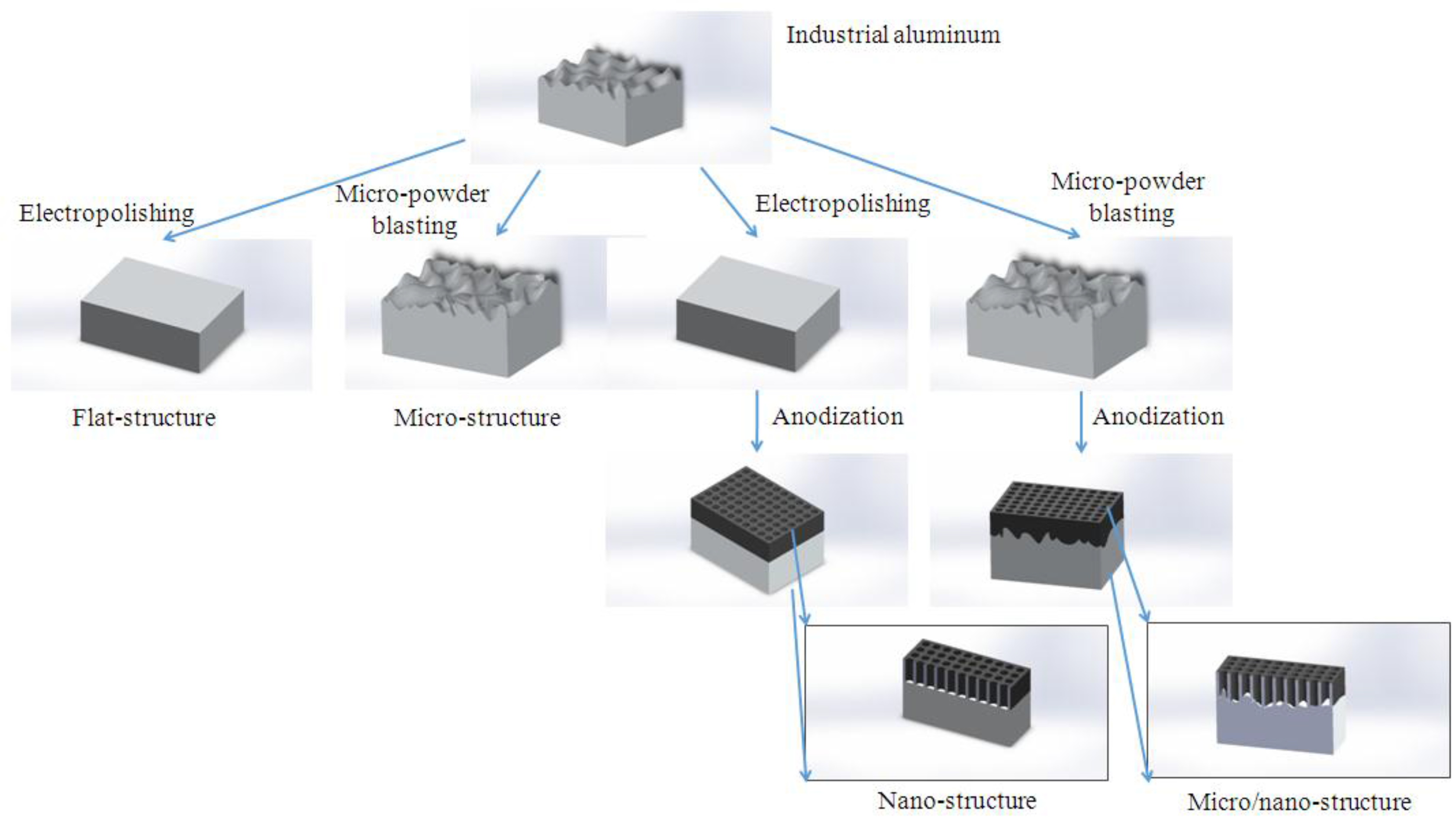
2.2. Micro-Structure of Al Template

A micro-structured Al template was prepared. A micro-blasting machine (MICROPEEN 1300 ZP/ZPD, Iepco, Leuggern, Switzerland) was used to perform micro-powder blasting on Al template (99.9%). The micro-powder blasting formed irregular concave micron-sized holes on the surface of the aluminum template. This method increased the surface roughness of the aluminum. The sands used for micro-powder blasting were MS 245A, MS 300A, MS 550A and MS 550BT (A means sharp sand, BT means round sand), with a diameters of 50–250, 30–70, 10–20, and 20–30 microns, respectively. To form an aluminum template with micro-structure, a blasting pressure of six bars was utilized; the distance between the blasting nozzle and the template was 3 cm; two blasting times were used in each case, and four kinds of sand particles were used.

2.3. Nano-Structure of Al Template (AAO)

A nano-structured Al template was formed, and anodic aluminum oxide (AAO) was prepared as follows:
  • Pre-treatment: Aluminum template with a purity of 99.9% was soaked in an alcohol solution and ultrasonically vibrated for 30 min. It was then placed in 5% NaOH and soaked for 3 min to remove surface oil. Following heat treatment (400 °C, 4 h), it was electrolytically polished using 85% perchloric acid (HClO4, Kanto Chemical Co., Ltd., Tokyo, Japan) and 15% ethanol (C2H5OH). It was then washed twice in deionized water.
  • Anode handling: (1) Aluminum template was firstly anodized using 0.5 M oxalic acid on 30 V at room-temperature for 1 h to do the anodic process for the first time; (2) Chemical etching: The aluminum was rinsed for the second time in deionized water, and placed in a solution of 1.5 wt % chromic acid (Katayama reagent Co., Ltd., Osaka, Japan) that had been mixed with 6 wt % phosphoric acid (Katayama reagent Co., Ltd.) at 70 °C. The reaction time was 1 h. The growth was etched to retain a few pit holes under its surface. It was then washed twice in deionized water, before being anodized for the second time; (3) The second anodic treatment was conducted using 0.5 M oxalic acid at 30 V and room temperature for 3 h.

2.4. Micro/Nano-Structure of Al Template

To generate a surface with micro/nano-structure, the micro-powder blasting method and an anodization process (voltage: 30 V, 0.5 M oxalic acid, room temperature, first anodized period time: 1 h, second anodized period time: 3 h) were utilized to form nano-holes in a micro-structured aluminum template. The novelty of this work is the use of an innovative method to fabricate an Al template with micro/nano-structure. This method is easy, fast, and cheap for the production of the micro/nano-structured Al template. The surface properties of the flat aluminum or aluminum templates with various structures (micro-structure, nano-structure, and micro/nano-structure) importantly affect the cell culture that is performed on these materials. Figure 1 displays the fabrication of Al template with various structures.

2.5. Surface Properties

A contact angle meter (DIGIDROP DGD-DI, GBX, Dublin, Ireland) was used to measure the contact angle of aluminum templates with various structures. The contact angles of the surfaces of Al template with flat-structure, micro-structure, nano-structure, and micro/nano-structure are discussed. 5 points were measured on each specimen. Deionized water (0.5 μL) was dropped on the template surface. The three states of solid/gas/liquid affected the liquiddrop stability, the use of computer-controlled photography (25/s) captured images and converted the image files. The obtained data of measured contact angle were made into charts.
A MultiMode 3D scanning probe atomic force microscope (DI 3100, Advanced Surface Microscopy, Indianapolis, IN, USA) was utilized to determine the surface roughness of Al templates with various structures. The atomic force microscope (AFM) was also applied to measure the surface profile of aluminum templates with different structures. The authors measured the surface roughness of each test template at 5 measurement points. The scanning range of each measurement point was 5 × 5 μm2. The surface morphology of Al templates with various structures was analyzed by SEM (JSM-6700F, JOEL, Peabody, MA, USA).

2.6. Cell Culture

In the phosphate-buffered saline (PBS) buffer allocation method, PBS and deionized water were mixed in ratio of 1:9. The PBS was put in a sterilized bottle and stored in a refrigerator at 4 °C. Dulbecco’s Modified Eagle Medium from HyClone Co. (South Logan, UT, USA) was added to 10% PBS and 1% penicillin (HyClone Co.). The MG63 cell line (ATCC CRL-1427) was used in the cell culture. MG63 is a human bone precursor cell (human osteogenic sarcoma). MG63 cells are utilized in experimental research of the in vitro attachment and proliferation of bone cells. The microculture tetrazolium test (MTT) is 3-(4,5-cimethylthiazol-2-yl)-2,5-diphenyl tetrazolium bromide. It is a yellow compound that accepts hydrogen ions. It acts on the respiratory chain of living cell lines. Cracking its tetrazolium ring using succinate dehydrogenase and cytochrome C yields a blue formazan crystal. In this study, the crystal was dissolved in dimethyl sulfoxide, and its optical density (OD) was measured using an ELISA machine (Anthos 2020, Biochrom, Cambridge, UK). The OD value indicated the cell activity.

2.7. Statistics

Measured data were subjected to statistical analysis. For any given experiment, each data point represented the mean ± standard deviation (SD) of six individual experiments. The Tukey-test was used to determine significance between groups in the contact angle and surface roughness. Statistical significance was indicated by * p < 0.05, ** p < 0.01, and *** p < 0.001.

3. Results and Discussion

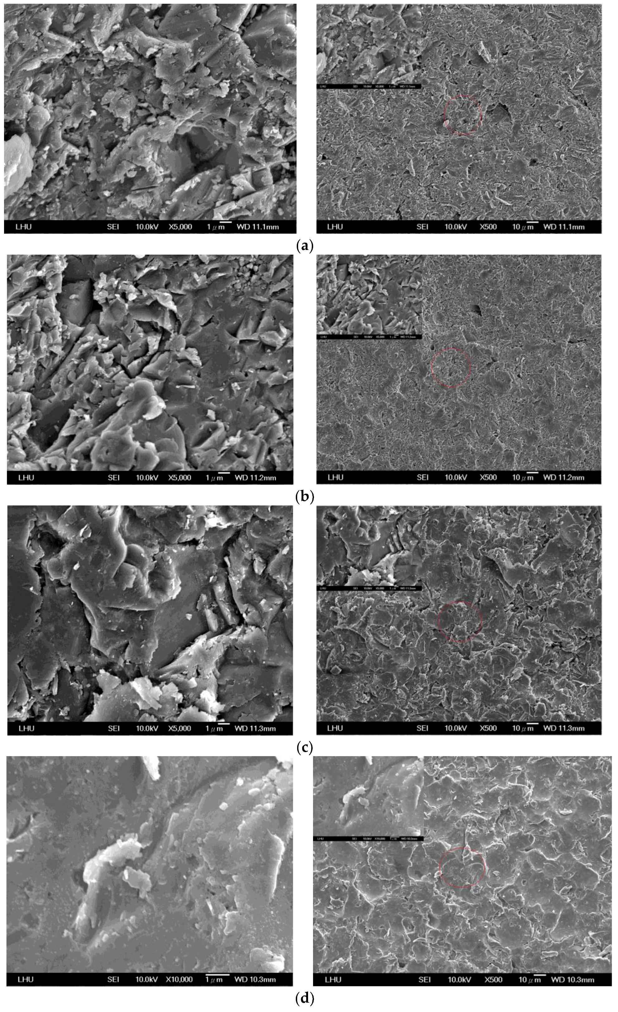
3.1. Surface Morphology of Micro-Structure of Al Template

In this work, micro-powder blasting was carried out to form a micron-scale surface on an aluminum template, which was then anodized to produce nanoholes in anodic alumina. Finally, the micro/nano-structure of the aluminum template was formed. The first goal was to form a suitable micro-structured surface of aluminum using various sand particles on micro-powder blasting (Figure 2). The depth of the surface of the aluminum micro-structure declined as the diameter of the sand particles declined (Figure 2a–c). The results also reveal that surfaces which had been impacted by larger sand particles were more concave and convex. Figure 2d demonstrates that the depth of the surface micro-structure of aluminum that underwent impact by round sand particles was less than that which underwent impact by sharp sand particles. The results also indicate that the micro-structure surface of aluminum that underwent impact by round sand particles was smoother than that by sharp sand particles.

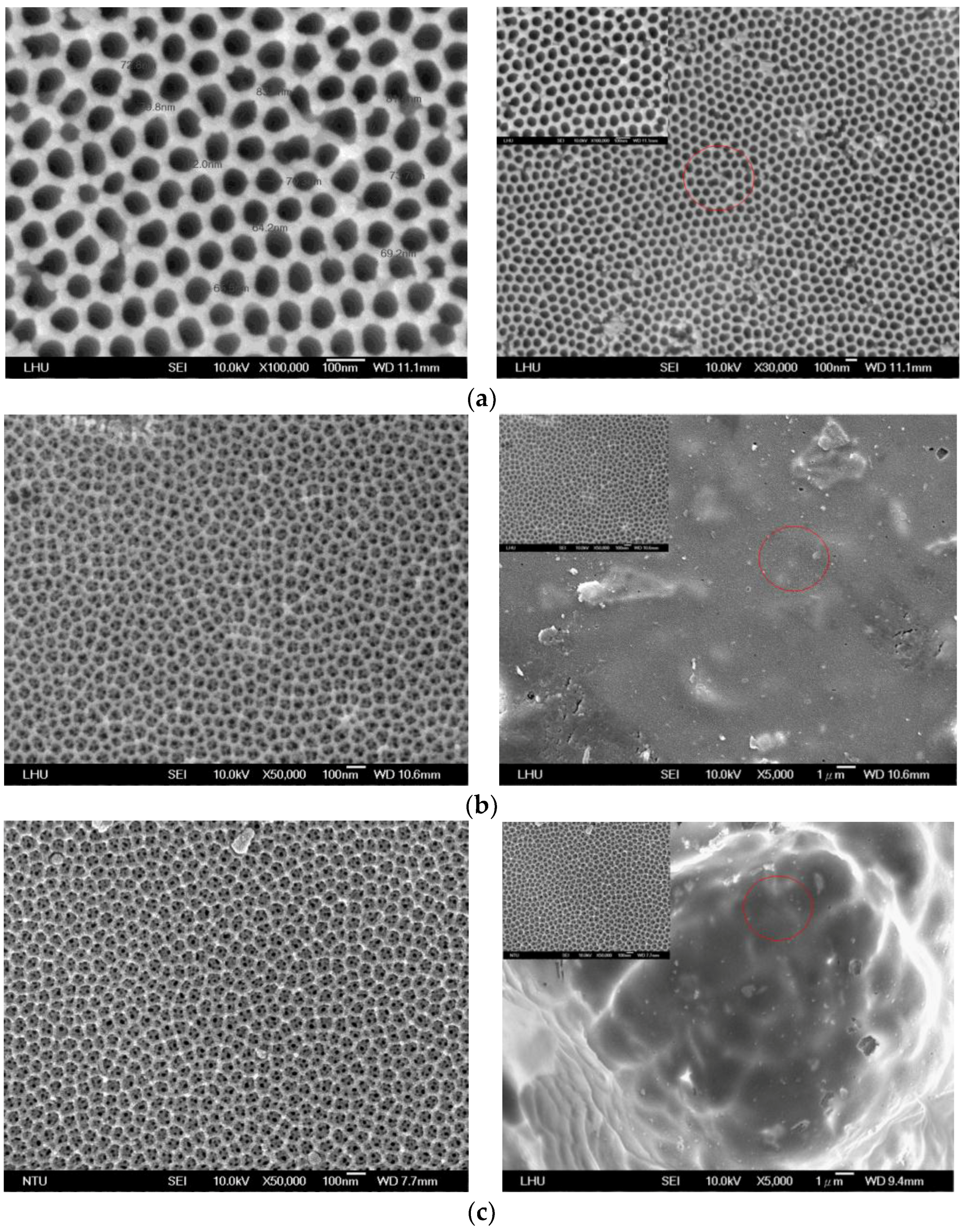
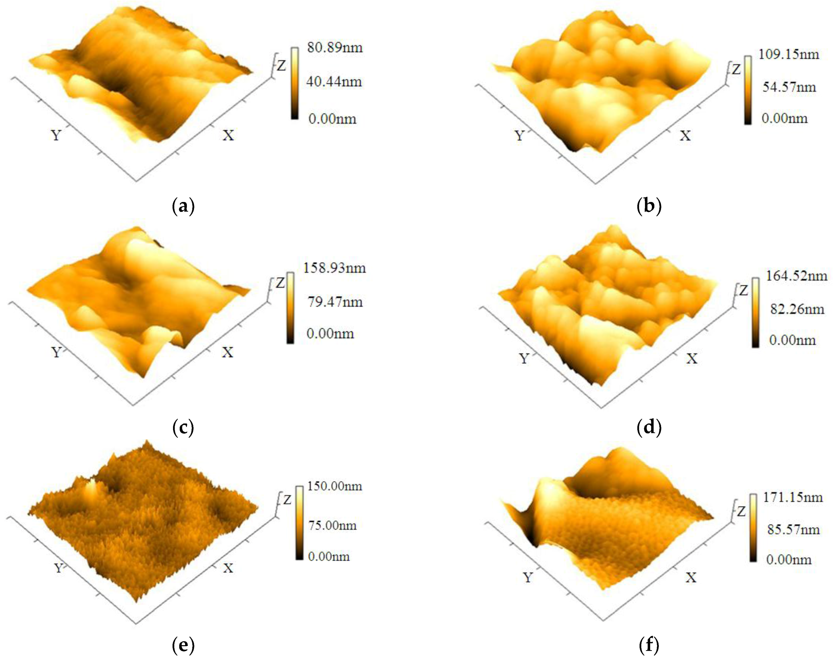
3.2. Surface Morphologies of Nano and Micro/Nano-Structures of Al Template

To assess the quality of the prepared anodized aluminum templates, they were observed using SEM and AFM, as presented in Figure 3. Figure 3a shows the SEM images of AAO that were formed by the anodization process. The results reveal that the mean pore size in AAO was approximately 100 nm. Additionally, the formed pore arrays of AAO were very uniform. The micro/nano-structure of the Al template obtained by micro-powder blasting and anodic oxidation process is discussed. Figure 3b presents an SEM image for the previous process with electrolytic polishing, followed by the anodic oxidation process. The results demonstrate that micro-powder blasting barely formed a micro-structure, but rather formed nanoholes in AAO, yielding a pore size of about 60–80 nm. Figure 3c shows the SEM images following anodic oxidation process without electrolytic polishing. The results indicate that the micro-structure formed on the Al template, and nanoholes formed in the micro-structure with sizes about 50–80 nm, suggesting that the micro/nano-structure formed on the Al template.

3.3. Surface Properties of Various Al Template Structures

The surface properties of the various structured templates importantly affect the cell culture thereon. The effects of contact angle and surface roughness of Al templates with various structures on their surface are considered. Table 1 displays measured contact angles on smooth Al template (flat-structure), a micro-structured Al template that was formed by micro-powder blasting (MS 245A, MS 300A and MS 550BT), the nano-structure (AAO) and the micro/nano-structured Al template, revealing that the contact angles of the Al templates with the different structures fall into three groups. The contact angles of the smooth Al template and the micro-structure of Al substrate formed by micro-powder blasting (MS 245A) were about 77°–88°. The contact angle of Al template by MS 245A did not decline very much as the size of the sand particles increased, yielding a larger micro-structure, so the surface properties of the Al template did not improve with an increase in the particle size. The contact angles of the Al template with micro-structure formed by micro-powder blasting (MS 300A, MS 550BT) were around 28°–36°, revealing that the surface of the Al template by micro-powder blasting changed from hydrophilic to more hydrophilic. Furthermore, the contact angles of Al templates with various structures were affected by the size of sand particles, and are independent of their shapes. Finally, the contact angles of the nano-structure (AAO) and micro/nano-structure on Al template were about 7°–21°, indicating that these structures are superhydrophilic. These results also show that the micro/nano-structured Al template had the lowest contact angles, and that the micro/nano-structure of the Al template was more hydrophilic than the other structures of the Al template. Contact angle values by Tukey-test are also listed in Table 1. The contact angle indicates that there was no statistically significant difference between the smooth Al template and the Al template with micro-structure (MS 245A). The results also show that the contact angle between Al template with micro-structure (MS 300A) and the Al template with micro-structure (MS 500BT) had no statistically significant difference. The other two had statistically significant differences from each other in terms of contact angle for different structured templates.
Table 1 also presents the surface average roughness (Ra) values of Al templates with various structures. The results of surface roughness (Ra) indicate that the Ra of the Al template after micro-powder blasting is larger than that of flat Al template. The Wenzel equation appears that the surface roughness of the template can improve the surface wetting property. The contact angle of the template decreased as its surface roughness increased [34]. The results indicate that micro-powder blasting with smaller sand particle yielded larger Ra values of the Al template, because larger sand eroded the Al template more strongly and it could not produce small bumps on the surface of the Al template. The Ra value of the Al template fell as the size of the sand in the micro-powder blasting increased. The results also demonstrated that rounder sand in micro-powder blasting yielded smaller Ra values of the Al template, because sharp sand could more easy produce bumps on the surface of the Al template. The micro/nano-structure of the Al template had the highest Ra value. The Tukey-test for surface roughness of Al templates with various structures is also listed on Table 1, indicating that there was no statistically significant difference between the nano-structured (AAO) template and the micro-structured template (MS 245A). The results also reveal that the surface roughness between the micro-structured template (MS 300A) and the micro/nano-structured template had no statistically significant difference. The other two had statistically significant differences with each other in surface roughness for different structured templates.
The results of this study also reveal that the micro-powder blasting + anodized method yielded the minimum contact angle and the maximum surface roughness in the Al template. The contact angle had a smaller value and the surface roughness had the smallest value via the anodized method. The contact angle was largest via micro-powder blasting with different sized particles.

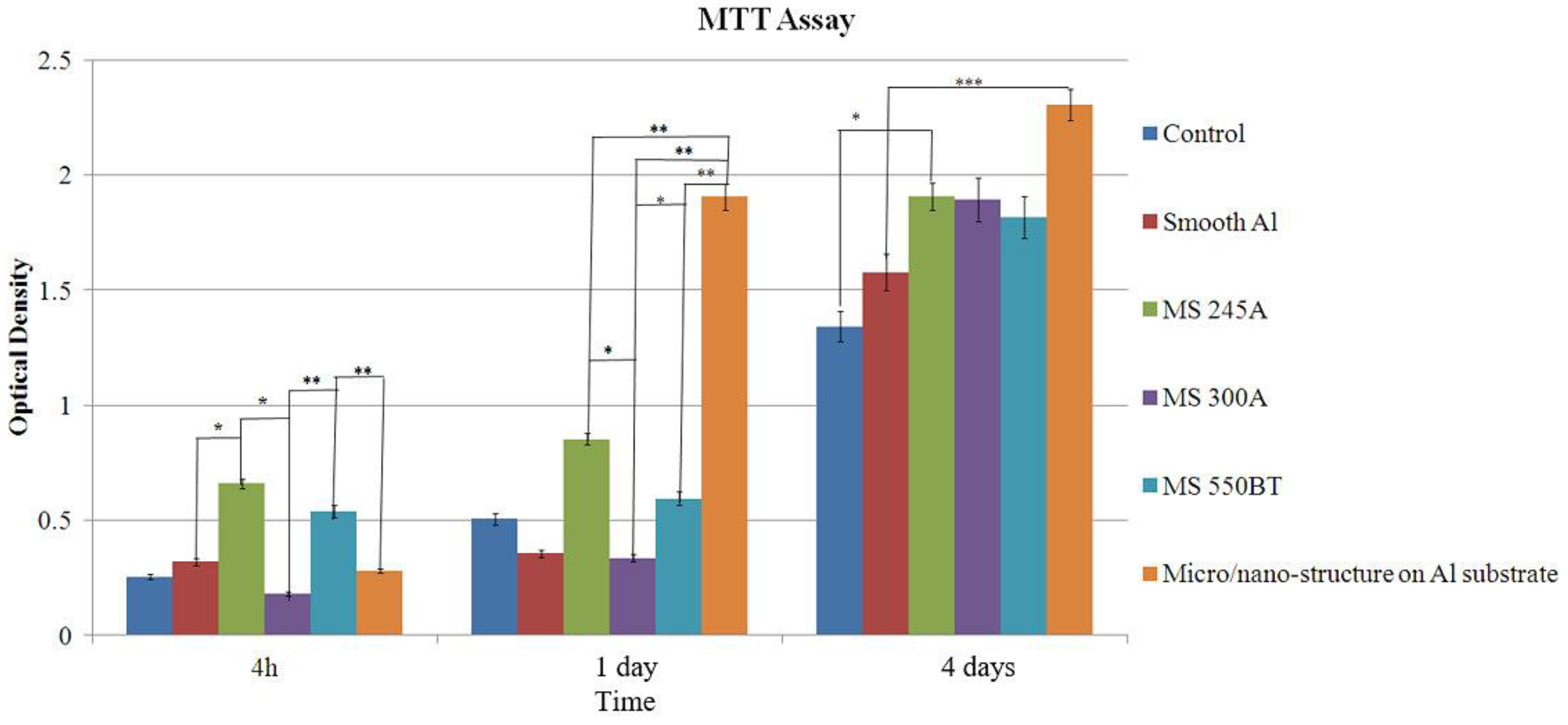
3.4. Cell Viability Evaluation in Vitro

Figure 4 displays the MTT assay on Al templates with various structures. The OD value was statistically significantly different between the smooth Al template and the micro/nano-structured Al template. The results show that the OD value increased with the cell time regardless of the template structure. The results also reveal that the micro/nano-structure of the Al template had the highest OD value because it had the largest surface area; this explains why its surface approaches superhydrophilicity. The results also show that the OD values among the smooth Al template and micro/nano-structured Al template were statistically significantly different at day 4. In vitro studies revealed that the growth response of specific cell types give insight into the surface properties of the substrate. The surface roughness affects the cell response. The growth behavior of osteoblast-like cells (MG63) demonstrates the phenotypic characteristics of roughness-dependence. The results herein demonstrate that surface roughness may play an important role in determining cell response [35,36,37]. The results also demonstrate that the OD value depends on the contact angle. There are many studies indicating that the suitable hydrophilic property of a template surface can improve the cell adhesion and spreading on the surface [35,38,39]. A smaller contact angle yields a larger OD value, indicating that the hydrophilic nature of the template favors the cell adhesion and proliferation.
The results also indicate that a good behavior of cell adhesion and proliferation appeared on the surface of the Al template obtained by micro-powder blasting + anodized method, followed by the use of micro-powder blasting. The OD value had the smallest value on the smooth Al template.

3.5. The Results of Null Hypothesis

The null hypothesis was that surface modification methods (micro-powder blasting, anodized process, micro-powder blasting + anodized process) only has an effect on the surface of the Al template. The authors wanted to determine the depth of the Al template micro-structure or micro/nano-structure after surface modification. Figure 5 shows the surface profile of different structures of aluminum templates measured by AFM. The depth of the Al template was about 80.89 nm. The results show that the depths of the micro-structure of the Al template were 109.15 nm, 158.93 nm, and 164.52 nm for sand particles MS 245A, MS 300A, and MS 550BT by micro-powder blasting, respectively. The depth of AAO was 150.00 nm for the anodized process. The depth of Al template micro/nano-structure was 171.15 nm by the micro-powder blasting + anodized process. The previous results can reveal that the surface modification methods influence the surface layer of the Al template. The experimental results fit the null hypothesis.

4. Conclusions

This study evaluates the effects of Al template with various structures on cell cultures. The results can be used for the reference on bone or dental implants. The Al template with the flat-structure was slightly hydrophilic; the Al template with micro-structure formed by micro-powder blasting was more hydrophilic. The Al template with nano-structure became superhydrophilic by the anodization method. The Al template with micro/nano-structure became superhydrophilic, and it had the maximum value of contact angle. The Al template with micro/nano-structure had the maximum value of surface roughness, followed by the Al template with micro-structure, followed by the smooth Al template, and the Al template with nano-structure had the minimum value. Osteoblast-like cells (MG63) were cultured on the variously structured templates for 4 h, 1 day, and 4 days, before the MTT assays were performed. The results revealed that the Al template with micro/nano-structure had the highest OD value. The reason is that this template had the superhydrophilic property and the maximum surface roughness. The Al template with micro/nano-structure was more suitable for cell culture in this study.

Acknowledgments

The authors would like to gratefully acknowledge of financial support from National Taipei University of Technology-Taipei Medical University Joint Research Program (NTUT-TMU-101-12).

Author Contributions

Ming-Liang Yen and Yung-Kang Shen conceived and design the experiments; Hao-Ming Hsiao and Chiung-Fang Huang performed the experiments; Yi Lin, Yu-Liang Tsai and Chun-Wei Chang analyzed the data; Hsiu-Ju Yen, Yi-Jung Lu and Yun-Wen Kuo contributed analysis tools; Ming-Liang Yen and Yung-Kang Shen wrote the paper.

Conflicts of Interest

The authors declare no conflict of interest.

References

  1. Ishikawa, K.; Miyamoto, Y.; Nagayama, M.; Asaoka, K. Blasting coating method: New method of coating titanium surface with hydroxyapatite at room temperature. J. Biomed. Mater. Res. Part A Appl. Biomater. 1997, 38, 129–134. [Google Scholar] [CrossRef]
  2. Mano, T.; Ueyama, Y.; Ishikawa, K.; Suzuki, K. Initial tissue response to a titanium implant coated with apatite at room temperature using a blast coating method. Biomaterials 2002, 23, 1931–1936. [Google Scholar] [CrossRef]
  3. O’Neill, L.; O’Sullivan, C.; O’Hare, P.; Sexton, L.; Keady, F.; O’Donoghue, I. Deposition of substituted apatites onto titanium surfaces using a novel blasting process. Surf. Coat. Technol. 2009, 204, 484–488. [Google Scholar] [CrossRef]
  4. O’Hare, P.; Meenan, B.J.; Barke, G.A.; Byrne, G.; Dowling, D.; Hint, J.A. Biological responses to hydroxyapatite surface deposited via a co-incident microblasting technique. Biomaterials 2010, 31, 515–522. [Google Scholar] [CrossRef] [PubMed]
  5. O’Sullivan, C.; O’Hare, P.; O’Leary, N.D.; Cream, A.M.; Ryan, K.; Dobson, A.D.W.; O’Neill, L. Deposition of substituted apatites with anticolonizing properties onto titanium surfaces using a novel blasting process. J. Biomed. Mater. Res. Part B Appl. Biomater. 2010, 95, 141–149. [Google Scholar] [CrossRef] [PubMed]
  6. Fleming, D.; O’Neill, L.; Byrne, G.; Barry, N.; Dowling, D.P. Wear resistance enhancement of the titanium alloy Ti-6Al-4V via a novel coincident microblasting process. Surf. Coat. Technol. 2011, 205, 4941–4945. [Google Scholar] [CrossRef]
  7. O’Sullivan, C.; O’Hare, P.; Byrne, G.; O’Neill, L.; Ryan, K.B.; Vrean, A.M. A modified surface on titanium deposited by a blasting process. Coatings 2011, 1, 53–71. [Google Scholar] [CrossRef]
  8. Tan, F.; Naciri, M.; Dowling, D.; Rubeai, M.A. Osteoconductirity and growth factor production by MG63 osteoblastic cells on bioglass-coated orthopedic implants. Biotechnol. Bioeng. 2011, 108, 454–464. [Google Scholar] [CrossRef] [PubMed]
  9. Tan, F.; Naciri, M.; Dowling, D.; Rubeai, M.A. In vitro and in vivo bioactivity of coblast hydroxyapatite coating and the effect of impaction on its osteoconductirity. Biotechol. Adv. 2012, 30, 352–362. [Google Scholar] [CrossRef] [PubMed]
  10. Bachle, M.; Kohal, R.J. A systematic review of the influence of different titanium surface on proliferation, differentiation and protein synthesis of osteoblast-like MG63 Cells. Clin. Oral Implant. Res. 2004, 15, 683–692. [Google Scholar] [CrossRef] [PubMed]
  11. Gentile, F.; Tirinato, L.; Battistam, E.; Causa, F.; Liberale, C.; Fabrizio, E.M.; Decuzzi, P. Cells preferentially grow on rough substrates. Biomaterials 2010, 31, 7205–7212. [Google Scholar] [CrossRef] [PubMed]
  12. Decuzzi, P.; Ferrari, M. Modulating cellular adhesion through nanotopography. Biomaterials 2010, 31, 173–179. [Google Scholar] [CrossRef] [PubMed]
  13. Zareidoost, A.; Yousefpour, M.; Ghaseme, B.; Amanzadeh, A. The relationship of surface roughness and cell response of chemical surface modification of titanium. J. Mater. Sci. Mater. Med. 2012, 23, 1479–1488. [Google Scholar] [CrossRef] [PubMed]
  14. Yousepour, M.; Zareidoost, A. Evaluation of the effect of two-step acid etching on the surface treatment and improved bioactivity of nitinol. J. Mash. Dent. Sch. 2016, 40, 281–296. [Google Scholar]
  15. Gittens, R.A.; Mclachlan, T.; Olivares-Navarrete, R.; Cai, Y.; Berner, S.; Tannenbaum, R.; Schwartz, Z.; Sandhage, K.H.; Boyan, B.D. The effects of combined micro-/submicro-scale surface roughness and nanoscale features on cell proliferation and differentiation. Biomaterials 2011, 32, 3395–3403. [Google Scholar] [CrossRef] [PubMed]
  16. Gittens, R.A.; Olivares-Navarrete, R.; Schwartz, Z.; Boyan, B.D. Implant osseointegration and the role of microroughness and nanostructures: Lessons for spine implants. Acta Biomater. 2014, 10, 3363–3371. [Google Scholar] [CrossRef] [PubMed]
  17. Wang, X.; Han, G.R. Fabrication and characterization of anodic aluminum oxide template. Microelectron. Eng. 2003, 14, 166–171. [Google Scholar] [CrossRef]
  18. Yuan, J.H.; He, F.Y.; Sun, D.C.; Xia, X.H. A simple method for preparation of through-hole porous anodic alumina membrane. Chem. Mater. 2004, 16, 1841–1844. [Google Scholar] [CrossRef]
  19. Firouzi, A.; Kumar, D.; Bull, L.M.; Besier, T.; Sieger, P.; Huo, Q.; Walker, S.A.; Zasadzinski, J.A.; Glinka, C.; Nicol, J. Cooperative organization of inorganic surfactants and biomimetic assemblies. Science 1995, 267, 1138–1143. [Google Scholar] [CrossRef] [PubMed]
  20. Kresge, C.T.; Leonowicz, M.E.; Roth, W.J.; Vartuli, J.C.; Beck, J.S. Ordered malodorous molecular sieves synthesized by a liquid crystal templates mechanism. Nature 1992, 359, 710–712. [Google Scholar] [CrossRef]
  21. Akmatsu, K.; Takeib, S.; Mizuhatab, M.; Kajinamib, A.; Dekib, S.; Takeokac, S.; Fujiid, M.; Hayashid, S.; Yamamotod, K. Preparation and characterization of polymer thin films containing silver and silver sulfide nano particles. Thin Solid Film 2000, 359, 55–60. [Google Scholar] [CrossRef]
  22. O’Sullivan, J.P.; Wood, G.C. The morphology and mechanism of formation of porous anodic films on aluminum. Proc. R. Soc. Lond. A 1970, 137, 511–543. [Google Scholar] [CrossRef]
  23. Keller, F.; Hunter, M.S.; Robinson, D.L. Structural features of oxide coatings on aluminum. J. Electrochem. Soc. 1953, 100, 411–419. [Google Scholar] [CrossRef]
  24. Laet, J.D.; Anhellemont, J.; Terryn, H.; Vereechen, J. Characterization of various aluminum oxide layers by means of spectroscopic ellipsometry. Appl. Phys. A. 1992, 54, 72–78. [Google Scholar] [CrossRef]
  25. Parkhutik, V.P.; Shershulsky, V.I. Theoretical modelling of porous oxide growth on aluminum. J. Phys. D Appl. Phys. 1992, 25, 1258–1263. [Google Scholar] [CrossRef]
  26. Ng, K.O.; Vanderbilt, D. Stability of periodic domain structures in a two-dimensional dipolar model. Phys. Rev. 1995, 2177, 13–52. [Google Scholar] [CrossRef]
  27. Chung, C.K.; Zhou, R.X.; Liu, T.Y.; Chang, W.T. Hybrid pulse anodization for the fabrication of porous anodic alumina films from commercial purity (99%) aluminum at room temperature. Nanotechnology 2009, 20, 005301–005304. [Google Scholar] [CrossRef] [PubMed]
  28. Den, E.B.; Ruijter, E.D.; Smits, J.H.; Ginsel, L.; Von, A.R.; Jansen, J. Quantitative analysis of cell proliferation and orientation on substrata with uniform parallel surface micro-grooves. Biomaterials 1996, 17, 1093–1099. [Google Scholar]
  29. Clark, P.; Connolly, P.; Curtis, A.; Dow, J.; Wilkinson, C. Topographical control of cell behaviour: II. Multiple grooved substrata. Development 1990, 108, 635–644. [Google Scholar] [PubMed]
  30. Wójciak, S.B.; Cutis, A.; Monaghan, W.; Macdonald, K.; Wilkinson, C. Guidance and activation of murine macrophages by nanometric scale topography. Exp. Cell Res. 1996, 223, 426–435. [Google Scholar] [CrossRef] [PubMed]
  31. Clark, P.; Connolly, P.; Curtis, S.A.; Dow, A.J.; Wilkinson, C.D. Topographical control of cell behavior. I. simple step cues. Development 1987, 99, 439–448. [Google Scholar] [PubMed]
  32. Popat, K.C.; Leary, E.E.S.; Mukhatyar, V.; Chatvanichkul, K.I.; Mor, G.K. Influence of nanoporous alumina membranes on long–term osteoblast response. Biomaterials 2005, 26, 4516–4522. [Google Scholar] [CrossRef] [PubMed]
  33. Hoess, A.; Teuscher, N.; Thormann, A.H.; Heilmann, A. Cultivation of hepatoma cell line HepG2 on nanoporous aluminum oxide membranes. Acta Biomater. 2007, 3, 43–50. [Google Scholar] [CrossRef] [PubMed]
  34. De Gennes, P.-G.; Brochard-Wyart, F.; Quere, D. Capillarity and Wetting Phenomena; Springer Inc.: New York, NY, USA, 2004. [Google Scholar]
  35. Lampin, M.; Warocquier, R.; Legris, C.; Degrange, M.; Sigot-Luizard, M.F. Correlation between substrate roughness and wettabililty, cell adhesion and cell migration. J. Biomed. Mater. Res. 1997, 36, 99–108. [Google Scholar] [CrossRef]
  36. Hallab, N.J.; Bundy, K.J.; O’Connor, K.; Moses, R.L.; Jacobs, J.J. Evaluation of metallic and polymeric biomaterial surface energy and surface roughness characterization for directed cell adhesion. Tissue Eng. 2001, 7, 55–71. [Google Scholar] [CrossRef] [PubMed]
  37. Deligianmi, D.D.; Katsala, N.D.; Koutsoukos, P.G.; Missirlis, Y.F. Effect of surface roughness of hydroxyapatite on human bone marrow cell adhesion, proliferation, differentiation and detachment strength. Biomaterials 2001, 22, 85–96. [Google Scholar]
  38. Van Wachem, P.B.; Bengelling, T.; Feijen, J.; Bantjes, A.; Detmers, J.P.; van Aken, W.G. Interaction of cultured human endothelial cells with polymeric surfaces of different wettabilities. Biomaterials 1985, 6, 403–408. [Google Scholar] [CrossRef]
  39. Webb, K.; Hlady, V.; Tresco, P.A. Relative importance of surface wettability and charged functional groups on NIH 3T3 fibroblast attachment, spreading and cytoskeletal organization. J. Biomed. Mater. Res. 1998, 41, 422–430. [Google Scholar] [CrossRef]
Figure 1. Fabrication process for different structures of aluminum (Al) templates.
Coatings 07 00179 g001
Figure 2. SEM images of Al template by powder blasting processing: (a) MS 245A (Φ = 50–250 μm); (b) MS 300A (Φ = 30–70 μm); (c) MS 550A (Φ = 10–20 μm); (d) MS 550BT (Φ = 20–30 μm).
Coatings 07 00179 g002
Figure 3. SEM images of nano-structure and micro/nano-structure: (a) Φ = 100 nm (anodic aluminum oxide (AAO)); (b) Φ = 60–80 nm (nano-hole on micro/nano-structure with electrolysis polishing); (c) Φ = 50–80 nm (nano-hole on micro/nano-structure, without electrolysis polishing).
Coatings 07 00179 g003
Figure 4. The microculture tetrazolium test (MTT) assay for different structured templates. (Values are the mean ± SD of six experiments (n = 6), * p < 0.05, ** p < 0.01, *** p < 0.001).
Coatings 07 00179 g004
Figure 5. Surface profile of different structures on aluminum templates: (a) Al template; (b) MS 245A; (c) MS 300A; (d) MS 550BT; (e) AAO; (f) micro/nano-structure without electrolysis polishing.
Coatings 07 00179 g005
Table 1. Contact angles and surface roughnesses for different structured templates by Tukey-test.
GroupContact Angle (°)Surface Roughness (nm)p-ValueTukey-Test
AAO18.76 ± 3.0912.65 ± 0.060.001 ***/0.001 ***A/A
MS 245A80.70 ± 3.8613.88 ± 0.07B/A
MS 300A33.58 ± 3.0451.67 ± 0.13C/B
MS 550BT31.22 ± 3.0240.07 ± 0.20C/C
Micro/nano-structure on Al substrate11.08 ± 4.1956.37 ± 0.28D/B
Smooth Al85.72 ± 3.5425.57 ± 0.13B/D
*** p < 0.001.

