Article

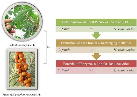
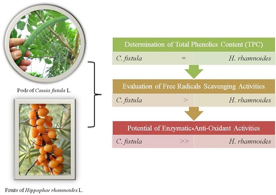
Relative Free Radicals Scavenging and Enzymatic Activities of Hippophae rhamnoides and Cassia fistula Extracts: Importance for Cosmetic, Food and Medicinal Applications

1
Department of Pharmacy, Faculty of Pharmacy and Alternative Medicine, The Islamia University of Bahawalpur, Punjab, Bahawalpur 63100, Pakistan
2
HYMETEC SA, Infection Control Unit, Isnes 5032, Belgium
3
Department of Aesthetic and Anti-Aging Medicine, Centre Médical des Guittières, Saint-Philbert-de-Grand-Lieu 44310, France
4
Department of Biotechnology, Biotechnology Center, Federal University of Paraiba, Castelo Branco, João Pessoa, Paraíba 58051-900, Brazil
5
Department of Pharmaceutical Sciences and Nanomedicine, Fluorotronics USA, Inc., Vista, CA 92081, USA
*
Authors to whom correspondence should be addressed.
These authors contributed equally to this work.
Cosmetics 2017, 4(1), 3; https://doi.org/10.3390/cosmetics4010003
Submission received: 2 November 2016 / Revised: 19 December 2016 / Accepted: 22 December 2016 / Published: 6 January 2017

Abstract

:
Hippophae rhamnoides L. and Cassia fistula L. extracts have great potential as food, medicinal, or cosmetic ingredients. The aim of our study was to assess their relative antioxidant activities and key enzymatic activities. Thereby, H. rhamnoides’ fruit and C. fistula’s pod extracts were evaluated by spectrophotometry, based on their respective total phenolic content (TPC), 2,2-diphenyl-1-picrylhydrazyl (DPPH) ferric-reducing power, capacity in nitric oxide, hydroxyl and superoxide radicals scavenging, as well as on their β-glucuronidase, α-glucosidase and α-tyrosinase inhibition activities. H. rhamnoides and C. fistula extracts exhibited similarly high TPC levels, hydroxyl ion [OH] quenching activity, and α-glucosidase and α-tyrosinase IC50 values (p > 0.05). However, their respective DPPH radical, nitric oxide radical [NO], and superoxide anion [O2−•] scavenging activities, as well as their IC50 values for β-glucuronidase, significantly differed (p ≤ 0.05), with results showcasing the highest values in C. fistula extracts. In sum, our in vitro data explicitly suggest that the pod extracts of C. fistula exert better antioxidant and enzymatic properties than those exhibited by the fruit extract of H. rhamnoides. They also implicitly encourage performing multiple in vitro assays in order to thoroughly select a plant extract destined to a given medicinal, dietetic, or esthetic application.

Graphical Abstract

1. Introduction

Antioxidants prevent oxidative damages (e.g., 8-hydroxyguanine, cell membrane lipid peroxidation) and subsequent diseases (e.g., inflammatory state-diseases such as cancers, Alzheimer’s, diabetes, and strokes), by effectively quenching or inhibiting free radicals (e.g., hydroxyl and superoxide radicals). For a long time, synthetic antioxidants have been employed as food additives, but safety concerns have restricted their use, due to their possible involvement in chronic diseases. Therefore, much attention has been directed toward the isolation of natural antioxidants from botanical sources, especially edible plants [1,2,3,4,5]. Furthermore, plant extracts that can inhibit specific enzymes (e.g., β-glucuronidase, α-glycosidase, and α-tyrosinase), particularly involved in the alteration of skin during aging, are appreciated for their potential use in original cosmetic formulations.
β-glucuronidase, an acid hydrolase, plays a crucial role in catalyzing the hydrolysis of glucuronide into glycones (i.e., alkyl, acyl, aryl groups) and free glucuronic acid [6,7]. This catalysis releases the terminal glucuronide unit linked through the β-configuration by Carbon 1 (C1) [8]. β-glucuronidase was first discovered in the rumen of sheep, although this enzyme has been recorded as present in all plants, animals, and bacteria so far studied [9]. The research and development (R&D) of specific β-glucuronidase inhibitors (e.g., baicalin) is growing [10], most notably in the fields of drug detoxification [11] and cancer therapy [12], as well as in its ability to overcome glycosaminoglycan (GAG) impaired metabolism during skin aging [13], treat skin conditions such as psoriasis [14] and formulate cosmetics [15].
α-glycosidase comprises a family of hydrolases, which are enzymes located in the brush-border surface membrane of small intestinal cells [16]. α-glycosidase is implicated in the hydrolysis of the 1,4 glycosidic linkage from the non-reducing end of the α-glucosides, α-linked oligosaccharide, and α-glucans substrates, producing α-d-glucose and other monosaccharides that are employed as carbon and energy sources [17,18]. Therefore, α-glucosidase inhibitors are not only considered in the management of type-2 diabetes (T2D) [19], tumorigenesis [20], but also in the prevention of skin aging and skin damages [21,22].
α-tyrosinase is found in plant and animal tissues, and represents a rate-limiting and copper-containing oxidase, involved in controlling melanin synthesis through two distinct chemical reactions [23,24,25]: (i) hydroxylation of a monophenol; and (ii) conversion of an o-diphenol into the corresponding o-quinone, which then undergoes several reactions, eventually forming melanin. In humans, the tyrosinase enzyme is encoded by the TYR gene expressed inside melanosomes [23,25,26]. When a mutation in the TYR gene results in impaired tyrosinase production, it is usually associated with type I oculocutaneous albinism, or increased melanin synthesis in skin cancer (e.g., melanoma) [27,28]. In addition to molecular strategies for controlling the tyrosinase activity [29], several polyphenols (e.g., flavonoids and stilbenoid substrate analogues), free radical scavengers and copper chelators, have been identified as potent tyrosinase inhibitors, capable of preventing skin hyperpigmentation and inducing skin whitening [24,30,31,32,33].
Hippophae rhamnoides L. (Elaeagnacea), commonly known as sea-buckthorn, is a flowering, spiny, deciduous shrub, native to fixed dunes and sea cliffs in Europe (e.g., Germany, France) and Asia (e.g., Nepal, India, China). It is both an agricultural and an ornamental plant. Although H. rhamnoides is a relatively expensive raw material, its fruits are quite beneficial due to their higher average content of ascorbic acid (aka vitamin C), in comparison to lemons and oranges [34]. Consequently, the fruits of H. rhamnoides are used in traditional Austrian medicine to fight infections (e.g., flu), in the form of tea, juice, and syrup [35]. H. rhamnoides exerts various pharmacological effects, including cytoprotection, protection against stress, immunomodulation, hepatoprotection, radioprotection, anti-atherogenicity, anti-tumorigenicity, anti-microbial activity, and tissue regeneration [36]. We have recently shown the beneficial effects of H. rhamnoides extracts in the prevention of premature aging of human skin and melasma [5,37].
Cassia fistula L. (Caesalpinaceae/Leguminosae), also known as the golden shower tree, is a famous yellow flowering plant from Asia (e.g., particularly found in the forests of India, Sri Lanka, Thailand and Pakistan), and displays numerous medicinal properties for protecting against skin conditions and inflammatory diseases [37,38,39]. Thereby, we recently demonstrated that C. fistula’s extracts are capable of preventing premature skin aging in healthy individuals [39], and melasma in a good number of patients when compared with treatment using a placebo (i.e., without the plant extract) [37]. These beneficial effects are probably due to their relatively rich content of bioactive ingredients (i.e., phenolic compounds, fatty acids, flavonoids, tannins and glycosides) [40,41], and their ability to significantly decrease the tyrosine activity-mediated melanin level.
The present study was designed to determine the TPC, antioxidant, and key enzymatic inhibition activities of cosmetic importance from H. rhamnoides and C. fistula extracts.

2. Materials and Methods

2.1. Key Chemicals, Reagents and Enzymes

Gallic acid (PubChem CID: 370), l-ascorbic acid (PubChem CID: 54670067), 2,2-diphenyl-1-picrylhydrazyl (DPPH) (PubChem CID: 2735032), trichloroacetic acid (TCA) (PubChem CID: 6421), ethyldia minetetraacetic (EDTA), methanol, and Folin-Ciocalteu reagent (FCR), were obtained from Sigma (St. Louis, MO, USA). Sodium acetate buffer pH 5.0, Sodium nitroprusside dehydrate, and Sodium carbonate (Na2CO3), were purchased from Merck (Darmstadt, Germany).
Substrate p-nitrophenyl-beta-d-glucuronide (NPG) (PubChem CID: 3291162), β-glucuronidase (E.C. 3.2.1.31-Type B-1 from bovine liver [42], yeast α-glucosidase [43] and mushroom tyrosinase [44], were purchased from Sigma Company (St. Louis, MO, USA) were all obtained for the purpose of this study.
All of the chemicals and enzymes used in this study were of analytical grade.

2.2. Plants

H. rhamnoides’s fruits were obtained from Pak Sea International Skardo, Pakistan. C. fistula pods were collected from Abbasia Campus, Islamia University, Bahawalpur, Pakistan. The respective voucher specimens (HR-FT-03-11-26 and CF-FT-03-11-27) were preserved for future reference at the herbarium of the Pharmacognosy Section, Faculty of Pharmacy, Islamia University, Bahawalpur, Pakistan. The plant names C. fistula L. [45] and H. rhamnoides L. [46] have been checked.

2.3. Analytical Apparatus

A dual-beam UV-VIS spectrophotometer (Uvikon XL, Bio-Tek Instruments, Bad Friedrichshall, Germany), Synergy™ HT Multi-Mode Microplate Reader (BioTek Instruments, Inc.), and rotary evaporator EYELA (CA-1111, Rikakikai Co. Ltd., Tokyo, Japan), were the three main devices used in this study.

2.4. Plant Extraction

A total of 150 g of C. fistula pods, pre-placed for three days at room temperature in a 5 L beaker containing 70% methanol, were extracted. A total of 320 g of H. rhamnoides berries were crushed and successively macerated in a mixture containing 2 L of analytical grade methanol and distilled water (1:1 v/v). The macerated plant material was filtered through muslin cloth for coarse filtration. The coarse filtrate was then filtered through a Whatman N 1 filter paper in order to produce particle-free extracts. The filtrate was evaporated under reduced pressure at 40 °C using a rotary vacuum evaporator, and it was reduced to almost one-fourth of the amount, into a semi-solid mass.

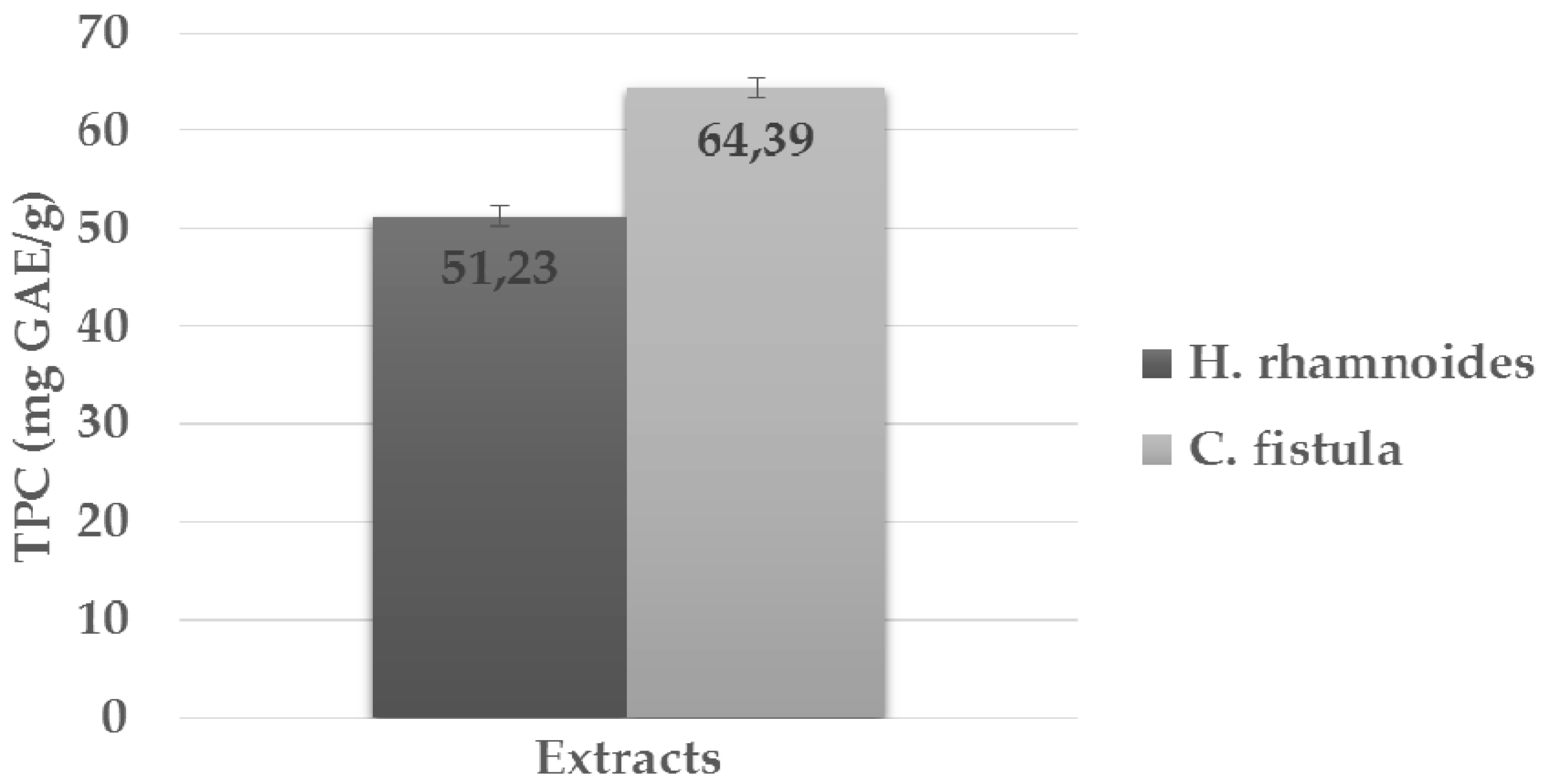
2.5. Total Phenolic Content (TPC) Determination

The TPC of the plant extracts was measured according to a previously described procedure [47]. A total of 200 µL of the respective plant extract was oxidized with 1 mL FCR (0.5 N). Following this, the reaction was neutralized with 1 mL of the saturated Na2CO3 (75 g/L). After incubation for 2 h at room temperature, optical absorbance of the resulting blue color was measured using spectrophotometry at 760 nm, against a blank of the reagent. The quantification was recorded based on the standard curve of gallic acid. All experiments were carried out in triplicate, and the results were expressed as milligrams of gallic acid equivalent per gram of plant extract (mg GAE/g).

2.6. DPPH Free Radical Scavenging Activity

The free radical scavenging assay was performed following the method of Elmastas et al. [48]. Stock solution of DPPH (33 mg/L) was prepared in methanol, which provided an initial optical absorbance of 0.493 when assessed using spectrophotometry. A total of 5 mL of the DPPH stock solution was added to 1 mL of diluted methanolic extract solution in order to obtain a proper range of concentrations (250–1500 μg/mL). After incubation at room temperature for 30 min, the optical absorbance was measured using spectrophotometry at 517 nm, against the reagent blank value. The IC50 values of compounds (i.e., concentration at which there is 50% scavenging) were then calculated using EZ-Fit Enzyme kinetics software version 5.03 (Perella Scientific Inc., Amherst, MA, USA). The DPPH scavenging activity was compared to that of natural antioxidants, such as ascorbic acid (i.e., vitamin C). All experiments were carried out in triplicate, and decreased absorbance of reaction mixture was indicative of increased DPPH free radical scavenging activity.

2.7. Nitric Oxide Radical [NO] Scavenging Assay

[NO] scavenging activity was assessed by mixing sodium nitroprusside (5 mM), pre-dissolved in phosphate buffered saline, with various concentrations of methanolic extract (250–1500 µg/mL). After incubation at room temperature for 30 min, 1.5 mL of the diluted solution was collected and diluted with 1.5 mL of Griess reagent, pre-dissolved in 1% sulphanilamide, 2% phosphoric acid and 0.1% N-1-naphthylethylenediamine dihydrochloride. During this reaction, diazotization of nitrite with sulphanilamide and subsequent coupling with N-1-naphthylethylene diaminedihydrochloride, resulted in the formation of active chromophore, the value of which was estimated using spectrophotometry at 546 nm, against the reagent blank value. The IC50 values of the compounds were then calculated using EZ-Fit Enzyme kinetics software version 5.03 (Perella Scientific Inc.). All experiments were carried out in triplicate, and decreased absorbance of reaction mixture was indicative of increased nitric oxide scavenging activity [40].

2.8. Hydroxyl Ion Radical [OH] Scavenging Assay

To perform [OH] scavenging activity, various concentrations of the respective methanolic plant extracts (250–1500 µg/mL) were placed in a test tube and evaporated to dryness. 1 mL of iron-EDTA solution (0.13% ferrous ammonium sulfate and 0.26% EDTA), 0.5 mL EDTA (0.018%), DMSO (0.85%, v/v in 0.1 M phosphate buffer, pH 7.4) and 0.5 mL ascorbic acid (0.22%), were then added to each tube. The tubes were subsequently tightly capped and heated in a water bath at 80–90 °C for 15 min. The reaction was terminated by adding 1 mL of ice-cold TCA (17.5% w/v). Finally, 3 mL of Nash reagent (75.0 g ammonium acetate, 3 mL glacial acetic acid and 2 mL acetyl acetone for a total volume of 1 L, mixed with pure water), was added to each tube and left at room temperature for 15 min for color development. The intensity of the resulting yellow color was measured using spectrophotometry at 412 nm, against the reagent blank value. The IC50 values of the compounds were then calculated using EZ-Fit Enzyme kinetics software version 5.03 (Perella Scientific Inc.). All experiments were carried out in triplicate, and decreased absorbance of reaction mixture was indicative of increased hydroxyl ion scavenging activity [49].

2.9. Superoxide Anion Radical [O2−•] Scavenging Assay

[O2−•] scavenging assay was carried out by the method described by Elmastas et al. [48]. Reaction mixtures included 1 mL nitroblue tetrazolium (pH = 7.4; 156 mM), 1 mL NADH solution (pH = 7.4; 468 mM), and various concentrations of methanolic plant extracts (250–1500 μg/mL). In order to initiate the reaction, 100 mL of 60 mM phenazine methosulfate (pH 7.4) was added to the mixture and incubated at 25 °C for 5 min. Optical absorbance was measured using spectrophotometry at 560 nm against the reagent blank value, and compared with pre-cited standards. The IC50 values of the compounds were then calculated using EZ-Fit Enzyme kinetics software (Perella Scientific Inc.). All experiments were carried out in triplicate, and decreased absorbance of reaction mixture was indicative of increased superoxide anion scavenging activity.

2.10. β-Glucuronidase Inhibition Assay

The ß-glucuronidase activity was determined by spectrophotometry using an optical absorbance measurement of p-nitrophenol, formed from the substrate using the method of Collins et al. [50], modified as follows: the reaction mixture made in a final volume of 100 µL contained 70 µL of 0.1 M acetate buffer pH 5.0 (3.402 g/250 mL of sodium acetate, pH was adjusted with 0.1 M acetic acid), 10 µL of test compound (0.5 mM), 10 µL of Enzyme (24 U) pre-incubated for 10 min, and 10 µL of 0.5 mM p-nitro phenyl-beta-d-glucuronide. The mix reaction was then incubated at 37 °C (38 ± 0.2 °C) for 30 min (30 ± 0.2 min). The reaction was stopped by the addition of 50 µL of 0.2 M Na2CO3. After the reaction had been stopped, the optical absorbance was measured against the reagent blank value by spectrophotometry at 405 nm, using a Synergy HT BioTek 96-well microplate reader. l-aspartic acid was used as a positive control. The percentage of enzyme inhibition was calculated using the following equation:
Inhibition (%) = [(A0 − A1) × 100]/A0
where A0 = Control (total enzyme activity without inhibitor); A1 = Test (activity in the presence of test compound).
All experiments were carried out in triplicate. The IC50 values of the compounds were calculated using EZ-Fit Enzyme kinetics software version 5.03 (Perella Scientific Inc.).

2.11. α-Glucosidase Inhibition Assay

The α-glucosidase inhibition activity was performed according to the slightly modified method of Tiwari et al. [51]. The final volume of the reaction mixture was 100 µL, which contained 70 µL of phosphate buffer saline (50 mM, pH 6.8), 10 µL of test compound (0.5 mM), and 10 µL (0.057 U) enzyme. The content was mixed, pre-incubated at 37 °C for 10 min, and pre-read against the reagent blank value by spectrophotometry at 400 nm. The reaction was initiated using 10 µL of 0.5 mM substrate (i.e., p-nitrophenol glucopyranoside). Acarbose was used as a positive control. After incubation at 37 °C for 30 min, optical absorbance was measured against the reagent blank value by spectrophotometry at 400 nm, using a Synergy HT BioTek 96-well microplate reader. The percentage of enzyme inhibition was calculated using the Equation (1).
All experiments were carried out in triplicate. The IC50 values of the compounds were calculated using EZ-Fit Enzyme Kinetics Software version 5.03 (Perrella Scientific Inc.).

2.12. α-Tyrosinase Inhibition Assay

The α-tyrosinase inhibition tests were carried out in a 96-well plate following the method described by [52]. The reaction mixture made in a final volume of 100 μL, contained 40 μL of phosphate buffer (100 mM, pH 6.8), 20 μL mushroom tyrosinase enzyme (6 U), and 10 μL test compound (0.5 mM). The content was pre-incubated at 37 °C for 5 min. 30 μL of l-dopamine (10 mM) was then added as a substrate. The final mixture was incubated at 37 °C for 10 min. Kojic acid was used as a positive control. Optical absorbance was read against the reagent blank value by spectrophotometry at 490 nm, using a Synergy HT BioTek 96-well microplate reader. The percentage of enzyme inhibition was calculated using the Equation (1).
All experiments were carried out in triplicate. The IC50 values of the compounds were calculated using EZ-Fit Enzyme Kinetics Software version 5.03 (Perrella Scientifich, Inc.).

2.13. Statistical Analysis

All of the analyses were performed in triplicate, in order to minimize the incidence of errors. The amount of extract needed to inhibit the concentration of free radicals by 50% (i.e., IC50), was graphically determined by a linear regression method using MS-Windows based graph pad instat software version 3 (GraphPad Software, Inc., San Diego, CA, USA).

3. Results and Discussion

Extracts from C. fistula and H. rhamnoides are gaining much attention in medicine and esthetics, because of their relative high content of antioxidants (e.g., vitamin C, polyphenols such as flavonoids and saponins, unsaturated fatty acids (UFAs)), known to counteract naturally-occurring or induced molecular and cellular damage-mediated oxidation [5,34,36,37,38,39,40,41,47,53].
Recent investigations led by our research groups have proven that great skin benefits (e.g., slower skin aging; anti-melasma) can be produced when C. fistula and H. rhamnoides extracts are topically applied [5,37,39].
The phyto-antioxidant arsenal is composed of several chemicals (i.e., free radical scavenging) and enzymatic reactions. Therefore, the specificity and sensitivity of a single method is insufficient for providing an inclusive examination of all phenolic compounds in a given plant extract. A combination of reliable tests is necessary in order to evaluate a complete antioxidant and enzymatic activity profile (i.e., free radical scavenging and key enzymatic inhibition capacities).
Following this original approach, our data provided a better qualitative and quantitative understanding of the anti-oxidant and enzymatic inhibition abilities of C. fistula and H. rhamnoides, two traditional plant extracts, valuable in modern medicine.
Therefore, the high concentration of total phenolic content (TPC) encountered in H. rhamnoides (51.23 ± 1.38 mg GAE/g) and C. fistula (64.39 ± 1.21 mg GAE/g) extracts (Figure 1), indicate an important anti-oxidant role of the two extracts [4]. Moreover, comparative TPC analysis of the two phyto-extracts showed insignificant variations (p > 0.05) (Figure 1). Interestingly, similar data have been reported by Korekar et al. [53], who demonstrated that the TPC in methanolic fruit extracts of H. rhamnoides is about 4056 ± 70.1 mg GAE/100 g (i.e., 40.56 mg GAE/g).
In a further step, we evaluated the quenching activities of the free radicals belong to H. rhamnoides and C. fistula. The resulting findings, expressed as IC50 (µg/mL), and presented in Table 1, indicated that: (i) DPPH radical, nitric oxide radical [NO] and superoxide anion [O2] scavenging activities of C. fistula extracts (89.07 ± 1.31, 116.82 ± 0.89 and 103.25 ± 1.37, respectively), are significantly more potent (p < 0.05) than those of H. rhamnoides extracts (107.26 ± 1.34, 139.38 ± 1.14 and 122.15 ± 1.09, respectively); (ii) hydroxyl ion [OH] quenching activity is similarly efficient (p > 0.05) in both extracts (91.04 ± 1.04 and 86.45 ± 0.93 for H. rhamnoides and C. fistula, respectively). However, these extract data were significantly (albeit expectedly) lower (p < 0.05) than the results produced when pure ascorbic acid was used as a standard (i.e., an external positive control). In sum, we state that similar TPC obtained from plants of different families does not necessarily result in similar specific chemical scavenging capacity, hence the requirement to perform a blend of assays in order to evaluate the antioxidant capacities of phyto-extracts.
Additionally, we decided to determine the enzymatic activities of the two extracts based on their ability to inhibit three major enzymes with great potential for the cosmetic industry (i.e., β-glucuronidase, α-glucosidase, and α-tyrosinase) [14,15,21,22,32]. Although it was initially thought that β-glucuronidase inhibition activity was absent in plants, later studies showed that it can be found in a number of plant families [54]. As shown in Table 2, our experimental analyses confirmed the presence of such enzymatic activity in both H. rhamnoides and C. fistula extracts, when compared to that of l-aspartic acid, used as external control.
Comparatively, C. fistula extracts significantly contained more potent (i.e., about two-fold, p < 0.05) anti-β-glucuronidase inhibition activity (IC50 = 33.80 ± 0.2 µg/mL) than H. rhamnoides extracts (IC50 = 61.21 ± 0.5 µg/mL) (Table 2). Furthermore, the anti-β-glucuronidase activity exerted by C. fistula extracts was not significantly different to that of pure l-asp acid (Table 2), indicating that the isolation and purification of the main bioactive compound contained in C. fistula pods could be promising as an adjuvant therapeutic for protecting against skin conditions such as psoriaris [14], or as a bioactive ingredient in cosmetic formulations (e.g., to prevent body malodor arising from sweat) [15]. Besides, although the anti-α-glucosidase activity, expressed as % inhibition at the extract dose of 0.05 mg, was significantly higher (i.e., about four-fold, p < 0.05) in C. fistula extracts (10.52% ± 0.53%), when compared to that of H. rhamnoides extracts (41.90% ± 0.25%) (Table 2), the related IC50 values were not significantly different (p > 0.05) (i.e., 55.31 ± 0.31 μg/mL and 51.09 ± 0.14 μg/mL for C. fistula and H. rhamnoides extracts, respectively) (Table 2). Also, these IC50 values are significantly higher (p < 0.05) than those produced when pure acarbose is used as an external control (Table 2), indicating that both extracts might be exploitable at a dose superior to 0.05 mg for the management of certain health conditions (e.g., T2D) [19], as well as in cosmetic formulations (e.g., to prevent precocious skin aging or skin damage) [21,22]. Eventually, the α-tyrosinase inhibition activity of both extracts demonstrated similar results (p > 0.05), but could potentially be high enough to prevent skin hyperpigmentation and induce skin whitening [32], either alone or in combination (i.e., IC50 = 39.2 ± 0.12 μg/mL and 45.9 ± 0.20 μg/mL for C. fistula and H. rhamnoides extracts, respectively) (Table 2). This effect was also stable at the dose of 0.05 mg, for which the percentage of inhibition has been measured (Table 2). Nevertheless, the corresponding IC50 results of both extracts are significantly (p < 0.05), but expectedly, too low compared to when pure Kojic acid is used as an external control. These results corroborate those published by Cho et al. [55], who performed a mass screening on a number of tropical plant species eliciting a tyrosinase inhibition activity above 50%. Nowadays, there are a number of reports exploring the tyrosinase inhibition activity effects of fatty acids (FAs). Thereby, it has been shown that topical application of linolenic, linoleic and oleic acids elicit a bleaching effect on guinea pig skin stimulated with UV light [56]. Therefore, the relatively high α-tyrosinase inhibition activity of exerted either by H. rhamnoides or C. fistula extracts, may be due to their relative content of unsaturated fatty acids (UFAs).
In sum, we postulate that similar TPC in plant extracts can not discriminate against key specific antioxidant potential and/or enzymatic activities.

4. Conclusions and Perspectives

Based on our various in vitro assays, which aimed to assess the antioxidant and key enzymatic activities of two major plant extracts used in traditional medicine, our results clearly confirmed that both H. rhamnoides and C. fistula extracts present potent antioxidant activities. More importantly, it was recorded that C. fistula’s pod extracts exert the best antioxidant and enzymatic activities, when compared to those of H. rhamnoides’ fruit extracts, which are known to contain a very high content of vitamin C (i.e., about 30 times more than orange fruit). Indeed, C. fistula’s pods represent the richest polyphenolic part of the plant and exert valuable enzymatic activities for cosmetic use. Overall, our data strongly state that TPC in a given plant extract shall be attributed to particular free radical scavenging or enzymatic activities, exploitable for cosmetics, medical, or food applications. This implicates the use of multiple antioxidant and enzymatic assays in order to select the most valuable plant extract or phyto-ingredient. Ongoing studies from our lab aim to explore C. fistula’s pod extracts in cells, animal models, and in humans of any age, including those suffering of chronic inflammatory-state diseases or premature skin aging. We also intend to characterize the safety and efficacy, both in vitro and in vivo, of nanoencapsulated C. fistula extracts.

Acknowledgments

The authors thank Farman Ullah Khan for its valuable contribution in this article. The authors did not receive funds to support our present research work or to cover the costs to publish in open access.

Author Contributions

Barkat Ali Khan and Farid Menaa conceived and designed the experiments; Barkat Ali Khan, Naveed Akhtar, and Farid Menaa performed the experiments; Barkat Ali Khan, Naveed Akhtar, Bouzid Menaa, Abder Menaa, and Valdir A. Braga analyzed the data; Naveed Akhtar contributed reagents/materials/analysis tools; Barkat Ali Khan and Farid Menaa wrote the paper.

Conflicts of Interest

The authors declare no conflict of interest.

Abbreviations

The following abbreviations are used in this manuscript:
C. fistulaCassia fistula
DPPH2,2-diphenyl-1-picrylhydrazyl
DMSODimethylsulfoxide
EDTAEthyldiaminetetraacetic
FAs:FCRFatty acids Folin-Ciocalteu reagent
GAEGallic acid equivalent
H. rhamnoidesHippophae rhamnoides
IC50Concentration requested to obtain 50% inhibition
Na2CO3Sodium bicarbonate
TCATrichloroacetic acid
TPCTotal phenolic content
TYRTyrosinase
UFAsUnsaturated fatty acids

References

  1. Menaa, F.; Badole, S.L.; Menaa, B.; Menaa, A. Promising plant extracts with in vivo anti-melanoma potential. In Bioactive Dietary Factors and Plant Extracts in Dermatology, 1st ed.; Watson, R.R., Preedy, V.R., Zibadi, S., Eds.; Humana Press Inc., Springer: New York, NY, USA, 2013; pp. 283–290. [Google Scholar]
  2. Menaa, F.; Menaa, A.; Tréton, J. Polyphenols against skin aging. In Polyphenols in Human Health and Disease, 1st ed.; Watson, R.R., Preedy, V.R., Zibadi, S., Eds.; Academic Press, Elsevier Publisher: Cambridge, MA, USA, 2013; pp. 819–829. [Google Scholar]
  3. Menaa, F.; Menaa, A. Skin photoprotection by polyphenols in animal models and humans. In Polyphenols in Human Health and Disease, 1st ed.; Watson, R.R., Preedy, V.R., Zibadi, S., Eds.; Academic Press, Elsevier Publisher: Cambridge, MA, USA, 2013; pp. 831–838. [Google Scholar]
  4. Zia-Ul-Haq, M.; Shakeel, A.; Mughal, Q.; Sezai, E. Compositional studies and anti-oxidant potential of Albizia lebbeck (L.) Benth. pods and seeds. Turk. J. Biol. 2013, 37, 25–32. [Google Scholar]
  5. Khan, B.A.; Akhtar, N.; Braga, V.A. Anti-Aging Effects of Hippophae rhamnoides Emulsion on Human Skin. Trop. J. Pharm. Res. 2012, 11, 955–962. [Google Scholar] [CrossRef]
  6. Zenser, T.V.; Lakshmi, V.M.; Davis, B.B. Human and Escherichia Coli β-glucuronidase hydrolysis of glucuronide conjugates of benzidine and 4-aminobiphenyl, and their hydroxy metabolites. Drug Metab. Dispos. 1999, 27, 1064–1067. [Google Scholar] [PubMed]
  7. Ho, K.J.; Ho, L.H. Determination of urinary beta-glucuronidase activity. Single-point versus enzyme kinetic measuring system. Enzyme 1980, 25, 361–370. [Google Scholar]
  8. Sanjeev, J.; William, B.D.; Zhi-wel, C.F.; Sly, W.S.; Jeffery, H.G. Structure of human β-glucuronidase reveals candidate lysosomal targeting and active site motifs. Nat. Struct. Biol. 1996, 3, 375–381. [Google Scholar]
  9. Marsh, C.A. A glucuronide-decomposing enzyme from rumen micro-organisms. 2. Purification and kinetics. Biochem. J. 1954, 58, 609–617. [Google Scholar] [CrossRef] [PubMed]
  10. Takasuna, K.; Kasai, Y.; Kitano, Y.; Mori, K.; Kobayashi, R.; Hagiwara, T.; Kakihata, K.; Hirohashi, M.; Nomura, M.; Nagai, E. Protective effects of kampo medicines and baicalin against intestinal toxicity of a new anticancer camptothecin derivative, irinotecan hydrochloride (CPT-11), in rats. Jpn. J. Cancer Res. 1995, 86, 978–984. [Google Scholar] [CrossRef] [PubMed]
  11. Sperker, B.; Backman, J.T.; Kroemer, H.K. The role of beta-glucuronidase in drug disposition and drug targeting in humans. Clin. Pharmacokinet. 1997, 33, 18–31. [Google Scholar] [CrossRef] [PubMed]
  12. Takasuna, K.; Hagiwara, T.; Hirohashi, M.; Kato, M.; Nomura, M.; Nagai, E.; Yokoi, T.; Kamataki, T. Inhibition of intestinal microflora beta-glucuronidase modifies the distribution of the active metabolite of the antitumor agent, irinotecan hydrochloride (CPT-11) in rats. Cancer Chemother. Pharmacol. 1998, 42, 280–286. [Google Scholar] [CrossRef]
  13. Schmiegelow, P.; Nüssgen, A.; Grasedyck, K.; Lindner, J. Skin changes in advanced age Biochemical findings corresponding to morphology? Z. Gerontol. 1986, 19, 179–189. [Google Scholar] [PubMed]
  14. Koehler, V. Topical Compositions e.g., Ointment Contg. Beta-Glucuronidase—For Treatment of Non-Infectious Skin Disorders e.g., Psoriasis. German Patent DE 2819129 A1, 15 November 1979. [Google Scholar]
  15. Holtzinger, G. Cosmetic Composition and Method for Suppressing Human Body Malodor Arise from Sweat. European Patent EP0714655 A1, 5 June 1996. [Google Scholar]
  16. Lembcke, B.; Löser, C.; Fölsch, U.R.; Wöhler, J.; Creutzfeldt, W. Adaptive responses to pharmacological inhibition of small intestinal alpha-glucosidases in the rat. Gut 1987, 28, 181–187. [Google Scholar] [CrossRef] [PubMed]
  17. Chiba, S. Molecular mechanism in α-glucosidase and glucoamylase. Biosci. Biotechnol. Biochem. 1997, 61, 1233–1239. [Google Scholar] [CrossRef] [PubMed]
  18. Lebovitz, H.E. Alpha glucosidase inhibitors. Endocrinol. Metab. Clin. N. Am. 1997, 26, 539–551. [Google Scholar] [CrossRef]
  19. Hirsh, A.J.; Yao, S.Y.; Young, J.D.; Cheeseman, C.I. Inhibition of glucose absorption in the rat jejunum: A novel action of alpha-d-glucosidase inhibitors. Gastroenterology 1997, 113, 205–211. [Google Scholar] [CrossRef]
  20. Walaszek, Z.M.; Hanausek, W. Inhibition of 7,12 dimethylbenzanthracene-induced rat mammary tumorigenesis by 2,5-di-o-acetyl-d-glucaro-1,4:6,3-dilactone, an in vivo ß-glucuronidase inhibitor. Carcinogenesis 1984, 5, 767–772. [Google Scholar] [CrossRef] [PubMed]
  21. Declercq, L.; Maes, D.H.; Corstjens, H.A. Cosmetic Compositions Containing Alpha Glucosidase Inhibitors and Methods of Use. U.S. Patent 9072717 B2, 7 July 2007. [Google Scholar]
  22. Declercq, L.; Maes, D.H.; Corstjens, H.A. Cosmetic Compositions Containing Alpha Glucosidase Inhibitors and Methods of Use. U.S. Patent 20090074822 A1, 19 March 2009. [Google Scholar]
  23. Menaa, F.; Menaa, A. Progressive genetic architecture of human skin pigmentation. J. Primatol. 2013, 3, 116. [Google Scholar] [CrossRef]
  24. Kumar, C.M.; Sathisha, U.V.; Dharmesh, S.; Rao, A.G.; Singh, S.A. Interaction of sesamol (3,4-methylenedioxyphenol) with tyrosinase and its effect on melanin synthesis. Biochimie 2011, 93, 562–569. [Google Scholar] [CrossRef] [PubMed]
  25. Barton, D.E.; Kwon, B.S.; Francke, U. Human tyrosinase gene, mapped to chromosome 11 (q14 → q21), defines second region of homology with mouse chromosome 7. Genomics 1988, 3, 17–24. [Google Scholar] [CrossRef]
  26. Theos, A.C.; Tenza, D.; Martina, J.A.; Hurbain, I.; Peden, A.A.; Sviderskaya, E.V.; Stewart, A.; Robinson, M.S.; Bennett, D.C.; Cutler, D.F.; et al. Functions of adaptor protein (AP)-3 and AP-1 in tyrosinase sorting from endosomes to melanosomes. Mol. Biol. Cell. 2005, 16, 5356–5372. [Google Scholar] [CrossRef] [PubMed]
  27. Witkop, C.J. Albinism: Hematologic-storage disease, susceptibility to skin cancer, and optic neuronal defects shared in all types of oculocutaneous and ocular albinism. Ala. J. Med. Sci. 1979, 16, 327–330. [Google Scholar] [CrossRef]
  28. Menaa, F. Latest Approved Therapies for Metastatic Melanoma: What Comes Next? J. Skin Cancer 2013, 2013, 735282. [Google Scholar] [CrossRef] [PubMed]
  29. Ubeid, A.A. Short-sequence oligopeptides with inhibitory activity against mushroom and human tyrosinase. J. Investig. Dermatol. 2009, 129, 2242–2249. [Google Scholar] [CrossRef] [PubMed]
  30. Fais, A. Tyrosinase inhibitor activity of coumarin-resveratrol hybrids. Molecules 2009, 14, 2514–2520. [Google Scholar] [CrossRef] [PubMed] [Green Version]
  31. Chang, T.S. An Updated Review of Tyrosinase Inhibitor. Int. J. Mol. Sci. 2009, 10, 2440–2475. [Google Scholar] [CrossRef] [PubMed]
  32. Choi, S. Aloesin inhibits the hyperpigmentation induced by UV radiation. Clin. Exp. Dermatol. 2002, 27, 513–515. [Google Scholar] [CrossRef] [PubMed]
  33. Chen, W.C.; Tseng, T.S.; Hsiao, N.W.; Lin, Y.L.; Wen, Z.H.; Tsai, C.C.; Lee, Y.C.; Lin, H.H.; Tsai, K.C. Discovery of Highly Potent Tyrosinase Inhibitor, T1, with Significant Anti-Melanogenesis Ability by zebrafish in vivo Assay and Computational Molecular Modeling. Sci. Rep. 2015, 5, 7995. [Google Scholar] [CrossRef] [PubMed]
  34. Hussain, I.; Khan, L.; Marwat, G.A.; Ahmed, N.; Saleem, M. Comparative study of Vitamin C contents in fruits and medicinal plants. J. Chem. Soc. Pak. 2008, 30, 406–409. [Google Scholar]
  35. Vogl, S.; Picker, P.; Mihaly-Bison, J.; Fakhrudin, N.; Atanasov, A.G.; Heiss, E.H.; Wawrosch, C.; Reznicek, G.; Dirsch, V.M.; Saukel, J.; et al. Ethnopharmacological in vitro studies on Austria’s folk medicine—An unexplored lore in vitro anti-inflammatory activities of 71 Austrian traditional herbal drugs. J. Ethnopharmacol. 2013, 49, 750–771. [Google Scholar] [CrossRef] [PubMed]
  36. Suryakumar, G.; Gupta, A. Medicinal and therapeutic potential of Sea buckthorn (Hippophae rhamnoides L.). J. Ethnopharmacol. 2011, 138, 268–278. [Google Scholar] [CrossRef] [PubMed]
  37. Khan, B.A.; Akhtar, N.; Hussain, I.; Abbas, K.A.; Rasul, A. Whitening efficacy of plant extracts including Hippophae rhamnoides and Cassia fistula extracts on the skin of Asian patients with melasma. Postep. Dermatol. Alergol. 2013, 30, 226–232. [Google Scholar] [CrossRef] [PubMed]
  38. Bahorun, T.; Neergheen, V.S.; Aruoma, O.I. Phytochemical constituents of Cassia fistula. Afr. J. Biotechnol. 2005, 4, 1530–1540. [Google Scholar] [CrossRef]
  39. Khan, B.A.; Akhtar, N.; Menaa, A.; Menaa, F. A Novel Cassia fistula (L)-Based Emulsion Elicits Skin Anti-Aging Benefits in Humans. Cosmetics 2015, 2, 368–383. [Google Scholar] [CrossRef]
  40. Bhalodia, N.R.; Nariya, P.B.; Acharya, R.N.; Shukla, V.J. In vitro anti-oxidant activity of hydro alcoholic extract from the fruit pulp of Cassia fistula Linn. Ayu 2013, 34, 209–214. [Google Scholar] [CrossRef] [PubMed]
  41. Manonmani, G.; Bhavapriya, V.; Kalpana, S.; Govindasamy, S.; Apparanantham, T. Anti-oxidant activity of Cassia fistula (Linn.) flowers in alloxan induced diabetic rats. J. Ethnopharmacol. 2005, 97, 39–42. [Google Scholar] [CrossRef] [PubMed]
  42. Universal Protein Resource ß-glucuronidase. Available online: http://www.uniprot.org/uniprot/A3KMY8 (accessed on 2 February 2014).
  43. Universal Protein Resource α-glucosidase. Available online: http://www.uniprot.org/uniprot/P38138 (accessed on 2 February 2014).
  44. Universal Protein Resource Tyrosinase. Available online: http://www.uniprot.org/uniprot/K9HSW6 (accessed on 2 February 2014).
  45. The Plant List, C. fistula L. Available online: http://www.theplantlist.org/1.1/browse/A/Leguminosae/Cassia/ (accessed on 2 February 2014).
  46. The plant list H. rhamnoides L. Available online: http://www.theplantlist.org/1.1/browse/A/Elaeagnaceae/Hippophae/ (accessed on 2 February 2014).
  47. Khan, B.A.; Akhtar, N. Phytochemical analysis and acute toxicity tests of two medicinal plant extracts. J. Med. Plant Res. 2012, 6, 3545–3548. [Google Scholar]
  48. Elmastas, M.; Gulcin, I.; Isildak, O.; Kufrevioglu, O.I.; Ibaoglu, K.; Aboul-Enein, H.Y. Radical scavenging activity and anti-oxidant capacity of Bay leaf extracts. J. Iran. Chem. Soc. 2006, 3, 258–266. [Google Scholar] [CrossRef]
  49. Ramakrishna, H.; Sushma, S.; Murthy, D.R.; Mamatharani, D.R.; Panduranga, M.G. Hydroxy radical and DPPH scavenging activity of crude protein extract of Leucas linifolia: A folk medicinal plant. Asian J. Plant Sci. Res. 2012, 2, 30–35. [Google Scholar]
  50. Collins, R.A.; Ng, T.B.; Fong, W.P.; Wan, C.C.; Yeung, H.W. Inhibition of glycohydrolase enzyme by aqueous extracts of Chines medicinal herbs in microplate format. Biochem. Mol. Biol. Int. 1997, 42, 1163–1169. [Google Scholar] [PubMed]
  51. Tiwari, A.K.; Swapna, M.; Ayesha, S.B.; Zehra, A.; Agawane, S.B.; Madhusudana, K. Identification of proglycemic and antihyperglycemic activity in anti-oxidant rich fraction of some common food grains. Int. Food Res. J. 2011, 18, 915–923. [Google Scholar]
  52. Lee, K.H.; Ab Aziz, F.H.; Syahida, A.; Abas, F.; Shaari, K.; Israf, D.A.; Lajis, N.H. Synthesis and biological evaluation of curcumin-like diarylpentanoid analogues for anti-inflammatory, antioxidant and antityrosinase activities. Eur. J. Med. Chem. 2009, 44, 3195–3200. [Google Scholar] [CrossRef] [PubMed]
  53. Korekar, G.; Stobdan, T.S.; Chaurasia, O.P.; Singh, S.B. Phenolic content and anti-oxidant capacity of various solvent extracts from Seabuckthorn (Hippophae rhamnoides L.) fruit pulp, seeds, leaves and stem bark. Acta Aliment. 2011, 40, 449–458. [Google Scholar] [CrossRef]
  54. Alwen, A.; Benito Moreno, R.M.; Vicente, O.; Heberle-Bors, E. Plant endogenous beta glucuronidase activity: How to avoid interference with the use of the E. coli beta glucuronidase as a reporter gene in transgenic plants. Transgenic Res. 1992, 1, 63–70. [Google Scholar] [CrossRef] [PubMed]
  55. Cho, S. N-benzylbenzamides a new class of potent tyrosinase inhibitors. Bioorganic Med. Chem. 2006, 16, 2682–2684. [Google Scholar] [CrossRef] [PubMed]
  56. Ando, H.; Ryu, A.; Hashimoto, A.; Oka, M.; Ichihashi, M. Linoleic acid and alpha-linolenic acid lightens ultraviolet-induced hyperpigmentation of the skin. Arch. Dermatol. Res. 1998, 290, 375–381. [Google Scholar] [CrossRef] [PubMed]
Figure 1. Total phenolic content (mg GAE/g) in Hippophae rhamnoidesfruit and Cassia fistula’s pod extracts. Data are expressed as means ± standard deviations (SD) based n = 3 independent experiments; Values were not significantly different (p > 0.05). TPC stands for total phenolic content; GAE denotes gallic acid equivalents.
Cosmetics 04 00003 g001
Table 1. In vitro comparative evaluation of the anti-oxidant activity (IC50 µg/mL) in Hippophae rhamnoidesfruit and Cassia fistula’s pod extracts.
Extracts: Scavenging AssayH. rhamnoides (IC50 in µg/mL)C. fistula (IC50 in µg/mL)Ascorbic Acid (IC50 in µg/mL)
Nitric oxide radical [NO]139.38 ± 1.14 a116.82 ± 0.89 c66.04 ± 1.01 b
DPPH radical107.26 ± 1.34 a89.07 ± 1.31 c31.34 ± 2.018 b
Hydroxyl radical [OH]91.04 ± 1.04 a86.45 ± 0.93 a74.91 ± 1.22 b
Superoxide radical [O2−•]122.15 ± 1.09 a 103.25 ± 1.37 c47.21 ± 1.14 b
Data are expressed as means ± standard deviations (SD) on the basis of n = 3 independent experiments; Values marked by the same letter mean that they were not significantly different (p > 0.05) when the value of a radical scavenging effect in a given plant extract was compared to the radical scavenging effect exerted by another plant extract or the compound used as control. Conversely, values marked by a different letter mean that the given radical scavenging effect was significantly different (p < 0.05) when the statistical analysis was performed between the extracts. IC50 denotes inhibitory concentration 50. Ascorbic acid was used a positive control (standard).
Table 2. β-Glucuronidase, α-Glucosidase and α-Tyrosinase inhibition activities elicited by Hippophae rhamnoidesfruit and Cassia fistula’s pod extracts.
Extractβ-Glucuronidaseα-Glucosidaseα-Tyrosinase
% Inhibition at 0.05 mgIC50 (µg)% Inhibition at 0.05 mgIC50 (µg)% Inhibition at 0.05 mgIC50 (µg)
H. rhamnoides65.55 ± 0.1 a61.21 ± 0.5 a41.90 ± 0.25 d51.09 ± 0.14 f72.57 ± 0.3 g45.9 ± 0.2 h
C. fistula61.23 ± 0.2 a33.80 ± 0.2 b10.52±0.53 e55.31 ± 0.31 f77.64 ± 0.6 g39.2 ± 0.12 i
Control-33.80 ± 0.1 b l-asp acid-38.61 ± 0.12 Acarbose-6.00 ± 0.01 Kojic acid
Data, including IC50, are expressed as means ± standard deviations (SD) on the basis of n = 3 independent experiments; Values marked by the same letter mean that they were not significantly different (p > 0.05) when the value of a given enzymatic activity in a given plant extract was compared to the enzymatic activity exerted by another plant extract or the compound used as control. Conversely, values marked by a different letter mean that the given enzymatic activity was significantly different (p < 0.05) when the statistical analysis was performed between the extracts. IC50 denotes inhibitory concentration 50.

