10-Hydroxy-2-Decenoic Acid in Royal Jelly Extract Induced Both Filaggrin and Amino Acid in a Cultured Human Three-Dimensional Epidermis Model


1
Bionics Program, Tokyo University of Technology Graduate School, Tokyo 192-0982, Japan
2
School of Bioscience and Biotechnology, Tokyo University of Technology, Tokyo 192-0982, Japan
*
Author to whom correspondence should be addressed.
Cosmetics 2017, 4(4), 48; https://doi.org/10.3390/cosmetics4040048
Submission received: 21 October 2017
/
Revised: 3 November 2017
/
Accepted: 6 November 2017
/
Published: 10 November 2017
Abstract
:Royal jelly (RJ) is a natural product which the honeybee secretes as a special diet for a queen bee. It is one of the natural products in which various functionalities, such as antibacterial effects, immunomodulating properties, and estrogen-like action, were reported. We investigated the effect of the RJ extract on the moisturizing effect by topical application in humans. The stratum corneum moisture was increased significantly after four weeks by using the RJ extract lotion compared to placebo lotion. RJ extract contained a characteristic ingredient, 10-hydroxy-2-decenoic acid (10H2DA) and 10-hydroxydecanoic acid (10HDAA), etc. However, the mechanism of stratum corneum moisture and its contributing ingredient have not yet been elucidated. We have investigated the effects of 10H2DA and 10HDAA on the free amino acids content in the stratum corneum using a cultured human three-dimensional epidermis model. Additionally, the effect of 10H2DA and 10HDAA on the amounts of filaggrin (FLG) and aquaporin 3 (AQP3) were investigated at the mRNA level and by immunohistochemistry using a cultured human epidermis model. It was determined that 10H2DA increases the free amino acids in the stratum corneum of the cultured human epidermis model, and that it increased FLG on both the mRNA and protein levels. On the other hand, these actions are not observed by treatment of 10HDAA. The mRNA and protein level of AQP3 did not increase with 10H2DA or 10HDAA use. It was thought that the increase in the amount of FLG and the increase in the free amino acids of the epidermis and the stratum corneum, respectively, by 10H2DA were participating in the moisturizing function of the stratum corneum by the continuous use of RJ extract lotion.
1. Introduction
Dry skin is caused by some factors including insufficient in the emollient oil of the skin surface, the decrease in the natural moisturizing factor (NMF) of the stratum corneum, and the fragility of the stratum corneum barrier function by reduction of intercellular lipids and ceramide, etc. [1,2]. It is thought that filaggrin (FLG) is a main protein which produces the NMF in the stratum corneum, and bears the water-retention ability of the stratum corneum [3]. FLG is a protein of epidermal keratinocytes; at first, a precursor of proFLG was made in the cells of the granular cell layer, which becomes the principal ingredient of keratohyalin granules [4]. When the keratinocyte of the granular cell layer turns into the stratum corneum, it is decomposed and the proFLG turns into FLG [4]. FLG has the action which condenses the keratin fiber of the keratinocyte. For this reason, morphologically and much unlike the granule cells, the stratum corneum cell is made flat [5]. Then, it is decomposed into a small molecule and the FLG serves as the NMF in the stratum corneum [6,7]. It was shown clearly by Smith and others in 2006 that the variation of the FLG gene which carries out the code of this FLG caused ichthyosis vulgaris [8]. It is well known that there are many mergers with ichthyosis vulgaris and atopic dermatitis (AD) [9]. FLG variation is recognized in the family line in which the patient suffers from ichthyosis vulgaris and AD. Therefore, examination was performed regarding the possibility that FLG variation is related to the onset of AD. As a result, although FLG variation caused ichthyosis vulgaris, it became clear that it is also a development-of-symptoms factor of AD [9,10]. Eight FLG variations are identified in Japanese people. When the Japanese AD patient was questioned based on the knowledge of these eight FLG variations, and FLG variation was accepted at 27% [11].
Aquaglyceroporin, AQP3, the most abundantly-expressed aquaporin in keratinocytes, plays an important role in the transport of water and glycerol in the epidermis. It is localized to the plasma membrane of the lower basal layer and spinous layer of the epidermis, but is absent from the upper granular layer of keratinocytes [12]. Increased AQP3 expression has been reported in atopic eczema and was proposed to mediate the enhanced trans-epidermal water loss (TEWL) seen in this disorder [13]. Elevated AQP3 expression and levels in AD lesions in patients observed increased TEWL, as well as in two mouse models of this disease [14].
We have reported that the topical application and oral administration of RJ extract improved dry skin in the double-blind placebo control study [15,16]. FLG mRNA rose significantly compared to the control by 10 μg/mL and 20 μg/mL RJ extract in the culture human epidermis model [15]. RJ is a natural product of the white milk which the honeybee secretes as special diet of a queen bee, and contains 10-hydroxy-2-decenoic acid (10H2DA) as a characteristic ingredient [17]. Furthermore, 10-hydroxydecanoic acid (10HDAA), proteins, amino acids, vitamins, minerals, etc., are contained in RJ [16], and it is widely used for food or cosmetics. RJ and 10H2DA reported anti-microbial effects [18], anti-tumor effects [19], immunomodulatory effects [20], anti-rheumatic effects [21], estrogen-like action [22,23,24], anti-angiogenesis [25], a stiff shoulder improvement effect [26], improvement of the lipid profile of postmenopausal women [27], and a poor circulation improvement effect in young women [28], etc. On the other hand, it is related to the skin, such as the inhibition of androgen-induced sebaceous gland hyperplasia [29], the improvement action of the skin condition of an atopic dermatitis mouse model [30], the anti-inflammatory effect in lipopolysaccharide-stimulated RAW 264.7 macrophages [31], the release control of the inflammatory cytokine of thymic stromal lymphopoietin-(TSLP)-induced inflamed epidermis by 10H2DA [32], and the collagen production promotion action of the cultivated human fibroblast by 10H2DA [33] are reported. However, there is no report the effect of 10H2DA or 10HDAA to the amino acid content of the stratum corneum, FLG, and AQP3 in the epidermis in cultures.
Thus, in order to clarify the mechanism of the moisturizing effect of RJ extract, we examined the effect of 10H2DA and 10HDAA on the amount of free amino acids, FLG, and AQP3 in the cultured human three-dimensional epidermis model.
2. Materials and Methods
2.1. Test Material
For the examination using the cultured human three-dimensional epidermis model, the RJ extract (Yamada Bee Company, Inc., Okayama Japan) was used. Moreover, 10H2DA and 10HDAA of more than 98% purity, supplied from Yamada Bee Company, Inc. were used. 10H2DA and 10HDAA were dissolved in 10 mmol/L, 20 mmol/L, and 40 mmol/L of ethanol. JTC-801 [N-(4-amino-2-methylquinolin-6-yl)-2-[(4-ethylphenoxy) methyl] benzamide] was used as a positive control (AdooQ BioScience, Irvine, CA, USA) [34] dissolved in 10 μmol/L of ethanol.
2.2. Measurement of the Amounts of FLG and AQP3 mRNA in the Human Three-Dimensional Epidermis Model Cultured with 10H2DA and 10HDAA
The human three-dimensional epidermis model was cultured for 24 h in the culture medium with 10H2DA and 10HDAA added at concentrations of 10, 20, and 40 μmol/L. JTC-801 was used as a positive control of the increase in FLG at the concentration of 10 nmol/L. Total RNA was isolated from cells after 24 h of culture using an mRNA extraction kit (QIAGEN K.K., Tokyo, Japan) according to the manufacturer’s instructions. The amounts of both FLG and AQP3 were measured using a real-Time RT-PCR Kit (Takara Bio Inc., Shiga, Japan) and ABI PRISM 7900HT system (Applied Biosystems, Forster City, CA, USA) according to the manufacturers’ instructions. The β-actin was used as an endogenous reference. The primers of FLG, AQP3, and β-actin were purchased from QIAGEN. Using the ΔΔCt method, relative changes in mRNA expression were calculated and normalized to that of β-actin. The examination was done with n = 3.
2.3. Measurement of the Amount of Amino Acids in the HumanThree-Dimensional Epidermis Model Cultured with 10H2DA and 10HDAA
The human three-dimensional epidermis model was cultured in the culture medium, which added 10H2DA and 10HDAA at concentrations of 20 and 40 μmol/L, and culturing was performed for five days. JTC-801 was used as a positive control for the increase in FLG at the concentration of 10 nmol/L. The epidermis model was processed with 0.25% trypsin-EDTA solution, and the residual sheet was made into the stratum corneum. The residual sheet was freeze-dried and its weight was measured. The residual sheet was immersed in 10 mmol/L HCl at room temperature 24 h, and free amino acid was extracted. The amino acid for analytical curves (Thermo Fisher Scientific, Waltham, MA, USA) and the extracted free amino acids were made to react by alt. ortho-phthalaldehyde (OPA; Thermo Scientific). Free amino acids were determined by a fluorescence plate reader (Ex. 335 nm, Em. 440 nm).
Free amino acid quantitative values were divided by the weight of the stratum corneum, and the amount of free amino acids per 1 mg of stratum corneum was calculated. Moreover, the residual substance to which the total amount of amino acids immersed the stratum corneum in 10 mmol/L HCl at room temperature for 24 h, was made to react to OPA after neutralization by an equivalent amount of 6 mol/L HCl after hydrolysis, and the amount of all amino acids was determined by the fluorescence plate leader (Ex. 335 nm, Em. 440 nm). Free amino acid quantitative values were divided by the total amino acid quantitative values, and the amount of free amino acids per all the amino acids was calculated. The examination was done with n = 3. A 1/1000 ethanol concentration in the culture medium was made.
2.4. Immunohistochemical Staining of FLG and AQP3 in the Human Three-Dimensional Epidermis Model Cultured with 10H2DA and 10HDAA
The epidermis model was cultured in assay medium. One hour later, culture medium was added to wells with or without (control) 40 μg/mL 10H2DA or 40 μg/mL 10HDAA. After five days of culture, an OCT compound (Sakura Finetek Japan Co., Ltd., Tokyo, Japan) was added to prepare the frozen sections. The frozen sections were immersed in methanol and allowed to stand for 5 min at 0 °C. The sections were sufficiently dried and washed three times with PBS containing 0.01% BSA. Next, goat serum diluted to 10% using PBS was dropped onto the sections. The glass slides were placed in a moisture chamber and allowed to stand for 1 h at room temperature. The slides were washed three times with PBS containing 0.01% BSA. Following this, anti-FLG polyclonal antibody (Biolegend Inc., San Diego, CA, USA) and anti-aquaporin 3 polyclonal antibody (LifeSpan Biosciences, Inc., Seattle, WA, USA), diluted 500-fold with PBS containing 0.01% BSA, was dropped onto the slides. The slides were allowed to stand overnight in a moisture chamber at 4 °C. They were then washed three times with PBS containing 0.05% Tween 20. Subsequently, Alexa Fluor 488 F(ab’) frag of goat anti-mouse IgG IgM(H + M) and Alexa Flour 546 goat anti-rabbit IgG, IgM (H + M) (Invitrogen, Life Technologies, Rockville, MD, USA), diluted 1000-fold with PBS containing 0.01% BSA, was dropped onto the slides. The slides were allowed to stand overnight in a moisture chamber at 4 °C. They were subsequently washed five times with PBS containing 0.05% Tween 20 and once with PBS. Fluoromount (Japan Tanner Corp., Osaka, Japan) was dropped onto the slides, and coverslips were mounted on them. Finally, the slides were examined using a fluorescence microscope (BX51, Olympus Corporation, Tokyo, Japan).
JTC-801 was used as a positive control of the increase in FLG at the concentration of 10 nmol/L. Nuclear dyeing (DAPI, Thermo Scientific) was also performed simultaneously. The examination was done with n = 3. A 1/1000 ethanol concentration in culture medium was made.
Moreover, we analyzed three fluorescence intensities of an antibody-positive part at a time in Image J from the picture of three sheets. Furthermore, we analyzed the area of the antibody-positive part of the picture of three sheets with Image J, which was divided by the area of the whole specimen except a membrane was compared as an area ratio.
2.5. Data Analysis
The amount of mRNA was analyzed by the 2−ΔΔCt method, which is expressed as the mean ± standard deviation. An unpaired t-test was performed and the result with p < 0.05 being considered statistically significant. The measurement of the amount of amino acid was expressed as the mean ± standard deviation. An unpaired t-test was performed to the result with p < 0.05 being considered statistically significant.
3. Results
3.1. Effects of 10H2DA and 10HDAA on mRNA Expression of FLG and AQP3 in the Cultured Human Three-Dimensional Epidermis Model
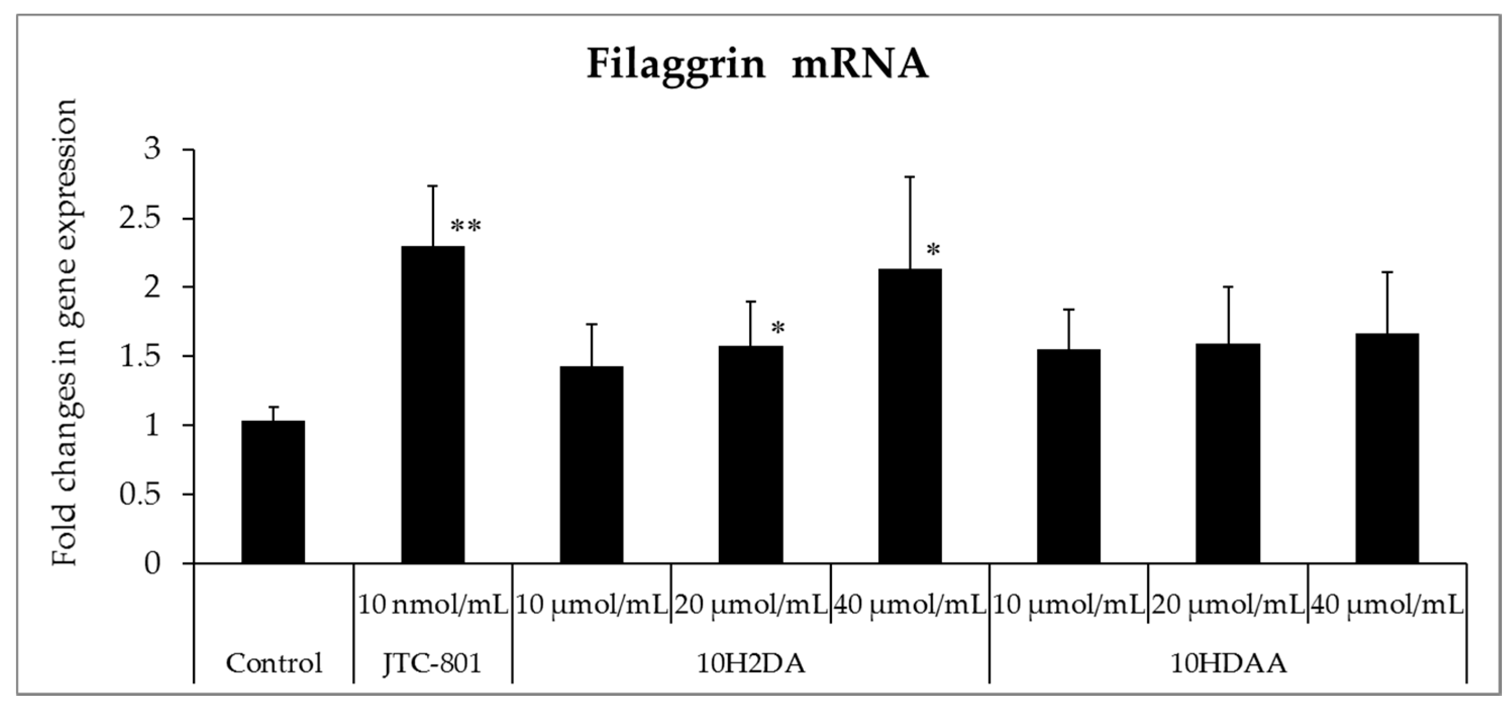
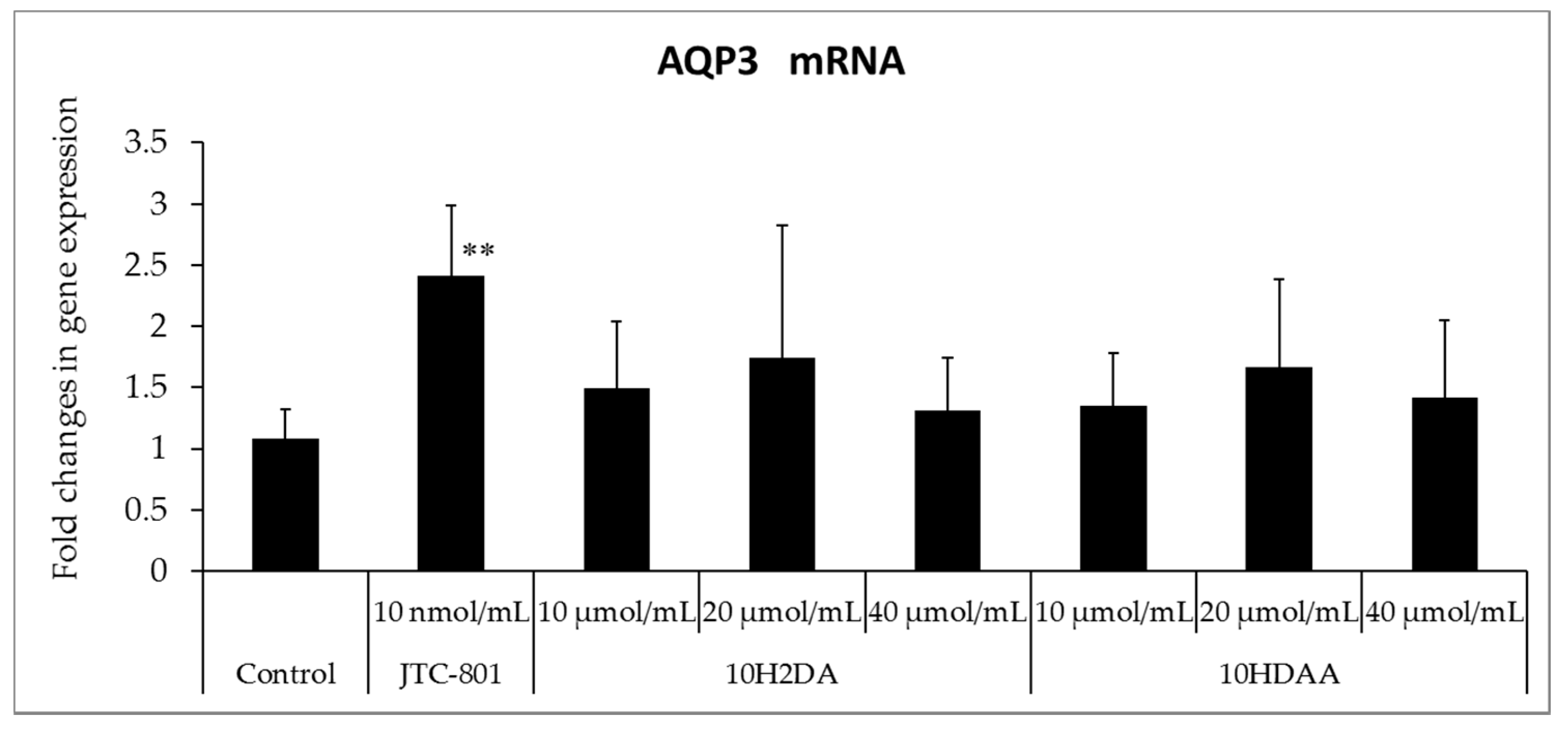
The result is shown in Figure 1. Although the amount of mRNA of FLG increased with 10H2DA at a concentration of 20 and 40 μmol/L, it did not increase with 10HDAA even at a concentration of 40 μmol/L. In addition, JTC-801, used as a positive contrast, made FLG increase by 10 nmol/L. Although AQP 3 mRNA also increased by 10 nmol/L with JTC-801, the increase was not seen with 10H2DA or 10HDAA at concentrations of 10, 20, or 40 μmol/L.
3.2. Effect of 10H2DA and 10HDAA on the Amino Acid Level in the Three-Dimensional Skin Model
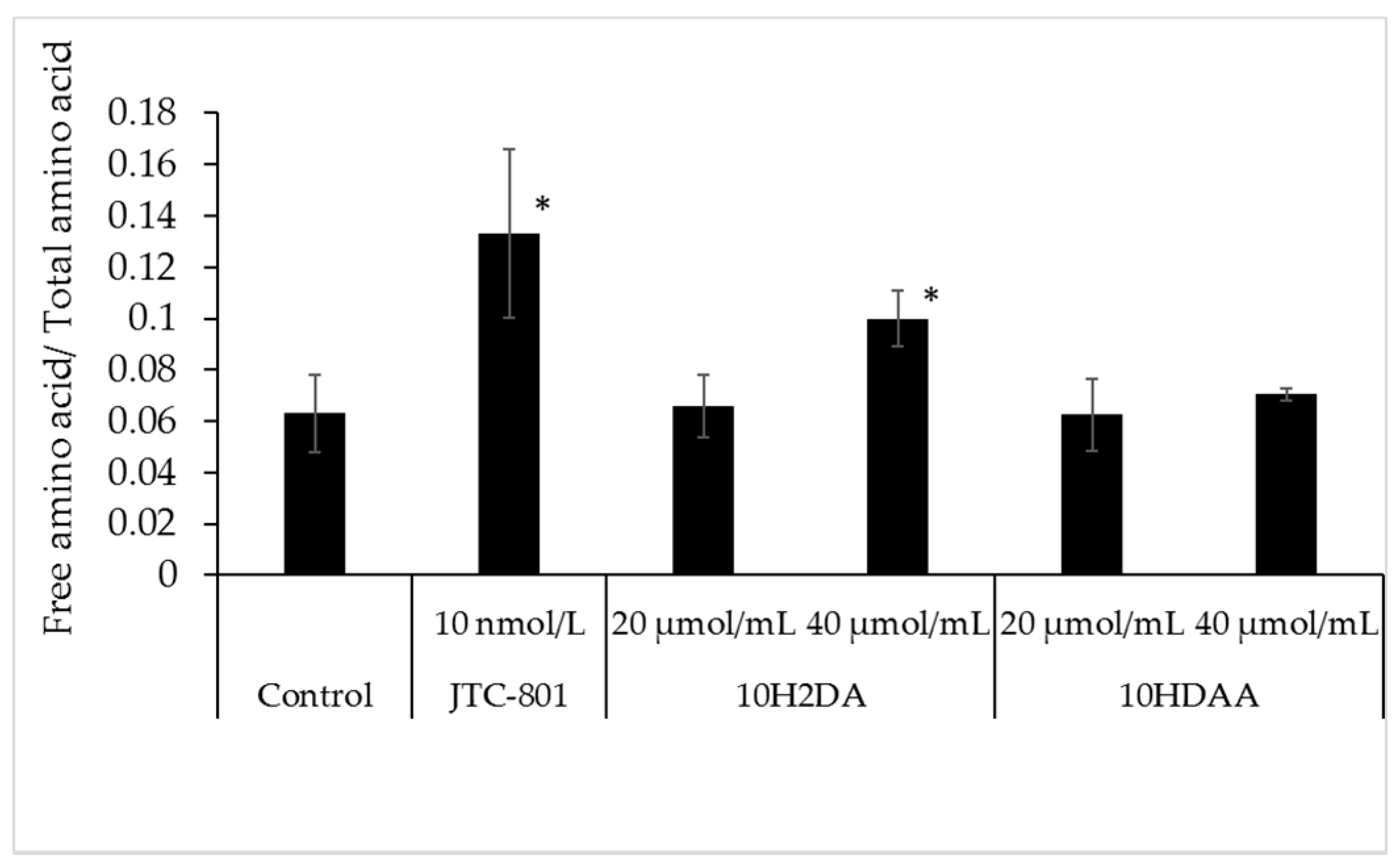
The result is shown in Figure 2. Although the amount of amino acid increased by 10H2DA at the concentration of 40 μmol/L, it did not increase by 10HDAA even at the concentration of 40 μmol/L. In addition, JTC-801, used as positive contrast, made the amount of amino acid increase by 10 nmol/L.
3.3. Immunohistochemical Staining of FLG in the Three-Dimensional Skin Model Treated with 10H2DA or 10HDAA
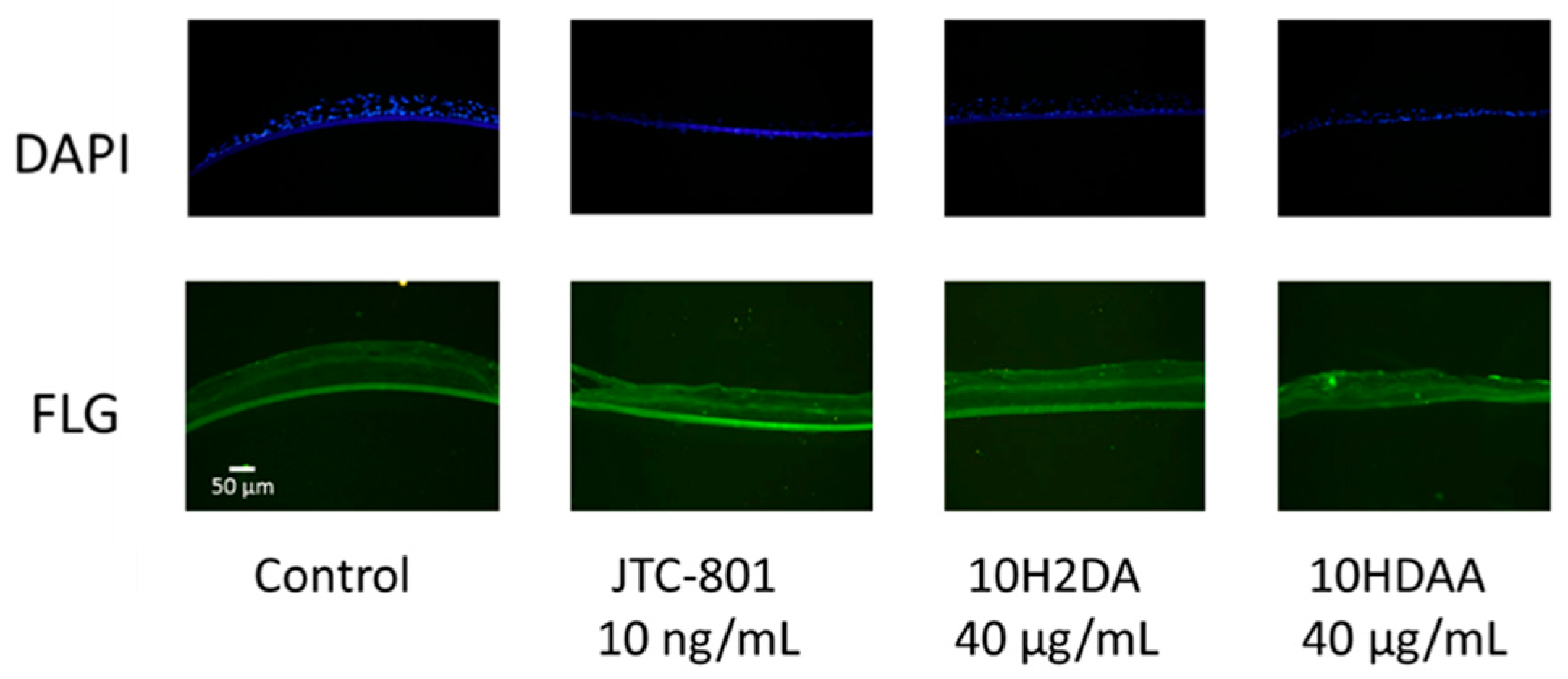
The effect of 10H2DA or 10HDAA on anti-FLG antibody-positive proteins in the cultured human skin model was examined and is shown in Figure 3. JTC-801 was used as a positive control. Anti-FLG antibody-positive protein mainly existed in the granular layer, and it increased with 40 μmol/L 10H2DA and 10 nmol/L JTC-801. It did not increase with 10HDAA at 40 μmol/L.
The fluorescence intensity and the area ratio of the anti-FLG antibody-positive part are shown in Figure 4. The fluorescence intensity of the granular layer increased intentionally, compared with the control, with 40 μmol/L 10H2DA and 10 nmol/L JTC-801. On the other hand, there was no significant difference between 40 μmol/L10HDAA and the control. In the area ratio of the anti-FLG antibody-positive part, the dyeing part of the granular layer increased intentionally, compared with the control, with 40 μmol/L 10H2DA, 40 μmol/L 10HDAA, and 10 nmol/L JTC-801.
3.4. Immunohistochemical Staining of AQP3 in the Three-Dimensional Skin Model Treated with 10H2DA or 10HDAA
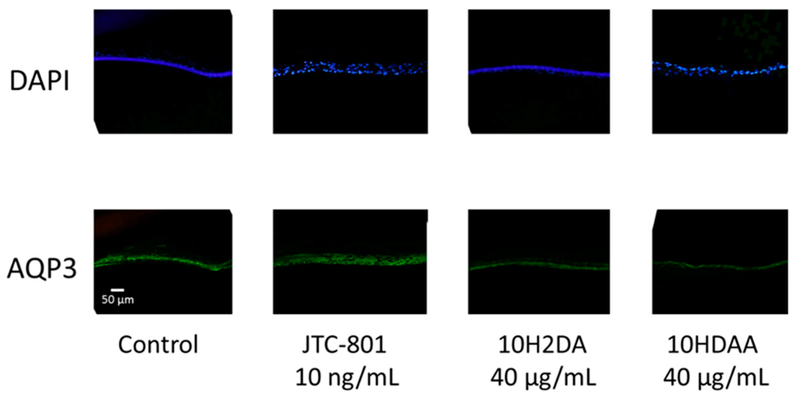
The effect of 10H2DA or 10HDAA on anti-AQP3 antibody-positive proteins in cultured human skin model was examined and is shown in Figure 5. The basal layer was mainly dyed by anti-AQP3 antibody, butthe increase was not seen with 40 μmol/L 10H2DA or 40 μmol/L 10HDAA. The protein levels of immunoreactive AQP3 was increased with 10 nmol/L JTC-801, in the same way as the increase was seen in mRNA levels by 10 nmol/L JTC-801.
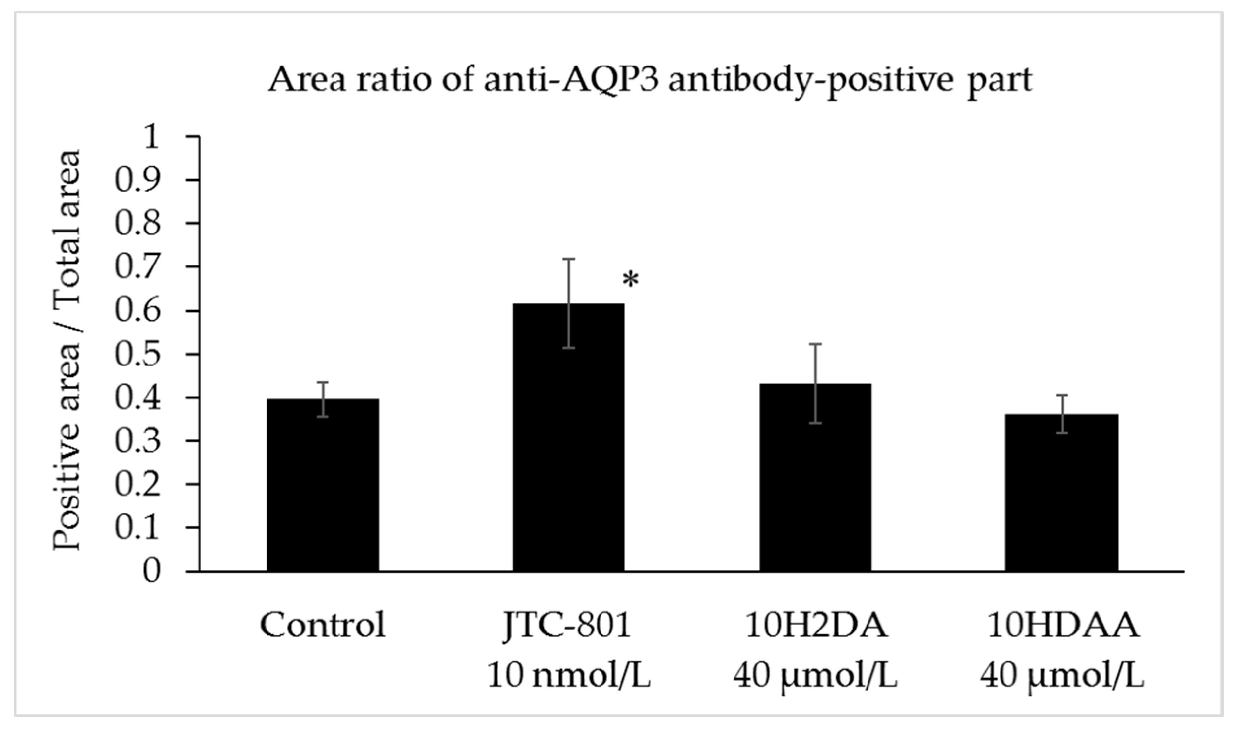
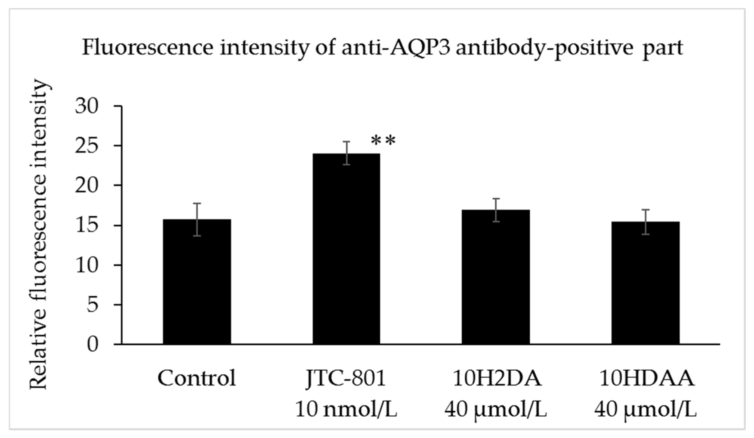
The fluorescence intensity and the area ratio of the anti-AQP3 antibody-positive part are shown in Figure 6. There was no significant difference between 10 nmol/L JTC-801 or 40 μmol/L 10H2DA, 40 μmol/L 10HDAA, and control. At an area ratio, there was no significant difference between 10 nmol/LJTC-801 or 40 μmol/L 10H2DA, 40 μmol/L 10HDAA, and control.
4. Discussion
Since the amount of stratum corneum moisture of the forearm skin had improved in four weeks by the application of RJ extract, it became clear that the consecutive use of the RJ extract increased the amount of moisture of the stratum corneum in humans and induced the amount of mRNA of FLG using the cultured human three-dimensional outer skin model [15]. The pro-FLG is expressed in the granule cell layer of the skin keratinocytes. FLG receives hydrolyzation by more than one enzyme, and becomes definitive in amino acids and its derivatives, which accounts for the majority of NMF. NMF shows an effect important for the moisture of the stratum corneum [3]. This mechanism is also supported with loss-of-function mutations in the FLG gene of atopic dermatitis patients’ decreased levels of the stratum corneum NMF [6,7]. It was revealed that RJ increased the FLG expression which participated in maintaining the stratum corneum moisture. Furthermore, in order to explore the ingredient which is participating in the increase in the amount of stratum corneum moisture by RJ extract, the effects of 10H2DA and 10HDAA on the amount of mRNA of FLG were investigated using the cultured human three-dimensional outer skin model. As a result, expression of the mRNA of FLG is increased by the addition of 10H2DA at a concentration of 40 μmol/L. On the other hand, the amount of mRNA of FLG did not increase by 10HDAA addition at the same concentration.
Since the amount of 10H2DA included in RJ extract is more than 2%, at least 3.6 μg of 10H2DA exists in 1 g of lotion containing 1.8% RJ extract [15]. The action concentration which 10H2DA promotes FLG in the epidermis and increases the amino acids in the stratum corneum is approximately at the same level as the concentration of 10H2DA contained in the RJ extract lotion. Moreover, the percutaneous effect of 10H2DA on the stratum corneum is good. From these aspects, it was thought that 10H2DA contained in RJ extract participated in the increase in the amount of stratum corneum moisture.
On the other hand, even when the oral administration of 7.2 g of RJ one time per day is carried out for eight weeks, the amount of stratum corneum moisture was increased [16]. Since the amount of blood of a 55 kg woman is 4.6 kg when intestinal absorption of 10H2DA is assumed to be the same 60% as general unsaturated fatty acid, the amount of 10H2DA in blood serves as 20 μg/mL. This concentration is the same level as the concentration of 10H2DA, which increased the FLG and stratum corneum amino acids of the cultured human epidermis model in this study. Therefore, even when a tablet containing 7.2 g RJ is taken, it is thought that 10H2DA is participating in the moisturizing of the stratum corneum in humans.
This study also examined the action of 10H2DA and 10HDAA to AQP3 mRNA in the cultured human epidermal model, and the amount of AQP3 is not affected by 10H2DA or 10HDAA. AQP3 is a water/glycerol-transporting protein expressed in keratinocytes of the epidermis. It is reported that AQP3 expression was increased in the AD skin [13] and AQP3 was involved in epidermal hyperplasia [14]. Therefore, it was thought that there was no concern of causing an epidermal hyperplasia by topical application of RJ extract.
JTC-801, used as a positive control, increased FLG in the granular layer at the mRNA level, and increased the free amino acids in the cultured epidermal model at the concentration of 10 nmol/L. In addition, JTC-801 increased AQP3 in the mRNA and protein levels at a concentration of l0 nmol/L in the cultured epidermal model.
5. Conclusions
The moisturizing function of the stratum corneum improved by the topical application of the RJ extract, and it became clear that 10H2DA, which is contained in RJ extract, is involved in its mechanism by increasing the amount of FLG of the epidermis and increasing the free amino acid of the stratum corneum in the skin.
Acknowledgments
The authors gratefully acknowledge the technical assistance of Midori Watanabe.
Author Contributions
Lihao Gu, Haifeng Zeng, and Kazuhisa Maeda performed the experiments. Kazuhisa Maeda designed the study and performed the data analysis. Lihao Gu, Haifeng Zeng, and Kazuhisa Maeda interpreted the data and drafted the manuscript; and Kazuhisa Maeda supervised the study and critically revised the manuscript. All authors read and approved the final manuscript.
Conflicts of Interest
The authors declare no conflicts of interest.
References
- Menon, G.K.; Norlén, L. Stratum corneum ceramides and their role in skin barrier function. In Skin Moisturization; Leyden, J.J., Rawlings, A.V., Eds.; Marcel Dekker, Inc.: New York, NY, USA, 2002; pp. 31–60. [Google Scholar]
- Tagami, H. Functional characteristics of the stratum corneum in photoaged skin in comparison with those found in intrinsic aging. Arch. Dermatol. Res. 2008, 300 (Suppl. 1), S1–S6. [Google Scholar] [CrossRef] [PubMed]
- Kezic, S.; Kemperman, P.M.; Koster, E.S.; de Jongh, C.M.; Thio, H.B.; Campbell, L.E.; Irvine, A.D.; McLean, W.H.; Puppels, G.J.; Caspers, P.J. Loss-of-function mutations in the filaggrin gene lead to reduced level of natural moisturizing factor in the stratum corneum. J. Investig. Dermatol. 2008, 128, 2117–2119. [Google Scholar] [CrossRef] [PubMed]
- Scharschmidt, T.C.; Man, M.Q.; Hatano, Y.; Crumrine, D.; Gunathilake, R.; Sundberg, J.P.; Silva, K.A.; Mauro, T.M.; Hupe, M.; Cho, S.; et al. Filaggrin deficiency confers a paracellular barrier abnormality that reduces inflammatory thresholds to irritant and haptens. J. Allergy Clin. Immunol. 2009, 124, 496–506. [Google Scholar] [CrossRef] [PubMed]
- Dale, B.A.; Holbrook, K.A.; Steinert, P.M. Assembly of stratum corneum basic protein and keratin filaments in macrofibrils. Nature 1978, 276, 729–731. [Google Scholar] [CrossRef] [PubMed]
- Kamata, Y.; Taniguchi, A.; Yamamoto, M.; Nomura, J.; Ishihara, K.; Takahara, H.; Hibino, T.; Takeda, A. Neutral cysteine protease bleomycin hydrolase is essential for the breakdown of deiminated FLG into amino acids. J. Biol. Chem. 2009, 284, 12829–12836. [Google Scholar] [CrossRef] [PubMed]
- Hoste, E.; Kemperman, P.; Devos, M.; Denecker, G.; Kezic, S.; Yau, N.; Gilbert, B.; Lippens, S.; De Groote, P.; Roelandt, R.; et al. Caspase-14 is required for FLG degradation to natural moisturizing factors in the skin. J. Investig. Dermatol. 2011, 131, 2233–2241. [Google Scholar] [CrossRef] [PubMed]
- Smith, F.J.; Irvine, A.D.; Terron-Kwiatkowski, A.; Sandilands, A.; Campbell, L.E.; Zhao, Y.; Liao, H.; Evans, A.T.; Goudie, D.R.; Lewis-Jones, S.; et al. Loss-of-function mutations in the gene encoding FLG cause ichthyosis vulgaris. Nat. Genet. 2006, 38, 337–342. [Google Scholar] [CrossRef] [PubMed]
- Palmer, C.N.; Irvine, A.D.; Terron-Kwiatkowski, A.; Zhao, Y.; Liao, H.; Lee, S.P.; Goudie, D.R.; Sandilands, A.; Campbell, L.E.; Smith, F.J.; et al. Common loss-of-function variants of the epidermal barrier protein filaggrin are a major predisposing factor for atopic dermatitis. Nat. Genet. 2006, 38, 441–446. [Google Scholar] [CrossRef] [PubMed]
- Rodríguez, E.; Baurecht, H.; Herberich, E.; Wagenpfeil, S.; Brown, S.J.; Cordell, H.J.; Irvine, A.D.; Weidinger, S. Meta-analysis of filaggrin polymorphisms in eczema and asthma: Robust risk factors in atopic disease. J. Allergy Clin. Immunol. 2009, 123, 1361–1370. [Google Scholar] [CrossRef] [PubMed]
- Nomura, T.; Akiyama, M.; Sandilands, A.; Nemoto-Hasebe, I.; Sakai, K.; Nagasaki, A.; Ota, M.; Hata, H.; Evans, A.T.; Palmer, C.N.; et al. Specific FLG mutations cause ichthyosis vulgaris and are significantly associated with atopic dermatitis in Japan. J. Investig. Dermatol. 2008, 128, 1436–1441. [Google Scholar] [CrossRef] [PubMed]
- Sougrat, R.; Morand, M.; Gondran, C.; Barré, P.; Gobin, R.; Bonté, F.; Dumas, M.; Verbavatz, J.M. Functional expression of AQP3 in human skin epidermis and reconstructed epidermis. J. Investig. Dermatol. 2002, 118, 678–685. [Google Scholar] [CrossRef] [PubMed]
- Olsson, M.; Broberg, A.; Jernas, M.; Carlsson, L.; Rudemo, M.; Suurküla, M.; Svensson, P.A.; Benson, M. Increased expression of aquaporin 3 in atopic eczema. Allergy 2006, 61, 1132–1137. [Google Scholar] [CrossRef] [PubMed]
- Nakahigashi, K.; Kabashima, K.; Ikoma, A.; Verkman, A.S.; Miyachi, Y.; Hara-Chikuma, M. Upregulation of aquaporin-3 is involved in keratinocyte proliferation and epidermal hyperplasia. J. Investig. Dermatol. 2011, 131, 865–873. [Google Scholar] [CrossRef] [PubMed]
- Oribe, E.; Koshiishi, Y.; Tatefuji, T.; Hashimoto, K.; Akimoto, M.; Maeda, K. Effect of royal jelly extract on water holding ability and elasticity of forearm skin. Jpn. Aesthet. Dermatol. Symp. 2013, 6, 10–14. (In Japanese) [Google Scholar]
- Oribe, E. Effect of long term intake of enzyme-treated royal jelly in normal skin. Fine Chem. 2013, 42, 20–25. (In Japanese) [Google Scholar]
- Howe, S.R.; Dimick, P.S.; Benton, A.W. Composition of freshly harvested and commercial royal jelly. J. Apic. Res. 1985, 24, 52–61. [Google Scholar] [CrossRef]
- Blum, M.S.; Novak, A.F.; Taber, S., 3rd. 10-Hydroxy-delta 2-decenoic acid, an antibiotic found in royal jelly. Science 1959, 130, 452–453. [Google Scholar] [CrossRef] [PubMed]
- Tamura, T.; Fujii, A.; Kuboyama, N. Antitumor effects of royal jelly (RJ). Nihon Yakurigaku Zasshi 1987, 89, 73–80. [Google Scholar] [CrossRef] [PubMed]
- Sver, L.; Orsolić, N.; Tadić, Z.; Njari, B.; Valpotić, I.; Basić, I. A royal jelly as a new potential immunomodulator in rats and mice. Comp. Immunol. Microbiol. Infect. Dis. 1996, 19, 31–38. [Google Scholar] [CrossRef]
- Yang, X.Y.; Yang, D.S.; Zhang, W.; Wang, J.M.; Li, C.Y.; Ye, H.; Lei, K.F.; Chen, X.F.; Shen, N.H.; Jin, L.Q.; Wang, J.G. 10-Hydroxy-2-decenoic acid from Royal jelly: A potential medicine for RA. J. Ethnopharmacol. 2010, 128, 314–321. [Google Scholar] [CrossRef] [PubMed]
- Mishima, S.; Suzuki, K.M.; Isohama, Y.; Kuratsu, N.; Araki, Y.; Inoue, M.; Miyata, T. Royal jelly has estrogenic effects in vitro and in vivo. J. Ethnopharmacol. 2005, 101, 215–220. [Google Scholar] [CrossRef] [PubMed]
- Suzuki, K.M.; Isohama, Y.; Maruyama, H.; Yamada, Y.; Narita, Y.; Ohta, S.; Araki, Y.; Miyata, T.; Mishima, S. Estrogenic activities of fatty acids and a sterol isolated from royal jelly. Evid. Based Complement. Altern. Med. 2008, 5, 295–302. [Google Scholar] [CrossRef] [PubMed]
- Moutsatsou, P.; Papoutsi, Z.; Kassi, E.; Heldring, N.; Zhao, C.; Tsiapara, A.; Melliou, E.; Chrousos, G.P.; Chinou, I.; Karshikoff, A.; et al. Fatty acids derived from royal jelly are modulators of estrogen receptor functions. PLoS ONE 2010, 5, e15594. [Google Scholar] [CrossRef] [PubMed]
- Ito, S.; Nitta, Y.; Fukumitsu, H.; Soumiya, H.; Ikeno, K.; Nakamura, T.; Furukawa, S. Antidepressant-like activity of 10-hydroxy-trans-2-decenoic Acid, a unique unsaturated fatty acid of royal jelly, in stress-inducible depression-like mouse model. Evid. Based Complement. Altern. Med. 2012, 2012, 139140. [Google Scholar] [CrossRef] [PubMed]
- Izuta, H.; Chikaraishi, Y.; Shimazawa, M.; Mishima, S.; Hara, H. 10-Hydroxy-2-decenoic acid, a major fatty acid from royal jelly, inhibits VEGF-induced angiogenesis in human umbilical vein endothelial cells. Evid. Based Complement. Altern. Med. 2009, 6, 489–494. [Google Scholar] [CrossRef] [PubMed]
- Lambrinoudaki, I.; Augoulea, A.; Rizos, D.; Politi, M.; Tsoltos, N.; Moros, M.; Chinou, I.; Graikou, K.; Kouskouni, E.; Kambani, S.; et al. Greek-origin royal jelly improves the lipid profile of postmenopausal women. Gynecol. Endocrinol. 2016, 32, 835–839. [Google Scholar] [CrossRef] [PubMed]
- Yamada, N.; Yoshimura, H. Effect of royal jelly on chills in Japanese young wamen. Jpn. Soc. Nutr. Food Sci. 2010, 63, 271–278. [Google Scholar] [CrossRef]
- Maeda, T.; Kuroda, H.; Motoyoshi, K. Effects of royal jelly and 10-hydroxy decenoic acid on the sebaceous glands of hamster ear. Nihon Hifuka Gakkai Zasshi 1988, 98, 469–475. (In Japanese) [Google Scholar] [PubMed]
- Taniguchi, Y.; Kohno, K.; Inoue, S.; Koya-Miyata, S.; Okamoto, I.; Arai, N.; Iwaki, K.; Ikeda, M.; Kurimoto, M. Oral administration of royal jelly inhibits the development of atopic dermatitis-like skin lesions in NC/Nga mice. Int. Immunopharmacol. 2003, 3, 1313–1324. [Google Scholar] [CrossRef]
- Chen, Y.F.; Wang, K.; Zhang, Y.Z.; Zheng, Y.F.; Hu, F.L. In vitro anti-Inflammatory effects of three fatty acids from royal jelly. Mediat. Inflamm. 2016, 2016, 3583684. [Google Scholar] [CrossRef] [PubMed]
- Duplan, H.; Questel, E.; Hernandez-Pigeon, H.; Galliano, M.F.; Caruana, A.; Ceruti, I.; Ambonati, M.; Mejean, C.; Damour, O.; Castex-Rizzi, N.; et al. Effects of Hydroxydecine® (10-hydroxy-2-decenoic acid) on skin barrier structure and function in vitro and clinical efficacy in the treatment of UV-induced xerosis. Eur. J. Dermatol. 2011, 21, 906–915. [Google Scholar] [PubMed]
- Koya-Miyata, S.; Okamoto, I.; Ushio, S.; Iwaki, K.; Ikeda, M.; Kurimoto, M. Identification of a collagen production-promoting factor from an extract of royal jelly and its possible mechanism. Biosci. Biotechnol. Biochem. 2004, 68, 767–773. [Google Scholar] [CrossRef] [PubMed]
- Otsuka, A.; Doi, H.; Egawa, G.; Maekawa, A.; Fujita, T.; Nakamizo, S.; Nakashima, C.; Nakajima, S.; Watanabe, T.; Miyachi, Y.; et al. Possible new therapeutic strategy to regulate atopic dermatitis through upregulating filaggrin expression. J. Allergy Clin. Immunol. 2014, 133, 139–146. [Google Scholar] [CrossRef] [PubMed]
Figure 1.
Effects of 10H2DA and 10HDAA on the mRNA expressions of FLG and AQP3 in the human epidermal model cultured for 24 h. The concentrations of 10H2DA and 10HDAA were 10 μg/mL, 20 μg/mL, and 40 μg/mL. JTC-801 (10 ng/mL) was used as a positive control. The result was analyzed by the 2−ΔΔCt method relative to the internal control gene (β-actin), fold changes in gene expression expressed with the mean ± standard deviation of three experiments. * p < 0.05 vs. control, and ** p < 0.01 vs. control.


Figure 2.
Effects of 10H2DA and 10HDAA on the amount of amino acid of stratum corneum in the human epidermal model cultured for five days. The concentrations of 10H2DA and 10HDAA were 20 μg/mL and 40 μg/mL. JTC-801 (10 ng/mL) was used as a positive control. The result was expressed in the ratio of free amino acid to total amino acid in the stratum corneum with the mean ± standard deviation of three experiments. * p < 0.05 vs. control.

Figure 3.
Effects of 10H2DA and 10HDAA on the anti-FLG antibody-positive protein in the human epidermal model cultured for five days. The concentrations of 10H2DA and 10HDAA were 40 μg/mL. JTC-801 (10 ng/mL) was used as a positive control. The nuclei are represented by blue staining. Green fluorescence in FLG in the granule cell layer appear as lines. The membrane was strongly nonspecifically stained in green.

Figure 4.
The fluorescence intensity and the area ratio of an anti-FLG antibody-positive part. Every three-fluorescence intensity per picture of one sheet were measured, the result of the picture of three sheets was calculated. In addition, the area ratio was calculated by having measured the area of the part which strongly exhibits fluorescence in the area of the whole specimen without the membrane. The result was expressed with the mean ± standard deviation. * p < 0.05 vs. control, and ** p < 0.01 vs. control.

Figure 5.
Effects of 10H2DA and 10HDAA on the anti-AQP3 antibody-positive protein in the human epidermal model cultured for five days. The concentrations of 10H2DA and 10HDAA were 40 μg/mL, and JTC-801 (10 ng/mL) was used as a positive control. The nuclei are represented by blue staining. Green fluorescence in AQP3 shows the presence of the basal cell layer.

Figure 6.
The fluorescence intensity and the area ratio of an anti-AQP3 antibody-positive part. Every three-fluorescence intensity per picture of one sheet were measured, and the result of the picture of three sheets was calculated. In addition, the area ratio was calculated by having measured the area of the part which is strongly exhibiting fluorescence in the area of whole specimen without the membrane. The result was expressed as the mean ± standard deviation. * p < 0.05 vs. control, and ** p < 0.01 vs. control.

© 2017 by the authors. Licensee MDPI, Basel, Switzerland. This article is an open access article distributed under the terms and conditions of the Creative Commons Attribution (CC BY) license (http://creativecommons.org/licenses/by/4.0/).
Share and Cite
MDPI and ACS Style
Gu, L.; Zeng, H.; Maeda, K. 10-Hydroxy-2-Decenoic Acid in Royal Jelly Extract Induced Both Filaggrin and Amino Acid in a Cultured Human Three-Dimensional Epidermis Model. Cosmetics 2017, 4, 48. https://doi.org/10.3390/cosmetics4040048
AMA Style
Gu L, Zeng H, Maeda K. 10-Hydroxy-2-Decenoic Acid in Royal Jelly Extract Induced Both Filaggrin and Amino Acid in a Cultured Human Three-Dimensional Epidermis Model. Cosmetics. 2017; 4(4):48. https://doi.org/10.3390/cosmetics4040048
Chicago/Turabian StyleGu, Lihao, Haifeng Zeng, and Kazuhisa Maeda. 2017. "10-Hydroxy-2-Decenoic Acid in Royal Jelly Extract Induced Both Filaggrin and Amino Acid in a Cultured Human Three-Dimensional Epidermis Model" Cosmetics 4, no. 4: 48. https://doi.org/10.3390/cosmetics4040048
Note that from the first issue of 2016, this journal uses article numbers instead of page numbers. See further details here.