Article

Schisandra chinensis Protects the Skin from Global Pollution by Inflammatory and Redox Balance Pathway Modulations: An In Vitro Study

1
Greentech, Biopôle Clermont-Limagne, 63360 Saint Beauzire, France
2
Syntivia, Place Pierre Potier, 31106 Toulouse CEDEX 1, France
3
Complexity, Innovation, and Sport Activity department (CIAMS), University of Paris-Saclay, 91405 Orsay CEDEX, France
4
CIAMS department, University of Orléans, 45067 Orléans, France
*
Author to whom correspondence should be addressed.
Cosmetics 2018, 5(2), 36; https://doi.org/10.3390/cosmetics5020036
Submission received: 3 April 2018 / Revised: 17 May 2018 / Accepted: 4 June 2018 / Published: 7 June 2018

Abstract

:
Epidemiological results show that airborne particulate matter (PM) induces health alterations in line with pulmonary and cardiovascular pathologies. Deleterious effects of PM on the skin have also been investigated. A possible approach to prevent Reactive Oxygen Species (ROS)-mediated disorders for both preventive and treatment means is based on the use of substances, which can be found in plants. These can act as secondary metabolites, and lignans are a promising candidate. Thus, the objective of this study was firstly to identify reconstructed human epidermis, using a transcriptomic approach, and also to identify the effects of Urban Dust and of Urban Dust and Schisandra chinensis (S.C.) extract on the expression of genes that are involved in the response to cellular protection mechanisms. Secondly, we examined the effect of an active extract from S.C. on the protection of human keratinocytes damages that were caused by pollution, through the evaluation of Nrf2 and AhR pathways, NF-kB, and DJ-1. Urban Dust included the over-expression of metalloproteinases MMP-1 and MMP-9 and an increase in Glutathione peroxidase 2 (GPX2). In the presence of Urban Dust, S.C. extract activated the over-expression of several genes that are involved in the antioxidant response and in the detoxification pathway, including Ferritin light chain (FTL) and GPX2. Exposure to urban dust activated the cytoplasmic expression of NF-kB and AhR, when compared to the control. Co-treatment of Urban Dust and S.C. extract increased DJ-1 protein levels, Nrf2 expression, and decreased AhR and NF-kB in the cytoplasm. At the same time, this co-treatment increased SOD2 expression (50%: p < 0.001) and catalase activity (120%: p < 0.05), when compared to Urban Dust alone. Thus, S.C. might be able to protect the Normal Human Epidermal Keratinocytes (NHEK) from environmental aggression, by fighting the harmful effects of urban pollution.

1. Introduction

Environmental pollution is increasing, especially in industrialized countries, and it represents a worldwide problem that cannot be avoided [1]. The air pollutants, which are produced by both natural and human sources, include nitrogen and sulfur oxides, carbon monoxide, ozone, volatile organic compounds, particulate matter (PM) and polycyclic aromatic hydrocarbons (PAHs.), and heavy metals [2]. Today, there is sufficient evidence that various air pollutants have adverse impacts on human and environmental health, inducing the development or the exacerbation of cardiovascular and respiratory diseases [3]. The link between skin diseases and pollution exposure was first published in 2010, when authors suggested that air pollution exposure was significantly correlated with pigment spots on the face and hands and, to a lesser extent, with wrinkles [4,5]. It has also been shown that environmental pollution is significantly associated with weakened barrier function, oxidative stress, and skin diseases [6,7]. It also has a negative effect on skin microflora, inducing an anaerobic favorable environment by blocking pores, and therefore leading to the growth of Cutibacterium acnes, which plays an important role in the development of acne [8]. The decreased content of vitamin E in the skin and an increased sebum secretion rate are also consequences of exposure to pollutants.
Generally speaking, pollutants penetrate the skin either through hair follicles or transdermally, and they exert their detrimental effects through the production of reactive oxygen species (ROS), which contribute to extrinsic skin ageing, which is particularly characterized by pigment spots on the face and nasolabial folds. Figure 1 summarizes the signaling pathway responses to global pollution, inducing the release of inflammatory cytokines, and the production of metalloproteinases (MMPs), which are closely related to inflammatory skin diseases and skin ageing [2]. The toxicity of pollutants is also linked, at least in part, to the activation of the aryl hydrocarbon receptor (AhR). Upon ligand binding, cytoplasmic AhR translocates to the nucleus and dimerizes with the AhR nuclear translocator (ARNT). The ligand-activated AhR/ARNT complex then binds to specific promoter elements called dioxin-responsive elements (DRE), and induces the expression of target genes, such as cytochrome P450 1A1 (CYP1A1).

1.1. Antioxidants that Inhibit ROS

Besides endogenous antioxidants that are present in the skin, such as vitamin A, vitamin C, and vitamin E, enzymes such as superoxide dismutase, catalase, and glutathione biosynthesizing enzymes protect the tissues from free radicals [9]. Other endogenous defense mechanisms, including a fundamental biochemical pathway (nuclear factor erythroid 2-related factor 2 (Nrf2)), are activated in order to fight the deleterious effects of all pollutants on the skin. It is able to help to eliminate and inactivate exogenous toxic agents by closely interconnected fundamental biological pathways. The involvement of the Nrf2 pathway in the skin is of high importance, as it plays a role in skin homeostasis and skin renewal, and the function of Nrf2 as a master regulation of cellular redox homeostasis is widely recognized. In fact, Nrf2 not only regulates a variety of antioxidant enzymes, such as NAD(P)H: quinone oxidoreductase (NQO1), thioredoxin, and heme oxygenase-1, but also several phase I and phase II drug metabolizing enzymes, for example, glutathione S-transferase. Phase-II protective enzymes are responsible for the antioxidant response, xenobiotic disposition, inflammatory response, metabolic programming cell proliferation, and survival through the antioxidant-response element (ARE) (Figure 1). Nrf2 is constitutively expressed in the cytoplasm, and its accumulation and activation in the nucleus are favored in oxidative injury. The activity of Nrf2 is regulated by various mechanisms. Under homeostatic conditions, Nrf2 is localized in the cytoplasm, where it is sequestered by its inhibitor, Kelch-like ECH-associated protein 1 (Keap1). It also appears that Nrf2 is directly regulated by AhR. Additionally, Nrf2 is stabilized by the Parkinson’s-associated protein (DJ-1), which is a multifunctional protein that is expressed in almost all tissues that are involved in various physiological processes, such as transcriptional regulation, anti-oxidative stress reaction, mitochondrial regulation, and signal transduction [10] (Figure 1). More precisely, DJ-1 promotes Nrf2 binding to antioxidant response elements, by which Nrf2 can regulate the expression of several endogenous antioxidative enzymes and can reduce ROS production to protect the mitochondria, and can also respond to oxidative stress. Under oxidative stress, DJ-1 plays a critical antioxidant defense role in several molecular processes. In addition to directly regulating some antioxidant gene expression, DJ-1 functions as an atypical ROS scavenger peroxiredoxin-like peroxidase, through the oxidization of conserved cysteine residue (Cys106). Additionally, it protects mitochondria by directly maintaining mitochondrial complex I activity [10] and by translocating into mitochondria as an endogenous antioxidant.

1.2. Plant-Derived Compounds to Limit ROS Production

A possible approach to attack ROS-mediated disorders for both preventive and treatment means, is based on the use of substances which can be found in plants, which act as secondary metabolites. Thus, plant-derived compounds, such as carotenoids and polyphenols, are molecules with antioxidant properties [9]. Catechins, including epicatechin, epicatechin-3-gallate, epigallocatechin, and epigallocatechin gallate are included in green tea. These polyphenols present interesting biological activities, such as antimicrobial, anti-inflammatory, anti-cancer, antioxidant, and radical scavenging activities [11]. Moreover, they are known for having a protective effect on oxidative stress and DNA damage that is induced by environmental pollutants (e.g., pesticides and smoking). Luteolin, apigenin, or terniflorin, which are molecules belonging to the flavonoids, and several terpenoid compounds, such as marrubiin, sacranoside, and phenylpropanoid compounds were identified in Marrubium vulgare [12]. The extract of Selaginella lepidophylla (Rose of Jericho or the Desert Rose) contains an adaptive moisture retention complex with film forming properties that improves skin barrier function, offering intense moisturizing benefits and reducing trans epidermal water loss from compromised skin. In addition, it also inhibits the accumulation of PM on the skin, provides antioxidant benefits, and enhances cellular proliferation [13]. It also appears that phyto-derived compounds from plants, which have estrogenic characteristics, such as equol, can increase the skin’s antioxidant defense [14]. Indeed, in vitro studies have reported that equol has a greater capacity for neutralizing oxidants compared to genistein, vitamin C, or other polyphenolic compounds like quercetin [15]. Moreover, equol seems to have beneficial effects on extracellular matrix proteins like collagen and elastin, and can protect against oxidative stress and inflammation.
Various phytochemicals and herbal extracts exert their antioxidant properties by activating the Nrf2 system in an AhR-dependent or AhR-independent manner in human epidermal keratinocytes [16]. Among them, Schisandra chinensis (S.C.), a traditional Chinese herbal medicine, includes Schisandra lignans, such as schisandrin A, deoxyshisandrin, and γ-schisandrin, which are the major constituents of S.C., and more than 40 of them have now been isolated [17]. Lignans were found to possess several beneficial pharmacological effects, exhibiting potent anti-oxidative and anti-inflammatory properties, detoxification, and anti-carcinogenic activity. In fact, Opletal et al. [18] reported that the lipid peroxidation of cell membranes could be inhibited, and that the ability of resisting the ROS of cells by enhancive superoxide dismutase (SOD) with catalase activities could both be reinforced by lots of Schisandra lignans. Besides, organic acids, sterols, vitamins, and carbohydrates were also reportedly extracted from S.C. [17]. Thus, it appears that S.C. possesses various biological effects which may activate the endogenous mechanisms of the defense and biological pathways to fight against pollutants. These findings led us to postulate that S.C. lignans might protect skin cell functions against urban pollution, and that they can be used as a skin protective agent against pollutants.
The Nrf2-Keap 1 system and AhR have not yet been explored as a strategy to enhance antioxidant defenses [16]. The role of DJ-1 is less explored, even if its role is essential in the physiological process to fight against pollutants. Thus, the objective of this study was firstly to identify the reconstructed human epidermis, by a transcriptomic approach, and also to identify the effects of Urban Dust and then the effect of Urban Dust and S.C. extract on the expression of genes that are involved in the response to cellular protection mechanisms, namely the detoxification pathways. Secondly, we examined the effect of the active extract from S.C. on the protection of human keratinocytes damages that were caused by pollution, through the evaluation of Nrf2 and AhR pathways, NF-kB, and DJ-1. As pollution is a complex mixture of particulate matter (PM2.5 and PM10), PAHs, VOCs, and nitrogen oxides, we decided to standardize the cell culture models using a Standard Reference Material® (Urban Dust 1649b, NIST), which is well characterized and represents global urban pollution stress.

2. Materials and Methods

2.1. Transcriptomic Analyses

The transcriptomic approach was conducted on a 3D model of reconstructed human epidermis (StratiCELL, Lyon, France, RHEs), and using qRT-PCR, it measured the effects of the S.C. extract on the expression of 93 genes that were involved in the response and cellular protection mechanisms against pollution, namely the detoxification pathways, inflammation, and the antioxidant defense.
Tissues were cultured at the air-liquid interface for 14 days in a suitable culture medium in a humid atmosphere at 37 °C with 5% CO2. The tissues were transferred into a 12-well plate before the treatments. The effect of the S.C. extract that was applied after 48 h in the culture medium of RHEs, was evaluated. Urban Dust (Standard Reference Material® 1649b, NIST) was either applied or not at 80 μg·mL−1 in the culture medium during the last 24 h of the treatment. At the end of the treatment, total RNAs were extracted, and the sample integrity was analyzed by spectrophotometry and capillary electrophoresis. cDNAs were then synthesized from mRNA by reverse transcription. Gene expression changes were addressed by qPCR. The total RNAs were extracted using the Quiagen RNeasy kit (74106, QIAGEN), Thermo Fisher Scientific, 91963 Courtaboeuf, France). The tissues were removed from their inserts and were immersed directly in the lysis buffer. The extraction of RNA was performed on the tissues according to the supplier’s recommendations. The collected RNAs were stored at −80 °C.
The RNA concentration was determined by the spectrophotometric measurement (ULtrospec 1100 Pro from Amersham, (Scinteck Instruments, Manassas, VA, USA), and the RNA quality was analyzed by capillarity electrophoresis (Agilent Bioanalyzer 2100—Agilent RNA 6000 Nano kit, 5067-1511, (Agilent, Santa Clara, CA, USA). The integrity of the total RNA was assessed by the visualization of intact ribosomal RNA bands. Reverse transcription was performed with the high capacity RNA-to-cDNA kit (Applied Biosystems, 91140 Villebon sur Yvette, France) from 2 μg of total RNA, according to the manufacturer’s instructions. The cDNAs were then stored at −20 °C. Gene expression was analyzed using 384-wells microfluidic TaqManq PCR arrays that were designed by StratiCELL and were manufactured on demand by Applied Biosystems (91140 Villebon sur Yvette, France). The TaqMan arrays were processed as described by the manufacturer’s instructions.
Threshold cycles (Ct) were obtained for each gene. The data were analyzed using the RQ application that was available on the Applied Biosystems website, and was designed to perform the relative quantification of gene expression using the comparative Ct (ΔΔCt) method, through a combination of statistical analysis. The data were analyzed with a combination of β2-microglobulin (b2M), glyceraldehyde-3-phosphate dehydrogenase (GAPDH), and tyrosine 3-monooxygenase/tryptophan 5-monooxygenase activation protein epsilon polypeptide (YWHAE) as housekeeping genes. The maximum Ct cut-off value was fixed at 36 cycles.

2.2. The effect of the Extract from S.C. on the Protection of Human Keratinocytes (NHEK) Damages that are Caused by Pollution

2.2.1. Viability and Cytotoxicity Determination

NHEK were seeded in a 96-well plate and were incubated for 24 h at 37 °C, 5% CO2. Viability was first assessed on NHEK with Urban Dust and/or the S.C. extract at 8 concentrations. The S.C. extract was diluted in NHEK culture medium (maximum DMSO concentration: 0.1%), and Urban Dust was dispersed in NHEK culture medium. S.C. was brought into contact with the cells after 24 h, and Urban Dust after 6 h. The combination of the two products was tested after 6 h. During the last 3 h, WST1 (Roche) was introduced into the medium. This reagent contains tetrazolium salts, a violet indicator that is cleaved in formazan, a yellow indicator, and metabolically active cells. The level of the yellow color is proportional to the number of living cells, and absorbance was measured at 450 nm. A viability value below 80% indicated significant cytotoxicity.

2.2.2. Effect of Urban Pollution and the S.C. Extract on Cell Pathways

The cells were seeded in a 96-well plate and were treated with S.C extract at 50 μg·mL−1 for 24 h. and were then exposed after 6 h to Urban Dust (Standard Reference Material® 1649b, NIST, 80 μg·mL−1) + S.C. extract (50 μg·mL−1). The cells were then fixed with formalin, and the expression of several biomarkers that were implicated in the cell response to pollution were detected by immunofluorescence in the cytoplasm and/or in the nucleus (AhR, Nrf2, DJ-1, Nf-kB and SOD2). After a step of permeabilization/saturation, staining of the treated cells with antibodies (anti-AhR polyclonal, anti-Nrf2 polyclonal, anti-DJ-1 polyclonal, anti-NF-kB polyclonal, and anti-SOD2 polyclonal) was performed overnight at 4 °C. The secondary antibody AlexaFluor 488 tagged goat antibody was applied after 1 h at room temperature.
Fluorescent labeling was imaged and quantified by automated microscopy (ArrayScan Cellomics™, Thermo Fisher Scientific, 91963 Courtaboeuf, France). The fluorescence was quantified by the bioapplication Compartmental Analysis in the nucleus and cytoplasm. The condition of more than 1200 cells was analyzed.
Catalase activity was quantified by colorimetry on cell lysate at 6 h after Urban Dust exposure using a Catalase activity assay kit, and measured at 570 nm on a microplate reader (Multiskan-Ascent MTX Lab Systems).
ROS production was measured at 6 h after Urban Dust, in order to evaluate the intracellular redox balance. Intracellular oxidation is quantified by flow cytometry (BD Accuri™ C6 Plus) with a DCF-DA (2′,7′-dichlorofluorescein diacetate) probe (Sigma Aldrich, 38297 Saint Quentin Fallavier, France). DCF-DA enters into the cells, where it is hydrolyzed in fluorescent DCFH in the presence of ROS.

2.3. Statistical Analysis

Results were expressed as mean value ± SEM and represent the means of triplicate determinations that were obtained in four separate experiments. An ANOVA, followed by a Bonferroni post-hoc test was performed using SPSS Software (version 16.00 for Windows, OR, USA), and statistical significance was considered at p < 0.05.

3. Results

3.1. Transcriptomic Results

The effect of Urban Dust () is consistent with historical standards. As shown in Table 1, it significantly increases the expression of Cytochrome P450, family 1, member A1 (CYP1A1), Cytochrome P450, family 1 Subfamily B member A1 (CYP1B1), and aldehyde dehydrogenase 3 family member A1 (ALDH3A1).
Other effects of Urban Pollution include the over-expression of metalloproteinases MMP-1 and MMP-9, and an increase in Glutathione peroxidase 2 (GPX2: p < 0.006) and NAD(P)H deshydrogenase (p < 0.001).
In the presence of Urban Dust, S.C. extract (50 μg·mL−1) activates the over-expression of several genes that are involved in the antioxidant response and in the detoxification pathway, including Ferritin light chain (FTL) and GPX2. It also induces the over expression of a small proline-rich protein (SPRR1A) that is a gene coding for cornifin-A, and functions as a cross-linked envelope precursor.

3.2. The Effect of Urban Dust and S.C. Extract on NF-kB Activation

In the NHEK model, Urban Dust significantly activated the cytoplasmic expression of NF-kB (+16%, p < 0.001), when compared to the control. The co-treatment of Urban Dust and S.C. extract significantly decreased NF-kB expression in the cytoplasm (−8%, p < 0.001) and nucleus (−6%, p < 0.001), when compared to Urban Dust alone.

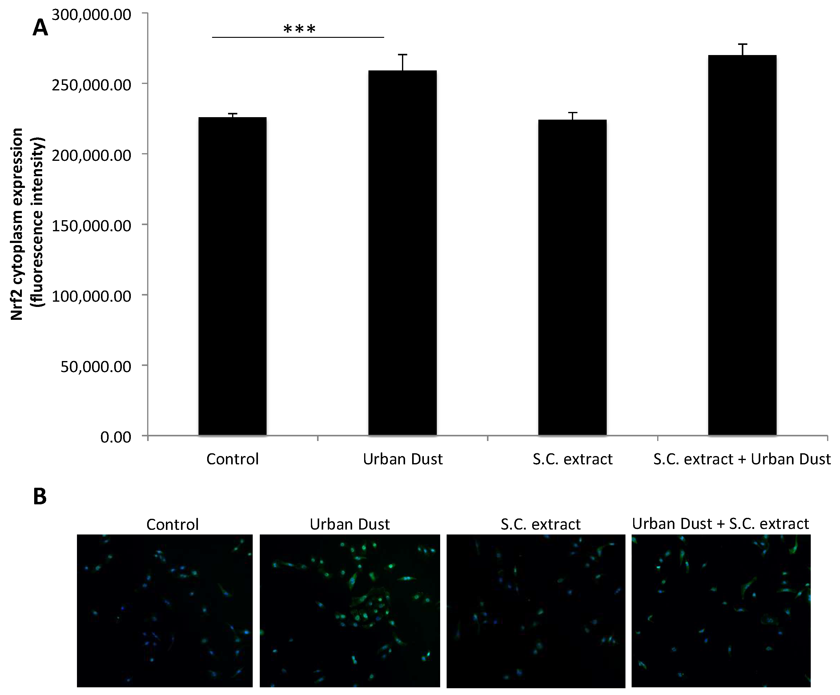
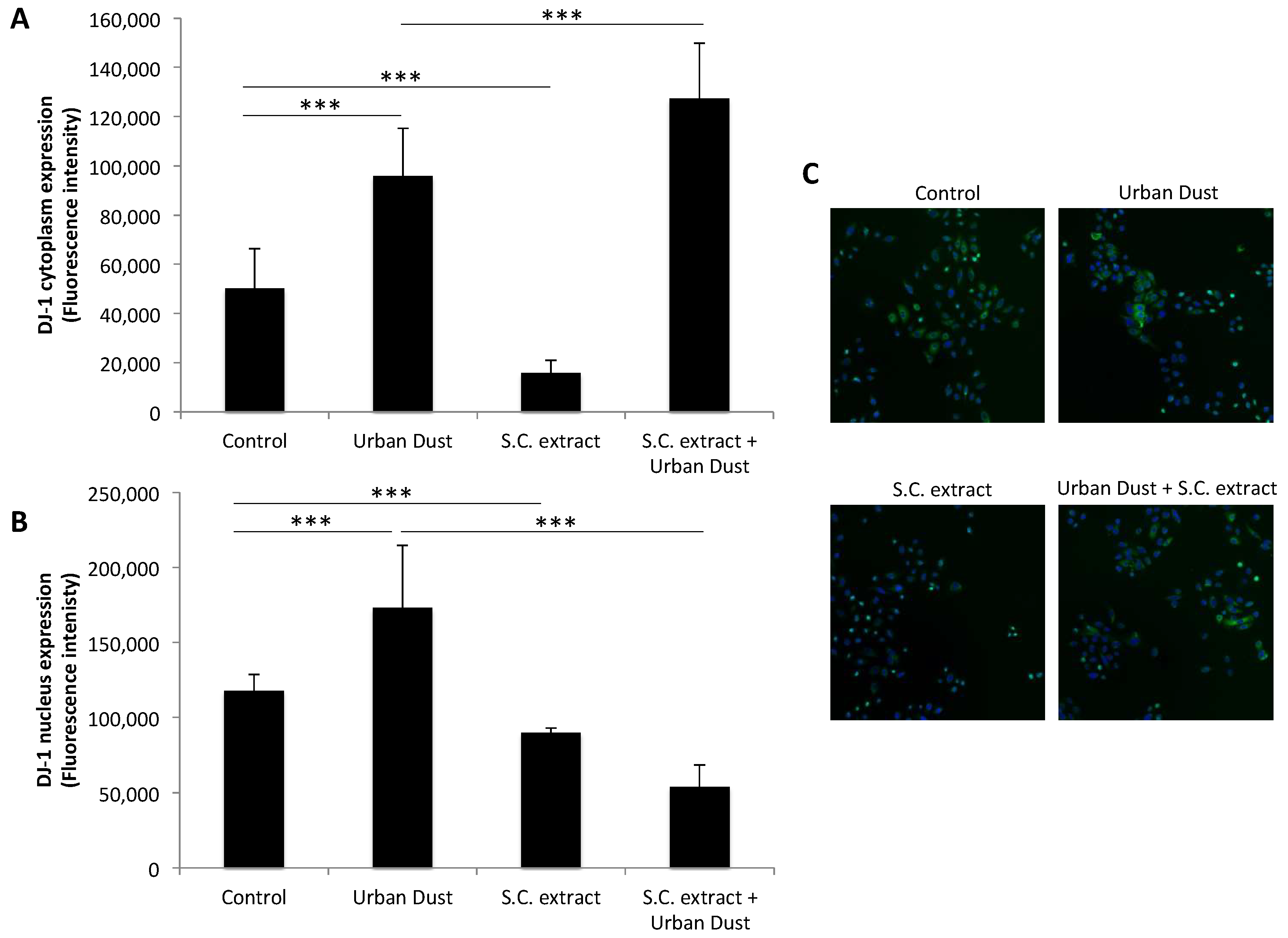
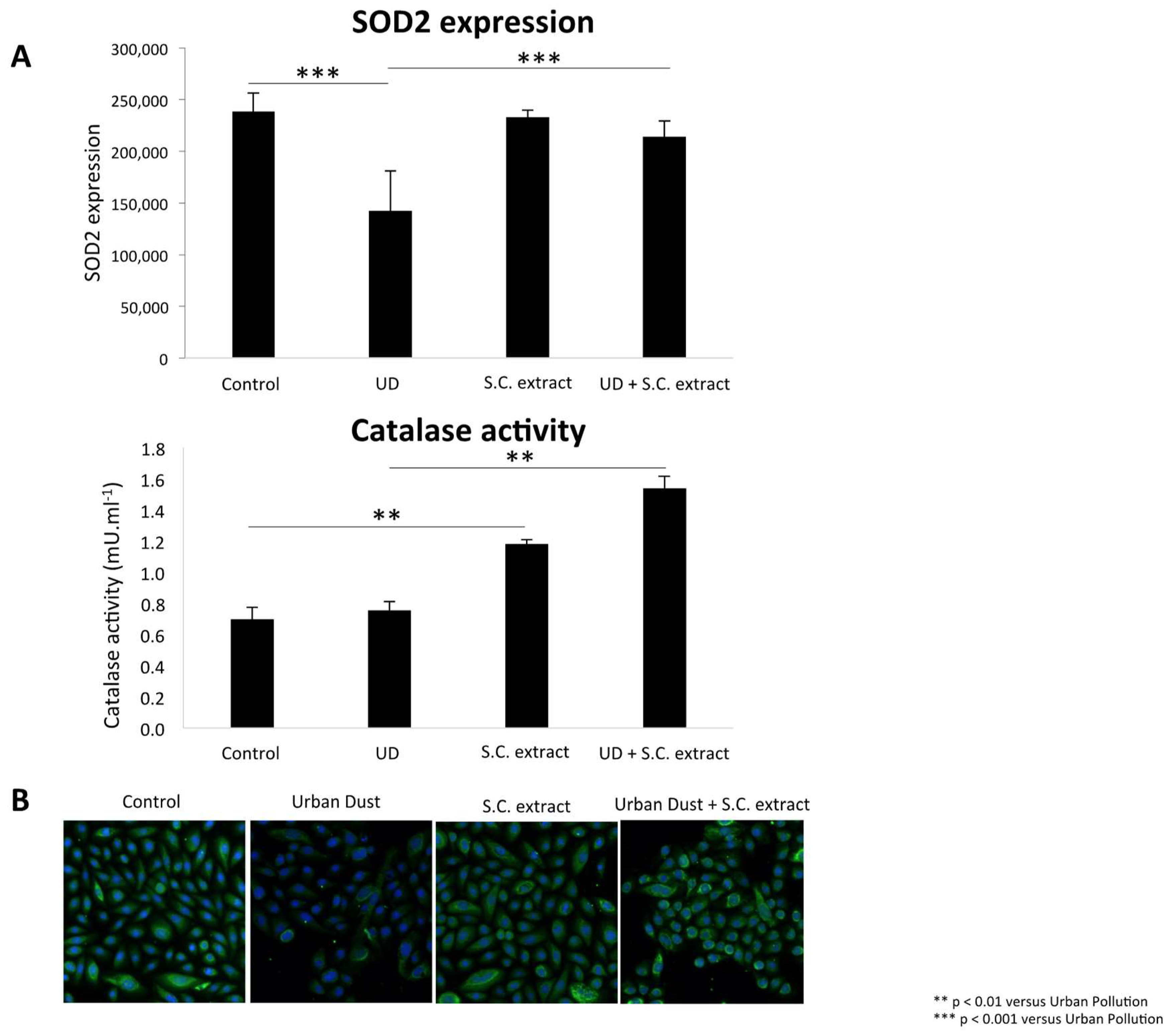
3.3. S.C. Extract Modulates Redox Pathways

Urban Dust induced an increase in Nrf2 cytoplasmic (+15%, p < 0.001) and an increase of nucleus (+28%, p < 0.001) expression in the cells. The co-treatment demonstrated a higher cytoplasmic expression of Nrf2 (Figure 2) and a significant decrease in the nucleus expression (−12%, p < 0.001), when compared to Urban Dust alone.
DJ-1 cytoplasmic (+91%, p < 0.001) and nucleus (+47%, p < 0.001) expression increased in the NHEK that were exposed to Urban Dust, when compared to the control (Figure 3). Co-treatment also induced a higher DJ-1 cytoplasmic expression when compared to Urban Dust alone (+33%: p < 0.001). However, DJ-1 nucleus activity decreased after co-treatment.
Co-treatment of Urban Dust and S.C. extract increased SOD2 expression (+50%: p < 0.001) and catalase activity (+120%: p < 0.05) (Figure 4).

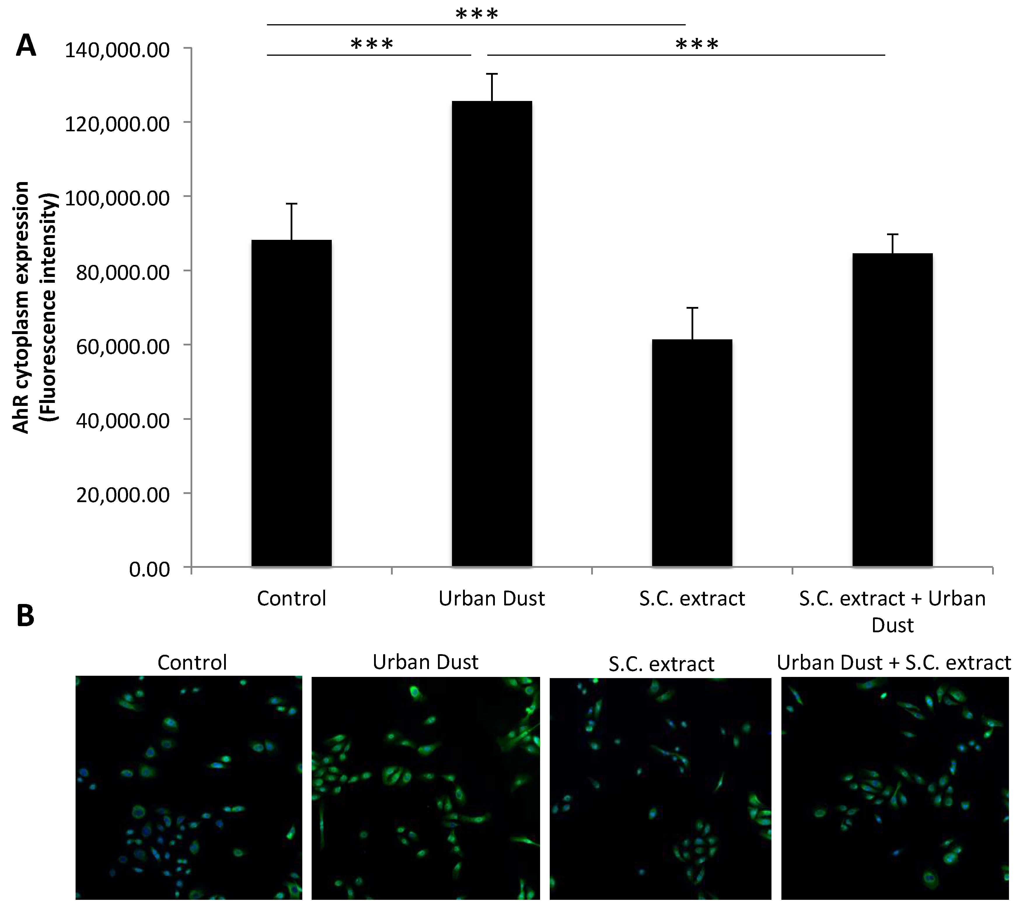
3.4. The Effect of Urban Dust and S.C. Extract on the Aryl Hydrocarbon Receptor (AhR)
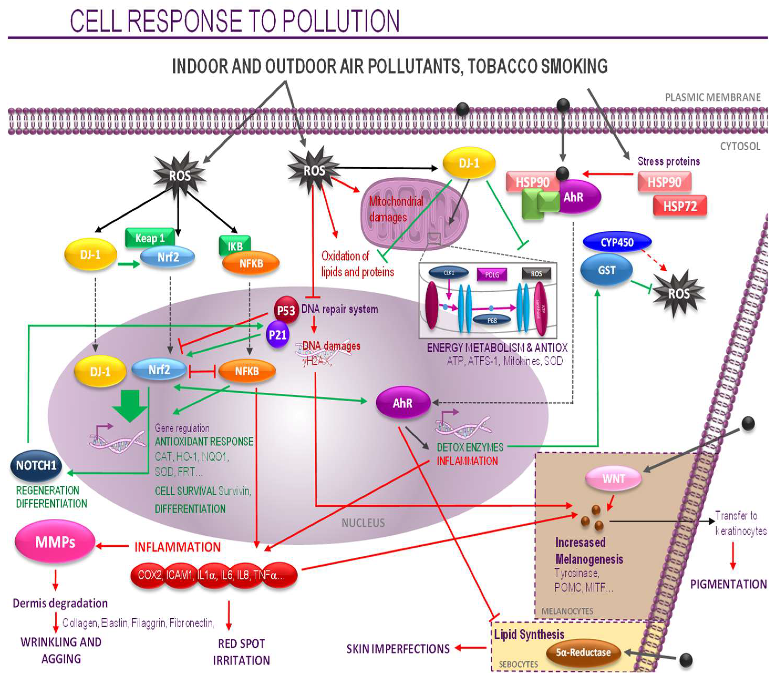
Urban Dust induced the expression of AhR in the cells. The presence of AhR increases in both the cytoplasm (+42%, p < 0.001) and the nucleus (+44%, p < 0.001) (Figure 5). Co-treatment with the S.C. extract induced a return to basal values (control).

4. Discussion

Global air pollution has become a major threat to human health. Being the largest organ of the human body, the skin is one of the major targets of air pollutants. Several studies have investigated the effect that air pollutants have on skin damage, focusing their interest especially on PM and ozone. These reported that outdoor air pollution is a relevant risk factor for the development of atopic dermatitis, a chronic inflammatory skin disease, and noted a cause-effect relationship between air pollution and skin quality [5,19]. At the cellular level, PM induces skin oxidative stress, leading to ROS and causing the secretion of pro-inflammatory cytokines. As a consequence, an increase of matrix metalloproteinases (MMPs) occurs, resulting in the degradation of mature dermal collagen, which contributes to skin aging.
Generally speaking, a myriad of extracts or isolated/purified substances from different parts of plants, including roots, leaves, flowers, seeds, etc., have traditionally been used to prevent skin damage, and the use of natural products to maintain skin integrity and health in the face of the deleterious effects of pollutants, attracts great interest. Many of these substances contain active principles of the polyphenol group (antioxidants) or other antioxidants with diverse chemical structures. Although they mainly function as antioxidants, they also display anti-inflammatory and immunomodulatory activities, and also control dermal extracellular matrix remodeling [20]. Among them, Schisandra chinensis is a traditional Chinese herbal medicine that has been used for treatments in Asia for thousands of years. It is known to possess antioxidant and anti-inflammatory properties, through high concentrations of lignans [21]. Previous studies have also shown the beneficial properties of S.C. on cigarette smoke-induced airway injury in animal models [22]. Therefore, the aim of our study was to better understand the cellular mechanisms that are induced by pollution in keratinocytes, and the effect of S.C extract in protecting the skin against ROS induced by Urban Dust. In this study, we have demonstrated the protective effect of S.C. extract, which increased DJ-1 protein levels, Nrf2 expression, and decreased AhR and NF-kB in the cytoplasm, even if the cells were under stress pollution. At the same time, co-treatment of Urban Dust and S.C. extract increased SOD2 expression (50%: p < 0.001) and catalase activity (120%: p < 0.05), when compared to Urban Dust alone (Figure 4). These results are in line with those of Lin et al., [23], who recently reported that Schisandrin B, an active ingredient extracted from S.C., blocked the activation of NF-κB, and activated Nrf2, which resulted in the inhibition of the inflammatory response.
Other compounds that contain the botanical active ingredient for skin applications have also been proposed and seem to have similar properties. Recently, equol, which is classified as a polyphenolic compound, has been proposed for its application to skin health, due to its effect on dermal parameters [24]. This compound is naturally present in some plants, such as beans, cabbages, and lettuces. In vitro and ex vivo experimentations show that equol protects the skin against environmental assaults [25,26]. More specifically, it has been found that equol protects against peroxide-induced endothelial cell apoptosis, due to the activation of the estrogen receptor and Nrf2/ARE signaling pathways [27]. It also increases the expression of the xenobiotic metabolizing enzyme quinone reductase (both mRNA and protein levels) [28]. Other molecules such as sulphoraphane, which is present in large amounts in broccoli, also induces the transcriptional activation of Nrf2 [24]. Generally speaking, Nrf2 is activated by ROS and is crucial for the transcriptional activation of an array of antioxidant and detoxification genes. In fact, it orchestrates major cellular defense mechanisms, including phase-II detoxification, inflammatory signaling, DNA repair, and recent experimental evidence supports the important role that Nrf2 plays as a skin barrier. Nrf2 has therefore emerged as a promising molecular target for the pharmacological prevention of human pathologies resulting from exposure to environmental toxicants, including solar UV-induced damages and carcinogenesis [29]. S.C. extract inhibited the ROS that were induced by Urban Dust through the activation of Nrf2 and SOD2, which clears mitochondrial reactive oxygen species (ROS) and, as a result, confers protection to the cell. ROS have also been reduced by intracellular antioxidant enzymes, including catalase. Indeed, mitochondria catalase was shown to protect cells from oxidative injury [30]. Moreover, the increase of DJ-1 noted in our study after the co-treatment of Urban Dust and S.C. extract, is an important result, with DJ-1 quenching the activity of ROS and protecting the mitochondrial function. It is also known that DJ-1 promotes Nrf2 binding to antioxidant response elements by which Nrf2 can regulate the expression of several endogenous antioxidative enzymes and can reduce ROS production to protect the mitochondria, and can also respond to oxidative stress. This mechanism is mediated by the fact that DJ-1 sequesters Keap1, an Nrf2 binding protein in the cytosol, promotes Nrf2 degradation. To our knowledge, no study has investigated the link between molecules derived from plants and this important regulatory protein DJ-1, which plays a key role in the health of skin. In fact, as suggested by Ishiwatari et al., DJ-1 in skin plays a significant role in the protection against oxidative stress, and might be an indicator of antioxidative defense against environmental stressor-induced damage [31].
Data in the literature shows that most of the phytochemicals upregulate Nrf2 signaling in association with AhR activation [32,33,34]. Besides having an effect on Nrf2, we also noted that S.C. extract has an effect on the AhR pathway. In fact, as shown in Figure 5, Urban Dust activates the AhR pathway, which is required for the biotransformation of pollutants, such as PAHs, through the different phases of detoxification. Phase I introduces chemically reactive groups to xenobiotics for these compounds to be conjugated to polar compounds in the phase II reactions. Finally, in the third phase, the conjugated xenobiotics are further processed to facilitate their excretion out of the cell. We demonstrated that Urban Dust induces the expression of the genes in phase I. It should be noted that the first enzymes of the detoxification process can produce intermediate metabolites that are even more reactive than their exogenous precursors [35], and it is therefore of paramount importance for the cell to achieve complete transformation and detoxification of such metabolites through the expression of gene coding for phase II enzymes. It is also important to note that phase II gene expression is dependent on the oxidative stress sensor Nrf2. Our observations confirm that Urban Dust activates the drug metabolism response, especially in phase I which is dependent on the CYP450 family members, as well as other enzymes such as MMP-1 and MMP-9. These results are in agreement with those of Li et al. [36]. The activation of the MMP-1 gene has been shown to be critically linked to mitogen-activated protein kinase activation. We found that, in addition to its Nrf2 effects, the co-treatment of Urban Dust and S.C. improved the anti-oxidant gene expression (Figure 4) and induced a significant decrease in AhR expression (Figure 5), as compared to Urban Dust alone. Recently, Woo et al. also noted that cyanidin-3-glucoside (C3G), a major active compound of mulberry extract, showed biological activities that protect the cells from pollutants, which can be attributed to the AhR signaling pathway being repressed [37]. Various compounds, such as plant polyphenols (e.g., resveratrol and curcumin) also directly or indirectly activate AhR, while other compounds are thought to be AhR antagonists [38,39]. Although further studies are needed, S.C. extract may be a promising compound to use in alleviating the symptoms that are related to AhR activation.

5. Conclusions

Recent studies showing deleterious effects of air pollution on skin health have piqued the interest of scientists to better characterize the mechanisms that are involved, in order to enable them to develop and market better protective strategies. Using an in vitro and a reconstructed human epidermis model, together with a standardized Urban Dust, we noted that such urban pollutants activate multiple stress-related pathways, and that treatment with S.C. extract attenuated the Urban Dust-induced oxidative stress. The protective mechanism was associated, at least in part, with the modulation of the Nrf2 and AhR pathways and the activation of DJ-1. The Ahr, Nrf2, and DJ-1-targeting strategy may promote our health, including that of our skin. These findings led us to postulate that S.C. might protect skin cell functions against urban pollution and that it could be used as a skin protection agent.

Author Contributions

E.B., E.R., E.F., and J.-Y.B. conceived and designed the experiments; C.B., O.B., and S.G., performed the experiments; E.F. analyzed the data; E.F., E.R. and A.L. wrote the paper.

Funding

This research received no external funding.

Acknowledgments

We acknowledge Philippe Bedos and Claire Leduc for their technical support.

Conflicts of Interest

The authors declare no conflicts of interest.

References

  1. Canova, C.; Heinrich, J.; Anto, J.M.; Leynaert, B.; Smith, M.; Kuenzli, N.; Zock, J.P.; Janson, C.; Cerveri, I.; De Marco, R.; et al. The influence of sensitization to pollens and moulds on seasonal variations in asthma attacks. Eur. Respir. J. 2013, 42, 935–945. [Google Scholar] [CrossRef] [PubMed] [Green Version]
  2. Rembiesa, J.; Ruzgas, T.; Engblom, J.; Holefors, A. The impact of pollution on skin and proper efficacy testing for anti-pollution claims. Cosmetics 2018, 5, 4. [Google Scholar] [CrossRef]
  3. Puri, P.; Nandar, S.K.; Kathuria, S.; Ramesh, V. Effects of air pollution on the skin: A review. Indian J. Dermatol. Venereol. Leprol. 2017, 83, 415–423. [Google Scholar] [PubMed]
  4. Lanuti, E.L.; Kirsner, R.S. Effects of pollution on skin aging. J. Investig. Dermatol. 2010, 130, 2696. [Google Scholar] [CrossRef] [PubMed]
  5. Vierkötter, A.; Schikowski, T.; Ranft, U.; Sugiri, D.; Matsui, M.; Krämer, U.; Krutmann, J. Airborne particle exposure and extrinsic skin aging. J. Investig. Dermatol. 2010, 130, 2719–2726. [Google Scholar] [CrossRef] [PubMed]
  6. Kampa, M.; Castanas, E. Human health effects of air pollution. Environ. Pollut. 2008, 151, 362–367. [Google Scholar] [CrossRef] [PubMed]
  7. Kim, K.E.; Cho, D.; Park, H.J. Air pollution and skin diseases: Adverse effects of airborne particulate matter on various skin diseases. Life Sci. 2016, 152, 126–134. [Google Scholar] [CrossRef] [PubMed]
  8. Krutmann, J.; Moyal, D.; Xiang, L.F.; Seité, S. Pollution and acne: Is there a link? Clin. Cosmet. Investig. Dermatol. 2017, 10, 199–204. [Google Scholar] [CrossRef] [PubMed]
  9. Mc Daniel, D.; Farris, P.; Valacchi, G. Atmospheric skin aging: Contributors and inhibitors. J. Cosmet. Dermatol. 2017, 17, 124–137. [Google Scholar] [CrossRef] [PubMed]
  10. Im, J.Y.; Lee, K.W.; Woo, J.M.; Junn, E.; Mouradian, M.M. DJ-1 induces thioredoxin 1 expression through the Nrf2 pathway. Hum. Mol. Genet. 2012, 21, 3013–3024. [Google Scholar] [CrossRef] [PubMed] [Green Version]
  11. Lambert, J.D.; Elias, R.J. The antioxidant and pro-oxidant activities of green tea polyphenols: A role in cancer prevention. Arch. Biochem. Biophys. 2010, 501, 65–72. [Google Scholar] [CrossRef] [PubMed] [Green Version]
  12. Juliano, C.; Magrini, G.A. Cosmetic functional ingredients from botanical sources for anti-pollution skincare products. Cosmetics 2018, 5, 19. [Google Scholar] [CrossRef]
  13. Mistry, N. Guidelines for formulating anti-pollution products. Cosmetics 2017, 4, 57. [Google Scholar] [CrossRef]
  14. Lephart, E.D. Equol’s anti-aging effects protect against environmental assaults by increasing skin antioxidant defense and ECM proteins while decreasing oxidative stress and inflamamtion. Cosmetics 2018, 5, 16. [Google Scholar] [CrossRef]
  15. Lephart, E.D. Skin aging and oxidative stress. Equol’s anti-aging effects via biochemical and molecular mechanisms. Ageing Res. Rev. 2016, 31, 36–54. [Google Scholar] [CrossRef] [PubMed]
  16. Matsumoto, Y.; Ide, F.; Kishi, R.; Akutagawa, T.; Sakai, S.; Nakamura, M.; Ishikawa, T.; Fuji-Kuriyama, Y.; Nakatsuru, Y. Aryl hydrocarbon receptor plays a significant role in mediating airborne particulate induced carcinogenesis in mice. Environ. Sci. Technol. 2017, 41, 3775–3780. [Google Scholar] [CrossRef]
  17. Casey, M.C.; McNally, R.S.; Conti, B.J.; Mak, T.W.; Ting, J.P.Y. DJ-1, a cancer- and Parkinson’s disease-associated protein, stabilizes the antioxidant transcriptional master regulator Nrf2. Proc. Natl. Acad. Sci. USA 2006, 103, 5091. [Google Scholar]
  18. Opletal, L.; Sovova, H.; Bartlova, M. Dibenzo[a,c]cyclooctadiene lignans of the genus Schisandra: Importance, isolation and determination. J. Chromatogr. B 2004, 812, 357–371. [Google Scholar] [CrossRef]
  19. Kim, J.; Kim, E.-H.; Oh, I.; Jung, K.; Han, Y.; Cheong, H.-K.; Ahn, K. Symptoms of atopic dermatitis are influenced by outdoor air pollution. J. Allergy Clin. Immunol. 2013, 132, 495–497. [Google Scholar] [CrossRef] [PubMed]
  20. Bosch, R.; Philips, N.; Suarez-Perez, J.A.; Juarranz, A.; Devmurari, A.; Chalensouk-Khaossat, J.; Gonzalez, M. Mechanisms of Photoaging and Cutaneous Photocarcinogenesis, and Photoprotective Strategies with Phytochemicals. Antioxidants 2015, 4, 248–268. [Google Scholar] [CrossRef] [PubMed] [Green Version]
  21. Gao, C.; Cong, C.; Hue, N.J.; Ciao, B. Protective effect of Schizandrin B against damage of UVB irradiated skin cells depend on inhibition of inflammatory pathways. Bioengineered 2017, 8, 36–44. [Google Scholar] [CrossRef] [PubMed]
  22. Jia, R.; Zhang, H.; Yang, Z.; Zhao, H.; Liu, F.; Wang, H.; Miao, M.; Wang, Q.; Liu, Y. Protective effects of Schisandrin B on cigarette smoke-induced airway injury in mice through Nrf2 pathway. Int. Immunopharmacol. 2017, 53, 11–16. [Google Scholar] [CrossRef] [PubMed]
  23. Lin, Q.; Qin, X.; Shi, M.; Qin, Z.; Meng, Y.; Qin, Z.; Guo, S. Schisandrin B inhibits LPS-induced inflammatory response in human umbilical vein endothelial cells by activating Nrf2. Int. Immunopharmacol. 2017, 49, 142–147. [Google Scholar] [CrossRef] [PubMed]
  24. Lephart, E.D. Resveratrol, 4′Acetoxy Resveratrol, R-Equol, Racemic Equol or S-Equol as Cosmeceuticals to improve dermal health. Int. J. Mol. Sci. 2017, 18, 1193. [Google Scholar] [CrossRef] [PubMed]
  25. Lephart, E.D. Protective effects of equol and their polyphenolic isomers against dermal aging: Microarray/protein evidence with clinical implications and unique delivery into human skin. Pharm. Biol. 2013, 51, 1393–1400. [Google Scholar] [CrossRef] [PubMed] [Green Version]
  26. Lephart, E.D. Human Skin Gene Expression Doesn’t Correlate with Protein Expression? Unless Both Parameters Are quantified. J. Cosmet. Dermatol. 2018, 17, 244–245. [Google Scholar] [CrossRef] [PubMed]
  27. Zhang, T.; Liang, X.Y.; Shi, L.Y.; Wang, L.; Chen, J.L.; Kang, C.; Zhu, J.D.; Mi, M.T. Estrogen receptor and PI3K/Akt signaling pathway involvement in S-equol-induced activation of Nrf2/ARE in endothelia cells. PLoS ONE 2013, 8, e79075. [Google Scholar]
  28. Froyen, E.B.; Steinberg, F.M. Soy isoflavones increase quinone reductase in heap-1c1c7 cells via estrogen receptor beta and nuclear factor erythroid 2-related factor 2 binding to the antioxidant response element. J. Nutr. Biochem. 2011, 22, 843–848. [Google Scholar] [CrossRef] [PubMed]
  29. Schafer, M.; Werner, S. Nrf2-A regulator of keratinocyte redox signaling. Free Radic. Biol. Med. 2015, 88, 243–252. [Google Scholar] [CrossRef] [PubMed]
  30. Bai, J.; Cederbaum, A.I. Mitochondrial catalase and oxidative injury. Biol. Signals Recept. 2001, 10, 189–199. [Google Scholar] [CrossRef] [PubMed]
  31. Ishiwatari, S.; Takahashi, M.; Yasuda, C.; Nakagawa, M.; Saito, Y.; Noguchi, N.; Matsukuma, S. The protective role of DJ-1 in ultraviolet-induced damage of human skin: DJ-1 levels in the stratum corneum as an indicator of antioxidative defense. Arch. Dermatol. Res. 2015, 307, 925–935. [Google Scholar] [CrossRef] [PubMed]
  32. Nakahara, T.; Mitoma, C.; Hashimoto-Hachiya, A.; Takahara, M.; Tsuji, G.; Uchi, H.; Yan, X.; Hachisuka, J.; Chiba, T.; Esaki, H.; et al. Antioxidant Opuntia ficus-indica extract activates AhR-Nrf2 signaling and upregulates filaggrin and loricrin expression in human keratinocytes. J. Med. Food. 2015, 18, 1143–1149. [Google Scholar] [CrossRef] [PubMed]
  33. Wu, Z.; Uchi, H.; Morino-Koga, S.; Shi, S.; Furue, M.Z. Ligustilide ameliorated ultraviolet B-induced oxidative stress and inflammatory cytokine production in human keratinocytes through upregulation of Nrf2/HO-1 and suppression of NF-kB pathway. Exp. Dermatol. 2015, 24, 703–708. [Google Scholar] [CrossRef] [PubMed]
  34. Takei, K.; Hashimoto-Hachiya, A.; Takahara, M.; Tsuji, G.; Nakahara, T.; Furue, M. Cynaropicrin attenuates UVB-induced oxidative stress via the AhR-Nrf2-Nqo1 pathway. Toxicol. Lett. 2015, 234, 74–80. [Google Scholar] [CrossRef] [PubMed]
  35. Xu, C.; Li, C.Y.; Kong, A.N. Induction of phase I, II and III drug metabolism/transport by xenobiotics. Arch. Pharm. Res. 2005, 28, 249–268. [Google Scholar] [CrossRef] [PubMed] [Green Version]
  36. Li, J.; Ghio, A.J.; Cho, S.H.; Brinckerhoff, C.E.; Simon, S.A.; Liedtke, W. Diesel Exhaust Particles Activate the Matrix-Metalloproteinase-1 Gene in Human Bronchial Epithelia in a β-Arrestin—Dependent Manner via Activation of RAS. Environ. Health Perspect. 2009, 117, 400. [Google Scholar] [CrossRef] [PubMed]
  37. Woo, H.; Lee, J.; Park, D.; Jung, D. Protective Effect of Mulberry (Morus alba L.) Extract against Benzo[a]pyrene Induced Skin Damage through Inhibition of Aryl Hydrocarbon Receptor Signaling. J. Agric. Food Chem. 2017, 20, 10925–10932. [Google Scholar] [CrossRef] [PubMed]
  38. Mohammadi-Bardbori, A.; Bengtsson, J.; Rannug, U.; Rannug, A.; Wincent, E. Quercetin resveratrol, and curcumin are indirect activators of the aryl hydrocarbon receptor (AhR). Chem. Res. Toxicol. 2012, 25, 1878–1884. [Google Scholar] [CrossRef] [PubMed]
  39. Tsuji, G.; Takahara, M.; Uchi, H.; Matsuda, T.; Chiba, T.; Takeuchi, S. Identification of ketoconazole as an AhR-Nrf2 activator in cultured human keratinocytes: The basis of its anti-inflammatory effect. J. Investig. Dermatol. 2012, 132, 59–68. [Google Scholar] [CrossRef] [PubMed]
Figure 1. Signaling pathway responses to global pollution. Air pollutants induce Reactive Oxygen Species (ROS) generation and pro-inflammatory cytokines. Upon ligation by pollutants, the activated AhR translocates from the cytoplasm into the nucleus. This translocated AhR binds with ANRT, resulting in the activation of Cytochrome P450, family 1, member A1 (CYP1A1) transcription. ROS generated by CYP1A1 stimulates the production of TNF-α and IL-8. ROS also activates beneficial cellular responses, including Nuclear factor (erythroid-derived 2)-like 2 (Nrf2) activation. Under normal conditions, Nrf2 localizes in the cytoplasm where it interacts with the actin binding protein, Kelch-like ECH associating protein 1 (Keap1), and is rapidly degraded by the ubiquitin-proteasome pathway. Signals from ROS target the Nrf2-Keap1 complex, dissociating Nrf2 from Keap1. The Parkinson’s-associated protein, DJ-1, is indispensable for Nrf2 stabilization, by affecting Nrf2association with its inhibitor Keap1. Stabilized Nrf2 translocates to the nuclei, binds to the antioxidant response element, and thereby regulates the expression of a large battery of genes that are involved in the cellular antioxidant protection, including NADPH quinine oxyreductase (NQO-1), heme oxygenase-1 (HO-1), glutathione (GSH)… ROS can also activate gene transcription via transcription factors, such as NF-kB, that can interact directly with specific DNA motifs on the promoters of target genes. The transcriptions of several MMP family members are strongly regulated by NF-kB. Increased activities of NF-kB leads to collagen breakdown, the downregulation of type I procollagen, and the upregulations of MMPs, resulting in premature skin ageing.
Cosmetics 05 00036 g001
Figure 2. (A) Nrf2 cytoplasm expression in Normal Human Epidermal Keratinocytes after Urban Dust exposure, or co-treatment with Urban dust and S.C. extract (50 μg·mL−1). *** p < 0.001. (B) Representative pictures of Nrf2 expression: Nrf2 in green, Nucleus in blue.
Cosmetics 05 00036 g002
Figure 3. DJ-1 cytoplasm (A) and nucleus (B) expression in the NHEK after Urban Dust exposure, or co-treatment with Urban dust and S.C. extract (50 μg·mL−1). *** p < 0.001. (C) Representative pictures of DJ-1 expression: DJ-1 in green, Nucleus in blue.
Cosmetics 05 00036 g003
Figure 4. (A) The effect of S.C. extract on SOD2 expression and catalase activity in the NHEK that were exposed to Urban Dust (80 μg·mL−1). ** p < 0.01 and *** p < 0.001. UB: Urban Dust (B) Representative pictures of SOD2 expression: SOD2 in green, Nucleus in blue.
Cosmetics 05 00036 g004
Figure 5. (A) AhR cytoplasm expression after Urban Dust exposure, or co-treatment with Urban Dust and S.C. extract. *** p < 0.001. (B) Representative pictures of AhR expression: AhR in green, Nucleus in blue.
Cosmetics 05 00036 g005
Table 1. The Effect of Urban Dust and S.C. extract on gene expression profiles.
Effect of Urban Dust (80 μg·mL−1) on Gene Expression
Gene SymbolTarget NameFold Changep Value
CYP1A1cytochrome P450, family 1, subfamily A, polypeptide 123.1860.001
CYP1B1cytochrome P450, family 1, subfamily B, polypeptide 17.7710.000
MMP1matrix metallopeptidase 1 3.4120.014
ALDH3A1aldehyde dehydrogenase 3 family, member A12.3640.011
NQO1NAD(P)H dehydrogenase, quinone 12.3390.000
GPX2glutathione peroxidase 22.1450.006
IL1Binterleukin 1, beta2.0530.046
GSTM1glutathione S-transferase mu 11.7840.037
MMP9matrix mettalopeptidase 91.6640.042
HSPA5/BiPheat shock 70kDa protein 51.5620.003
GADD45Agrowth arrest and DNA-damage-inducible, alpha1.4840.021
FTHferritin, heavy polypeptide 11.1830.035
COL7A1collagen, type VII, alpha 11.3370.029
GSTP1glutathione S-transferase pi11.3020.040
SQSTM1sequestosome 11.2030.034
CLN3ceroid-lipofuscinosis, neuronal 31.1720.013
SPRR1Asmall proline-rich protein 1A1.1710.035
GSRglutathione reductase1.0840.035
FMO4flavin containing monooxygenase 40.8100.012
MT4metallothionin 40.4340.040
Effect of S.C extract (50 μg·mL−1) on gene expression
CYP1B1cytochrome P450, family 1, subfamily A, polypeptide 11.5350.014
HSPA5/Bipheat shock 70kDa protein 51.4150.014
Effect of S.C extract (50 μg·mL−1) in presence of Urban Dust (80 μg·mL−1) on gene expression
GPX2glutathione peroxidase 21.3650.007
EPHX2epoxide hydrolase 2, cytoplasmic 1.1150.000
FTLferritin, light polypeptide1.2050.031
SPRR1ASmall proline-rich protein 1A1.2010.027

