Metabolome Consistency: Additional Parazoanthines from the Mediterranean Zoanthid Parazoanthus Axinellae


Abstract
:1. Introduction

2. Results and Discussion
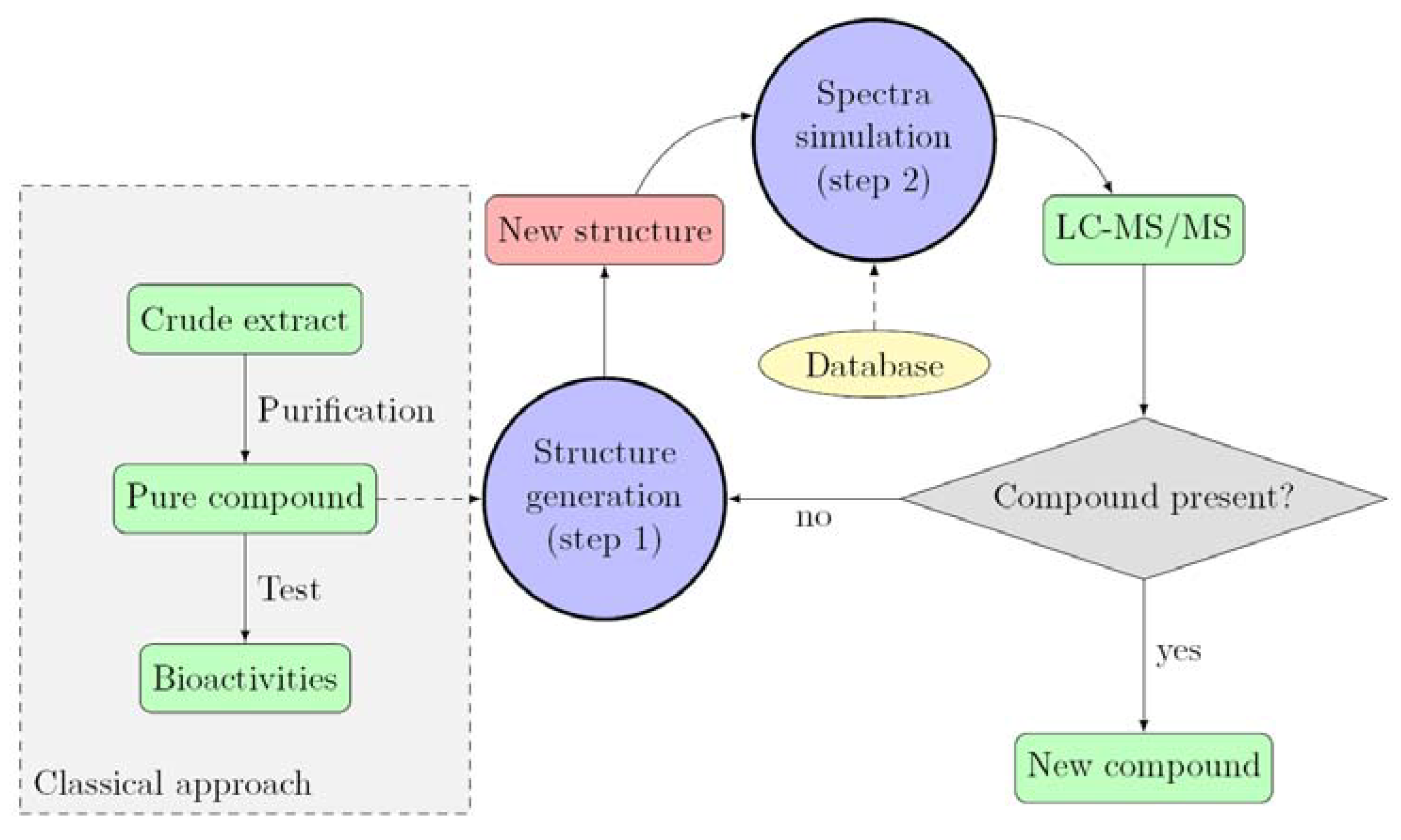
2.1. Description of the Approach

2.2. Identification of the Fragmentation Pattern


| Compound | Empirical Formula | m/z (∆ ppm) | |||||
|---|---|---|---|---|---|---|---|
| [M+H]+ | [M-NH3+H]+ | [M-CH5N3+H]+ | [M-C2H6N4O+H]+ | [M-C6H10N4O+H]+ | [M-C7H10N4O2+H]+ | ||
| 1 | C15H20N5O3 | 318.1558 (0.8) | 301.1290 (1.7) | 259.1081 (-1.5) | 216.1012 (3.3) | 162.0553 (-2.1) | 136.0749 (5.8) |
| 2 | C15H18N5O3 | 316.1410 (-1.8) | 299.1135 (1.2) | 257.0920 (0.3) | 214.0867 (-2.1) | 162.0551 (-0.9) | 136.0725 (-6.2) |
| 3 | C16H20N5O3 | 330.1562 (-0.4) | 313.1303 (-2.5) | 271.1082 (-1.8) | 228.1023 (-1.4) | 176.0707 (-0.5) | 150.0912 (-1.8) |
| 4 | C16H20BrN5O3 | 410.0829 (-1.6) | 393.0555 (0.5) | 351.0338 (0.2) | 308.0277 (1.2) | 253.9782 (1.2) | 228.0022 (1.5) |
| 5 | C16H18BrN5O3 | 408.0676 (-2.5) | 391.0406 (-1.5) | 349.0192 (-2.8) | 306.0115 (3.01) | 253.9809 (0.9) | 228.0031 (5.5) |
2.3. Identification of New Parazoanthines

| Compound | Empirical Formula | m/z (∆ ppm) | |||||
|---|---|---|---|---|---|---|---|
| [M+H]+ | [M-NH3+H]+ | [M-CH5N3+H]+ | [M-C2H6N4O+H]+ | [M-C6H10N4O+H]+ | [M-C7H10N4O2+H]+ | ||
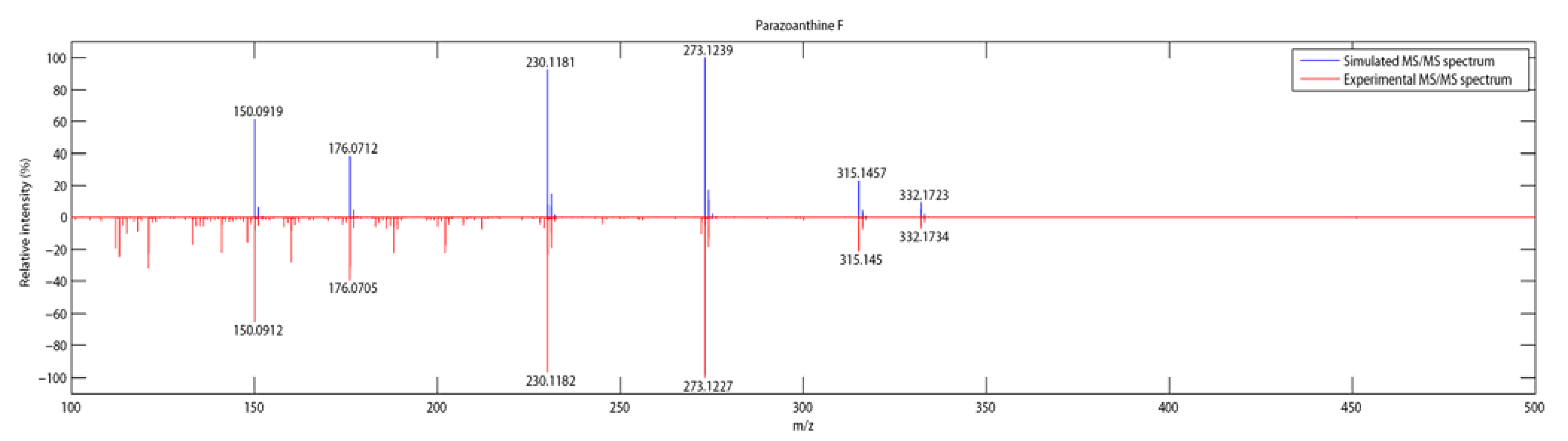
| 6 | C16H21N5O3 | 332.1734 (-5.1) | 315.1450 (0.5) | 273.1227 (2.5) | 230.1182 (-2.8) | 176.0705 (0.6) | 150.0912 (-0.94) |
| 7 | C15H18BrN5O3 | 396.0611 (13.9) | 379.0361 (10.4) | 337.0158 (7.24) | 294.0071 (-19.9) | - | - |
| 8 | C15H16BrN5O3 | 394.0496 (3.4) | 377.0239 (1.3) | 335.0011 (4.4) | 291.9931 (12.6) | 239.9640 (6.14) | - |
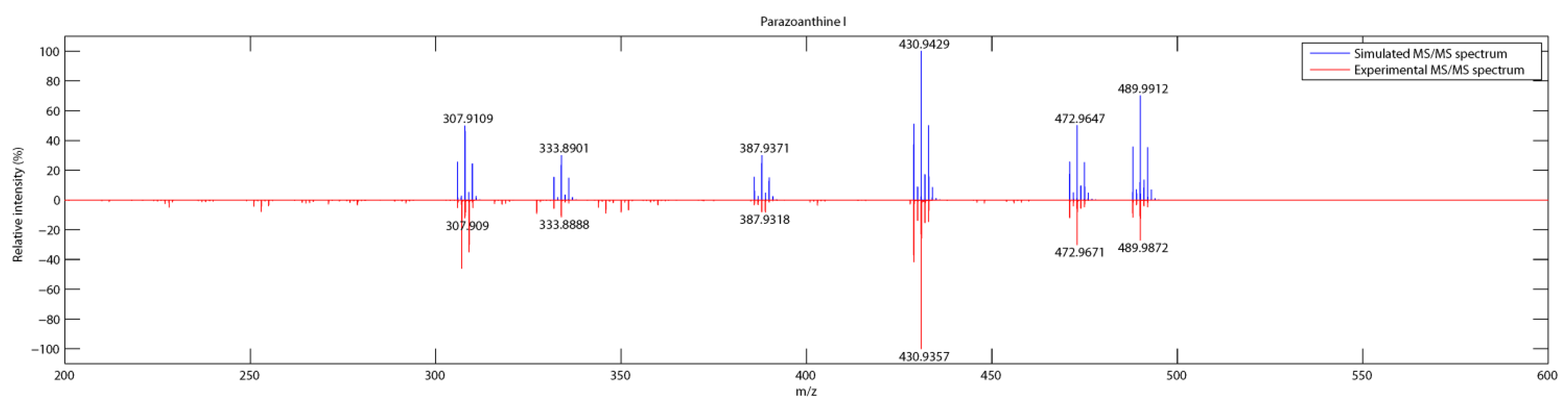
| 9 | C16H19Br2N5O3 | 487.9903 (5.0) | 470.9640 (4.7) | 438.9438 (1.4) | 385,9386 (-0.1) | 331.8931 (-4.7) | 305.9108 (5.1) |
| 10 | C16H17Br2N5O3 | 485.9761 (2.0) | 468.9540 (-7.4) | 426.9300 (-2.9) | - | 331.8917 (-0.2) | 305.9025 (32.4) |


2.4. Determination of the Absolute Configuration

3. Experimental Section
3.1. General Procedure
3.2. Biological Material
3.3. Sample Preparation and Data Acquisition
3.4. MS/MS Spectra Simulation
4. Conclusions
Supplementary Files
Acknowledgments
Author Contributions
Conflicts of Interest
References
- Cachet, N.; Genta-Jouve, G.; Regalado, E.L.; Mokrini, R.; Amade, P.; Culioli, G.R.; Thomas, O.P. Parazoanthines A–E, hydantoin alkaloids from the mediterranean sea anemone parazoanthus axinellae. J. Nat. Prod. 2009, 72, 1612–1615. [Google Scholar] [CrossRef]
- Blunt, J.W.; Copp, B.R.; Keyzers, R.A.; Munro, M.H.G.; Prinsep, M.R. Marine natural products. Nat. Prod. Rep. 2014, 31, 160–258. [Google Scholar] [CrossRef]
- Laville, R.; Thomas, O.P.; Berrué, F.; Marquez, D.; Vacelet, J.; Amade, P. Bioactive guanidine alkaloids from two caribbean marine sponges. J. Nat. Prod. 2009, 72, 1589–1594. [Google Scholar] [CrossRef]
- Bondu, S.; Genta-Jouve, G.; Leiros, M.; Vale, C.; Guigonis, J.-M.; Botana, L.M.; Thomas, O.P. Additional bioactive guanidine alkaloids from the mediterranean sponge crambe crambe. RSC Adv. 2012, 2, 2828–2835. [Google Scholar]
- Genta-Jouve, G.; Cachet, N.; Holderith, S.; Oberhänsli, F.; Teyssié, J.-L.; Jeffree, R.; Al Mourabit, A.; Thomas, O.P. New insight into marine alkaloid metabolic pathways: Revisiting oroidin biosynthesis. ChemBioChem 2011, 12, 2298–2301. [Google Scholar] [CrossRef]
- Ridder, L.; Wagener, M. Sygma: Combining expert knowledge and empirical scoring in the prediction of metabolites. ChemMedChem 2008, 3, 821–832. [Google Scholar] [CrossRef]
- Steinbeck, C. Recent developments in automated structure elucidation of natural products. Nat. Prod. Rep. 2004, 21, 512–518. [Google Scholar] [CrossRef]
- Ridder, L.; van der Hooft, J.J.J.; Verhoeven, S.; de Vos, R.C.H.; van Schaik, R.; Vervoort, J. Substructure-based annotation of high-resolution multistage MSn spectral trees. Rapid Commun. Mass Spectrom. 2012, 26, 2461–2471. [Google Scholar] [CrossRef]
- Cariello, L.; Crescenzi, S.; Prota, G.; Zanetti, L. New zoanthoxanthins from the mediterranean zoanthid parazoanthus axinellae. Experientia 1974, 30, 849–850. [Google Scholar] [CrossRef]
- Griffiths, M.Z.; Alkorta, I.; Popelier, P.L.A. Predicting pKa values in aqueous solution for the guanidine functional group from gas phase ab initio bond lengths. Mol. Inform. 2013, 32, 363–376. [Google Scholar] [CrossRef]
- Mouls, L.; Subra, G.; Aubagnac, J.-L.; Martinez, J.; Enjalbal, C. Tandem mass spectrometry of amidated peptides. J. Mass Spectrom. 2006, 41, 1470–1483. [Google Scholar] [CrossRef]
- Rojas-Chertó, M.; Kasper, P.T.; Willighagen, E.L.; Vreeken, R.J.; Hankemeier, T.; Reijmers, T.H. Elemental composition determination based on MSn. Bioinformatics 2011, 27, 2376–2383. [Google Scholar] [CrossRef]
- Manzo, E.; Pagano, D.; Nuzzo, G.; Gavagnin, M.; Ciavatta, M.L. First synthesis of parazoanthine-A and its O-Me derivative. Tetrahedron Lett. 2012, 53, 7083–7084. [Google Scholar] [CrossRef]
- Boumendjel, A.; Sotoing Taïwe, G.; Ngo Bum, E.; Chabrol, T.; Beney, C.; Sinniger, V.; Haudecoeur, R.; Marcourt, L.; Challal, S.; Ferreira Queiroz, E.; et al. Occurrence of the synthetic analgesic tramadol in an african medicinal plant. Angew. Chem. Int. Ed. 2013, 52, 11780–11784. [Google Scholar] [CrossRef]
- Bringmann, G.; Lang, G. Full absolute stereostructures of natural products directly from crude extracts: The HPLC-MS/MS-NMR-CD “triad”. In Sponges (Porifera); Müller, W.G., Ed.; Springer: Berlin/Heidelberg, Germany, 2003; Volume 37, pp. 89–116. [Google Scholar]
- Bringmann, G.; Götz, D.; Bruhn, T. The online stereochemical analysis of chiral compounds by HLPC-ECD coupling in combination with quantum-chemical calculations. In Comprehensive Chiroptical Spectroscopy; John Wiley & Sons, Inc.: Hoboken, NJ, USA, 2012; pp. 355–386. [Google Scholar]
- Genta-Jouve, G.; Weinberg, L.; Cocandeau, V.; Maestro, Y.; Thomas, O.P.; Holderith, S. Revising the absolute configurations of coatlines via density functional theory calculations of electronic circular dichroism spectra. Chirality 2013, 25, 180–184. [Google Scholar] [CrossRef]
- Cachet, N. Metabolites secondaires d’invertébrés marins et biosynthèse in vivo d’alcaloides d’Agelas oroides. Ph.D. Thesis, University of Nice-Sophia Antipolis, Nice, France, 2009. [Google Scholar]
- Schummer, J. The end of silent rites. HYLE Int. J. Philos. Chem. 2006, 12, 157–159. [Google Scholar]
- Bachelard, G. Le Nouvel Esprit Scientifique; Presses universitaires de France: Paris, France, 1983. [Google Scholar]
© 2014 by the authors; licensee MDPI, Basel, Switzerland. This article is an open access article distributed under the terms and conditions of the Creative Commons Attribution license (http://creativecommons.org/licenses/by/3.0/).
Share and Cite
Audoin, C.; Cocandeau, V.; Thomas, O.P.; Bruschini, A.; Holderith, S.; Genta-Jouve, G. Metabolome Consistency: Additional Parazoanthines from the Mediterranean Zoanthid Parazoanthus Axinellae. Metabolites 2014, 4, 421-432. https://doi.org/10.3390/metabo4020421
Audoin C, Cocandeau V, Thomas OP, Bruschini A, Holderith S, Genta-Jouve G. Metabolome Consistency: Additional Parazoanthines from the Mediterranean Zoanthid Parazoanthus Axinellae. Metabolites. 2014; 4(2):421-432. https://doi.org/10.3390/metabo4020421
Chicago/Turabian StyleAudoin, Coralie, Vincent Cocandeau, Olivier P. Thomas, Adrien Bruschini, Serge Holderith, and Grégory Genta-Jouve. 2014. "Metabolome Consistency: Additional Parazoanthines from the Mediterranean Zoanthid Parazoanthus Axinellae" Metabolites 4, no. 2: 421-432. https://doi.org/10.3390/metabo4020421