Review

Particle-Rich Cytoplasmic Structure (PaCS): Identification, Natural History, Role in Cell Biology and Pathology

1
Department of Molecular Medicine, University of Pavia, Pavia 27100, Italy
2
Pathologic Anatomy Unit, IRCCS Policlinico San Matteo, Pavia 27100, Italy
3
Centro Grandi Strumenti, University of Pavia, Pavia 27100, Italy
*
Author to whom correspondence should be addressed.
Biomolecules 2014, 4(3), 848-861; https://doi.org/10.3390/biom4030848
Submission received: 28 March 2014 / Revised: 13 August 2014 / Accepted: 5 September 2014 / Published: 22 September 2014
(This article belongs to the Special Issue Proteasomes and Its Regulators)

Abstract

:
Cytoplasmic structures showing a selective concentration of both polyubiquitinated proteins and proteasome have been described in various epithelial, hematopoietic, mesenchymal and neural cells in vitro or in fetal tissues, as well as in chronically-infected, mutated preneoplastic and neoplastic tissues. These cytoplasmic structures differ from other ubiquitin-reactive cytoplasmic bodies, like sequestosomes, aggresome-like-induced structures in dendritic cells (DALIS)/non-dendritic cells (ALIS) and aggresomes in showing distinctive ultrastructural organization (particle-rich cytoplasmic structure or PaCS), a cytochemical pattern and a functional profile. Their formation can be induced in vitro in dendritic or natural killer cells by trophic factors and interleukin treatment. They originate in close connection with ribosomes, while, as a result of their growth, the cytoskeleton and other surrounding organelles are usually dislocated outside their core. Interestingly, these particulate cytoplasmic structures are often found to fill cytoplasmic blebs forming proteasome- and polyubiquitinated protein-discharging vesicles, called ectosomes, which are found to detach from the cell and freely float in the extracellular space. To clearly point out the importance of the polyubiquitinated proteins and proteasome containing cytoplasmic structures, their role in cell biology and pathology has been carefully analyzed.

1. Introduction

Various ubiquitin-storing cytoplasmic bodies or structures, with or without proteasome colocalization, have been reported in cell lines under stress conditions, leading to the aggregation of misfolded proteins. The “proteolytic centers” developing in cells under proteasome function inhibitors [1], the pericentriolar “aggresomes” accumulating mutated proteins [2], the “aggresome-like-induced structures” in dendritic cells (DALIS) [3,4] or in non-dendritic cells (ALIS) [5], the amorphous to fibrillary “sequestosomes” [6], the yeast “juxtanuclear quality control compartment” (JUNQ) and the perivacuolar “insoluble protein deposit” (IPOD) [7] and the “particle-rich cytoplasmic structure” (PaCS) [8] accumulating ubiquitin proteasome system (UPS) components are among such bodies. Comparable structures have been also observed in ex vivo pathological tissues, including nerve, muscle and hepatocellular diseases [9,10,11] and neoplastic [12] or infectious conditions [8,13].
The wide spectrum of cells, treatments and pathologic conditions involved and the variety of technical procedures employed make a comparison among the different structures quite difficult. However, recent correlative electron/confocal microscopy investigations and direct ultrastructural immunogold observations [6,14,15] have allowed for the characterization in mammalian cells of at least four types of structurally- and cytochemically-different bodies with apparently different roles in cell biology and pathology: sequestosomes, DALIS/ALIS, pericentriolar aggresomes and PaCSs. These are briefly discussed in the following chapter.

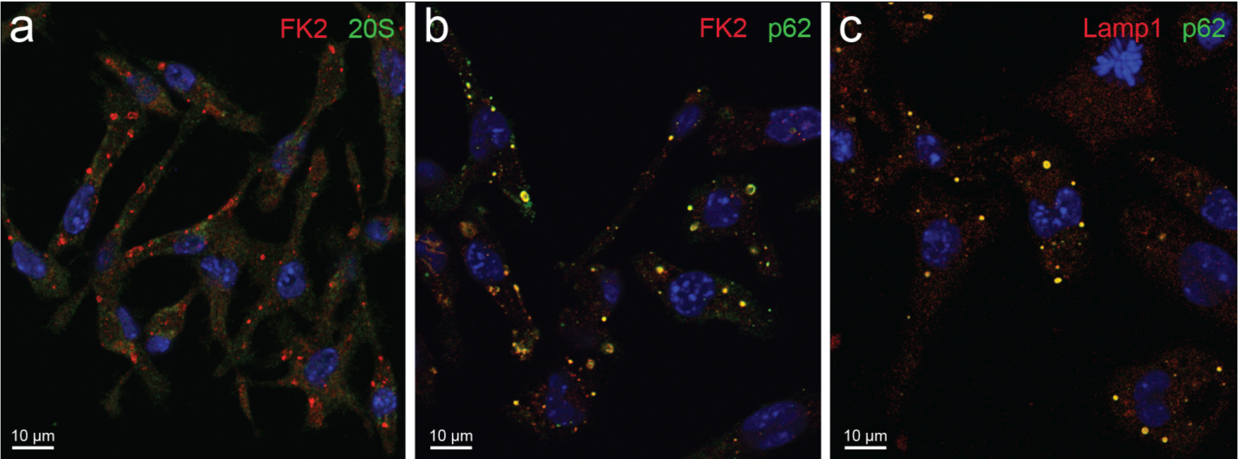
2. Types and Function of Ubiquitin-Reactive Cytoplasmic Structures

The sequestosomes have been characterized as insoluble, cytoplasmic aggregates of proteins, which in their soluble precursor form, may be cytotoxic and potentially amyloidogenic, while in their aggregated form, they are amorphous to fibrillary, apparently inert, deposits, though still amenable to degradation, by the autophagic-lysosomal pathway [6,15,16,17,18] (Figure 1 and [19]). Their sequestration from the cytosol may represent a general protective mechanism for cells harboring potentially cytotoxic misfolded proteins [6]. The yeast IPOD [7] is likely to be related to mammalian sequestosomes, given its content of insoluble, dense protein aggregates, the lack of a proteasome and its relationship with autophagy.
Figure 1. PaCSs and sequestosomes in HeLa cells. (a) Ultrastructural identification of a sequestosome (white asterisk) adherent to a large PaCS (black asterisk) in the cytoplasm of a HeLa cell cultured under basal conditions. Note the presence of several small PaCSs (some with arrowheads). N, nucleus; (b,c) The boxed area in (a) is enlarged to show PaCS distinctive barrel-like particles and FK1 antibody reactivity for polyubiquitinated proteins [19] (see the immunogold particles on light gray areas) as opposed to the thin granulo-fibrillary structure and FK1 unreactivity of the sequestosome (no immunogold particles on dark gray area); (d) Example of PaCS (black asterisk) showing proteasome immunogold reactivity next to an unreactive sequestosome (white asterisk).
Biomolecules 04 00848 g001
A similar function and natural history is suggested for aggresomes, whose localization in a juxtanuclear pericentriolar area, where molecular determinants of autophagy and lysosomes, proteasomes and chaperone molecules are recruited through the activation of the microtubular transport system [20,21,22], may facilitate degradation of their contents [2,23]. Thus, aggresomes may differ from sequestosomes for their juxtanuclear topography, resulting from the selective activation of the microtubule-dependent transport system by minute, peripheral cytoplasmic aggregates.
In light of recent morphological and functional findings [6,14,15], the need to separate (D)ALIS (and related endosomal structures) from sequestosomes (and aggresomes) seems evident. DALIS were first characterized by confocal microscopy in dendritic cells (DCs) stimulated with lipopolysaccharide (LPS), as transient deposits of newly synthesized, ubiquitinated proteins with a role in antigen processing and presentation [3,4]. Later, similar cytoplasmic bodies were found in macrophages [24], another type of professional immunocompetent cell, as well as in a variety of non-professional cells, including epithelial cells and fibroblasts, either under stimuli eliciting an immune response or after proteasome function inhibition or treatments altering protein synthesis [5].
Figure 2. Cytoplasmic bodies in LPS-treated RAW 264.7 macrophage observed by confocal microscopy. Cells briefly fixed in formaldehyde and permeabilized with saponin show (a) immunofluorescent cytoplasmic bodies intensely reactive for ubiquitin (FK2 antibody specific for both mono- and poly-ubiquitinated proteins [19]) and unreactive for the 20S proteasome. Colocalizations (yellow) between FK2 and p62 protein, as well as between the late endosome/lysosome marker, LAMP1 and p62 are shown (b,c). Blue, nuclei.
Biomolecules 04 00848 g002
Meanwhile, sequestosomes were identified and characterized at both the light and electron microscopy level in various cell lines by Bjørkøy and coworkers [6,17,18]. In HeLa cells under proteasome inhibition, they clearly observed smaller, membrane-bound, CD63-positive, late endosome/lysosome structures of low pH content, as well as larger, membrane-free, CD63-negative, amorphous to fibrillar sequestosomes of neutral pH content, both structures being ubiquitin and p62 reactive [6]. On the other hand, a recent electron immunocytochemical investigation of LPS-stimulated DCs directly proved the vesicular nature of DALIS (as opposed to the solid-fibrillary pattern of sequestosomes [6]) and its close “connection with the elaborate tubular/reticular network that is formed by MIIC (MHC class II components) upon LPS stimulation” [14]. In addition, we observed very similar vesicular aggregates, positive for ubiquitin and p62, as well as for the endosome/lysosome marker LAMP1 in LPS-stimulated macrophages, where neither sequestosomes nor PaCSs were found (Figure 2 and Figure 3). Thus, it seems clear that (D)ALIS are ultrastructurally and cytochemically different from sequestosomes and, likely, serve a different function, with special reference to antigen processing and presentation [3,4,25]. At present, both (D)ALIS and sequestosomes have been appropriately characterized under light and electron microscopy in immunocompetent cells and in epithelial cells, respectively. Although it is possible that the two structures coexist in some cells, to the best of our knowledge, up to now, no published investigation has addressed this issue.
Figure 3. Cytoplasmic bodies of RAW 264.7 macrophage observed by electron microscopy (see [15] for technical details). (a) Untreated cell. N, nucleus; (b,c) LPS-treated cells show large, p62-immunoreactive aggregates of vesicles and enveloping membranes partly filled with amorphous electron-dense material. These structures ultrastructurally and cytochemically resemble the DALIS and endosome-mediated autophagic structures described by Kondylis et al. [14] in LPS-treated DCs. They are likely to be the same as the proteasome unreactive, andp62, FK2 and LAMP1 reactive bodies of Figure 2, considering that no sequestosome was found by TEM in LPS-treated RAW 264.7 cells.
Biomolecules 04 00848 g003
The resulting four structures are briefly summarized as follows.
(a)
Sequestosomes are reactive for p62 protein (also known as sequestosome 1 or SQSTM1) and ubiquitin and unreactive for the proteasome. They accumulate aggregated insoluble proteins in amorphous to thinly fibrillar (with 5–7 nm-thick fibrils) cytoplasmic bodies mostly devoid of enveloping membranes, though apparently destined to autophagy in a pathway also involving NBR1 (neighbor of BRCA1 gene 1) and ALFY (autophagy-linked FYVE) proteins [6,15,16,17,18].
(b)
DALIS [3,4] and ALIS [5] are reactive for ubiquitin and p62 and unreactive for the proteasome. They differ from sequestosomes in their ultrastructure, as recently characterized by Kondylis and coworkers [14] in LPS-activated DCs. Indeed, DALISs contain vesicular membranes and are more or less completely engulfed by LC3-positive double membranes, connected with the late endosomal MHC class II compartment in an “unconventional autophagic pathway” [14].
(c)
Pericentriolar aggresomes result from the histone deacetylase 6 (HDAC6)- and dynein-dependent microtubular transport of smaller and peripheral aggregates (often combined with components of the UPS and/or the autophagic-lysosomal pathway) toward the microtubule organizing center (MTOC) [2]. Here, they may associate loosely with each other and form a distinctive “aggregate of aggregates”, rather than a single larger coalescing body [23].
(d)
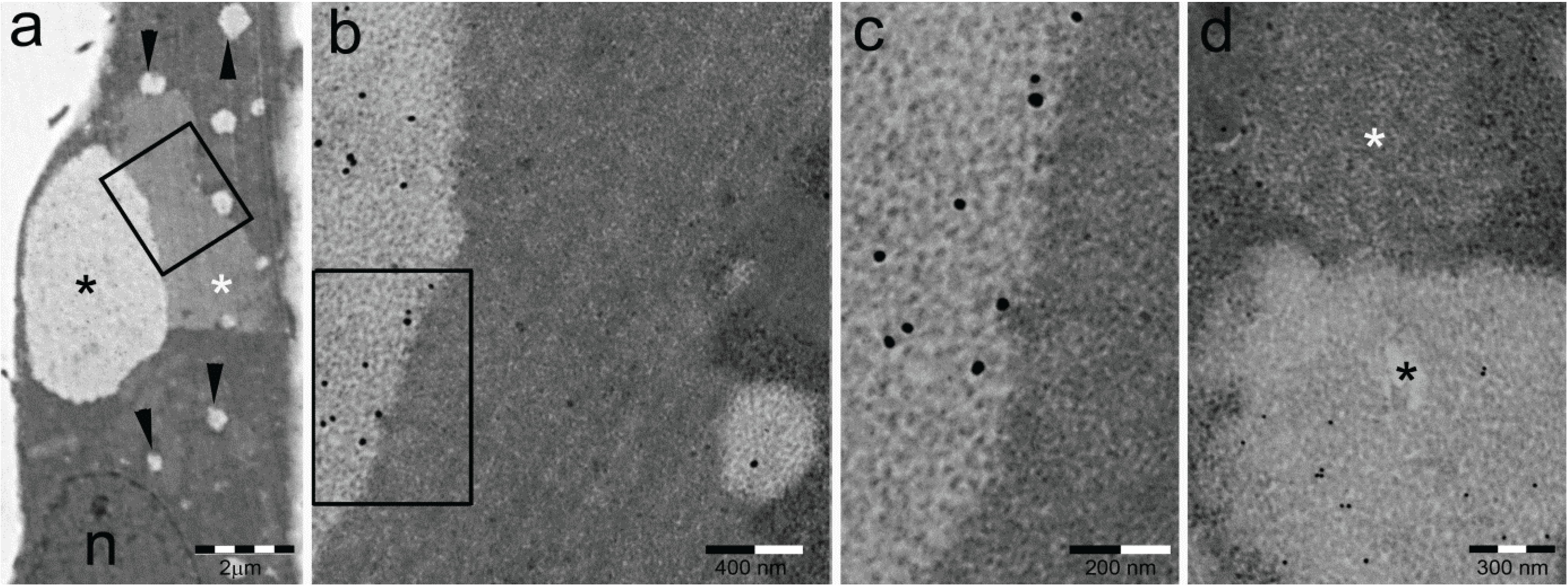
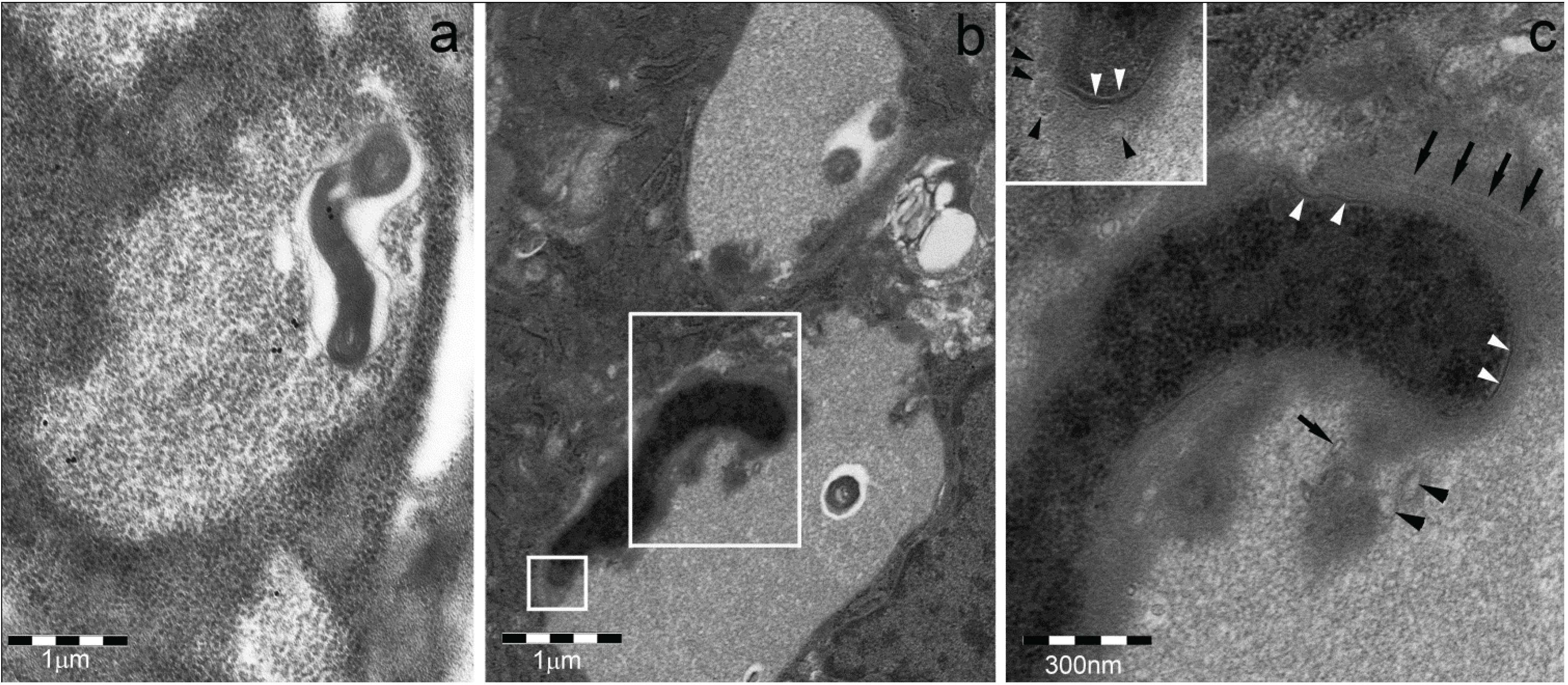
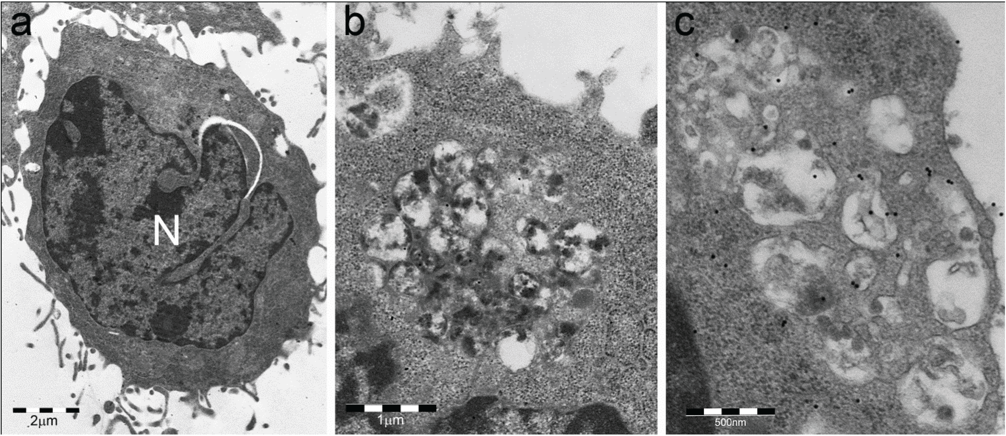
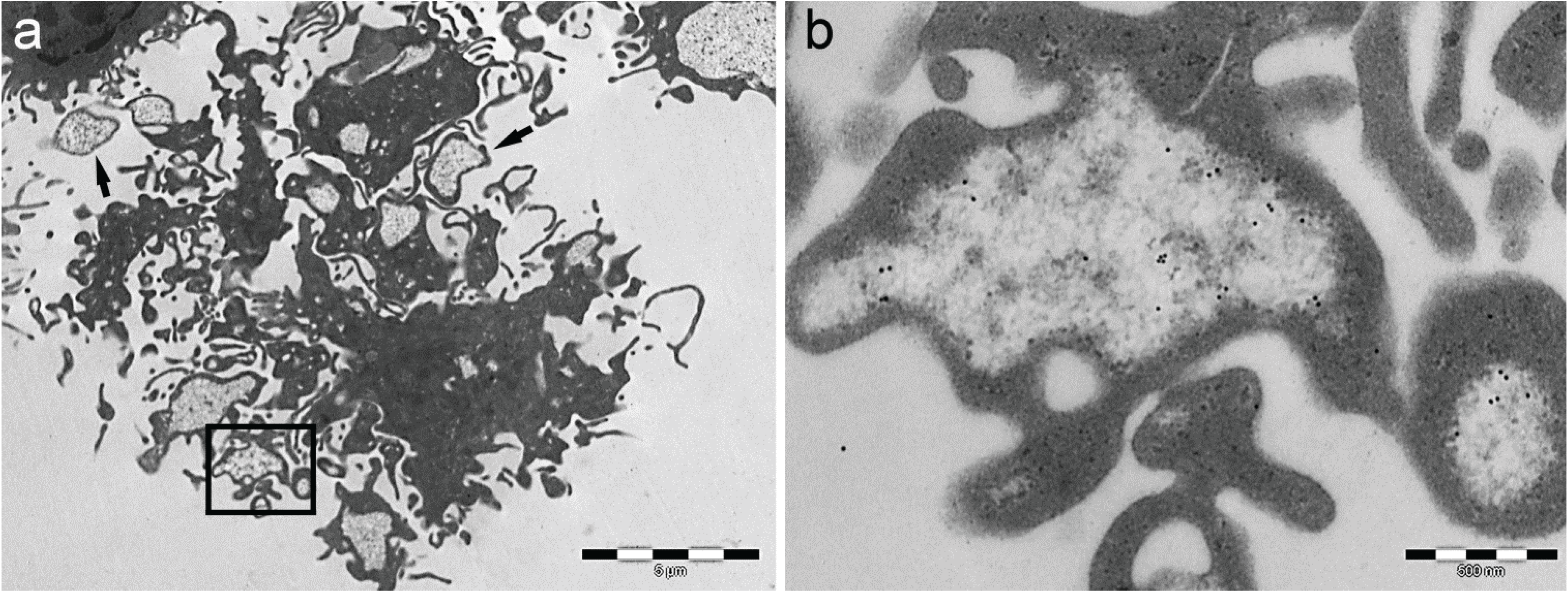
Particle-rich cytoplasmic structures (PaCSs) contain proteasome, polyubiquitinated proteins and glycogen and are characterized ultrastructurally by a collection of barrel-like particles of about 13 nm thick and 13–20 nm (infrequently up to 45) long (Figure 1 and Figure 4; see also [8,15,26]). As PaCSs store highly soluble components poorly preserved by common aldehyde fixatives, their detection requires stronger fixation, such as, for instance, by combined aldehyde and osmium solutions [12,15].
Inclusion bodies are typical of many pathological conditions, from neurodegenerative to muscle and liver diseases [9,10,11,16,27]. Both sequestosomes and aggresomes are likely to have a role in their genesis, but unfortunately, most information on such inclusions deal with their advanced developmental stages, making it difficult to reconstruct their precise intracellular origin. Only in the case of hepatocellular hyaline bodies described by Denk and coworkers [16], the thinly fibrillar ultrastructure reactive for ubiquitin and p62 closely resembles that of sequestosomes in HeLa cells.
In the following chapters, we will concentrate mostly on structures, like PaCS, regularly storing both proteasome and polyubiquitinated proteins and characterized at both the confocal fluorescence microscopy and ultrastructural immunogold level in cell lines, as well as in ex vivo tissues. Indeed, their high concentration of UPS components points to these structures as an attractive target for functional, as well as histopathological and pharmacological investigations.

3. Cytochemical and Ultrastructural Characterization of PaCS

In addition to polyubiquitinated proteins (FK1 antibody), 20S and 19S proteasome reactivity, PaCS was found to show positivity for antibodies directed against free ubiquitin, the immunoproteasome (20Sβ5i component), the E1 ubiquitin-activating enzyme, glycogen and glycogen-related proteins, such as glycogen synthase [8,15,26]. Under transmission electron microscopy (TEM) of aldehyde-osmium fixed, resin-embedded sections (Figure 1), PaCS appears as a collection of barrel-like particles in a relatively clear cytoplasmic area void of cytoskeleton fibrils and usually surrounded by ribosomes, with or without rough endoplasmic reticulum (RER) cisternae. It should be stressed that in cells cultured under basal conditions, PaCS is poorly preserved by the short (10–15 min) formaldehyde fixation commonly used for confocal microscopy immunofluorescence or even by a 24–48-h formaldehyde fixation followed by paraffin or resin embedding. As a consequence, no clear PaCS was visible when cells or tissue sections were investigated by conventional or confocal light microscopy immunofluorescence. However, semi-thin (~1 μm thick) resin sections obtained from TEM aldehyde-osmium fixed samples gave valuable immunofluorescence with many antibodies [12,15].

4. PaCS Distribution in Cells and Tissues

PaCSs have been detected in a variety of cultured cell lines, either neoplastic or non-neoplastic, and ex vivo tissue cells, including normal fetal, chronically-infected, mutated and neoplastic cells [8,12,15,26,28,29]. However, PaCSs were substantially missing in normal adult unstimulated, uninfected cells and tissues. An obvious cell type restriction was observed. PaCSs were found in cells, like many, though not all, epithelial and several mesenchymal (chondroblasts and osteoblasts, but not fibroblasts), hematopoietic (neutrophils and megakaryocytes/platelets, but not lymphocytes or erythrocytes and their precursors) and neuroid (neuroblasts and choroid plexus) cells. PaCS distribution in both fetal and neoplastic tissues, with a remarkable correspondence of cell types in the two conditions, depicts a sort of oncofetal distributive pattern [12,26]. The well-known increased expression of proteasome and ubiquitin molecules in both fetal and neoplastic cells [26,30,31,32] and their crucial role in both types of growth [33,34] may well account for some of these findings; however, they would not explain why PaCSs were not seen, for instance, in erythroblasts and reticulocytes, lymphocytes, plasma cells or fibroblasts and related neoplasms, despite their known proteasome expression. The focal concentration of UPS components and activity in a non-compartmentalized cytosolic center, allowing structured interaction with several other cytoplasmic factors (including, for instance, chaperone molecules, deubiquitinating enzymes and glycogen), is probably also important, besides accumulation per se of increased UPS components.
Of interest is also the relationship of PaCS development with the differentiation state of the cell. Highly immature embryonic/fetal or neoplastic cells show scarce or no PaCS, which, on the other hand, are extensively developed in cells undergoing differentiation, while progressively disappearing in fully-differentiated cells.
In addition, PaCSs were found in SDS gene-mutated neutrophils from the Shwachman-Diamond neutropenia [28] and in ANKRD26 gene-mutated megakaryocytes and platelets from type 2 thrombocytopenia [29], independently from the leukemic disease for which both conditions are at risk. Thus, it can be concluded that PaCS development shows a clear cell type, differentiation stage and pathologic state dependence.

5. PaCS Origin and Development

It is important to stress that PaCSs are constitutively expressed in some cell lines, as for instance HeLa cells. However, cell activation by trophic factors and interleukins is an important inducer of PaCS, as seen in CD14+ blood mononuclear cells stimulated with GM-CSF and IL-4 to obtain dendritic cells, blood NK (natural killer) cell precursors under IL-2 or IL-15 treatment [15] or ANKRD26 gene-mutated megakaryocyte precursor cells under treatment with thrombopoietin, IL-6 and IL-11 [29]. This behavior is likely to have some counterpart in the genesis of PaCS of fetal, neoplastic or inflammatory cells.
We took advantage of the possibility to induce PaCS in vitro by GM-CSF and IL-4 stimulation of blood mononuclear DC precursors to investigate under TEM the exact cytoplasmic site of PaCS origin. After 16 h of treatment, thin and minute collections of moderately electron-dense barrel-like particles coupled with all cytochemical markers of PaCS, including polyubiquitinated proteins and the proteasome appeared inside ribosome-rich cytoplasmic areas of a minority of cells. With a longer treatment (up to 5–7 days), PaCSs progressively extended to the majority of cells and increased in size (up to 4–5 μm in maximum diameter), by dislocating cell organelles, as well as the cytoskeleton and ribosomes, which often accumulated immediately around the PaCS periphery [8,15]. Thus, while the origin of PaCS in connection with ribosomes seems likely, the interaction with molecular chaperones and newly synthesized proteins as they sort from the poly-ribosome machine deserves further investigation.
In infected cells, the early presence inside PaCS of bacterial virulence products, such as H. pylori VacA, CagA and outer membrane proteins, or pertinent intracellular receptors, like NOD1 [8], suggests a direct role of bacterial products’ cytosolic accumulation in eliciting UPS colocalization and PaCS development.
The strong accumulation of polyubiquitinated proteins in the presence of the proteasome is rather surprising as, in vitro, the 26S proteasome is known to degrade polyubiquitinated proteins very rapidly. However the exact molecular form(s) of proteasome inside PaCS is presently unknown. Indeed, despite the coexisting presence of 20S and 19S, its two main molecular components, we do not know which level of proteasome assembly is reached [35,36,37]. The fact that within PaCS, most barrel-like particles are in a length range of 13–20 nm suggests that we are dealing mostly with uncoupled 19S and 20S molecules (with limited ability to rapidly degrade polyubiquitinated proteins), rather than with the fully-assembled 45 nm long 26S, uncommonly found inside PaCS [8,15]. It has been shown that the unassembled 20S molecule may process non-ubiquitinated, partially unfolded and oxidized proteins, showing an activity that might be suitable for bulk, unselective, slow degradation of altered proteins [38,39,40]. On the other hand, the full 26S molecule, known to be predominant in the nucleus (where PaCSs are not found) and likely present in PaCS-free cytoplasm, seems more apt to the tightly regulated task of modulating the specific activity of factors crucial for basic cellular functions, such as proliferation, cell cycling, gene expression, signal transduction or apoptosis.
The high concentration of glycogen inside PaCS may seem, by itself, surprising. However, it may be recalled that glycogen is the main source of cellular energy and ATP, which is an obligatory requirement of the 26S proteasome and chaperone function. In addition, proteins crucial for glycogen metabolism, like, for instance, glycogen synthase, are also present inside PaCS [15], thus raising the possibility that PaCS-related glycogen synthesis and degradation is at least in part functional to local ATP needs. Interestingly, the main source of cellular ATP in neoplastic and fetal cells, where PaCSs are highly represented, is known to be aerobic glycolysis, essentially cytosolic, as PaCSs are. In addition, it should be mentioned that AMP-dependent kinase, the main energy sensor of the cell, has also been localized in cytosolic glycogen particles [41].

6. Intracellular and Extracellular Fate of PaCS

Although, in general, PaCSs were found to lack evidence of autophagy, including the typical isolation membranes, their frequent topographic relationship with sequestosomes, largely destined to autophagy, has been noted in HeLa cells [15]. In addition, PaCS remnants have been found occasionally inside autophagic vesicles of pathologic, usually neoplastic cells [12]. Thus, it seems possible that, at least in severely-stressed cells, PaCS contents unfit for proteasome degradation could be handled by the autophagic-lysosomal pathway.
However, many observations in neoplastic and non-neoplastic cells strongly suggest a prominent mechanism of extracellular PaCS discharge through PaCS-filled cytoplasmic blebs, which, once severed from the cell, form isolated plasma membrane-enriched vesicles (so-called ectosomes), freely floating in culture media (Figure 4) or body interstitial spaces [15,26,29]. These findings may be relevant in many aspects, considering that PaCSs accumulate large amounts of ubiquitinated proteins and proteasome, potential sources of antigenic class I-presented molecules [42]. Indeed, a major role for both ectosomes and exosomes (the minute vesicles of multivesicular body origin carrying MIIC molecules and related antigens [43]) in intercellular communication seems likely, especially for eliciting immune responses [44]. In addition, PaCS-storing ectosome formation may well account for the increased proteasome and ubiquitin plasma levels observed in several pathologic conditions, from neoplasia to autoimmune diseases [31,45,46]. This finding is potentially of relevance in diagnostic and therapeutic analysis.
Figure 4. Extracellular fate of PaCS. (a,b) LPS-treated dendritic cells obtained in vitro with five days of GM-CSF and IL-4 treatment of blood mononuclear precursors show PaCS-filled blebs, some of which are detached from the cell and float in the surrounding space as free, cytoplasmic membrane enveloped vesicles (ectosomes; the black arrows point at some of them), as confirmed by serial section examination.
Biomolecules 04 00848 g004

7. Role of PaCS in Pathologic Conditions

7.1. Infectious Diseases

Ubiquitin-reactive protein aggregates have been found in the cytoplasm of infected gastrointestinal epithelia [8,13]. In H. pylori gastritis, the bacterium can enter foveolar cells from the lumen or lateral intercellular space and remain segregated from the cytosol by a host-derived enveloping membrane within which it retains its proliferative capacity [47]. However, H. pylori denuded of the enveloping membrane has been also found in the cytoplasm, surrounded by barrel-like particles associated with proteasome, polyubiquitinated proteins and bacterial virulence products, a pattern highly suggestive of bacterium-induced PaCS (Figure 5a; see also [8]). In addition, bacteria have also been found inside amorphous to thinly-fibrillar sequestosomes (Figure 5b,c) and membrane-delimited autophagic vesicles, in keeping with available evidence of VacA-mediated H. pylori-induced autophagy through p62 activation [48,49].
Figure 5. PaCS and infectious diseases. Electron microscopy of H. pylori colonized foveolar epithelium in a biopsy from human chronic gastritis (see [8] for more details). (a) A bacterium is shown inside PaCS; note the sparse CagA toxin immunogold reactivity of both the bacterium and its surrounding PaCS structure; (b) Longitudinally or cross-sectioned bacteria are found in two sequestosomes; the larger boxed area is enlarged in (c) and the smaller one in its inset to show partly preserved bacterial membrane and periplasmic space (white arrowheads) as well as flagella (arrows) and clubbed ends (black arrowheads); also note the poorly fibrillar, thinly granular content of the sequestosome.
Biomolecules 04 00848 g005

7.2. Neoplastic Growths

Polyubiquitinated proteins and proteasome-rich PaCSs have been detected sparsely in many epithelial neoplasms from stomach, intestine, lung, liver, kidney, ovary, thyroid, salivary glands and, more extensively, in clear cell neoplasms, including cancer of kidney and ovary, pancreatic serous microcystic adenoma and choroid plexus papilloma [12,26]. In addition, PaCSs have been regularly found in some neoplastic cell lines, including, for instance, HeLa, HL60 promyelocytic leukemia and SH-SY5Y neuroblastoma cell lines. PaCSs were not found in MDA-MB-231 breast carcinoma or Jurkat T-cell lymphoma [15]. A considerable homology between cell types of ex vivo neoplastic or fetal tissues and in vitro cell lines has been found [26], concerning PaCS presence/absence. This homology suggests some akin mechanism in PaCS development inside such cells. For example, increased expression/activity of trophic factors and their receptors are among common PaCS inducers to be considered in this respect.

8. Conclusions and Perspectives

In conclusion, the PaCS is a well-defined, distinctive cytoplasmic structure concentrating UPS components, which is shown in developing fetal or growth factors/ILs stimulated cells and in a variety of pathologic cells, from chronically-infected to mutated, preneoplastic or neoplastic. It differs ultrastructurally and cytochemically from other types of ubiquitin-reactive cytoplasmic structures, like sequestosomes, (D)ALIS and aggresomes, closely linked to the autophagic-lysosomal pathway.
PaCSs have been shown to display proteasome-type proteolytic activity toward a small test peptide [15]. However, in vitro and in vivo, PaCS invariably accumulates high concentrations of polyubiquitinated proteins. Although this finding may suggest proteasome malfunction in degrading such proteins, the possibility that the PaCS-associated proteasome retains the proteolytic activity against non-ubiquitinated, naturally unfolded or oxidized proteins remains to be investigated.
PaCS-storing cells may set up an alternative way to get rid of excessive accumulations of ubiquitinated misfolded proteins with their toxic and antigenic potential by releasing PaCS-filled cytoplasmic ectosomes. This is likely to be a relevant process in neoplastic and immunocompetent cells, and in vivo, it may account for the highly increased plasma levels of such products found in patients bearing neoplastic or autoimmune diseases [31,45,46]. Whether this has diagnostic relevance deserves further clinical investigation.
Work also remains to be done on the origin and natural history of a variety of cytoplasmic “inclusion” bodies formed by protein aggregates in several pathologic conditions, with special reference to neurodegenerative, liver or muscular diseases [9,10]. While ubiquitin conjugates and autophagy-linked proteins, as p62, are found in most of such structures, together with specific pathology-linked, mutated/misfolded proteins, evidence for proteasome component accumulation or proteasome malfunction has also been obtained for some of them, e.g., Parkinson’s Levy bodies [50,51]. Ubiquitin and proteasome enriched structures, like PaCS, should be investigated as a possible starting or intermediate step in their development.

Abbreviations

ALFY
autophagy-linked FYVE
ALIS
aggresome-like induced structures
DALIS
dendritic cell aggresome-like induced structures
DCs
dendritic cells
GM-CSF
granulocyte macrophage colony stimulating factor
HDAC6
histone deacetylase 6
IL
interleukin
IPOD
insoluble protein deposit
JUNQ
juxtanuclear quality control
LAMP1
lysosome-associated membrane protein 1
LPS
lipopolysaccharide
MHC
major histocompatibility complex
MIIC
MHC class II
MTOC
microtubule organizing center
NBR1
neighbor of BRCA1 gene 1
NK cells
natural killer cells
PaCS
particle-rich cytoplasmic structure
RER
rough endoplasmic reticulum
SQSTM1
sequestosome 1
TEM
transmission electron microscopy
UPS
ubiquitin proteasome system

Acknowledgments

This study was supported by grants from the Italian Ministry of Health to Fondazione IRCCS Policlinico San Matteo and University of Pavia (Grant No. RF-2010-2310098) and from Fondazione Cariplo (Grant Nos. 2011-0485 and 2012-0529).

Conflicts of Interest

The authors declare no conflict of interest.

References

  1. Wójcik, C.; Schroeter, D.; Wilk, S.; Lamprecht, J.; Paweletz, N. Ubiquitin-mediated proteolysis centers in HeLa cells: Indication from studies of an inhibitor of the chymotrypsin-like activity of the proteasome. Eur. J. Cell Biol. 1996, 71, 311–318. [Google Scholar]
  2. Johnston, J.A.; Ward, C.L.; Kopito, R.R. Aggresomes: A cellular response to misfolded proteins. J. Cell Biol. 1998, 143, 1883–1898. [Google Scholar] [CrossRef]
  3. Lelouard, H.; Gatti, E.; Cappello, F.; Gresser, O.; Camosseto, V.; Pierre, P. Transient aggregation of ubiquitinated proteins during dendritic cell maturation. Nature 2002, 417, 177–182. [Google Scholar] [CrossRef]
  4. Lelouard, H.; Ferrand, V.; Marguet, D.; Bania, J.; Camossetto, V.; David, A.; Gatti, E.; Pierre, P. Dendritic cell aggresome-like induced structures are dedicated areas for ubiquitination and storage of newly synthesized defective proteins. J. Cell Biol. 2004, 164, 667–675. [Google Scholar] [CrossRef]
  5. Szeto, J.; Kaniuk, N.A.; Canadien, V.; Nisman, R.; Mizushima, N.; Yoshimori, T.; Bazett-Jones, D.P.; Brumell, J.H. ALIS are stress-induced protein storage compartments for substrates of the proteasome and autophagy. Autophagy 2006, 2, 189–199. [Google Scholar] [CrossRef]
  6. Bjørkøy, G.; Lamark, T.; Brech, A.; Outzen, H.; Perander, M.; Øvervatn, A.; Stenmark, H.; Johansen, T. p62/SQSTM1 forms protein aggregates degraded by autophagy and has a protective effect on huntingtin-induced cell death. J. Cell Biol. 2005, 171, 603–614. [Google Scholar] [CrossRef]
  7. Kaganovich, D.; Kopito, R.; Frydman, J. Misfolded proteins partition between two distinct quality control compartments. Nature 2008, 454, 1088–1095. [Google Scholar] [CrossRef]
  8. Necchi, V.; Sommi, P.; Ricci, V.; Solcia, E. In vivo accumulation of Helicobacter pylori products, NOD1, ubiquitinated proteins and proteasome in a novel cytoplasmic structure. PLoS One 2010, 5, e9716. [Google Scholar] [CrossRef]
  9. Kuusisto, E.; Salminen, A.; Alafuzoff, I. Ubiquitin-binding protein p62 is present in neuronal and glial inclusions in human tauopathies and synucleinopathies. Neuroreport 2001, 12, 2085–2090. [Google Scholar] [CrossRef]
  10. Zatloukal, K.; Stumptner, C.; Fuchsbichler, A.; Heid, H.; Schnoelzer, M.; Kenner, L.; Kleinert, R.; Prinz, M.; Aguzzi, A.; Denk, H. p62 Is a common component of cytoplasmic inclusion in protein aggregation diseases. Am. J. Pathol. 2002, 160, 255–263. [Google Scholar] [CrossRef]
  11. Willis, M.S.; Patterson, C. Proteotoxicity and cardiac dysfunction—Alzheimer’s disease of the heart? N. Engl. J. Med. 2013, 368, 455–464. [Google Scholar]
  12. Necchi, V.; Sommi, P.; Vanoli, A.; Manca, R.; Ricci, V.; Solcia, E. Proteasome particle-rich structures are widely present in human epithelial neoplasms: Correlative light, confocal and electron microscopy study. PLoS One 2011, 6, e21317. [Google Scholar] [CrossRef]
  13. Kaniuk, N.A.; Lam, G.Y.; Ma, C.; Galindo-Mata, E.; Jones, N.; Vallance, B.A.; Brumell, J.H. Citrobacterrodentium infection induces MyD88-dependent formation of ubiquitinated protein aggregates in the intestinal epithelium. J. Innate Immun. 2011, 3, 83–98. [Google Scholar] [CrossRef]
  14. Kondylis, V.; van Nispen Tot Pannerden, H.E.; van Dijk, S.; Ten Broeke, T.; Wubbolts, R.; Geerts, W.J.; Seinen, C.; Mutis, T.; Heijnen, H.F. Endosome-mediated autophagy: An unconventional MIIC-driven autophagic pathway operational in dendritic cells. Autophagy 2013, 9, 861–880. [Google Scholar] [CrossRef]
  15. Sommi, P.; Necchi, V.; Vitali, A.; Montagna, D.; de Luigi, A.; Salmona, M.; Ricci, V.; Solcia, E. PaCSis a novel cytoplasmic structure containing functional proteasome and inducible by cytokines/trophic factors. PLoS One 2013, 8, e82560. [Google Scholar] [CrossRef]
  16. Denk, H.; Stumptner, C.; Fuchsbichler, A.; Müller, T.; Farr, G.; Müller, W.; Terracciano, L.; Zatloukal, K. Are the Mallory bodies and intracellular hyaline bodies in neoplastic and non-neoplastic hepatocytes related? J. Pathol. 2006, 208, 653–661. [Google Scholar] [CrossRef]
  17. Pankiv, S.; Clausen, T.H.; Lamark, T.; Brech, A.; Bruun, J.A.; Outzen, H.; Øvervatn, A.; Bjørkøy, G.; Johansen, T. p62/SQSTM1 binds directly to Atg8/LC3 to facilitate degradation of ubiquitinated protein aggregates by autophagy. J. Biol. Chem. 2007, 282, 24131–24145. [Google Scholar] [CrossRef]
  18. Kirkin, V.; Lamark, T.; Sou, Y.S.; Bjørkøy, G.; Nunn, J.L.; Bruun, J.A.; Shvets, E.; McEwan, D.G.; Clausen, T.H.; Wild, P.; et al. A role for NBR1 in autophagosomal degradation of ubiquitinated substrates. Mol. Cell 2009, 33, 505–516. [Google Scholar] [CrossRef] [Green Version]
  19. Fujimuro, M.; Sawada, H.; Yokosawa, H. Production and characterization of monoclonal antibodies specific to multi-ubiquitin chains of polyubiquitinated proteins. FEBS Lett. 1994, 349, 173–180. [Google Scholar] [CrossRef]
  20. Iwata, A.; Riley, B.E.; Johnston, J.A.; Kopito, R.R. HDAC6 and microtubules are required for autophagic degradation of aggregated huntingtin. J. Biol. Chem. 2005, 280, 40282–40292. [Google Scholar] [CrossRef]
  21. Wigley, W.C.; Fabunmi, R.P.; Lee, M.G.; Marino, C.R.; Muallem, S.; DeMartino, G.N.; Thomas, P.J. Dynamic association of proteasomal machinery with the centrosome. J. Cell Biol. 1999, 145, 481–490. [Google Scholar] [CrossRef]
  22. Waelter, S.; Boeddrich, A.; Lurz, R.; Scherzinger, E.; Lueder, G.; Lehrach, H.; Wanker, E.E. Accumulation of mutant huntingtin fragments in aggresome-like inclusion bodies as a result of insufficient protein degradation. Mol. Biol. Cell 2001, 12, 1393–1407. [Google Scholar] [CrossRef]
  23. García-Mata, R.; Bebök, Z.; Sorscher, E.J.; Sztul, E.S. Characterization and dynamics of aggresome formation by a cytosolic GFP-chimera. J. Cell Biol. 1999, 146, 1239–1254. [Google Scholar]
  24. Canadien, V.; Tan, T.; Zilber, R.; Szeto, J.; Perrin, A.J.; Brumell, J.H. Cutting edge: Microbial products elicit formation of dendritic cell aggresome-like induced structures in macrophages. J. Immunol. 2005, 174, 2471–2475. [Google Scholar] [CrossRef]
  25. Pierre, P. Dendritic cells, DRiPs, and DALIS in the control of antigen processing. Immunol. Rev. 2005, 207, 184–190. [Google Scholar] [CrossRef]
  26. Necchi, V.; Sommi, P.; Vitali, A.; Vanoli, A.; Savoia, A.; Ricci, V.; Solcia, E. Polyubiquitinated proteins, proteasome, and glycogen characterize the particle-rich cytoplasmic structure (PaCS) of neoplastic and fetal cells. Histochem. Cell Biol. 2014, 141, 483–497. [Google Scholar] [CrossRef]
  27. Olzmann, J.A.; Li, L.; Chin, L.S. Aggresome formation and neurodegenerative diseases: Therapeutic implications. Curr. Med. Chem. 2008, 15, 47–60. [Google Scholar]
  28. Necchi, V.; Minelli, A.; Sommi, P.; Vitali, A.; Caruso, R.; Longoni, D.; Frau, M.R.; Nasi, C.; de Gregorio, F.; Zecca, M.; et al. Ubiquitin-proteasome-rich cytoplasmic structures in neutrophils of patients with Shwachman-Diamond syndrome. Haematologica 2012, 97, 1057–1063. [Google Scholar] [CrossRef]
  29. Necchi, V.; Balduini, A.; Noris, P.; Barozzi, S.; Sommi, P.; di Buduo, C.; Balduini, C.L.; Solcia, E.; Pecci, A. Ubiquitin/proteasome-rich particulate cytoplasmic structures (PaCSs) in the platelets and megakaryocytes of ANKRD26-related thrombo-cytopenia. Thromb. Haemost. 2013, 109, 263–271. [Google Scholar]
  30. Kanayama, H.; Tanaka, K.; Aki, M.; Kagawa, S.; Miyaji, H.; Satoh, M.; Okada, F.; Sato, S.; Shimbara, N.; Ichihara, A. Changes in expressions of proteasome and ubiquitin genes in human renal cancer cells. Cancer Res. 1991, 51, 6677–6685. [Google Scholar]
  31. Lavabre-Bertrand, T.; Henry, L.; Carillo, S.; Guiraud, I.; Ouali, A.; Dutaud, D.; Aubry, L.; Rossi, J.F.; Bureau, J.P. Plasma proteasome level is a potential marker in patients with solid tumors and hemopoietic malignancies. Cancer 2001, 92, 2493–2500. [Google Scholar] [CrossRef]
  32. Bazzaro, M.; Lee, M.K.; Zoso, A.; Stirling, W.L.; Santillan, A.; Shih, IeM.; Roden, R.B. Ubiquitin-proteasome system stress sensitizes ovarian cancer to proteasome inhibitor-induced apoptosis. Cancer Res. 2006, 66, 3754–3763. [Google Scholar]
  33. Ryu, K.-Y.; Maehr, R.; Gilchrist, C.A.; Long, M.A.; Bouley, D.M.; Mueller, B.; Ploegh, H.L.; Kopito, R.R. The mouse polyubiquitin gene UbC is essential for fetal liver development, cell-cycle progression and stress tolerance. EMBO J. 2007, 26, 2693–2706. [Google Scholar] [CrossRef]
  34. Richardson, P.G.; Mitsiades, C.; Hideshima, T.; Anderson, K.C. Proteasome inhibition in the treatment of cancer. Cell Cycle 2005, 4, 290–296. [Google Scholar]
  35. Baumeister, W.; Walz, J.; Zühl, F.; Seemüller, E. The proteasome: Paradigm of a self-compartmentalizing protease. Cell 1998, 92, 367–380. [Google Scholar] [CrossRef]
  36. Hirano, Y.; Hendil, K.B.; Yashiroda, H.; Iemura, S.; Nagane, R.; Hioki, Y.; Natsume, T.; Tanaka, K.; Murata, S. A heterodimeric complex that promotes the assembly of mammalian 20S proteasomes. Nature 2005, 437, 1381–1385. [Google Scholar] [CrossRef]
  37. Finley, D. Recognition and processing of ubiquitin-protein conjugates by the proteasome. Annu. Rev. Biochem. 2009, 78, 477–513. [Google Scholar] [CrossRef]
  38. Liu, C.-W.; Corboy, M.J.; DeMartino, G.N.; Thomas, P.J. Endoproteolytic activity of the proteasome. Science 2003, 299, 408–411. [Google Scholar] [CrossRef]
  39. Baugh, J.M.; Viktorova, E.G.; Pilipenko, E.V. Proteasomes can degrade a significant proportion of cellular proteins independent of ubiquitination. J. Mol. Biol. 2009, 386, 814–827. [Google Scholar] [CrossRef]
  40. Pickering, A.M.; Koop, A.L.; Teoh, C.Y.; Ermark, G.; Grune, T.; Davies, K.J. The immunoproteasome, the 20S proteasome, and the PA28αβ proteasome regulator are oxidative-stress-adaptive proteolytic complexes. Biochem. J. 2010, 432, 585–594. [Google Scholar] [CrossRef]
  41. Bendayan, M.; Londono, I.; Kemp, B.E.; Hardie, G.D.; Ruderman, N.; Prentki, M. Association of AMP-activated protein kinase subunits with glycogen particles as revealed in situ by immunoelectron microscopy. J. Histochem. Cytochem. 2009, 57, 963–971. [Google Scholar] [CrossRef]
  42. Kloetzel, P.M. The proteasome and MHC class I antigen processing. Biochim. Biophys. Acta 2004, 1695, 225–233. [Google Scholar]
  43. Raposo, G.; Nijman, H.W.; Stoorvogel, W.; Liejendekker, R.; Harding, C.V.; Melief, C.J.; Geuze, H.J. B lymphocytes secrete antigen-presenting vesicles. J. Exp. Med. 1996, 183, 1161–1172. [Google Scholar] [CrossRef]
  44. Théry, C.; Ostrowski, M.; Segura, E. Membrane vesicles as conveyors of immune responses. Nat. Rev. Immunol. 2009, 9, 581–593. [Google Scholar] [CrossRef]
  45. Egerer, K.; Kuckelkorn, U.; Rudolph, P.E.; Rückert, J.C.; Dörner, T.; Burmester, G.R.; Kloetzel, P.M.; Feist, E. Circulating proteasomes are markers of cell damage and immunologic activity in autoimmune diseases. J. Rheumatol. 2002, 29, 2045–2052. [Google Scholar]
  46. Sixt, S.U.; Dahlmann, B. Extracellular, circulating proteasomes and ubiquitin—Incidence and relevance. Biochim. Biophys. Acta 2008, 1782, 817–823. [Google Scholar]
  47. Necchi, V.; Candusso, M.E.; Tava, F.; Luinetti, O.; Ventura, U.; Fiocca, R.; Ricci, V.; Solcia, E. Intracellular, intercellular, and stromal invasion of gastric mucosa, preneoplastic lesions, and cancer by Helicobacter pylori. Gastroenterology 2007, 132, 1009–1023. [Google Scholar] [CrossRef]
  48. Terebiznik, M.R.; Raju, D.; Vázquez, C.L.; Torbricki, K.; Kulkarni, R.; Blanke, S.R.; Yoshimori, T.; Colombo, M.I.; Jones, N.L. Effect of Helicobacter pylori’s vacuolatingcytotoxin on the autophagy pathway in gastric epithelial cells. Autophagy 2009, 5, 370–379. [Google Scholar] [CrossRef]
  49. Zheng, Y.T.; Shahnazari, S.; Brech, A.; Lamark, T.; Johansen, T.; Brumell, J.H. The adaptor protein p62/SQSTM1 targets invading bacteria to the autophagy pathway. J. Immunol. 2009, 183, 5909–5916. [Google Scholar] [CrossRef]
  50. Kwak, S.; Masaki, T.; Ishiura, S.; Sugita, H. Multicatalytic proteinase is present in Lewy bodies and neurofibrillary tangles in diffuse Lewy body disease brains. Neurosci. Lett. 1991, 128, 21–24. [Google Scholar] [CrossRef]
  51. McNaught, K.S.; Björklund, L.M.; Belizaire, R.; Isacson, O.; Jenner, P.; Olanow, C.W. Proteasome inhibition causes nigral degeneration with inclusion bodies in rats. Neuroreport 2002, 13, 1437–1441. [Google Scholar] [CrossRef]

