Review

The Role of miRNAs in Common Inflammatory Arthropathies: Osteoarthritis and Gouty Arthritis

by
Panagiota Papanagnou
1,*,†,
Theodora Stivarou
1,2,† and
Maria Tsironi
1
1
Department of Nursing, Faculty of Human Movement and Quality of Life Sciences, University of Peloponnese, Orthias Artemidos and Plateon St, GR-23100 Sparti, Greece
2
Immunology Laboratory, Immunology Department, Hellenic Pasteur Institute, P.O Box 115 21, Athens, Greece
*
Author to whom correspondence should be addressed.
These authors contributed equally to this work.
Biomolecules 2016, 6(4), 44; https://doi.org/10.3390/biom6040044
Submission received: 8 August 2016 / Revised: 29 October 2016 / Accepted: 2 November 2016 / Published: 11 November 2016

Abstract

:
MicroRNAs (miRNAs) are small, non-coding RNA species that are highly evolutionarily conserved, from higher invertebrates to man. Up to 1000 miRNAs have been identified in human cells thus far, where they are key regulators of the expression of numerous targets at the post-transcriptional level. They are implicated in various processes, including cell differentiation, metabolism, and inflammation. An expanding list of miRNAs is known to be involved in the pathogenesis of common, non-autoimmune inflammatory diseases. Interestingly, osteoarthritis (OA) is now being conceptualized as a metabolic disease, as there is a correlation among hyperuricemia and metabolic syndrome (MetS). Experimental evidence suggests that metabolic deregulation is a commonality between these different pathological entities, and that miRNAs are key players in the modulation of metabolic routes. In light of these findings, this review discusses the role of miRNAs in OA and gouty arthritis, as well as the possible therapeutic targetability of miRNAs in these diseases.

1. Introduction

Functionally, critical responses, such as inflammatory ones, are subjected to strict control, not only through the “supervising” role of modulatory proteins, but also by distinct RNA species, which, although they do not code for any protein, are key regulators for many functional systems, including the immune system. These non-coding, regulatory RNA species are the so-called microRNAs (hereafter referred to as “miRNAs”), which function as post-transcriptional regulators of gene expression. miRNAs can variably regulate the expression of numerous, even hundreds of different transcripts. In turn, the expression of a certain transcript may be controlled by multiple miRNAs [1,2,3,4,5].
miRNAs are short, single-stranded molecules displaying remarkable evolutionary conservation, from fruit fly (Drosophila melanogaster) to man [6]. Cells of human origin express up to 1000 miRNAs, with their expression patterns being cell/tissue-specific or not [7,8,9,10]. Genes coding for miRNAs may be found as independent genes, or they may map to intronic regions of other genes, and they are transcribed by RNA polymerases II and III. Adding another level of complexity, miRNA transcription units may be either mono- or poly-cistronic [11].
Biogenetically, miRNAs are first produced in the nucleus, in the form of primary miRNAs (pri-miRNAs), which undergo two consecutive steps of ribonucleolytic processing by two different RNases III in a cellular compartment-specific manner, i.e., by Drosha, within the nucleus, and then by Dicer, in the cytosol [12]. The first Drosha-mediated step results in the production of a hairpin-like precursor miRNA (pre-miRNA) that is exported to the cytosol in an exportin-5-dependent fashion. In the cytosol, Dicer, along with other factors, further processes pre-miRNA into a shorter (up to 24 nt) double-stranded molecule, of which the “guide strand” is eventually incorporated into a multicomponent ribonucleoprotein complex termed the RNA-induced silencing complex (RISC) and the other strand (symbolized as miRNA*) is degraded [11,13,14,15,16]. As part of the RISC, the “seed” region of a miRNA imperfectly forms base pairs with the 3′ untranslated region (UTR) of mRNA target(s) to shut off expression in various ways, including inhibition of translation and mRNA destabilization through deadenylation, or even the removal of the protective 5′ cap [17]. Importantly, miRNAs play an active role in a series of (patho)physiological mechanisms, including the control of inflammatory responses, which can be associated with autoimmunity or not [12,18,19,20,21,22,23].
Over 100 different types of arthritis have been described and miRNAs are implicated in some of them, as the literature, thus far, indicates [24,25]. However, osteoarthritis (OA) has increasingly attracted research interest because it is the most common form of arthritis [26]. Furthermore, the role of miRNAs in experimental models of arthritis and autoimmune diseases, including rheumatoid arthritis (RA), has been reported [27,28,29] and reviewed elsewhere [30,31,32]. Hence, in this review, we sought to discuss the role of miRNAs in the pathogenesis of two common types of non-autoimmune inflammatory arthropathies, OA and gouty arthritis, based on commonalities and correlations between these two different pathological entities: Gouty arthritis is associated with disturbed metabolic pathways, i.e., hyperuricemia, due to unbalanced purine nucleotide metabolism or excretion of uric acid by the kidney [33]. On the other hand, OA has been characterized as the fifth component of metabolic syndrome (MetS) [34]. More importantly, a positive correlation among hyperuricemia and MetS in different ethnicities has been found [35,36,37]. miRNAs are key controllers of chondrogenesis and the pathogenetic process of OA development [38,39], and they are interrelated with different metabolic routes [40,41,42]. In light of these data, the following paragraphs illustrate the involvement of miRNAs in pathways associated with both pathogenetic aspects of OA and gouty arthritis (from joint damage to pain), as well as their possible future exploitation in therapeutics for these inflammatory arthropathies.

2. OA and Gouty Arthritis: Overview of Pathophysiological Features

OA is a degenerative arthropathy associated with being overweight [43], which affects multiple tissues of the joints, including the cartilage, the subchondral bone, as well as the synovial membrane [44,45,46]. Consequently, in what follows, we will illustrate the role of miRNAs in different cell types in order to provide a global view of the pathogenesis of OA. Cartilage erosion and osteophytes at the cartilage–bone interface (osteophytosis) are pathognomonic features of this disease [47,48]. Synovial membrane inflammation, endochondral ossification, and subchondral bone sclerosis are well-known radiographic findings of OA, while the existence of a self-sustained hypoxia-inducible factor 2 alpha/Zn2+/zinc transporter ZIP8/metal-regulatory transcription factor 1 (HIF-2a/Zn2+/ZIP8/MTF1) catabolic circuit is a key molecular characteristic in this arthropathy [49,50,51]. Decreased levels of a major component of cartilage, the proteoglycan aggrecan [52], is a prominent biochemical feature of OA.
At the cellular level, chondrocytes and subchondral bone osteoblasts (SBOs) undergo numerous physiological alterations in OA: The regenerative capacity of OA chondrocytes is lost and OA chondrocytes acquire a “senescence-associated secretory phenotype” (SASP) characterized by the secretion of interleukin-1 (IL-1) and interleukin-6 (IL-6), as well as matrix metalloproteinase-3 (MMP-3) and -13 (MMP-13), which is largely induced by reactive oxygen species (ROS) [53]. In addition, lipid homeostasis is deregulated in chrondrocytes [54]. On the other hand, OA SBOs display a mineralization defect and altered profile of gene expression, which can be restored through miRNA mimics (i.e., synthetic miRNA-590-5p, miRNA-211-5p, miRNA-199a-5p, and miRNA-199a-3p), as evidenced in a rat model for OA. In fact, comparison among sclerotic and non-sclerotic bone specimens from OA sufferers demonstrated the existence of a molecular “signature” of eight miRNAs which possibly target 732 different transcripts, according to Ingenuity Pathway Analysis (IPA) bioinformatic analysis [55].
For many years, the prevailing view has been that inflammation and immune system components do not largely contribute to the pathogenesis of OA, which was thought to result from “wear and tear” processes. Yet, this was proven to be completely wrong [56]. Not only are human OA chondrocytes positive for components in miRNA biosynthesis (Dicer-1 and argonaute 2–4 (AGO2-4)) [57], but it is widely accepted that OA pathogenesis not only involves alterations in structural components of the joints, but also inflammatory and miRNA-dependent routes affecting different cellular processes, including lipid metabolism and autophagy [34,58,59,60]. Of note, RNA sequencing analysis revealed that human OA chondrocytes express several miRNAs that might play a role in OA, including the recently-identified miRNA-3085 [61]. Interestingly, miRNAs are implicated in pathways associated with altered lipid metabolism [54], as well as in routes possibly explaining the molecular basis of diabetes-induced OA [62].
Importantly, in silico analysis revealed the existence of distinct miRNA expression profiles, distinguishing OA from RA; even in the case that a single molecule (e.g., the transcription factor Ets-1) is predicted to be commonly targeted by miRNAs that display differential expression in OA and RA, this commonly-regulated molecule is postulated to be implicated in the pathogenesis of these different arthropathies through the modulation of different proteins [63].
On the other hand, gouty arthritis is a crystal-induced arthritis, i.e., a disease associated with the sterile inflammation of articular structures due to the deposition of crystals of monosodium urate (MSU). It may involve a single or multiple joints, and it progresses over four phases, i.e., asymptomatic hyperuricaemia, acute gout, intercritical gout, and chronic tophaceous gout [64,65]. Gouty arthritis can be misdiagnosed as rheumatoid arthritis, putting the lives of patients, simultaneously suffering from gout and other severe diseases, in jeopardy if it is not treated [66]. The identification of novel diagnostic markers with discriminative potential is, therefore, eagerly awaited.
Toll-like receptor 2 (TLR2) and 4 (TLR4) are critical players in the phagocytic ingestion of MSU crystals, while cluster of differentiation 14 (CD14), a TLR2/4-interacting adaptor molecule, which is engaged by MSU crystals, is crucial for the MSU-induced secretion of interleukin-1 beta (IL-1β) and their inflammagenicity, without impacting their uptake by macrophages. Production of IL-1β involves the proteolytic maturation of pro-IL-1β by caspase-1 upon activation of a macromolecular complex known as NACHT, LRR and PYD domains-containing protein 3 (NLRP3) inflammasome [67,68]. In turn, ROS formed by nicotinamide adenine dinucleotide phosphate (NADPH) oxidase and purinergic receptor P2X7-dependent signaling can positively regulate NLRP3 inflammasome [68]. The uptake of MSU crystals by residual macrophages in inflamed tissue stimulates a series of inflammatory pathways that lead to the local recruitment of leukocytes (chemotaxis), possibly through endothelial activation [68]. Although the involvement of TLRs, NLRP3 inflammasome, and IL-1β in the MSU-induced inflammatory reaction is well-known, there are no miRNAs discovered, thus far, directly targeting TLRs, IL-1β, chemokines, and NLRP3 in gout [69].

3. OA and Overweight: A Perplexed Interplay

High throughput analysis of OA cartilage samples, employing miRNA array analysis, revealed that the expression of distinct miRNAs, exemplified by miRNA-22 and miRNA-103, is deregulated in OA. In fact, a set of 16 miRNAs, undergoing either up- or down-regulation, characterizes this pathological condition. The physiological importance of the deregulated miRNA world came into the foreground when these miRNAs were found to correlate with other clinicopathological parameters, or to modulate target genes associated with different processes, including biomechanical transduction, bone morphogenesis, and lipid metabolism. Specifically, a positive correlation among the body mass index (BMI, a measure of obesity) and the up-regulated miRNAs, miRNA-22 and miRNA-103, was reported. On the contrary, an inverse correlation for those miRNAs of which expression is negatively modulated in OA (miRNA-25, miRNA-29a, and miRNA-337) and BMI was observed [70]. These findings pinpoint the pathogenetic link between obesity and articular cartilage erosion. Elevated expression of miRNA-483 was demonstrated to be another characteristic feature of OA, which was validated using Northern blotting [70].
The aforementioned apparently “simplistic” interconnection between overweight and OA can be further molecularly subdivided into (i) OA related to biomechanical signaling routes; (ii) OA related to obesity and inflammation, and, finally; (iii) OA related to obesity and deregulated lipid metabolism. In fact, miRNAs are known to be implicated in all of these molecular subdivisions related to excess weight. First, the expression patterns of several miRNAs (miRNA-221, miRNA-222, and miRNA-365) display mechanoresponsiveness [71,72], while a mechanoregulatory protein, integrin alpha-5, the levels of which are correlated with BMI, was identified as a target of miRNA-25 that is down-regulated in OA specimens [70]. Second, miRNAs and their targets, adipokines, are key mediators of “metaflammation”, a type of inflammation triggered by imbalanced cellular energetics [73]. Third, proteomic profiling, conducted by Iliopoulos and colleagues [70], identified peroxisome proliferator-activated receptor alpha (PPAR-alpha), a miRNA-22 target, as a protein that is negatively regulated in OA. Importantly, it was found that the expression of PPAR-alpha, and those of IL-1β (a cytokine that seems to be repressed by PPAR-alpha in chondrocytes), also correlate with BMI [70]. These data, along with the fact that PPAR-alpha functions as a crucial regulator of lipid metabolism and obesity [74], suggest that miRNAs control all physiological aspects through which overweight pathogenetically contributes to OA, i.e., the biomechanics associated with joint overloading, as well as inflammatory pathways and deregulated lipid metabolism.
Interestingly, a negative and positive correlation of the BMI of OA patients with miR-26a and nuclear factor kappa B (NF-kB), respectively, has been reported. In primary chondrocytes derived from mice fed with a high fat diet, levels of miR-26a were decreased in comparison to mice fed with a standard chow diet, whereas, in contrast, the levels of the transcripts of pro-inflammatory cytokines were increased. In obesity-related chondrocytes, miR-26a counteracts the positive regulation of NF-kB by saturated non-esterified fatty acid (NEFA), while NF-κB functions as a suppressor of miR-26a [75].
The levels of miRNA-27a are found to be decreased in OA chondrocytes in comparison to normal chondrocytes [76]. Intriguingly, decreased expression of miRNA-27a has been reported to be a molecular feature of mature adipocytes in obesity [77]. Therefore, one might presume that deregulated miRNA-27a-dependent signaling accounts for a mechanistic link between OA and overweight. Additional studies are needed to address whether this hypothesis holds true, and to investigate a possible connection between miRNA-27a and miRNA-33a, which, as outlined above, is related to the altered metabolic pathways observed in OA [34]. In addition, it would be interesting to investigate whether miRNA-driven alterations in chondrocyte lipid metabolism are associated with changes in adipose tissue homeostasis, which could collectively contribute to the osteoarthritic phenotype.

4. miRNA-Associated Pathways in OA

4.1. Metabolic Pathways

In OA, chondrocytes are characterized by the decreased expression of proteins taking part in reverse cholesterol transport (RCT), such as apolipoprotein A1 (ApoA1), and by lipid accumulation [54,78]. Transcription factor sterol regulatory element binding protein 2 (SREBP-2) and miRNA-33a are fundamental in the orchestration of sterol metabolism [79]. A SREBP-2 variant, namely SREBP-2 G/C, has been found to occur at high frequencies in Greek patients with OA. In normal chondrocytes, this variant dampens the transforming growth factor beta (TGF-β)-induced expression of aggrecan, thereby highlighting the importance of metabolic circuits in OA pathogenesis [52].
miRNA-33a, a key modulator of lipid metabolism [80], is a central player, linking OA pathogenesis with deregulated metabolism. As a matter of fact, miRNA-33a is produced by a nested gene, lying within intron 16 of human SREBP2, and both of these molecules are up-regulated in OA chondrocytes. miRNA-33a and SREBP-2 are subjected to common positive transcriptional regulation by TGF-β1, while miRNA-33a is essential for the TGF-β1-dependent expression of SREBP-2 itself. When normal chondrocytes are transfected with a miRNA-33a mimic, MMP-13 expression is increased, whereas, in contrast, levels of ApoA1 drop, although ApoA1 is not a direct miRNA-33a target. ATP binding cassette subfamily A member 1 (ABCA1), which is implicated in cholesterol efflux, is also decreased by miRNA-33a [34]. Given the importance of ApoA1 in RCT [81], and the role of MMP-13 in the acquisition of the osteoarthritic phenotype by chondrocytes [82], miRNA-33a emerges as a central player linking OA pathogenesis to deregulated metabolism [34].
Taking another step towards linking miRNAs with the modulation of metabolic networks and degenerative skeletal disorders, these post-transcriptional regulators have been postulated to be somehow involved in the progression of diabetic OA; however, rigorous evidence supporting this notion is lacking [62].

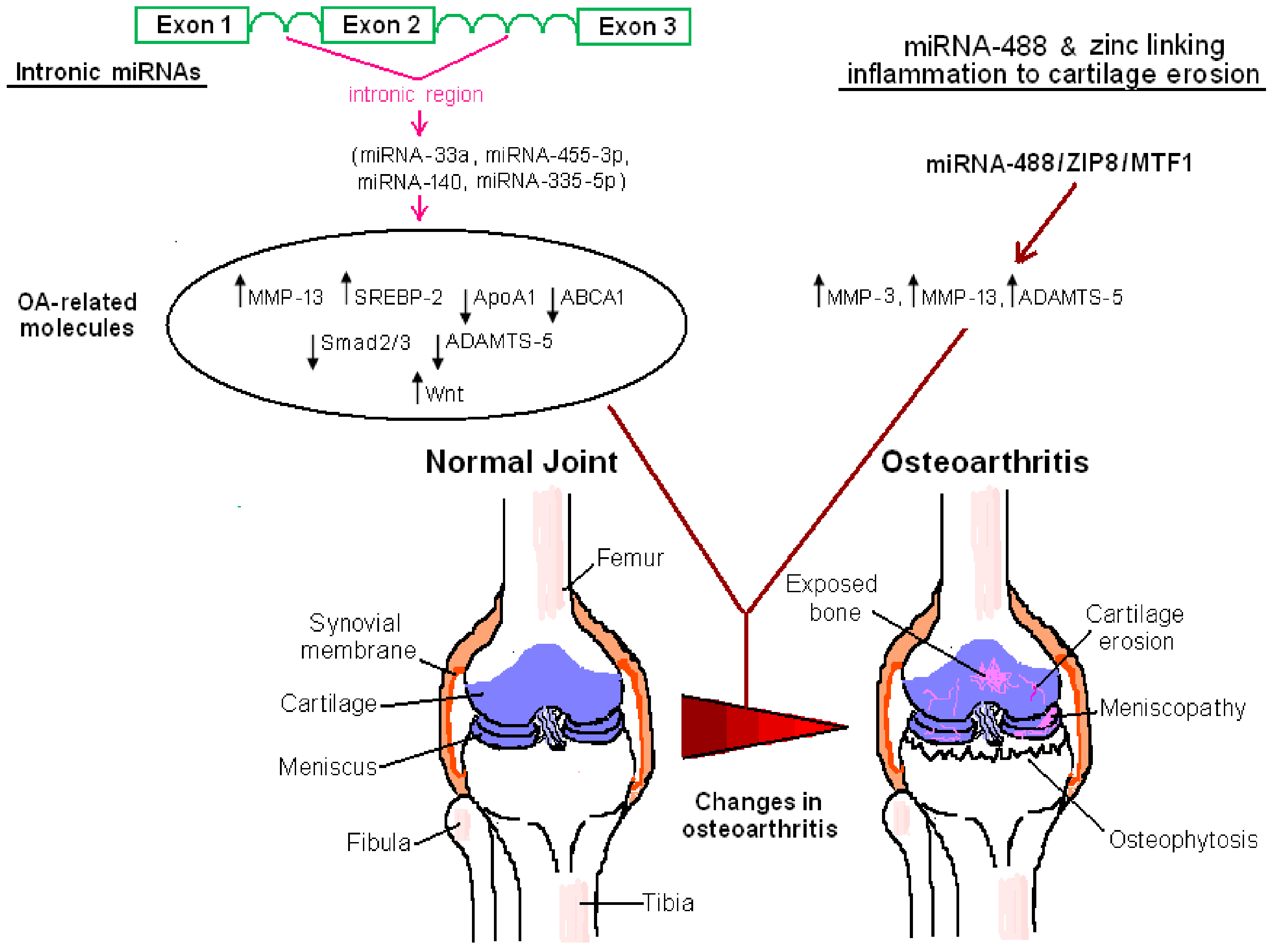
4.2. Inflammatory Networks, Degenerative Enzymes, and Zinc as the Molecular Link among Them

4.2.1. Inflammatory Signaling Pathways, MMPs, and A Disintegrin and Metalloproteinase with Thrombospondin Motifs Enzymes

Comparison of the miRNA expression profiles among cartilage, as well as bone obtained from OA and non-OA individuals, indicated that there are certain miRNAs that undergo a striking OA-specific up- or down-regulation. Specifically, in both cartilage and bone specimens from OA patients, miRNA-9 and miRNA-98 were detected at 8- and 23-fold higher levels compared to control samples, while miRNA-146 was found to be 14 times more abundant in OA samples than in control specimens. Bioinformatics predicted that all of the miRNAs mentioned above affect gene targets, which are implicated in inflammatory signaling pathways, such as the complement system, the NF-kB pathway, and leukocyte extravasation [83]. In a more recent study employing a surgical model of rat OA, it was found that miRNA-210 levels drop in diseased animals in comparison to sham-operated animals. When miRNA-210 is ectopically expressed via the administration of lentiviral constructs in the joints of OA rats, both the secretion of pro-inflammatory mediators and activation of the NF-kB pathway are prevented by miRNA-210 [84]. Similarly, miRNA-502-5p has also been found, through in vitro experimentation, to counteract NF-kB signaling. Mechanistically, this involves miRNA-502-5p-mediated inhibitory targeting of tumor necrosis factor receptor-associated factor 2 (TRAF2) [85]. The miRNA-29 family, of which expression is regulated throughout the development/progression of OA, also dampens NF-kB-mediated signalling [86].
An inflammatory circuit which seems to be etiopathologically linked to OA, is a miRNA-149-dependent one, where a drop in miRNA-149 levels in chondrocytes is correlated with an elevation in the expression of a set of cytokines (IL-6, IL-1β, and tumor necrosis factor alpha (TNF-α) [87]. A microarray-based analysis of miRNAs in human OA chondrocytes, stimulated by IL-1β, showed that the expression of 36 miRNAs were altered, with the majority of them (35 out of 36) being downregulated and affecting inflammatory networks [88].
Several studies have demonstrated that there are certain miRNAs that influence the expression of degenerative enzymes, such as MMP-13 and a disintegrin and metalloproteinase with thrombospondin motifs (ADAMTS) extracellular matrix proteases. A set of ADAMTS enzymes (ADAMTS4, ADAMTS5, ADAMTS7, as well as ADAMTS12) are controlled by miRNA-105, plausibly via targeting Runt-related transcription factor 2 (Runx2). In turn, miRNA-105 is negatively modulated by the p65 subunit of NF-kB upon stimulation of chondrocytes with fibroblast growth factor 2 (FGF2), suggesting that an FGF2/p65/miRNA-105/Runx2/ADAMTS pathway is critically implicated in the etiopathogenesis of OA. Significantly, it has been reported that there is an inverse correlation among miRNA-105 and Runx2, as well as ADAMTS [89]. Catabolic effects on the cartilage matrix, through the regulation of ADAMTS and MMPs, as well as regulation of Mothers against decapentaplegic homolog 3 (Smad3) synthesis, are also possibly exerted by miRNA-16-5p, which is found to be upregulated in cartilage specimens from OA sufferers compared to control samples [90].
A large-scale analysis demonstrated that miRNA-27b, as well as 41 other miRNAs undergo down-regulation in human OA chondrocytes in response to IL-1β, a key cytokine in OA development. IL-1β-dependent regulation of miRNA-27b is of great pathophysiological importance, as in silico analysis and luciferase reporter assays indicated that MMP-13 is targeted by this miRNA, with NF-kB functioning as a negative and positive modulator of miRNA-27b synthesis and MMP-13 secretion, respectively [57]. miRNA221-3p is another miRNA that can be suppressed by IL-1β. Interestingly, miRNA221-3p has recently been reported to be downregulated in synovial fibroblasts derived from subjects suffering from OA of the temporomandibular joint (TMJOA). This results in the derepression of the expression of its target, transcription factor Ets-1, and the consequent stimulation of the synthesis of the degradative enzymes, matrix metalloproteinase-1 (MMP-1) and 9 (MMP-9) [91]. miRNA-222, the expression of which is decreased in OA chondrocytes, is known to downregulate MMP-13 via targeting of histone deacetylase 4 (HDAC-4), and in mice where the osteoarthritic phenotype is surgically induced through destabilization of the medial meniscus (DMM), the overexpression of miRNA-222 in the knee in these animal models restrains cartilage damage [92].
The findings of another study imply that miRNA-602 and miRNA-608 could be exploited therapeutically in OA to suppress MMP-13. In fact, experimentation in animal models, as well as in different cell types (including chondrocytes stimulated with IL-1β), showed that the expression of MMP-13 is actually positively regulated by sonic hedgehog (SHH), the expression of which, in turn, is repressed by miRNA-602 and miRNA-608 [93]. ADAMTS5 (aggrecanase) is a miRNA-140 target, which finds application in modelling OA in animals. Silencing the expression of miRNA-140 in mice results in the establishment of an age-related mouse model of OA, where the aggrecan content in the cartilage matrix is decreased by the age of 12 months in miR-140−/− mice due to the derepression of the expression of ADAMTS5. On the other hand, miR-140-overexpressing transgenes are endowed with resistance to the development of arthropathy [94]. Part of the bone-protecting role of miRNA-140 is also possibly attributed to its ability to suppress the expression of MMP-13 in chondrocytes in a negative-feedback regulatory circuit [95].
miRNA-139 is an IL-1β-inducible miRNA that displays a chondral lesion-specific pattern of expression in OA, which seems to stimulate cartilage matrix catabolic processes via positively regulating the expression of the enzymes MMP-13 and ADAMTS4. Additionally, miRNA-139 exerts pro-apoptotic effects in human OA chondrocytes [96]. Employing the mono-iodoacetate-induced arthritis (MIA) rat model for OA, it was demonstrated that miRNA-101 stimulates the expression of a series of genes associated with extracellular matrix (ECM) degradation, including ADAMTS1 and ADAMTS5, as well as the expression of several cytokines. The usage of this animal model, which faithfully recapitulates human OA, showed that the intra-articular delivery of miRNA-101 mimic or miRNA-101 inhibitors by means of adenoviral vectors is a promising strategy to modulate cartilage erosion [97].
More recently, it was reported that miRNA-142-3p impedes the progression of OA in animals by acting as a negative modulator of the high mobility group box 1 protein (HMGB1)/NF-kB pathway [98]. Consistent with the anti-arthritic and chondroprotective properties of the bioactive compounds of green tea, exemplified by epigallocatechin-3-O-gallate (EGCG) [99,100], this green tea polyphenol engages a miR-199a-3p-dependent pathway to repress the expression of cyclooxygenase-2 (COX-2) and the synthesis of prostaglandin E2 (PGE2) in chondrocytes treated with IL-1β [101].
Interestingly, an inverse correlation among the expression pattern of miRNA-146α and MMP-13 is observed when the levels of these two molecules are evaluated in grade I and grade II OA, while miRNA-146α expression drops when OA progresses to more advanced stages, according to the Mankin scoring system. In vitro, it was demonstrated that miRNA-146α is increased in response to IL-1β. A role of miRNA-146α in cartilage homeostatic circuits (modulation of anabolic versus catabolic signals) or the induction of early OA by miRNA-146α have therefore been proposed as possible scenarios [102]. In line with these findings, in human chondrocytes, miRNA-146α suppresses the expression of MMP-13, as well as those of ADAMTS5 [103]. On the contrary, a positive correlation among miRNA-483 and MMP-13 in an experimental mouse model of OA has been found, implying the role of miRNA-483 in OA pathogenesis [104].

4.2.2. Zinc at the Crossroads of Inflammation and Cartilage Erosion

miRNA-488 is a TGF-β3-inducible miRNA, of which expression is decreased in OA chondrocytes compared to normal chondrocytes, and that targets ZIP8 (SLC39A8), a Zn2+ importer. Significantly, the suppression of ZIP8 expression contributes to the preservation of cartilage integrity in experimental models of OA [105]. This is consistent with the link between zinc homeostasis and OA. In fact, a zinc/ZIP8/MTF1 pathway that functions as a “molecular bridge” among inflammation and cartilage erosion has been described, as ZIP8 is an IL1β-inducible transporter, while the Zn2+-responsive transcription factor MTF1 positively regulates the expression of MMP-3, MMP-13, and ADATMTS5 [50,106,107]. Hence, the miRNA-488/ZIP8/MTF1/MMPs-ADAMTS5 axis is critical for the pathogenesis of OA.

4.3. TGF-β/Smad, Wingless , and Vascular Endothelial Growth Factor Pathway

A microarray-based, large-scale analysis identified the signatures of six miRNA, of which levels are increased in normal chondrocytes of human origin relative to OA chondrocytes (miRNA-1227, miRNA-576-5p, miRNA-149*, miRNA-634, miRNA-641, and miRNA-582-3p) and a single miRNA, namely miRNA-483-5p, which is selectively up-regulated in OA chondrocytes [108]. The finding of increased miRNA-483-5p in OA chondrocytes is in line with the results of Iliopoulos et al., who reported that miRNA-483 appeared to be up-regulated in diseased cartilage derived from OA patients [70]. The signalling circuits that these miRNA:target pairs participate in include Wingless (Wnt) and TGF-β/Smad pathways, which are pathogenetically related to OA, or are essential for the differentiation of chrondrocytes and in the onset/progression of OA [108]. Intriguingly, a negative correlation among miRNA-483 and the expression of TGF-β, as well as those of the TGF-β superfamily member bone morphogenic protein 7 (BMP7) in murine experimental OA, has been reported. The levels of both miRNA-483 and miRNA-483*, which reside within an intronic region of the insulin-like growth factor 2 (IGF2) locus, undergo an increase upon surgical intervention in an animal model of OA, and, hence, they are thought to be pathogenetically implicated in OA, irrespective of IGF2 [104]. A more recent study demonstrated that many of the Wnt pathway components (e.g., dishevelled segment polarity protein 3 (DVL3) and frizzled class receptor 5 (FZD5)) are directly targeted by miRNA-29 family members, of which the regulation of expression in cartilage seems to be important during the course of OA development. In fact, the miRNA-29 family is suppressed by transcription factor SRY-box 9 (SOX9), whereas, in turn, the miRNA-29 family dampens Wnt- and Smad-mediated signalling [86]. In human chondrocytes, Smad3 has been identified as a target of miRNA-16-5p, which has been incriminated in the development of OA [90]. According to a more recent study, Smad3 is also directly targeted by miRNA-23a-3p, of which expression in OA cartilage is elevated due to CpG island promoter hypomethylation. The net result of this miRNA-23a-3p/Smad3-dependent pathway is the decreased type II collagen/aggrecan content of the cartilage extracellular matrix in OA [109].
Another study demonstrated that in OA a prominent feature is the altered expression of two miRNAs, miRNA-140-5p and miRNA-455-3p [110]. A reciprocal regulation among miRNA-455-3p and TGF-β pathways—TGF-β influences miRNA-455-3p levels, while miRNA-455-3p dampens Smad2/3-mediated signaling, thereby possibly contributing to cartilage erosion—has been reported. Smad2, Chordin-Like 1 (CHRDL1), and Activin Receptor Type IIB (ACVR2B) have been identified as direct miRNA-455-3p targets, which are related with the TGF-β pathway [110]. On the other hand, miRNA-140 displays an almost chondrocyte-specific pattern of expression and controls pathways that are essential for unperturbed bone morphogenetic factor (BMP)/Smad signaling in an evolutionarily conserved manner, from mouse to zebrafish [111]. Intriguingly, both miRNA-455-3p and miRNA-140-5p are derived from intronic miRNA precursors that reside within collagen type XXVII alpha 1 (COL27A1) and WW domain containing E3 ubiquitin protein ligase 2 (WWP2) locus), respectively [110,111]. Of note, the spatiotemporal pattern of COL27 expression is known to play a crucial role in human skeletogenesis [112], while WWP2 is critical for craniofacial development [113]. In other words, the coding regions of miRNAs that are thought to be implicated in OA pathogenesis reside within host introns of genes that are critical for skeletal development.
In a study employing IL-1β-stimulated primary rat chondrocytes and a surgical rat model of OA where OA develops due to joint destabilization, miRNA-146α was found to be elevated in OA rats in comparison to sham-operated animals [114]. Smad4 transcription factor was characterized as a direct target of miRNA-146α, and its targeting by miRNA-146α was found to be essential for the ability of miRNA-146α to upregulate the expression of vascular endothelial growth factor (VEGF) in vitro and in vivo. In addition, miRNA-146α was found to suppress TGF-β signaling and to trigger chondrocyte apoptosis. Hence, it is postulated that miRNA-146α contributes to the OA phenotype by controlling TGF-β and VEGF via inhibiting their common mediator, Smad4. miRNA-146α-induced increase in VEGF in OA cartilage could contribute to abnormal angiogenesis and endochondral ossification during OA progression [114].

4.4. Autophagy

As evidenced by experiments in different mouse models of OA, autophagy is a homeostatic mechanism in cartilage, where it displays a constitutive pattern of expression, but is deregulated in favor of apoptotic cell death in OA [115]. Interestingly, a recent study identified miRNA-155 (a miRNA upregulated in OA) as a key negative modulator of the autophagic flux in human chondrocytes. miRNA-155 suppresses a series of autophagy-associated molecules exemplified by Atg proteins participating in the formation of the autophagosome, forkhead box O3 (FoxO3), as well as the autophagy inducer Unc-51-like kinase 1 (Ulk1)). Hence, miRNA-155 has been linked to the development of OA [116]. Since aging increases the risk for OA development, the decrease in the autophagic flux observed in OA chondrocytes is an aging-associated event [53].

4.5. Nociception

In human joint synoviocytes, miRNA-146α controls not only the expression of both cartilage matrix-degrading enzymes, but also those of inflammatory mediators associated with nociception. More importantly, it was reported for the first time that, in comparison to non-OA rats, OA animals experiencing pain express lower levels of miRNA-146α in their dorsal root ganglion (DRG) and spinal dorsal horn [103]. In addition, the role of miRNA-146α, not only in the pathogenesis of OA, but also in its clinical manifestations (i.e., chronic pain) was substantiated by the fact that miRNA-146α dampens the expression of a set of inflammatory molecules associated with pain perception in human glial cells, as well as the expression of transient receptor potential cation channel subfamily V member 1 (TRPV-1) [103]; an ion channel, which has attracted the interest of many pharmaceutical companies as a putative target of analgesic agents [117].

4.6. Mesenchymal Stem Cell-Associated Pathways

A large-scale study investigating the differential pattern of miRNAs expressed in mesenchymal stem cells (MSCs) derived from the bone marrow of OA and healthy donors provides insight into the possible role of MSCs in OA. A distinguishing feature of OA MSCs is the down-regulation of miRNA-335-5p. This is of major functional importance, since this miRNA targets the Wnt pathway antagonist Dickkopf Wnt signaling pathway inhibitor 1 (DKK1), thereby stimulating Wnt; a pathway that is interrelated with osteogenesis [118]. Given the role of unbalanced subchondral bone remodeling in the pathogenesis of OA, and the putative therapeutic value of manipulating MSCs in this process [119], miRNA-335-5p-dependent pathways could possibly be therapeutically harnessed in regenerative medicine. Of note, the miRNA-335-5p-coding region is actually an intronic miRNA (Figure 1). It is harbored within an intron by a gene, mesoderm specific transcript (MEST) [118]. On the other hand, miRNA-30a directly targets the Notch ligand Delta-like 4 (DLL4), thereby stimulating MSCs to express indicators of chondrogenic differentiation [120]. The usage of miRNAs for the stimulation of MSCs, to engage in a differentiation program, is a concept that is gaining ground in regenerative medicine and OA therapeutics [121].

5. miRNA-Associated Pathways in Gouty Arthritis

Analysis in mononuclear cells isolated from individuals suffering from gouty arthritis showed that miRNA-155 is expressed at higher levels in synovial fluid mononuclear cells (SFMCs) of patients in comparison to peripheral blood mononuclear cells (PBMCs) derived from healthy subjects. With the aim of unveiling a possible etiological link between miRNA-155 and gouty arthritis, it was found that the exposure of PBMCs from healthy participants to MSU crystals resulted in the induction of the expression of this miRNA. Src homology 2 (SH2) domain-containing inositol-5-phosphatase 1 (SHIP-1) levels were found to be lower, both in SFMCs of patients and in PBMCs of healthy controls on treatment with MSU crystals than in MSU-untreated PBMCs of healthy individuals. This is consistent with the fact that the immunoregulatory protein SHIP-1 is directly targeted by miRNA-155. SHIP-1 displays anti-inflammatory actions and the miRNA-155/SHIP-1 route is thought to fuel inflammation in gouty arthritis [122].
It is noteworthy that in THP-1 human monocytic cells, the decrease in SHIP-1 levels was found to coincide with a decrease of a molecule that is critically involved in inflammation—NF-kB inhibitor (IkB)—upon challenge with MSU crystals. miRNA-155-transfected or MSU-challenged THP-1 cells display a profound alteration in their pro-inflammatory secretory activity, exemplified by the elevation of IL-1β and TNF-α extracellular release, as evidenced by a multiplex immunoassay, termed Luminex. In line with the in vitro data, miRNA-155 is increased in the murine gout model. Importantly, in gouty arthritis clinical synovial specimens, the expression of SHIP-1 was reported to be scarce, which is in striking contrast to OA samples, which were positive for SHIP-1 [122]. The aforementioned data favor the notion that miRNA-155-dependent signaling positively regulates inflammation in gouty arthritis. In addition, these data pave the road for the identification of novel discriminative biomarkers for diagnosing gouty arthritis and other inflammatory arthropathies. A more recent study suggests that miRNA-146α halts inflammation evoked by MSU crystals; in fact, it suppresses the expression of a series of inflammatory mediators (monocyte chemoattractant protein-1 (MCP-1), TNF-α, interleukin 8 (IL-8), IL-1β). However, miRNA-146α is somehow lost throughout the acute response to these pro-inflammatory irritant particles [123].

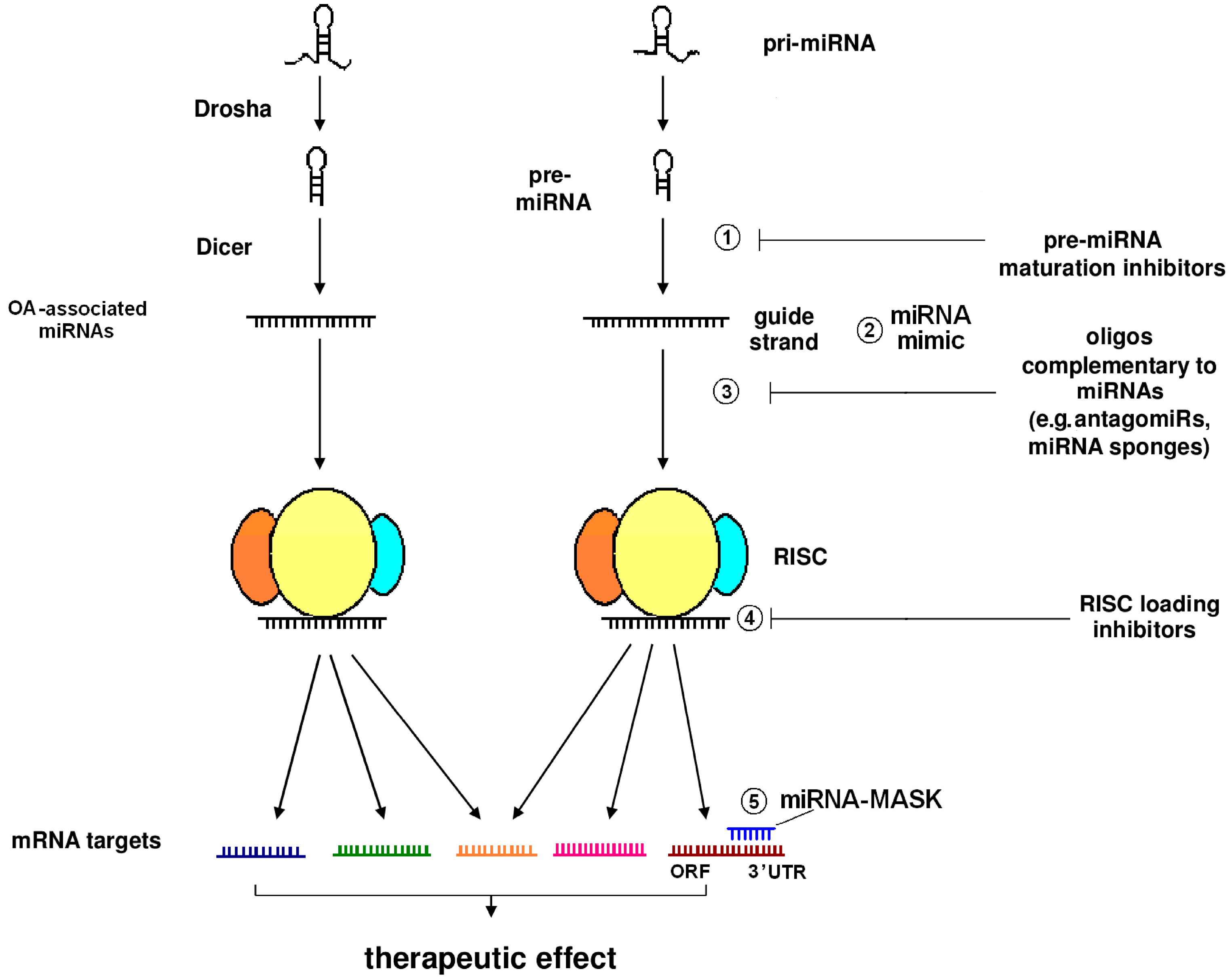
6. miRNA-Dependent Pathways as Therapeutic Targets in OA and Gouty Arthritis: At the Juncture of Biotechnology and Traditional Medicine

Agents used for the management of pain and inflammation, or as preoperative regimens in OA patients, may cause undesired side effects [124,125,126]. Hopefully, miRNA-based strategies hold promise for OA therapeutics [127], and could be used as an alternative, and perhaps safer, treatment option. miRNAs are actually nucleic acids that can be targeted using oligos that exhibit complementarity with the corresponding targets, to a lesser or greater extent in a similar way to antisense oligonucleotides (ASOs), which are used to silence mRNA targets. This stategy is termed miRNA antagonism, and complementary oligos are known as “antagomiRs”. Silencing miRNAs typically leads to the derepression of genes that are negatively modulated by miRNAs. The liver-specific miRNA-122 was actually among the first miRNAs to be targeted for silencing. In vitro, miRNA-122 was first targeted by an 2′-O-methylated ribonucleotide [128], while in vivo, miRNA-122 has been effectively and long-lastingly silenced by Krützfeldt and colleagues through enrolling a phosphorothioate (PS)-modified, cholesterol-conjugated 2′-O-methylated RNA molecule [129]. In the latter case, and from a historical perspective, the aforementioned researchers named the cholesterol conjugates of phosphorothioated, 2′-O-methylated RNA as “antagomiRs”. Since 2005, various chemically-modified complementary oligos have been designed to knock down the expression of miRNAs. Consequently, the term “antimiRs”, which includes antagomiRs, can be used to describe all the chemically divergent molecules targeting miRNAs.
The pharmacokinetic properties (e.g., binding affinity and resistance to nucleases) of antimiRs can be optimized via a series of modifications, such as ribose sugar modification at the 2′ position, the introduction of PS linkages into the phosphodiester backbone, the replacement of the sugar moieties with morpholino rings, or even the introduction of a 2′-O,4′-C-methylene bridge to generate bicyclic ribonucleotides, termed “locked nucleic acid” (LNA) molecules. The latter RNA analogues are characterized by the formation of particularly stable duplexes with their targets, exhibiting high melting temperatures (Tm values) [130]. Of note, miravirsen (SPC3649), an agent inhibiting hepatitis C virus (HCV), was the first anti-miRNA drug to enter clinical trials. It is a nuclease-resistant phosphorothioate-modified LNA oligo, which is complementary to the 5′ end of miRNA-122; a liver-specific miRNA, which is essential for HCV virulence [131]. A phase IIa clinical trial shows that miravirsen displays satisfactory, long-term beneficial effects in chronically genotype I, HCV-infected subjects in a dose-dependent manner, without any signs of development of viral resistance [132]. Interestingly, small-length LNAs, the so-called “tiny LNAs”, are complementary to the seed sequence of miRNAs, and can achieve the selective, long-term inhibition of a whole family of miRNAs sharing common seeds [133].
Moreover, antimiRs can be delivered either as naked species or as nanoparticles. The usage of nanotechnology may largely affect the biodistribution of an antimiR agent [134]. Another strategy to sequester miRNAs is the usage of the so-called “miRNA sponges”, introduced to biotechnology in 2007. miRNA sponges exhibit multiple (4–10), tandem binding sites for the miRNA target, separated by spacers. A whole miRNA family can be targeted by miRNA sponge constructs, since they block the functions of miRNAs with a common seed sequence. miRNA sponges can be delivered to cells via viral vectors, such as lentiviruses. For this reason, miRNA sponges can achieve long-term expression of the miRNA construct, as has been shown in different animal species. In contrast, antagomiRs have to be repeatedly administered at high doses in order to achieve long-term miRNA inhibition. An additional advantage of miRNA sponge technology is that, unlike cholesterol-conjugated antagomiR oligos, which are accumulated in the hepatic parenchyma, the design of miRNA sponges may allow their expression in any tissue of interest [135,136].
Another miRNA-based treatment strategy involves the employment of the so-called “miRNA-MASKs”. These are single-stranded oligos carrying 2′-O-methyl modifications, which are designed to be complementary to the 3′ UTR of the target transcript, thereby occupying it and hindering its interaction with the corresponding miRNA. The net result is the blockage of miRNA function and the derepression of the expression of a gene of interest [137]. Another approach is the usage of “miRNA mimics”. These synthetic molecules are double-stranded oligos, which in contrast to miRNA-MASKs, mimic the sequence of a mature miRNA molecule, or even the sequence of pre-miRNA [138]. Alternatively, a pri-miRNA mimic can be designed to mimic the primary miRNA molecule. The latter constructs engage the endogenous miRNA biogenetic pathway upon their delivery, and they are effective in gene silencing [139]. Noticeably, miRNA mimics have been used to uncover the role of the miRNA-30b/ERG pathway in OA [140], as well as the regulatory function of miRNA-634 in chondrocyte metabolism [141].
Pre-clinical experimentation in animals suggests that some miRNA species are possible targets for therapeutic intervention in OA. For instance, lentiviral delivery of miRNA-222 into the knee of a surgical DMM mouse model of OA yielded encouraging results [92]. On the contrary, miRNA-21, which suppresses the TGF-β superfamily member growth/differentiation factor 5 (GDF-5), has been incriminated in the development of OA due to its anti-chondrogenic effects [142]. This finding is of major importance, since miRNA-21 has already been successfully employed in an in vitro assay evaluating the RISC loading inhibitory potency of different compounds [143]. Hence, the notion that miRNA-21 may serve as a therapeutic target in OA is being reinforced. Other miRNA species critically implicated in OA pathogenesis, such as miRNA-155, do not seem to be “targetable”: miRNA-155 integrity is required for the unperturbed primary and memory immune responses, as evidenced in mice [144]. Conceivably, the putative targeting of a specific miRNA for therapeutic purposes is not always an easy matter and raises various concerns.
Recently described biotechnological techniques, such as the clustered regularly interspaced short palindromic repeats (CRISPR), can advance the characterization of miRNAs, which specifically operate in chondrocytes and OA pathogenesis, as well as the perplexed signalling networks in which they participate. Further, enrolling robust methodologies for the validation of direct miRNA targets, such as the high-throughput sequencing of RNA isolated by cross-linking immunoprecipitation (HITS-CLIP), will hopefully contribute to the minimization of hybridization-dependent phenomena, which are major hurdles in clinical applications [145]. These methodologies in combination with certain drug delivery modes, such as the use of synthetic materials, can enable the development of novel and efficacious therapeutic approaches in OA and/or gouty arthritis and the improvement of different parameters that are crucial for miRNA-based therapeutics. These parameters include hybridization-independent issues, e.g., the phagocytic uptake of circulating miRNA-containing particles, or even the off-target effects observed when miRNAs carry chemical modifications [146].
Interestingly, experiments have been carried out in order to study the molecular pathways underlying the therapeutic effects of herbal decoctions commonly used by Chinese medicine practitioners, such as the Xie-Zhuo-Chu-Bi-Fang (XZCBF) and the Xiezhuo Chubi Decoction (XZCBD). Data demonstrate that there are distinct miRNAs associated with herb-based therapeutics of hyperuricaemia, at least in a pre-clinical setting (hyperuricemic models). Certain miRNAs affect the expression of molecules critically implicated in uric acid handling, i.e., urate transporter 1 (URAT1)and xanthine oxidase (XO). In fact, traditional Chinese herb-based treatment with XZCBD has been reported to exert a suppressive effect on the expression of URAT1. This is possibly attributed to the XZCBD-induced upregulation of miRNA-34a, although more than 30 other miRNAs are also modulated by this formula at high doses [147]. According to a more recent study, miRNA-34a has been shown to counteract URAT1-mediated renal reabsorption of uric acid, thereby mitigating the disease phenotype in animals treated with XZCBF [148]. Furthermore, evidence has been provided that miRNA-448 modulates XO levels in an allopurinol-dependent manner [149]. In substantiation of the plausible importance of miRNAs in circuits associated with gout pathophysiology, URAT1 is crucial for the regulation of serum levels of uric acid [150] and XO is currently the mainstay pharmacological target in gout [151]. Overall, experimental evidence suggests that miRNA-dependent pathways are targeted by herbal formulations used in traditional medicine. In addition, miRNAs are possible targets for therapeutic intervention in OA, where their functions can be modulated by virtue of biotechnology advances, as depicted in Figure 2.

7. Conclusions and Future Perspectives

Inflammation and cartilage erosion in OA are linked by a miRNA-488-regulated Zn2+/ZIP8/MTF1 pathway, which affects the expression of MMPs-ADAMTS5. The prevailing view is that OA is a “wear and tear” process, which, in an oversimplified manner, is solely associated with cartilage erosion due to the cartilage matrix degeneration by catabolic enzymes (MMPs, ADAMTS enzymes). By taking advantage of high throughput screening and RNA sequencing technologies, and via experimental models of OA (surgical instability models or genetically engineered animals), it has been demonstrated that miRNAs not only simply act as regulators of inflammatory processes in the affected joints (e.g., chemotaxis), but also as key metabolic regulators at the juncture of the modulation of metabolism and inflammation (metaflammation), as well as in the control of metabolic pathways, which mechanistically link obesity to OA. Downregulated RCT, critically modulated by miRNA-33a, and deregulated autophagic flux, modulated by miRNA-155, are prominent molecular features of OA. From a structural genomics point of view, several miRNAs reside within the intronic regions of the human genome [152,153]. Certain OA-related miRNAs, i.e., miRNA-33a, miRNA-455-3p, miRNA-140, and miRNA-335-5p, are actually embedded within the intronic regions of human SREBP-2, COL27A1, WWP2, and MEST, respectively. The intronic class of miRNAs possibly displays biogenetic differences with regard to the exonic class of miRNAs in mammals [152]. Whether this intronic class of miRNAs differentially influences OA pathogenesis, compared to the exonic class of miRNAs, is a very interesting topic for future research. Another hallmark of OA is miRNA-335-5p downregulation in OA MSCs; something that can be therapeutically harnessed in the future, in the field of regenerative medicine.
In addition to OA, miRNAs are also central players in pathways associated with MSU-induced inflammation and gouty arthritis. The identification of miRNA(s) possibly targeting TLRs, IL-1β, chemokines, and NLRP3 in gouty arthritis is another major goal for the future. The etiopathological role of miRNA-155 is a commonality between OA and gouty arthritis, although, in OA, miRNA-155 functions as a suppressor of autophagy, whereas a miRNA-155/SHIP-1 pathway has been incriminated in gouty arthritis. Thus far, no such metabolism-associated miRNA has been discovered that links OA and gouty arthritis, although there is a positive correlation among hyperuricemia and MetS, and OA is being conceptualized as the fifth component of MetS. Thus, it would be interesting for researchers to address whether there is/are any common metabolism-related miRNA(s) playing a role in the pathogenesis of OA and gouty arthritis. This is a challenging task for the future.
Another future challenge for OA therapeutics would be to address whether there are OA-related miRNAs displaying evolutionary conservation of seed sequences, so that they could possibly be targeted by “tiny LNAs”. Moreover, interfering with miRNA-146α-dependent pathways controlling nociception in OA [103] might serve as a strategy to relieve pain. In fact, the idea that miRNA-mediated axes could be targeted for the management of pain has been gaining ground [154].
The studies mentioned above unequivocally indicate that miRNAs are interrelated with the pathogenesis of OA, as well as gouty arthritis. Even in the case that miRNAs are not etiopathologically associated with OA, their expression is somehow influenced throughout the OA development process, either as single non-coding molecules or as part of a miRNA “signature”. miRNAs could be clinically used (e.g., in a microarray-based format) as a feasible procedure for the specific assessment of OA, which could be less expensive than magnetic resonance imaging (MRI). More experimentation is required in this direction [155]. Validating a certain miRNA or miRNA sets, as non-invasive biomarker(s) in patients suffering from OA or gouty arthritis, using an established statistical method, such as receiver operating characteristic (ROC) curve analysis as has been attempted in other diseases [156,157], would therefore be another future goal. For instance, miRNA let-7e seems to be a negative predictor for severe OA, at least in Caucasians [155], and its validation as a biomarker is of great interest. Valuable data regarding the usefulness of distinct miRNAs as biomarkers at the pre-clinical level in animal models have been already provided [158]. However, ROC analyses are nodal statistical tools in clinical studies. Clinical data, properly processed in order to be statistically refined, are much-awaited, in order to propel the introduction of possible miRNA-based therapies into clinical practice.
It is worth mentioning that miRNAs are part of the underlying mechanism by which herbal decoctions, used by traditional Chinese medicine, act in the management of OA. A significant “take-home message” is that miRNA-mediated control plays a key role in the cellular responses to medical practices applied all over the world, from the West to the East. In addition, miRNAs are involved in the modulation of the inflammatory pathways activated in different types of arthropathies, whether they are evoked by crystal deposits or not.

Acknowledgments

The authors would like to thank the Emeritus Professor of Genetics and Human Genetics at the Aristotle University of Thessaloniki, Costas Triantaphyllidis, for his valuable comments on the manuscript. This theoretical study was self-funded. None of the authors received grants in support of this work or any funds for covering the costs to publish in open access.

Conflicts of interest

The authors declare no conflicts of interest.

References

  1. Saetrom, P.; Snove, O., Jr.; Rossi, J.J. Epigenetics and microRNAs. Pediatr. Res. 2007, 61, 17R–23R. [Google Scholar] [CrossRef] [PubMed]
  2. Selbach, M.; Schwanhausser, B.; Thierfelder, N.; Fang, Z.; Khanin, R.; Rajewsky, N. Widespread changes in protein synthesis induced by microRNAs. Nature 2008, 455, 58–63. [Google Scholar] [CrossRef] [PubMed]
  3. Baek, D.; Villen, J.; Shin, C.; Camargo, F.D.; Gygi, S.P.; Bartel, D.P. The impact of microRNAs on protein output. Nature 2008, 455, 64–71. [Google Scholar] [CrossRef] [PubMed]
  4. O’Connell, R.M.; Rao, D.S.; Chaudhuri, A.A.; Baltimore, D. Physiological and pathological roles for microRNAs in the immune system. Nat. Rev. Immunol. 2010, 10, 111–122. [Google Scholar] [CrossRef] [PubMed]
  5. Lin, S.; Gregory, R.I. MicroRNA biogenesis pathways in cancer. Nat. Rev. Cancer 2015, 15, 321–333. [Google Scholar] [CrossRef] [PubMed]
  6. Pasquinelli, A.E.; Reinhart, B.J.; Slack, F.; Martindale, M.Q.; Kuroda, M.I.; Maller, B.; Hayward, D.C.; Ball, E.E.; Degnan, B.; Muller, P.; et al. Conservation of the sequence and temporal expression of let-7 heterochronic regulatory RNA. Nature 2000, 408, 86–89. [Google Scholar] [PubMed]
  7. Chen, C.Z.; Li, L.; Lodish, H.F.; Bartel, D.P. MicroRNAs modulate hematopoietic lineage differentiation. Science 2004, 303, 83–86. [Google Scholar] [CrossRef] [PubMed]
  8. Chang, J.; Nicolas, E.; Marks, D.; Sander, C.; Lerro, A.; Buendia, M.A.; Xu, C.; Mason, W.S.; Moloshok, T.; Bort, R.; et al. miR-122, a mammalian liver-specific microRNA, is processed from hcr mrna and may downregulate the high affinity cationic amino acid transporter CAT-1. RNA Biol. 2004, 1, 106–113. [Google Scholar] [CrossRef] [PubMed]
  9. Chen, J.F.; Mandel, E.M.; Thomson, J.M.; Wu, Q.; Callis, T.E.; Hammond, S.M.; Conlon, F.L.; Wang, D.Z. The role of microRNA-1 and microRNA-133 in skeletal muscle proliferation and differentiation. Nat. Genet. 2006, 38, 228–233. [Google Scholar] [CrossRef] [PubMed]
  10. Bartel, D.P. MicroRNAs: Target recognition and regulatory functions. Cell 2009, 136, 215–233. [Google Scholar] [CrossRef] [PubMed]
  11. Saini, H.K.; Enright, A.J.; Griffiths-Jones, S. Annotation of mammalian primary microRNAs. BMC Genom. 2008, 9, 564. [Google Scholar] [CrossRef] [PubMed]
  12. Baulina, N.M.; Kulakova, O.G.; Favorova, O.O. MicroRNAs: The role in autoimmune inflammation. Acta Nat. 2016, 8, 21–33. [Google Scholar]
  13. Hutvagner, G.; McLachlan, J.; Pasquinelli, A.E.; Balint, E.; Tuschl, T.; Zamore, P.D. A cellular function for the RNA-interference enzyme Dicer in the maturation of the let-7 small temporal RNA. Science 2001, 293, 834–838. [Google Scholar] [CrossRef] [PubMed]
  14. Bernstein, E.; Caudy, A.A.; Hammond, S.M.; Hannon, G.J. Role for a bidentate ribonuclease in the initiation step of RNA interference. Nature 2001, 409, 363–366. [Google Scholar] [CrossRef] [PubMed]
  15. Grishok, A.; Pasquinelli, A.E.; Conte, D.; Li, N.; Parrish, S.; Ha, I.; Baillie, D.L.; Fire, A.; Ruvkun, G.; Mello, C.C. Genes and mechanisms related to RNA interference regulate expression of the small temporal RNAs that control C. elegans developmental timing. Cell 2001, 106, 23–34. [Google Scholar] [CrossRef]
  16. Lee, Y.; Ahn, C.; Han, J.; Choi, H.; Kim, J.; Yim, J.; Lee, J.; Provost, P.; Radmark, O.; Kim, S.; et al. The nuclear RNase III Drosha initiates microRNA processing. Nature 2003, 425, 415–419. [Google Scholar] [CrossRef] [PubMed]
  17. Gu, S.; Kay, M.A. How do miRNAs mediate translational repression? Silence 2010, 1, 11. [Google Scholar] [CrossRef] [PubMed]
  18. Hsu, S.H.; Ghoshal, K. MicroRNAs in liver health and disease. Curr. Pathobiol. Rep. 2013, 1, 53–62. [Google Scholar] [CrossRef] [PubMed]
  19. Singh, R.P.; Massachi, I.; Manickavel, S.; Singh, S.; Rao, N.P.; Hasan, S.; Mc Curdy, D.K.; Sharma, S.; Wong, D.; Hahn, B.H.; et al. The role of miRNA in inflammation and autoimmunity. Autoimmun. Rev. 2013, 12, 1160–1165. [Google Scholar] [CrossRef] [PubMed]
  20. Cruz-Santos, M.C.; Aragon-Raygoza, A.; Espinal-Centeno, A.; Arteaga-Vazquez, M.; Cruz-Hernandez, A.; Bako, L.; Cruz-Ramirez, A. The role of microRNAs in animal cell reprogramming. Stem Cells Dev. 2016, 25, 1035–1049. [Google Scholar] [CrossRef] [PubMed]
  21. Otsuka, M.; Kishikawa, T.; Yoshikawa, T.; Yamagami, M.; Ohno, M.; Takata, A.; Shibata, C.; Ishibashi, R.; Koike, K. MicroRNAs and liver disease. J. Hum. Genet. 2016. [Google Scholar] [CrossRef] [PubMed]
  22. Cao, D.D.; Li, L.; Chan, W.Y. Micrornas: Key regulators in the central nervous system and their implication in neurological diseases. Int. J. Mol. Sci. 2016, 17, 842. [Google Scholar] [CrossRef] [PubMed]
  23. Asghari, F.; Haghnavaz, N.; Baradaran, B.; Hemmatzadeh, M.; Kazemi, T. Tumor suppressor microRNAs: Targeted molecules and signaling pathways in breast cancer. Biomed. Pharmacother. 2016, 81, 305–317. [Google Scholar] [CrossRef] [PubMed]
  24. Sanmarti, R.; Canete, J.D.; Salvador, G. Palindromic rheumatism and other relapsing arthritis. Best Pract. Res. Clin. Rheumatol. 2004, 18, 647–661. [Google Scholar] [CrossRef] [PubMed]
  25. Sun, J.; Feng, M.; Wu, F.; Ma, X.; Lu, J.; Kang, M.; Liu, Z. Plasma miR-26a as a diagnostic biomarker regulates cytokine expression in systemic juvenile idiopathic arthritis. J. Rheumatol. 2016. [Google Scholar] [CrossRef] [PubMed]
  26. Murphy, L.; Helmick, C.G. The impact of osteoarthritis in the united states: A population-health perspective. Am. J. Nurs. 2012, 112, S13–S19. [Google Scholar] [CrossRef] [PubMed]
  27. Nakasa, T.; Shibuya, H.; Nagata, Y.; Niimoto, T.; Ochi, M. The inhibitory effect of microRNA-146a expression on bone destruction in collagen-induced arthritis. Arthritis Rheum. 2011, 63, 1582–1590. [Google Scholar] [CrossRef] [PubMed]
  28. Li, Y.T.; Chen, S.Y.; Wang, C.R.; Liu, M.F.; Lin, C.C.; Jou, I.M.; Shiau, A.L.; Wu, C.L. Brief report: Amelioration of collagen-induced arthritis in mice by lentivirus-mediated silencing of microRNA-223. Arthritis Rheum. 2012, 64, 3240–3245. [Google Scholar] [CrossRef] [PubMed]
  29. Peng, J.S.; Chen, S.Y.; Wu, C.L.; Chong, H.E.; Ding, Y.C.; Shiau, A.L.; Wang, C.R. Amelioration of experimental autoimmune arthritis through targeting of synovial fibroblasts by intraarticular delivery of microRNAs 140-3p and 140-5p. Arthritis Rheumatol. 2016, 68, 370–381. [Google Scholar] [CrossRef] [PubMed]
  30. Ceribelli, A.; Nahid, M.A.; Satoh, M.; Chan, E.K. MicroRNAs in rheumatoid arthritis. FEBS Lett. 2011, 585, 3667–3674. [Google Scholar] [CrossRef] [PubMed]
  31. Churov, A.V.; Oleinik, E.K.; Knip, M. MicroRNAs in rheumatoid arthritis: Altered expression and diagnostic potential. Autoimmun. Rev. 2015, 14, 1029–1037. [Google Scholar] [CrossRef] [PubMed]
  32. Garo, L.P.; Murugaiyan, G. Contribution of microRNAs to autoimmune diseases. Cell. Mol. Life Sci. 2016, 73, 2041–2051. [Google Scholar] [CrossRef] [PubMed]
  33. Riches, P.L.; Wright, A.F.; Ralston, S.H. Recent insights into the pathogenesis of hyperuricaemia and gout. Hum. Mol. Genet. 2009, 18, R177–R184. [Google Scholar] [CrossRef] [PubMed]
  34. Kostopoulou, F.; Malizos, K.N.; Papathanasiou, I.; Tsezou, A. MicroRNA-33a regulates cholesterol synthesis and cholesterol efflux-related genes in osteoarthritic chondrocytes. Arthritis Res. Ther. 2015, 17, 42. [Google Scholar] [CrossRef] [PubMed]
  35. Chen, L.Y.; Zhu, W.H.; Chen, Z.W.; Dai, H.L.; Ren, J.J.; Chen, J.H.; Chen, L.Q.; Fang, L.Z. Relationship between hyperuricemia and metabolic syndrome. J. Zhejiang Univ. Sci. B 2007, 8, 593–598. [Google Scholar] [CrossRef] [PubMed]
  36. Choi, H.K.; Ford, E.S. Prevalence of the metabolic syndrome in individuals with hyperuricemia. Am. J. Med. 2007, 120, 442–447. [Google Scholar] [CrossRef] [PubMed]
  37. Yang, T.; Chu, C.H.; Bai, C.H.; You, S.L.; Chou, Y.C.; Chou, W.Y.; Chien, K.L.; Hwang, L.C.; Su, T.C.; Tseng, C.H.; et al. Uric acid level as a risk marker for metabolic syndrome: A chinese cohort study. Atherosclerosis 2012, 220, 525–531. [Google Scholar] [CrossRef] [PubMed]
  38. Le, L.T.; Swingler, T.E.; Clark, I.M. Review: The role of microRNAs in osteoarthritis and chondrogenesis. Arthritis Rheum. 2013, 65, 1963–1974. [Google Scholar] [CrossRef] [PubMed]
  39. Sondag, G.R.; Haqqi, T.M. The role of microRNAs and their targets in osteoarthritis. Curr. Rheumatol. Rep. 2016, 18, 56. [Google Scholar] [CrossRef] [PubMed]
  40. Plummer, J.; Park, M.; Perodin, F.; Horowitz, M.C.; Hens, J.R. Methionine-restricted diet increases miRNAs that can target RUNX2 expression and alters bone structure in young mice. J. Cell. Biochem. 2016. [Google Scholar] [CrossRef] [PubMed]
  41. Vinod, M.; Patankar, J.V.; Sachdev, V.; Frank, S.; Graier, W.F.; Kratky, D.; Kostner, G.M. miR-206 is expressed in pancreatic islets and regulates glucokinase activity. Am. J. Physiol. Endocrinol. Metab. 2016, 311, E175–E185. [Google Scholar] [CrossRef] [PubMed]
  42. Massart, J.; Katayama, M.; Krook, A. Micromanaging glucose and lipid metabolism in skeletal muscle: Role of microRNAs. Biochim. Biophys. Acta 2016. [Google Scholar] [CrossRef]
  43. Reyes, C.; Leyland, K.M.; Peat, G.; Cooper, C.; Arden, N.K.; Prieto-Alhambra, D. Association between overweight and obesity and risk of clinically diagnosed knee, hip, and hand osteoarthritis: A population-based cohort study. Arthritis Rheumatol. 2016. [Google Scholar] [CrossRef] [PubMed]
  44. Sellam, J.; Berenbaum, F. The role of synovitis in pathophysiology and clinical symptoms of osteoarthritis. Nat. Rev. Rheumatol. 2010, 6, 625–635. [Google Scholar] [CrossRef] [PubMed]
  45. Li, G.; Yin, J.; Gao, J.; Cheng, T.S.; Pavlos, N.J.; Zhang, C.; Zheng, M.H. Subchondral bone in osteoarthritis: Insight into risk factors and microstructural changes. Arthritis Res. Ther. 2013, 15, 223. [Google Scholar] [CrossRef] [PubMed]
  46. Mancarella, L.; Addimanda, O.; Cavallari, C.; Meliconi, R. Synovial inflammation drives structural damage in hand osteoarthritis: A narrative literature review. Curr. Rheumatol. Rev. 2016, in press. [Google Scholar] [CrossRef]
  47. Aspden, R.M.; Scheven, B.A.; Hutchison, J.D. Osteoarthritis as a systemic disorder including stromal cell differentiation and lipid metabolism. Lancet 2001, 357, 1118–1120. [Google Scholar] [CrossRef]
  48. Lieben, L. Osteoarthritis: Osteophyte formation involves PAR2. Nat. Rev. Rheumatol. 2016, 12, 70–71. [Google Scholar] [CrossRef] [PubMed]
  49. Hosaka, Y.; Saito, T.; Sugita, S.; Hikata, T.; Kobayashi, H.; Fukai, A.; Taniguchi, Y.; Hirata, M.; Akiyama, H.; Chung, U.I.; et al. Notch signaling in chondrocytes modulates endochondral ossification and osteoarthritis development. Proc. Natl. Acad. Sci. USA 2013, 110, 1875–1880. [Google Scholar] [CrossRef] [PubMed]
  50. Kim, J.H.; Jeon, J.; Shin, M.; Won, Y.; Lee, M.; Kwak, J.S.; Lee, G.; Rhee, J.; Ryu, J.H.; Chun, C.H.; et al. Regulation of the catabolic cascade in osteoarthritis by the zinc-ZIP8-MTF1 axis. Cell 2014, 156, 730–743. [Google Scholar] [CrossRef] [PubMed]
  51. Lee, M.; Won, Y.; Shin, Y.; Kim, J.H.; Chun, J.S. Reciprocal activation of hypoxia-inducible factor (HIF)-2alpha and the zinc-ZIP8-MTF1 axis amplifies catabolic signaling in osteoarthritis. Osteoarthr. Res. Soc. 2016, 24, 134–145. [Google Scholar] [CrossRef] [PubMed]
  52. Kostopoulou, F.; Gkretsi, V.; Malizos, K.N.; Iliopoulos, D.; Oikonomou, P.; Poultsides, L.; Tsezou, A. Central role of SREBP-2 in the pathogenesis of osteoarthritis. PLoS ONE 2012, 7, e35753. [Google Scholar] [CrossRef] [PubMed]
  53. Portal-Nunez, S.; Esbrit, P.; Alcaraz, M.J.; Largo, R. Oxidative stress, autophagy, epigenetic changes and regulation by miRNAs as potential therapeutic targets in osteoarthritis. Biochem. Pharmacol. 2016, 108, 1–10. [Google Scholar] [CrossRef] [PubMed]
  54. Tsezou, A.; Iliopoulos, D.; Malizos, K.N.; Simopoulou, T. Impaired expression of genes regulating cholesterol efflux in human osteoarthritic chondrocytes. J. Orthop. Res. 2010, 28, 1033–1039. [Google Scholar] [CrossRef] [PubMed]
  55. Prasadam, I.; Batra, J.; Perry, S.; Gu, W.; Crawford, R.; Xiao, Y. Systematic identification, characterization and target gene analysis of microRNAs involved in osteoarthritis subchondral bone pathogenesis. Calcif. Tissue Int. 2016, 99, 43–55. [Google Scholar] [CrossRef] [PubMed]
  56. Wang, Q.; Rozelle, A.L.; Lepus, C.M.; Scanzello, C.R.; Song, J.J.; Larsen, D.M.; Crish, J.F.; Bebek, G.; Ritter, S.Y.; Lindstrom, T.M.; et al. Identification of a central role for complement in osteoarthritis. Nat. Med. 2011, 17, 1674–1679. [Google Scholar] [CrossRef] [PubMed]
  57. Akhtar, N.; Rasheed, Z.; Ramamurthy, S.; Anbazhagan, A.N.; Voss, F.R.; Haqqi, T.M. MicroRNA-27b regulates the expression of matrix metalloproteinase 13 in human osteoarthritis chondrocytes. Arthritis Rheum. 2010, 62, 1361–1371. [Google Scholar] [CrossRef] [PubMed]
  58. Katz, J.D.; Agrawal, S.; Velasquez, M. Getting to the heart of the matter: Osteoarthritis takes its place as part of the metabolic syndrome. Curr. Opin. Rheumatol. 2010, 22, 512–519. [Google Scholar] [CrossRef] [PubMed]
  59. Song, J.; Ahn, C.; Chun, C.H.; Jin, E.J. A long non-coding RNA, GAS5, plays a critical role in the regulation of miR-21 during osteoarthritis. J. Orthop. Res. 2014, 32, 1628–1635. [Google Scholar] [CrossRef] [PubMed]
  60. Nugent, M. MicroRNAs: Exploring new horizons in osteoarthritis. Osteoarthr. Res. Soc. 2016, 24, 573–580. [Google Scholar] [CrossRef] [PubMed]
  61. Crowe, N.; Swingler, T.E.; Le, L.T.; Barter, M.J.; Wheeler, G.; Pais, H.; Donell, S.T.; Young, D.A.; Dalmay, T.; Clark, I.M. Detecting new microRNAs in human osteoarthritic chondrocytes identifies miR-3085 as a human, chondrocyte-selective, microRNA. Osteoarthr. Res. Soc. 2016, 24, 534–543. [Google Scholar] [CrossRef] [PubMed] [Green Version]
  62. Jingsheng, S.; Yibing, W.; Jun, X.; Siqun, W.; Jianguo, W.; Feiyan, C.; Gangyong, H.; Jie, C. MicroRNAs are potential prognostic and therapeutic targets in diabetic osteoarthritis. J. Bone Miner. Metab. 2015, 33, 1–8. [Google Scholar] [CrossRef] [PubMed]
  63. Xu, Y.; Huang, Y.; Cai, D.; Liu, J.; Cao, X. Analysis of differences in the molecular mechanism of rheumatoid arthritis and osteoarthritis based on integration of gene expression profiles. Immunol. Lett. 2015, 168, 246–253. [Google Scholar] [CrossRef] [PubMed]
  64. Lawry, G.V.; Fan, P.T.; Bluestone, R. Polyarticular versus monoarticular gout: A prospective, comparative analysis of clinical features. Medicine 1988, 67, 335–343. [Google Scholar] [CrossRef] [PubMed]
  65. Burns, C.M.; Wortmann, R.L. Latest evidence on gout management: What the clinician needs to know. Ther. Adv. Chronic Dis. 2012, 3, 271–286. [Google Scholar] [CrossRef] [PubMed]
  66. Sewell, K.L.; Petrucci, R.; Keiser, H.D. Misdiagnosis of rheumatoid arthritis in an elderly woman with gout. J. Am. Geriatr. Soc. 1991, 39, 403–406. [Google Scholar] [CrossRef] [PubMed]
  67. Scott, P.; Ma, H.; Viriyakosol, S.; Terkeltaub, R.; Liu-Bryan, R. Engagement of CD14 mediates the inflammatory potential of monosodium urate crystals. J. Immunol. 2006, 177, 6370–6378. [Google Scholar] [CrossRef] [PubMed]
  68. Busso, N.; So, A. Mechanisms of inflammation in gout. Arthritis Res. Ther. 2010, 12, 206. [Google Scholar] [CrossRef] [PubMed]
  69. Wang, Y.; Xu, D.; Wang, B.; Hou, X. Could microRNAs be regulators of gout pathogenesis? Cell. Physiol. Biochem. Int. J. Exp. Cell. Physiol. Biochem. Pharmacol. 2015, 36, 2085–2092. [Google Scholar] [CrossRef] [PubMed]
  70. Iliopoulos, D.; Malizos, K.N.; Oikonomou, P.; Tsezou, A. Integrative microRNA and proteomic approaches identify novel osteoarthritis genes and their collaborative metabolic and inflammatory networks. PLoS ONE 2008, 3, e3740. [Google Scholar] [CrossRef]
  71. Dunn, W.; DuRaine, G.; Reddi, A.H. Profiling microRNA expression in bovine articular cartilage and implications for mechanotransduction. Arthritis Rheum. 2009, 60, 2333–2339. [Google Scholar] [CrossRef] [PubMed]
  72. Guan, Y.J.; Yang, X.; Wei, L.; Chen, Q. miR-365: A mechanosensitive microRNA stimulates chondrocyte differentiation through targeting histone deacetylase 4. FASEB J. 2011, 25, 4457–4466. [Google Scholar] [CrossRef] [PubMed]
  73. Ge, Q.; Brichard, S.; Yi, X.; Li, Q. MicroRNAs as a new mechanism regulating adipose tissue inflammation in obesity and as a novel therapeutic strategy in the metabolic syndrome. J. Immunol. Res. 2014, 2014, 987285. [Google Scholar] [CrossRef] [PubMed]
  74. Yoon, M. The role of PPARalpha in lipid metabolism and obesity: Focusing on the effects of estrogen on PPARalpha actions. Pharmacol. Res. 2009, 60, 151–159. [Google Scholar] [CrossRef] [PubMed]
  75. Xie, Q.; Wei, M.; Kang, X.; Liu, D.; Quan, Y.; Pan, X.; Liu, X.; Liao, D.; Liu, J.; Zhang, B. Reciprocal inhibition between miR-26a and NF-kappaB regulates obesity-related chronic inflammation in chondrocytes. Biosci. Rep. 2015, 35, e00204. [Google Scholar] [PubMed]
  76. Tardif, G.; Hum, D.; Pelletier, J.P.; Duval, N.; Martel-Pelletier, J. Regulation of the IGFBP-5 and MMP-13 genes by the microRNAs miR-140 and miR-27a in human osteoarthritic chondrocytes. BMC Musculoskelet. Dis. 2009, 10, 148. [Google Scholar] [CrossRef] [PubMed]
  77. Kim, S.Y.; Kim, A.Y.; Lee, H.W.; Son, Y.H.; Lee, G.Y.; Lee, J.W.; Lee, Y.S.; Kim, J.B. miR-27a is a negative regulator of adipocyte differentiation via suppressing PPARgamma expression. Biochem. Biophys. Res. Commun. 2010, 392, 323–328. [Google Scholar] [CrossRef] [PubMed]
  78. Horton, J.D.; Goldstein, J.L.; Brown, M.S. SREBPs: Activators of the complete program of cholesterol and fatty acid synthesis in the liver. J. Clin. Investig. 2002, 109, 1125–1131. [Google Scholar] [CrossRef] [PubMed]
  79. Marquart, T.J.; Allen, R.M.; Ory, D.S.; Baldan, A. miR-33 links SREBP-2 induction to repression of sterol transporters. Proc. Natl. Acad. Sci. USA 2010, 107, 12228–12232. [Google Scholar] [CrossRef] [PubMed]
  80. Ramirez, C.M.; Goedeke, L.; Fernandez-Hernando, C. “Micromanaging” metabolic syndrome. Cell Cycle 2011, 10, 3249–3252. [Google Scholar] [CrossRef] [PubMed]
  81. Huang, L.H.; Elvington, A.; Randolph, G.J. The role of the lymphatic system in cholesterol transport. Front. Pharmacol. 2015, 6, 182. [Google Scholar] [CrossRef] [PubMed]
  82. Blaney Davidson, E.N.; Remst, D.F.; Vitters, E.L.; van Beuningen, H.M.; Blom, A.B.; Goumans, M.J.; van den Berg, W.B.; van der Kraan, P.M. Increase in ALK1/ALK5 ratio as a cause for elevated MMP-13 expression in osteoarthritis in humans and mice. J. Immunol. 2009, 182, 7937–7945. [Google Scholar] [CrossRef] [PubMed]
  83. Jones, S.W.; Watkins, G.; Le Good, N.; Roberts, S.; Murphy, C.L.; Brockbank, S.M.; Needham, M.R.; Read, S.J.; Newham, P. The identification of differentially expressed microRNA in osteoarthritic tissue that modulate the production of TNF-alpha and MMP13. Osteoarthr. Res. Soc. 2009, 17, 464–472. [Google Scholar] [CrossRef] [PubMed]
  84. Zhang, D.; Cao, X.; Li, J.; Zhao, G. miR-210 inhibits NF-kappaB signaling pathway by targeting DR6 in osteoarthritis. Sci. Rep. 2015, 5, 12775. [Google Scholar] [CrossRef] [PubMed]
  85. Zhang, G.; Sun, Y.; Wang, Y.; Liu, R.; Bao, Y.; Li, Q. miR-502–5p inhibits IL-1beta-induced chondrocyte injury by targeting TRAF2. Cell. Immunol. 2016, 302, 50–57. [Google Scholar] [CrossRef] [PubMed]
  86. Le, L.T.; Swingler, T.E.; Crowe, N.; Vincent, T.L.; Barter, M.J.; Donell, S.T.; Delany, A.M.; Dalmay, T.; Young, D.A.; Clark, I.M. The microRNA-29 family in cartilage homeostasis and osteoarthritis. J. Mol. Med. 2016, 94, 583–596. [Google Scholar] [CrossRef] [PubMed] [Green Version]
  87. Santini, P.; Politi, L.; Dalla Vedova, P.; Scandurra, R.; d’Abusco, A.S. The inflammatory circuitry of miR-149 as a pathological mechanism in osteoarthritis. Rheumatol. Int. 2014, 34, 711–716. [Google Scholar] [CrossRef] [PubMed]
  88. Rasheed, Z.; Al-Shobaili, H.A.; Rasheed, N.; Al Salloom, A.A.; Al-Shaya, O.; Mahmood, A.; Alajez, N.M.; Alghamdi, A.S.; Mehana el, S.E. Integrated study of globally expressed microRNAs in IL-1beta-stimulated human osteoarthritis chondrocytes and osteoarthritis relevant genes: A microarray and bioinformatics analysis. Nucleosides Nucleotides Nucl. Acids 2016, 35, 335–355. [Google Scholar] [CrossRef] [PubMed]
  89. Ji, Q.; Xu, X.; Xu, Y.; Fan, Z.; Kang, L.; Li, L.; Liang, Y.; Guo, J.; Hong, T.; Li, Z.; et al. miR-105/Runx2 axis mediates FGF2-induced ADAMTs expression in osteoarthritis cartilage. J. Mol. Med. 2016, 94, 681–694. [Google Scholar] [CrossRef] [PubMed]
  90. Li, L.; Jia, J.; Liu, X.; Yang, S.; Ye, S.; Yang, W.; Zhang, Y. MicroRNA-16–5p controls development of osteoarthritis by targeting SMAD3 in chondrocytes. Curr. Pharm. Des. 2015, 21, 5160–5167. [Google Scholar] [CrossRef] [PubMed]
  91. Xu, J.; Liu, Y.; Deng, M.; Li, J.; Cai, H.; Meng, Q.; Fang, W.; Long, X.; Ke, J. MicroRNA221–3p modulates Ets-1 expression in synovial fibroblasts from patients with osteoarthritis of temporomandibular joint. Osteoarthr. Res. Soc. 2016, 24, 2003–2011. [Google Scholar] [CrossRef] [PubMed]
  92. Song, J.; Jin, E.H.; Kim, D.; Kim, K.Y.; Chun, C.H.; Jin, E.J. MicroRNA-222 regulates MMP-13 via targeting HDAC-4 during osteoarthritis pathogenesis. BBA Clin. 2015, 3, 79–89. [Google Scholar] [CrossRef] [PubMed]
  93. Akhtar, N.; Makki, M.S.; Haqqi, T.M. MicroRNA-602 and microRNA-608 regulate sonic hedgehog expression via target sites in the coding region in human chondrocytes. Arthritis Rheumatol. 2015, 67, 423–434. [Google Scholar] [CrossRef] [PubMed]
  94. Miyaki, S.; Sato, T.; Inoue, A.; Otsuki, S.; Ito, Y.; Yokoyama, S.; Kato, Y.; Takemoto, F.; Nakasa, T.; Yamashita, S.; et al. MicroRNA-140 plays dual roles in both cartilage development and homeostasis. Genes Dev. 2010, 24, 1173–1185. [Google Scholar] [CrossRef] [PubMed]
  95. Liang, Z.J.; Zhuang, H.; Wang, G.X.; Li, Z.; Zhang, H.T.; Yu, T.Q.; Zhang, B.D. MiRNA-140 is a negative feedback regulator of MMP-13 in IL-1beta-stimulated human articular chondrocyte C28/I2 cells. Inflamm. Res. 2012, 61, 503–509. [Google Scholar] [CrossRef] [PubMed]
  96. Makki, M.S.; Haqqi, T.M. niR-139 modulates MCPIP1/IL-6 expression and induces apoptosis in human OA chondrocytes. Exp. Mol. Med. 2015, 47, e189. [Google Scholar] [CrossRef] [PubMed]
  97. Dai, L.; Zhang, X.; Hu, X.; Liu, Q.; Man, Z.; Huang, H.; Meng, Q.; Zhou, C.; Ao, Y. Silencing of miR-101 prevents cartilage degradation by regulating extracellular matrix-related genes in a rat model of osteoarthritis. Mol. Ther. 2015, 23, 1331–1340. [Google Scholar] [CrossRef] [PubMed]
  98. Wang, X.; Guo, Y.; Wang, C.; Yu, H.; Yu, X.; Yu, H. MicroRNA-142-3p inhibits chondrocyte apoptosis and inflammation in osteoarthritis by targeting HMGB1. Inflammation 2016, 39, 1718–1728. [Google Scholar] [CrossRef] [PubMed]
  99. Katiyar, S.K.; Raman, C. Green tea: A new option for the prevention or control of osteoarthritis. Arthritis Res. Ther. 2011, 13, 121. [Google Scholar] [CrossRef] [PubMed]
  100. Leong, D.J.; Choudhury, M.; Hanstein, R.; Hirsh, D.M.; Kim, S.J.; Majeska, R.J.; Schaffler, M.B.; Hardin, J.A.; Spray, D.C.; Goldring, M.B.; et al. Green tea polyphenol treatment is chondroprotective, anti-inflammatory and palliative in a mouse post-traumatic osteoarthritis model. Arthritis Res. Ther. 2014, 16, 508. [Google Scholar] [CrossRef] [PubMed]
  101. Rasheed, Z.; Rasheed, N.; Al-Shobaili, H.A. Epigallocatechin-3-O-gallate up-regulates microRNA-199a-3p expression by down-regulating the expression of cyclooxygenase-2 in stimulated human osteoarthritis chondrocytes. J. Cell. Mol. Med. 2016. [Google Scholar] [CrossRef] [PubMed]
  102. Yamasaki, K.; Nakasa, T.; Miyaki, S.; Ishikawa, M.; Deie, M.; Adachi, N.; Yasunaga, Y.; Asahara, H.; Ochi, M. Expression of microRNA-146a in osteoarthritis cartilage. Arthritis Rheum. 2009, 60, 1035–1041. [Google Scholar] [CrossRef] [PubMed]
  103. Li, X.; Gibson, G.; Kim, J.S.; Kroin, J.; Xu, S.; van Wijnen, A.J.; Im, H.J. MicroRNA-146a is linked to pain-related pathophysiology of osteoarthritis. Gene 2011, 480, 34–41. [Google Scholar] [CrossRef] [PubMed]
  104. Qi, Y.; Ma, N.; Yan, F.; Yu, Z.; Wu, G.; Qiao, Y.; Han, D.; Xiang, Y.; Li, F.; Wang, W.; et al. The expression of intronic miRNAs, miR-483 and miR-483*, and their host gene, IGF2, in murine osteoarthritis cartilage. Int. J. Biol. Macromol. 2013, 61, 43–49. [Google Scholar] [CrossRef]
  105. Song, J.; Kim, D.; Lee, C.H.; Lee, M.S.; Chun, C.H.; Jin, E.J. MicroRNA-488 regulates zinc transporter SLC39A8/ZIP8 during pathogenesis of osteoarthritis. J. Biomed. Sci. 2013, 20, 31. [Google Scholar] [CrossRef]
  106. Kraus, V.B. Osteoarthritis: The zinc link. Nature 2014, 507, 441–442. [Google Scholar] [CrossRef] [PubMed]
  107. Rushton, M.D.; Young, D.A.; Loughlin, J.; Reynard, L.N. Differential DNA methylation and expression of inflammatory and zinc transporter genes defines subgroups of osteoarthritic hip patients. Ann. Rheum. Dis. 2015, 74, 1778–1782. [Google Scholar] [CrossRef] [PubMed]
  108. Diaz-Prado, S.; Cicione, C.; Muinos-Lopez, E.; Hermida-Gomez, T.; Oreiro, N.; Fernandez-Lopez, C.; Blanco, F.J. Characterization of microRNA expression profiles in normal and osteoarthritic human chondrocytes. BMC Musculoskelet. Dis. 2012, 13, 144. [Google Scholar] [CrossRef] [PubMed]
  109. Kang, L.; Yang, C.; Song, Y.; Liu, W.; Wang, K.; Li, S.; Zhang, Y. MicroRNA-23a-3p promotes the development of osteoarthritis by directly targeting SMAD3 in chondrocytes. Biochem. Biophys. Res. Commun. 2016, 478, 467–473. [Google Scholar] [CrossRef] [PubMed]
  110. Swingler, T.E.; Wheeler, G.; Carmont, V.; Elliott, H.R.; Barter, M.J.; Abu-Elmagd, M.; Donell, S.T.; Boot-Handford, R.P.; Hajihosseini, M.K.; Munsterberg, A.; et al. The expression and function of microRNAs in chondrogenesis and osteoarthritis. Arthritis Rheum. 2012, 64, 1909–1919. [Google Scholar] [CrossRef] [PubMed]
  111. Nakamura, Y.; Inloes, J.B.; Katagiri, T.; Kobayashi, T. Chondrocyte-specific microRNA-140 regulates endochondral bone development and targets Dnpep to modulate bone morphogenetic protein signaling. Mol. Cell. Biol. 2011, 31, 3019–3028. [Google Scholar] [CrossRef] [PubMed]
  112. Hjorten, R.; Hansen, U.; Underwood, R.A.; Telfer, H.E.; Fernandes, R.J.; Krakow, D.; Sebald, E.; Wachsmann-Hogiu, S.; Bruckner, P.; Jacquet, R.; et al. Type XXVII collagen at the transition of cartilage to bone during skeletogenesis. Bone 2007, 41, 535–542. [Google Scholar] [CrossRef] [PubMed]
  113. Zou, W.; Chen, X.; Shim, J.H.; Huang, Z.; Brady, N.; Hu, D.; Drapp, R.; Sigrist, K.; Glimcher, L.H.; Jones, D. The E3 ubiquitin ligase WWP2 regulates craniofacial development through mono-ubiquitylation of goosecoid. Nat. Cell Biol. 2011, 13, 59–65. [Google Scholar] [CrossRef] [PubMed]
  114. Li, J.; Huang, J.; Dai, L.; Yu, D.; Chen, Q.; Zhang, X.; Dai, K. miR-146a, an IL-1beta responsive miRNA, induces vascular endothelial growth factor and chondrocyte apoptosis by targeting Smad4. Arthritis Res. Ther. 2012, 14, R75. [Google Scholar] [CrossRef] [PubMed]
  115. Carames, B.; Taniguchi, N.; Otsuki, S.; Blanco, F.J.; Lotz, M. Autophagy is a protective mechanism in normal cartilage, and its aging-related loss is linked with cell death and osteoarthritis. Arthritis Rheum. 2010, 62, 791–801. [Google Scholar] [CrossRef] [PubMed]
  116. D’Adamo, S.; Alvarez-Garcia, O.; Muramatsu, Y.; Flamigni, F.; Lotz, M.K. MicroRNA-155 suppresses autophagy in chondrocytes by modulating expression of autophagy proteins. Osteoarthr. Res. Soc. 2016, 24, 1082–1091. [Google Scholar] [CrossRef] [PubMed]
  117. Premkumar, L.S.; Sikand, P. TRPV1: A target for next generation analgesics. Curr. Neuropharmacol. 2008, 6, 151–163. [Google Scholar] [CrossRef] [PubMed]
  118. Tornero-Esteban, P.; Rodriguez-Rodriguez, L.; Abasolo, L.; Tome, M.; Lopez-Romero, P.; Herranz, E.; Gonzalez, M.A.; Marco, F.; Moro, E.; Fernandez-Gutierrez, B.; et al. Signature of microRNA expression during osteogenic differentiation of bone marrow MSCs reveals a putative role of miR-335-5p in osteoarthritis. BMC Musculoskelet. Dis. 2015, 16, 182. [Google Scholar] [CrossRef] [PubMed]
  119. Wen, C.; Lu, W.W.; Chiu, K.Y. Importance of subchondral bone in the pathogenesis and management of osteoarthritis from bench to bed. J. Orthop. Trans. 2014, 2, 16–25. [Google Scholar] [CrossRef]
  120. Tian, Y.; Guo, R.; Shi, B.; Chen, L.; Yang, L.; Fu, Q. MicroRNA-30a promotes chondrogenic differentiation of mesenchymal stem cells through inhibiting delta-like 4 expression. Life Sci. 2016, 148, 220–228. [Google Scholar] [CrossRef] [PubMed]
  121. Ham, O.; Lee, C.Y.; Kim, R.; Lee, J.; Oh, S.; Lee, M.Y.; Kim, J.; Hwang, K.C.; Maeng, L.S.; Chang, W. Therapeutic potential of differentiated mesenchymal stem cells for treatment of osteoarthritis. Int. J. Mol. Sci. 2015, 16, 14961–14978. [Google Scholar] [CrossRef] [PubMed]
  122. Jin, H.M.; Kim, T.J.; Choi, J.H.; Kim, M.J.; Cho, Y.N.; Nam, K.I.; Kee, S.J.; Moon, J.B.; Choi, S.Y.; Park, D.J.; et al. MicroRNA-155 as a proinflammatory regulator via SHIP-1 down-regulation in acute gouty arthritis. Arthritis Res. Ther. 2014, 16, R88. [Google Scholar] [CrossRef] [PubMed]
  123. Dalbeth, N.; Pool, B.; Shaw, O.M.; Harper, J.L.; Tan, P.; Franklin, C.; House, M.E.; Cornish, J.; Naot, D. Role of miR-146a in regulation of the acute inflammatory response to monosodium urate crystals. Ann. Rheum. Dis. 2015, 74, 786–790. [Google Scholar] [CrossRef] [PubMed]
  124. Marsland, D.; Mumith, A.; Barlow, I.W. Systematic review: The safety of intra-articular corticosteroid injection prior to total knee arthroplasty. Knee 2014, 21, 6–11. [Google Scholar] [CrossRef] [PubMed]
  125. Lapi, F.; Piccinni, C.; Simonetti, M.; Levi, M.; Lora Aprile, P.; Cricelli, I.; Cricelli, C.; Fanelli, A. Non-steroidal anti-inflammatory drugs and risk of cerebrovascular events in patients with osteoarthritis: A nested case-control study. Intern. Emerg. Med. 2016, 11, 49–59. [Google Scholar] [CrossRef] [PubMed]
  126. Pelletier, J.P.; Martel-Pelletier, J.; Rannou, F.; Cooper, C. Efficacy and safety of oral NSAIDs and analgesics in the management of osteoarthritis: Evidence from real-life setting trials and surveys. Semin. Arthritis Rheum. 2016, 45, S22–S27. [Google Scholar] [CrossRef] [PubMed]
  127. Yu, X.M.; Meng, H.Y.; Yuan, X.L.; Wang, Y.; Guo, Q.Y.; Peng, J.; Wang, A.Y.; Lu, S.B. MicroRNAs’ involvement in osteoarthritis and the prospects for treatments. Evid. Based Complement. Altern. Med. 2015, 2015, 236179. [Google Scholar] [CrossRef] [PubMed]
  128. Jopling, C.L.; Yi, M.; Lancaster, A.M.; Lemon, S.M.; Sarnow, P. Modulation of hepatitis C virus RNA abundance by a liver-specific microRNA. Science 2005, 309, 1577–1581. [Google Scholar] [CrossRef] [PubMed]
  129. Krutzfeldt, J.; Rajewsky, N.; Braich, R.; Rajeev, K.G.; Tuschl, T.; Manoharan, M.; Stoffel, M. Silencing of microRNAs in vivo with ‘antagomirs’. Nature 2005, 438, 685–689. [Google Scholar] [CrossRef] [PubMed]
  130. Stenvang, J.; Petri, A.; Lindow, M.; Obad, S.; Kauppinen, S. Inhibition of microRNA function by antimiR oligonucleotides. Silence 2012, 3, 1. [Google Scholar] [CrossRef] [PubMed]
  131. Garcia-Sastre, A.; Evans, M.J. miR-122 is more than a shield for the hepatitis C virus genome. Proc. Natl. Acad. Sci. USA 2013, 110, 1571–1572. [Google Scholar] [CrossRef] [PubMed]
  132. Janssen, H.L.; Reesink, H.W.; Lawitz, E.J.; Zeuzem, S.; Rodriguez-Torres, M.; Patel, K.; van der Meer, A.J.; Patick, A.K.; Chen, A.; Zhou, Y.; et al. Treatment of HCV infection by targeting microRNA. N. Engl. J. Med. 2013, 368, 1685–1694. [Google Scholar] [CrossRef] [PubMed]
  133. Obad, S.; dos Santos, C.O.; Petri, A.; Heidenblad, M.; Broom, O.; Ruse, C.; Fu, C.; Lindow, M.; Stenvang, J.; Straarup, E.M.; et al. Silencing of microRNA families by seed-targeting tiny LNAs. Nat. Genet. 2011, 43, 371–378. [Google Scholar] [CrossRef] [PubMed]
  134. Cheng, C.J.; Saltzman, W.M.; Slack, F.J. Canonical and non-canonical barriers facing antimiR cancer therapeutics. Curr. Med. Chem. 2013, 20, 3582–3593. [Google Scholar] [CrossRef] [PubMed]
  135. Ebert, M.S.; Neilson, J.R.; Sharp, P.A. MicroRNA sponges: Competitive inhibitors of small RNAs in mammalian cells. Nat. Methods 2007, 4, 721–726. [Google Scholar] [CrossRef] [PubMed]
  136. Ebert, M.S.; Sharp, P.A. MicroRNA sponges: Progress and possibilities. RNA 2010, 16, 2043–2050. [Google Scholar] [CrossRef] [PubMed]
  137. Garzon, R.; Marcucci, G.; Croce, C.M. Targeting microRNAs in cancer: Rationale, strategies and challenges. Nat. Rev. Drug Dis. 2010, 9, 775–789. [Google Scholar] [CrossRef] [PubMed]
  138. Wang, Z. The guideline of the design and validation of miRNA mimics. Methods Mol. Biol. 2011, 676, 211–223. [Google Scholar] [PubMed]
  139. Van den Berg, F.T.; Rossi, J.J.; Arbuthnot, P.; Weinberg, M.S. Design of effective primary microRNA mimics with different basal stem conformations. Mol. Ther. Nucl. Acids 2016, 5, e278. [Google Scholar] [CrossRef] [PubMed]
  140. Li, L.; Yang, C.; Liu, X.; Yang, S.; Ye, S.; Jia, J.; Liu, W.; Zhang, Y. Elevated expression of microRNA-30b in osteoarthritis and its role in ERG regulation of chondrocyte. Biomed. Pharmacother. 2015, 76, 94–99. [Google Scholar] [CrossRef] [PubMed]
  141. Cui, X.; Wang, S.; Cai, H.; Lin, Y.; Zheng, X.; Zhang, B.; Xia, C. Overexpression of microRNA-634 suppresses survival and matrix synthesis of human osteoarthritis chondrocytes by targeting PIK3R1. Sci. Rep. 2016, 6, 23117. [Google Scholar] [CrossRef] [PubMed]
  142. Zhang, Y.; Jia, J.; Yang, S.; Liu, X.; Ye, S.; Tian, H. MicroRNA-21 controls the development of osteoarthritis by targeting GDF-5 in chondrocytes. Exp. Mol. Med. 2014, 46, e79. [Google Scholar] [CrossRef] [PubMed]
  143. Tan, G.S.; Chiu, C.H.; Garchow, B.G.; Metzler, D.; Diamond, S.L.; Kiriakidou, M. Small molecule inhibition of RISC loading. ACS Chem. Biol. 2012, 7, 403–410. [Google Scholar] [CrossRef] [PubMed]
  144. Vigorito, E.; Perks, K.L.; Abreu-Goodger, C.; Bunting, S.; Xiang, Z.; Kohlhaas, S.; Das, P.P.; Miska, E.A.; Rodriguez, A.; Bradley, A.; et al. MicroRNA-155 regulates the generation of immunoglobulin class-switched plasma cells. Immunity 2007, 27, 847–859. [Google Scholar] [CrossRef] [PubMed]
  145. Asahara, H. Current status and strategy of microRNA research for cartilage development and osteoarthritis pathogenesis. J. Bone Metab. 2016, 23, 121–127. [Google Scholar] [CrossRef] [PubMed]
  146. Zhang, Y.; Wang, Z.; Gemeinhart, R.A. Progress in microRNA delivery. J. Control. Release 2013, 172, 962–974. [Google Scholar] [CrossRef] [PubMed]
  147. Sun, W.F.; Zhang, X.X.; Sun, F.Y.; Xu, W.; Liang, J.; Feng, S.M.; Wang, T. MicroRNA expression patterns of the kidney in hyperuricemia mice treated with Xiezhuo Chubi decoction. Chin. J. Integr. Med. 2011, 17, 35–42. [Google Scholar] [CrossRef] [PubMed]
  148. Sun, W.F.; Zhu, M.M.; Li, J.; Zhang, X.X.; Liu, Y.W.; Wu, X.R.; Liu, Z.G. Effects of Xie-Zhuo-Chu-Bi-Fang on miR-34a and URAT1 and their relationship in hyperuricemic mice. J. Ethnopharmacol. 2015, 161, 163–169. [Google Scholar] [CrossRef] [PubMed]
  149. Knake, C.; Stamp, L.; Bahn, A. Molecular mechanism of an adverse drug-drug interaction of allopurinol and furosemide in gout treatment. Biochem. Biophys. Res. Commun. 2014, 452, 157–162. [Google Scholar] [CrossRef] [PubMed]
  150. Sakiyama, M.; Matsuo, H.; Shimizu, S.; Nakashima, H.; Nakamura, T.; Nakayama, A.; Higashino, T.; Naito, M.; Suma, S.; Hishida, A.; et al. The effects of URAT1/SLC22A12 nonfunctional variants, R90H and W258X, on serum uric acid levels and gout/hyperuricemia progression. Sci. Rep. 2016, 6, 20148. [Google Scholar] [CrossRef] [PubMed]
  151. Dalbeth, N.; Stamp, L. Allopurinol dosing in renal impairment: Walking the tightrope between adequate urate lowering and adverse events. Semin. Dial. 2007, 20, 391–395. [Google Scholar] [CrossRef] [PubMed]
  152. Rodriguez, A.; Griffiths-Jones, S.; Ashurst, J.L.; Bradley, A. Identification of mammalian microRNA host genes and transcription units. Genome Res. 2004, 14, 1902–1910. [Google Scholar] [CrossRef] [PubMed]
  153. Franca, G.S.; Vibranovski, M.D.; Galante, P.A. Host gene constraints and genomic context impact the expression and evolution of human micrornas. Nat. Commun. 2016, 7, 11438. [Google Scholar] [CrossRef] [PubMed]
  154. Bali, K.K.; Kuner, R. Noncoding RNAs: Key molecules in understanding and treating pain. Trends Mol. Med. 2014, 20, 437–448. [Google Scholar] [CrossRef] [PubMed]
  155. Beyer, C.; Zampetaki, A.; Lin, N.Y.; Kleyer, A.; Perricone, C.; Iagnocco, A.; Distler, A.; Langley, S.R.; Gelse, K.; Sesselmann, S.; et al. Signature of circulating microRNAs in osteoarthritis. Ann. Rheum. Dis. 2015, 74, e18. [Google Scholar] [CrossRef] [PubMed]
  156. Jiang, L.; Cheng, Q.; Zhang, B.H.; Zhang, M.Z. Circulating microRNAs as biomarkers in hepatocellular carcinoma screening: A validation set from China. Medicine 2015, 94, e603. [Google Scholar] [CrossRef] [PubMed]
  157. Huang, J.T.; Liu, S.M.; Ma, H.; Yang, Y.; Zhang, X.; Sun, H.; Zhang, X.; Xu, J.; Wang, J. Systematic review and meta-analysis: Circulating miRNAs for diagnosis of hepatocellular carcinoma. J. Cell. Physiol. 2016, 231, 328–335. [Google Scholar] [CrossRef] [PubMed]
  158. Kung, L.H.; Zaki, S.; Ravi, V.; Rowley, L.; Smith, M.M.; Bell, K.M.; Bateman, J.F.; Little, C.B. Utility of circulating serum miRNAs as biomarkers of early cartilage degeneration in animal models of post-traumatic osteoarthritis and inflammatory arthritis. Osteoarthr. Res. Soc. 2016, in press. [Google Scholar] [CrossRef] [PubMed]
Figure 1. Intronic microRNAs (miRNAs) and the miRNA-488/ZIP8/MTF1 pathway are critically involved in the pathogenesis of osteoarthritis (OA). Distinct miRNAs (miRNA-33a, miRNA-455-3p, miRNA-140, and miRNA-335-5p) reside within intronic regions of human genes and they modulate the expression of a series of molecules either positively (upward pointing arrows) or negatively (downward pointing arrows) associated with the development of osteoarthritis. These molecules include proteases responsible for the degradation of cartilage matrix (matrix metalloproteases (MMPs), A Disintegrin and Metalloproteinase with Thrombospondin Motifs 5 (ADAMTS5)), molecules related with cholesterol synthesis and efflux in OA chondrocytes (sterol regulatory element binding protein 2 (SREBP-2), apolipoprotein A1 (ApoA1), and ATP binding cassette subfamily A member 1 (ABCA1)), or they affect Mothers against decapentaplegic homolog 2/3 (Smad2/3)- and Wingless (Wnt)-dependent signalling. On the other hand, miRNA-488 influences a zinc transporter ZIP8/ metal-regulatory transcription factor 1 (ZIP8/MTF1)-mediated route, which eventually upregulates a set of molecules that are implicated in the enzymatic erosion of articular cartilage, i.e., MMP-3, MMP-13, and ADAMTS5. These molecular events largely contribute to the OA phenotype through promoting pathological changes; erosion of cartilage and meniscus, denudation of the subchondral bone and osteophytosis (see the text for details). Other miRNA-dependent, as well as miRNA-independent, pathways are involved in the pathogenesis of OA. However, they are not depicted here for reasons of simplicity. Intronic miRNAs reside in introns from different loci. A common graph of three exons has been drawn here for reasons of clarity. Key anatomic domains of the knee and their alterations during OA development are indicatively depicted. These molecular events are not only associated with knee osteoarthritis but they may also be localized in other joints.
Biomolecules 06 00044 g001
Figure 2. OA-associated miRNA-dependent pathways may be therapeutically targeted at multiple levels. Dicer-mediated processing of certain pre-miRNAs of the corresponding mature miRNA (guide strand) can be blocked via small molecules that serve as maturation inhibitors (level 1). miRNA mimics can be used to mimic the function of endogenous miRNA molecules (level 2). Oligonucleotides (oligos) complementary to miRNAs, such as antagomiRs and miRNA sponges, can interfere with miRNA:mRNA interactions through hybridization with target miRNAs (level 3). Chemically divergent small molecules, which block the assembly of RNA-induced silencing complex (RISC), designated as RISC loading inhibitors, have been also identified. These inhibitors do not affect argonaute 2 (Ago2) activity or pre-formed miRNA:Ago2 complexes (level 4). On the other hand, miRNA-masking (miRNA MASK) technology (level 5) involves the usage of single-stranded chemically modified oligos, which are complementary to the 3′ untranslated region (UTR) of a target transcript. Therefore, the interaction among miRNA and its target is prevented, and the expression of a gene of interest is derepressed. Overall, the net result of all of the above-mentioned putative therapeutic strategies is the modulation of the expression of different mRNAs that are associated with OA pathogenesis. Note that a miRNA may regulate multiple miRNA targets, while a distinct mRNA may be controlled by more than one miRNA. This phenomenon contributes to the generation of a complicated network of interplaying pathways. ORF: open reading frame; pri-miRNA: primary miRNA.
Biomolecules 06 00044 g002

