Review

Molecular Control of Interdigital Cell Death and Cell Differentiation by Retinoic Acid during Digit Development

by
Martha Elena Díaz-Hernández
,
Alberto Jesús Rios-Flores
,
René Fernando Abarca-Buis
,
Marcia Bustamante
and
Jesús Chimal-Monroy
*
Instituto de Investigaciones Biomédicas, Universidad Nacional Autónoma de México, Ciudad Universitaria, Apartado Postal 70228, México DF 04510, Mexico
*
Author to whom correspondence should be addressed.
Present Address: Laboratorio de Tejido Conjuntivo, Centro de Investigación y Atención al Quemado, Instituto Nacional de Rehabilitación, Av. México-Xochimilco 289, México DF 14389, Mexico
J. Dev. Biol. 2014, 2(2), 138-157; https://doi.org/10.3390/jdb2020138
Submission received: 31 January 2014 / Revised: 14 April 2014 / Accepted: 15 April 2014 / Published: 29 April 2014
(This article belongs to the Special Issue Retinoids in Development)

Abstract

:
The precise coordination of cell death and cell differentiation during the formation of developing digits is essential for generating properly shaped limbs. Retinoic acid (RA) has a fundamental role in digit development; it promotes or inhibits the molecular expression of several critical genes. This control of gene expression establishes molecular cascades that enable both the commencement of cell death and the inhibition of cell differentiation. In this review, we focus on the antagonistic functions between RA and fibroblast growth factor (FGF) signaling in the control of cell death and between RA and transforming growth factor beta (TGFβ) signaling in the control of cell differentiation.

1. Introduction

During formation of definitively shaped embryonic limbs, molecular control of cell differentiation and death directs the number of digits and digit separation. During evolution, the precise control of these processes resulted in the manifestation of a great diversity of appendages in all tetrapod species, which are adapted to the habitat in which they live. The number of digits varies from five to one, and they can be webbed or not. Notably, presumptive digits originate from cartilage condensations that give rise to digital rays that are always separated by interdigital tissue. In species with free digits, cell death occurs in the interdigital tissue. In contrast, in species with webbed digits, interdigital membranes are permanent, because the molecular cascades responsible for cell death are inhibited.
At advanced stages of limb development, cells from the undifferentiated zone underneath the apical ectodermal ridge (AER) either differentiate or die, according to signals that they receive once they move away from the zone [1]. The AER is a specialized region of the ectoderm located between the dorsal and ventral ectoderm that rims the distal margin of the limb bud [2]. The regulation of the maintenance of mesenchymal cells in an undifferentiated state depends on fibroblast growth factors (FGFs) and Wingless-Type MMTV Integration Site Family (WNT) proteins released from AER [3,4]. In embryonic chick limbs, Transforming Growth Factor beta (TGFβ)/activin signaling promotes cartilage differentiation and interdigital cell death has been shown to depend on retinoic acid (RA) signaling [5,6,7]. The integration, in time and space, of these signaling mechanisms is important for directing the definitive shapes of embryonic limbs. The main aim of this review is to summarize the current knowledge on the molecular control of programmed cell death by RA during digit development.

2. Digit Development

The tridimensional organization of limbs during embryonic development depends on three signaling centers of control. First, the AER directs proximal-distal growth. Second, there is the dorsal and ventral ectoderm. Finally, there is the Zone of Polarizing Activity (ZPA), which corresponds to a group of mesodermal cells located at the posterior region of the limb. The ZPA directs anterior-posterior axis formation [8,9,10]. RA induces the expressions of homeobox B8 (Hoxb8) and dHand, and then Sonic hedgehog (Shh); this establishes the ZPA [11,12,13,14]. ZPA transplants and SHH-soaked beads placed in the anterior region of a limb are able to generate limb duplications with mirror-image symmetry [15]. Once SHH binds to transmembrane receptors of the Patched (Ptc) family, GLI proteins become transcriptional activators instead of transcriptional repressors (as occurs in the absence of SHH signaling). At early stages of limb development, the high activity of SHH in the ZPA is reflected by the presence of the GLI3 activator, whereas low SHH activity is reflected by the presence of the GLI3 repressor [16]. It is well known that Gli3 mutant and double (Gli3 and Shh) mutant mice experience polydactyly with unpatterned digits [17,18]. Remarkably, in all cases, extra-digit formation is always accompanied with the formation of extra interdigital tissue. These results indicate that SHH/GLI3 signaling controls the digit number.

2.1. Cartilage Differentiation

During digit development, cartilage differentiation initiates when mesenchymal cells from the undifferentiated zone are recruited by chondrogenic factors to the chondrocyte lineage. The onset of cell recruitment occurs between the condensed cartilage and the AER, in the boundary of the growing digital region; the recruitment occurs in a zone that has been called either the phalanx-forming region (PFR) or the digit crescent (DC) [7,19]. The PFR/DC consists of mesenchymal cells that are characterized by the expression of Sox9, Bmpr1b, and Activin βa genes. These cells are also positive for pSMAD 1/5/8, which indicates that Bone Morphogenetic Protein (BMP) signaling occurs [7,19]. Members of the TGFβ superfamily, such as TGFβ and Activins, are also expressed in these digit regions. They are able to trigger the molecular cascade that stimulates the formation of cartilage tissue, because they induce the expression of Sox9 [20,21,22,23]. BMPs are able to maintain Sox9 expression at the tip of the developing digits in the PFR/DC, but are unable to recruit the cells underneath the AER for chondrocyte development. Instead, BMPs promote massive cell death.
Interdigital tissue is destined to either die or become permanent. This depends on whether the molecular cascade for cell death is activated or not [6,24,25]. Moreover, cell death has an important role in establishing digit identity [26]. In chick embryos, digital identity is associated with the number of phalanges. Members of the BMP family in interdigital tissue control the different levels of SMAD1/5/8 activity in the PFR/DC; these activities determine digit identity [7,26]. Remarkably, if interdigital tissue is removed and cultured in vitro (or disaggregated) and the resulting cells are cultured as a micromass, the fate of the tissue is changed. The tissue either forms cartilage that resembles a digit or the cultured cells differentiate generating cartilage nodules (instead of dying) [27]. These results suggest that interdigital tissue is able to become cartilage. Also, when a TGFβ-soaked bead is implanted, the tissue is able to grow an ectopic digit as a consequence of the molecular cascade (chondrogenesis) initiated by TGFβ. Under these conditions, the inhibition of cell death and the induction of Sox9 expression occurs [21,23,27]. Interestingly, formation of cartilage is antagonistic to the initiation of cell death. Although the formation of digits and digit separation requires the crosstalk of several signaling molecules, in this review, we will focus on the roles of the antagonism between RA and FGF and the antagonism between RA and TGFβ signaling in the control of cell death and cell differentiation.

3. Programmed Cell Death in the Limb Mesoderm

Apoptosis is the most abundant type of cell death that takes place during limb morphogenesis. Spatio‒temporal control of apoptosis during morphogenesis in mesenchymal cells is an important mechanism for shaping limbs [28]. In the embryonic limbs of avians, apoptosis is observed in the anterior and posterior margins of the limbs, and in the interdigital tissue. Although apoptosis has been observed in these regions, they have traditionally been called the anterior, posterior, and interdigital necrotic zones (ANZ, PNZ, and INZ), respectively, because they were recognized before the identification of apoptosis. However, with the current status quo, these areas should correctly be called the anterior, posterior, and interdigital apoptotic zones (AAZ, PAZ, and IAZ), respectively (Figure 1) [28,29,30].
Figure 1. Regions of cell death during chicken limb development. (A) Hind limb bud at stage 22HH showing the anterior apoptotic zone (AAZ) (arrow). (B) Anterior limb bud at stage 24HH exhibiting the posterior apoptotic zone (PAZ) (arrow). (C,D) Cell death is observed in the interdigital apoptotic zone (IAZ) of the hind limb at stages 31HH and 32HH, respectively (arrows). Neutral red staining is shown in A–D. (E) Tissue section of hind limb at stage 23HH demonstrating the presence of the Patch Opaque (arrowhead) and the AAZ (arrow), which are TUNEL-positive. (F,G) Tissue sections from the interdigital regions of hind limbs at stages 31HH (F) and 32 HH (G) exhibiting apoptotic cells (as revealed by the TUNEL assay). In (E), the proximal region is at the left, and in (F) and (G), the proximal regions are at the bottom.
Jdb 02 00138 g001
Molecular control of apoptosis occurs through at least two broad pathways, an “intrinsic” and an “extrinsic” one [31]. In both pathways, signaling results in the activation of caspases. In the intrinsic pathway, once cell death signals are triggered, proapoptotic BH3-only proteins (BID, BIM, and PUMA) activate BAX and BAK proteins to promote the release of cytochrome c from the mitochondria into the cytosol, thereby inducing apoptosome formation [32]. The apoptosome involves the interaction of cytochrome c with dATP, APAF-1, and procaspase 9; these interactions activate caspase 9. Caspase 9 in turn activates executioner caspases, such as caspase 3 [31]. Also, the activation of caspases 9 and 3 occurs when the protein SMAC/DIABLO, released from the mitochondria, inhibits Inhibitors of Apoptosis (IAP); IAP inhibits caspases [33,34]. Likewise, apoptosis inducing factor (AIF) and endonuclease G (endoG), two proteins released by mitochondria, are translocated to the nucleus to promote chromatin condensation and the formation of high-molecular DNA fragments [31]. In this situation, no cytochrome c‒mediated activation of caspases occurs [32]. The inhibition of apoptosis occurs by members of the BCL2 family proteins. Under survival conditions, these proteins bind to BAX or BAK to avoid the release of cytochrome c from mitochondria.
The extrinsic pathway of apoptosis is triggered by the activation of death receptors by their specific ligands. This leads to caspase 8 activation. As an initiator caspase, caspase 8 activates caspase 7 and caspase 3 via proteolytic processing. In addition, caspase 8 promotes the activation of BID, which together with BIM and PUMA, induces the release of cytochrome c from mitochondria [32]. Cathepsins are also involved in the extrinsic pathway of apoptosis. They are released into the cytosol after lysosome membrane permeabilization activates BID and degrades BCL-2. The release of cytochrome c from mitochondria is subsequently triggered, which leads to the initiation of the intrinsic pathway of apoptosis [35].
Mice deficient in either Bax and Bak or in Bim, Bid, and Puma (which code for pro-apoptotic proteins) display syndactyly, because no interdigital cell death occurs [32,36,37]. However, if caspase activity in the interdigital tissue of chick limbs is blocked by broad-spectrum caspase inhibitors, such as Z-VAD-FMK, or in Apaf1−/− single mutant mice or in compound mice for caspases, interdigital cell death is not inhibited, suggesting a mechanism of caspase-independent apoptosis [38,39,40,41,42]. On the other hand, it has been demonstrated that the expression of lysosomal cathepsins commits mesenchymal cells to die [43]. Interestingly, blocking the function of cathepsin does not inhibit interdigital cell death (Table 1) [44]. However, it has been suggested that cooperation between the extrinsic and intrinsic pathways of apoptosis during interdigital cell death occurs, because the simultaneous experimental inhibition of both cathepsins and caspases inhibits interdigital cell death [44]. Recently, it was suggested that the canonical caspase pathway leads to an initial activation of neutral endonucleases to initiate interdigital cell death. The second step, the cleavage of Serpin B1, promotes the expression of acidic nucleases, coinciding with interdigital pH acidification during tissue regression. In conclusion, interdigital cell death occurs by the coordinated and sequential activation of the caspase and lysosomal degenerative molecular cascades [45].
Table 1. Phenotypes observed in mutant mice for genes related to cell death or in embryonic chick limbs treated with inhibitors of caspases and cathepsins or both (see text for details).
Mutant /TreatmentModelPhenotypeReference
ApoptosisBax−/− Bak−/−MouseSyndactyly[32]
Bim−/− Bid−/− Puma−/−MouseSyndactyly[32]
Apaf−/−MouseNormal apoptosis[35]
Z-VAD-FMK treatmentChickenNormal apoptosis[40]
Inhibition of cathepsinChickenNormal apoptosis[44]
Inhibition of cathepsin and caspasesChickenSyndactyly[44]
Interestingly, interdigital tissue regression also involves intense remodeling of extracellular matrix (ECM) by ADAMTS5 and presumably by Stromelysin3. One substrate of ADAMTS5 is versican, which is cleaved by this metalloproteinase. In the absence of Adamts5, versican is not processed and mice develop syndactyly. However, the fragment of versican processed by ADAMTS5 is able to reestablish cell death in interdigital tissue [46]. There are other components of the ECM that are involved in cell death in the interdigital tissue, such as the α5 chain of Laminin, Fibrillin2, Fibulin1, Nidogen-1, Nidogen-2, and Reelin. Inhibition of cell death in interdigital tissue is observed in Laminin α5, Fibrillin2, and Nidogen1 and Nidogen2 deficient mice or in gene silencing experiments of the Reelin gene or its intracellular effector Dab1 (Table 2). Additionally, Fibulin1 is associated with the generation of synpolydactyly in humans [46,47,48,49,50]. Reelin activates the PI3 K-Akt pathway and it is known that Akt phosphorylates the pro-apoptotic molecules BAD, GSK-3b, Procaspase-9, and Forkhead transcription factors, which results in the inhibition of apoptosis [51]. Such cellular interactions between cells and ECM components are important for survival of interdigital tissue.
Table 2. Phenotypes observed in mutant mice for genes related to Extracellular Matrix or in embryonic chick limbs treated with RNAi of Reelin or Dab-1 (see text for details).
Mutant/TreatmentModelPhenotypeReference
Extracellular MatrixAdamts5−/−MouseSyndactyly[46]
Versican treatmentMouseRescues normal phenotype[46]
Fibrillin2−/−MouseSyndactyly[47]
Nidogen1−/−MouseSyndactyly[48]
Nidogen2−/−MouseSyndactyly[48]
Fibulin+/−MouseSynpolydactyly[46,50]
Reelin silencingChickenPromotes cell death[49,51]
Dab-1 silencingChickenPromotes cell death[49]

4. Retinoic Acid Signaling

Studies on rodents fed with a Vitamin A-deficient (VAD) diet showed that many organ systems were negatively affected, which reflects the importance of RA during embryonic development [52,53]. RA is derived from retinol (vitamin A) and is obtained from food as carotenoids and retinyl esters. b-carotene is transformed into retinal and then to retinol, whilst the retinyl ester is directly converted into retinol. Retinol binds Retinol Binding Protein 4 (RBP4) and is subsequently transported into the cell by STRA6 and then metabolized into retinaldehyde by retinol dehydrogenases (RDH). In embryonic development, RDH10 is purported to be the most important RDH involved in the metabolism of retinol [54,55]. Additionally, b-carotene can be converted into retinaldehyde by b-carotene oxygenase. Retinaldehyde transforms to RA by action of the retinaldehyde dehydrogenases RALDH1, 2 and 3. Retinoic-acid binding protein II (CRABPII) binds RA and is directed to the nucleus, where CRABPII delivers RA to the RA-receptors (RARs). RARs form heterodimers with retinoid receptors (RXRs) to regulate the transcription of specific genes [53]. The intracellular levels of active RA are regulated by CYP26 (also known as P450RA), a member of the cytochrome P450 family, which converts RA by oxidation into a wide variety of polar metabolites, such as 4-oxo RA and 18-OH-RA [56,57]. The intracellular levels of active RA are under the control of the balance between its synthesis and its degradation.
At advanced stages of limb development, during digit formation, the genes Rdh10 and Raldh2 are coexpressed in the interdigital mesenchyme, as well as Rarα and Rarβ. Although Rarαa is also expressed ubiquitously during digit formation, Cyp26b1 is expressed in digit primordia (Figure 2) [58,59,60]. Also, the Hammertoe mutant mouse has syndactyly in all four limbs because interdigital cell death is inhibited. RA treatment of pregnant Hammertoe females is able to rescue the normal phenotype [61,62]. Studies on Rdh10 mutant mice demonstrate that in the absence of RA, normal limb patterning occurs, but with syndactyly [59]. Similarly, Raldh2−/− rescued mutants at advanced stages show that RA synthesis is important to control cell death [63]. Regarding the participation of Rar and Rxr during development, the function of these genes has been evaluated in mutant mice with individual mutations or in combination. Single mutants for any Rar or Rxr show no effect on interdigital cell death. Double mutants for Rarγ−/− and RxrRarβ−/− show syndactyly as a consequence of the inhibition of interdigital cell death. Interdigital webbing is also observed in heterodimeric Rxr/Rar compound mutants. Conversely, Rarα+/−/Rarα+/− and Rarα+/−/Rarγ+/− show mild syndactyly, whereas Rarα+/−/Rarα+/−/Rarγ+/− show severe syndactyly indicating that RXRa:RARa and RXRa:RARg heterodimers are involved in the separation of the digits (Table 3) [64,65,66,67].
Figure 2. Expression of Raldh2, Rarβ, Cyp26b1 and Fgf8 in developing chick hind limbs. Whole mount in situ hybridization of Raldh2, Rarβ, Cyp26b1 and Fgf8. (A) Raldh2 expression pattern at stages 27–29HH and 31HH. (B) Rarβ expression pattern at stages 27–30HH. (C) Cyp26b1 expression pattern at developing stages 26–29HH. (D) Fgf8 expression pattern at stages 27–29HH and 32HH. Note that expression of Raldh2 and Rarβ is observed in the interdigital tissue, which is down-regulated at stages 30–31HH. Cyp26b1 is expressed in digital rays. Fgf8 is down‒regulated in the apical ectodermal ridge (AER), corresponding to developing stages in which cell death is observed in the interdigital tissue (compare with Figure 1). Also note that Raldh2 is expressed in the tendons (arrows) in developing stages 27–29HH. At 31HH, expression is still present in the tendons (arrows), but disappears from the interdigits when cell death begins.
Jdb 02 00138 g002
Figure 3. Retinoic acid (RA) participates in interdigital cell death. (A) A 50 mM RA-soaked bead was placed in the undifferentiated zone in the tip of the digit at stage 27HH and induced cell death (arrowhead) after 24 h; cell death was not observed in the contralateral limb. (B) A 50 mM RA-soaked bead was placed in the interdigital tissue at stage 28HH and induced cell death (arrowhead) after 24 h. Note a wider region of cell death in comparison with the contralateral limb. (C) A 50 mM RA-soaked bead was placed in the interdigital zone at stage 29HH and after 48 h accelerated the regression of the interdigital tissue (arrowhead) compared with the contralateral limb. (D) A 20 mM AGN193109 (RARs antagonist)-soaked bead was placed in the interdigital region at stage 30HH and after 24 h of treatment interdigital cell death (arrowhead) was inhibited. Neutral red staining is shown in A, B and D.
Jdb 02 00138 g003
Remarkably, in chick embryos, the inhibition of RA signaling by the use of a pan-RAR antagonist leads to the inhibition of interdigital cell death and the induction of an ectopic digit (Figure 3 and Figure 5) [68]. In contrast, in mutant mice mentioned above no formation of extra-digit is achieved, just interdigital webbing is seen. The mutant mouse model for synpolydactyly has a mutation in the homeotic gene HoxD13, resulting in duplication of one or more digits and the fusion of two or more digits [69]. HoxD13 is expressed during autopod development and regulates the expression of Raldh2; in this mutant the levels of this enzyme are diminished in interdigital tissue, as well as RA production and expression of RA downstream targets genes, such as RARb. Although this mutant has been classified as an animal with polydactyly, extra cartilage formation does not represent true ectopic digits, but rather represents uncontrolled chondrocyte differentiation in the interdigital tissue. The intrauterine treatment with RA in these mutants rescues the normal phenotype [69]. Altogether the results from Rdh10, Raldh2, Rar, Rxr mutant mice suggest that RA is important in promoting cell death, in chick embryos and synpolydactyly mutant mice. RA also inhibits chondrogenesis in addition to promoting cell death (Table 3).
Table 3. Phenotypes observed in mutant mice for genes related to retinoic acid (RA) signaling or in embryonic chick limbs treated with RA or pan-RAR antagonist (see text for details).
Mutant/TreatmentModelPhenotypeReference
RA signalingHammertoe mutant treated with RAMouseRescues normal phenotype[52]
Rdh10−/−MouseSyndactyly[49]
Raldh2−/−MouseSyndactyly[54]
Rarγ−/− Rxrβ−/−MouseSyndactyly[57,58]
Rxrα+/− Rarα+/−MouseMild syndactyly[57,58]
Rxrα+/− Rarγ+/−MouseMild syndactyly[57,58]
Rxrα+/−Rarα+/− Rarγ+/−MouseSevere syndactyly[57,58]
HoxD13−/−MouseSynpolydactyly[60]
HoxD13−/− intrauterin treatment with RAMouseRescues normal phenotype[60]
pan-RAR antagonistChickenInhibition of interdigital cell death[59]
Interdigital treatment with RAMouse/ChickenPromotes cell death[29,59,70]
Fused toes+/−MouseMassive cell death[71]

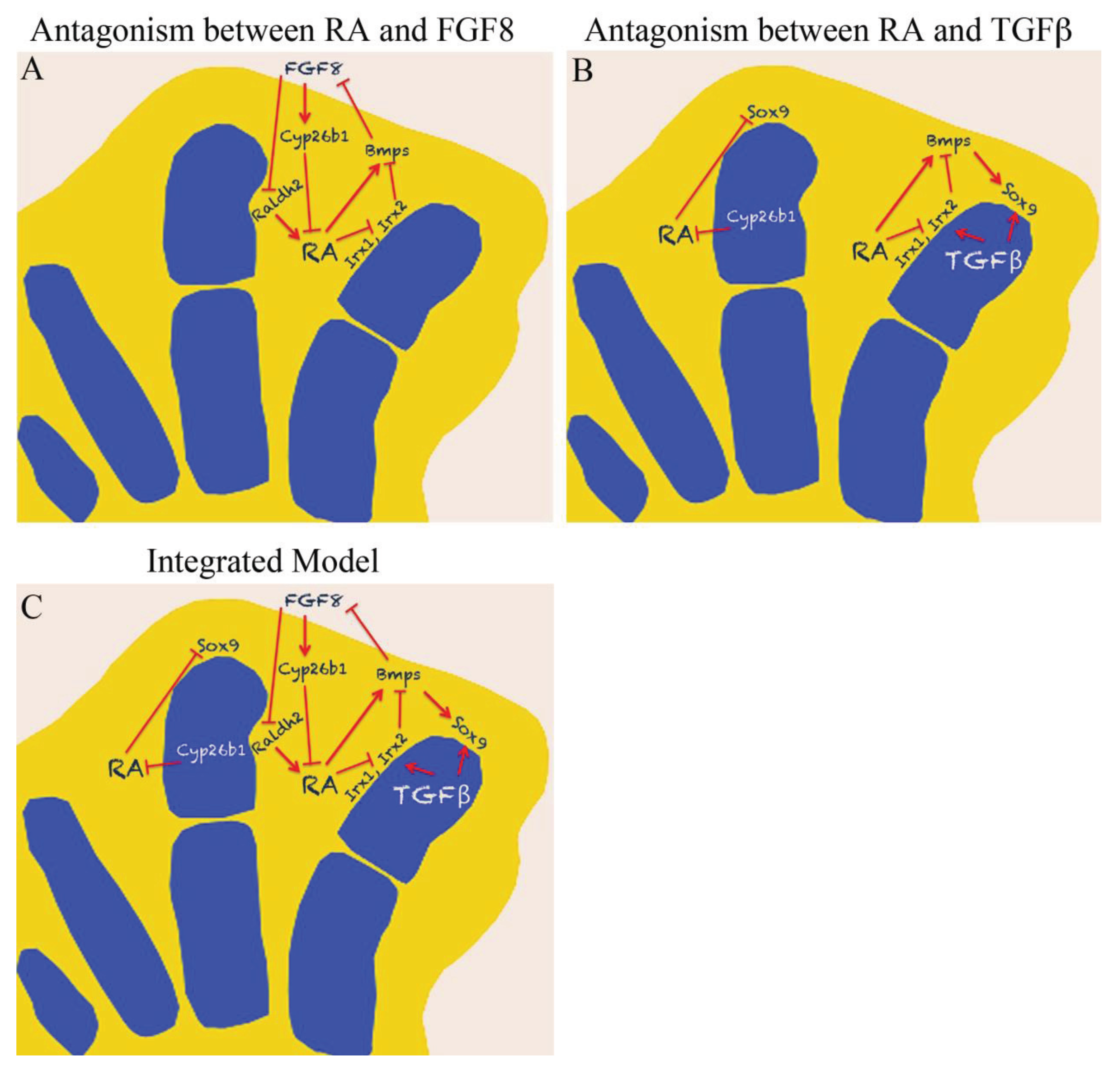
5. Antagonism between RA and FGF

Although RA is an important factor that potentially induces cell death, there are other factors that work in coordination with it to promote apoptosis. It was mentioned earlier that cells from the undifferentiated zone beneath the AER receive signals that direct them to differentiate or to die. The presence of the AER is important for controlling cell death, it releases the survival factor FGF8 that protects the mesenchyme from cell death [2,3,4,72]. It is known that the activity of BMP is necessary to inhibit Fgf8 expression in the AER and to promote cell death [73]. In contrast, activating FGF signaling in the interdigital tissue inhibits cell death, as occurs with the inactivation of BMP signaling (under these conditions expression of Fgf8 is not inhibited in the AER) [70,74,75]. Interestingly, cooperative functions between BMP and FGF signaling are observed when FGF signaling is inhibited in interdigital tissue and under these conditions BMPs cannot promote cell death [75]. Expression of BMPs depends on RA and expression of Bmp2, Bmp4, Bmp5 and Bmp7 is observed in interdigital tissue, whereas blocking of RA activity inhibits Bmp expression [68]. In contrast, FGF8 is able to antagonize RA signaling, since FGF8 inhibits expression of Raldh2 and induces Cy26b1 expression [76]. However, RA is also able to antagonize FGF signaling by inhibiting Fgfr1 expression [76]. Besides, as mentioned above, RA induces Bmp expression and BMP inhibits Fgf8 expression [68,73]. It has been suggested that down-regulation of Fgf8 in the AER could be the first event that triggers cell death [76].
Figure 4. RA inhibits Irx gene expression before the onset of cell death by a BMP-independent mechanism. (A–F) Irx1 expression is down‒regulated in the interdigital area at stage 27HH in chicken hind limbs after 8 hours of RA-treatment and RA/NOGGIN-double treatment. Note that the BMP antagonist NOGGIN does not inhibit the effect exerted by RA on Irx1 expression. (B,C) Active caspase 3 (green) is analyzed in the interdigital region of hind limbs after RA treatment for 8 h (B) and 12 h (C). (C) The first signs of cell death induced by RA were observed at 12 h. Active caspase 3 was inhibited after RA/NOGGIN-double treatment (E-F), but Irx1 gene expression remained unaffected (D), indicating that RA inhibits Irx expression by a BMP-independent mechanism. (G–I) RA inhibits Irx1 at the digit tip before the induction of cell death. Sox9 (G) and Irx1 (H) expression in hind limbs at stage 27HH after 4 h of treatment with RA, and the presence of active caspase 3 (I) at 12 h after the same treatment in the digit tip. Note that down-regulation of Sox9 and Irx1 expression begins before the presence of active caspase 3. In all in situ hybridizations, the experimental samples (A, D, G, H) are presented on the left, whereas controls are on the right. Black arrows in A and D indicate the area of Irx1 inhibition. Black arrows in G and H indicate the bead position. Autofluorescence of ionic-exchange beads used for RA-treatment (black asterisks) is observed as green color. The natural red color of Cy3, used to detect active caspase 3, was changed to green for better visualization of the images. (White asterisks indicate beads soaked in NOGGIN). Modified from [29].
Jdb 02 00138 g004
There is another group of genes regulated by RA during digit development: the Iroquois genes (Irx), which encode homeoproteins that belong to the TALE superclass of homeobox transcription factors with an Iroquois box that corresponds to an atypical homeodomain in the protein [71]. In vertebrates, their genomic organization is in two cognate clusters of three genes each; cluster “A” includes Irx1, Irx2, and Irx4 and cluster “B”, Irx3, Irx5, and Irx6 [71]. It has been observed that during limb development in chick embryos, Irx1 and Irx2 belonging to cluster “A” are coordinately expressed in the interdigital tissue and are down regulated by RA; remarkably by a BMP-independent mechanism. Irx expression is down-regulated before the observation of first signs of active caspase 3 (Figure 4) [29]. In this sense, it is possible to speculate that IRX1 and IRX2 might play a protective role against cell death. Thus, their down‒regulation by RA may be a prerequisite to promote the molecular cascade that ends in cell death. In other models, IRX proteins repress the expression of Bmp4 or Msx; two genes involved in cell death during limb development [71,77,78,79,80,81,82,83]. Accordingly, the mutant mouse fused toes (characterized by deletion of six genes, which include cluster B of the Irx genes) exhibits massive cell death concomitant with up‒regulation of Bmp4 and Dkk1, and down-regulation of Fgf8 and Fgf10 [71].

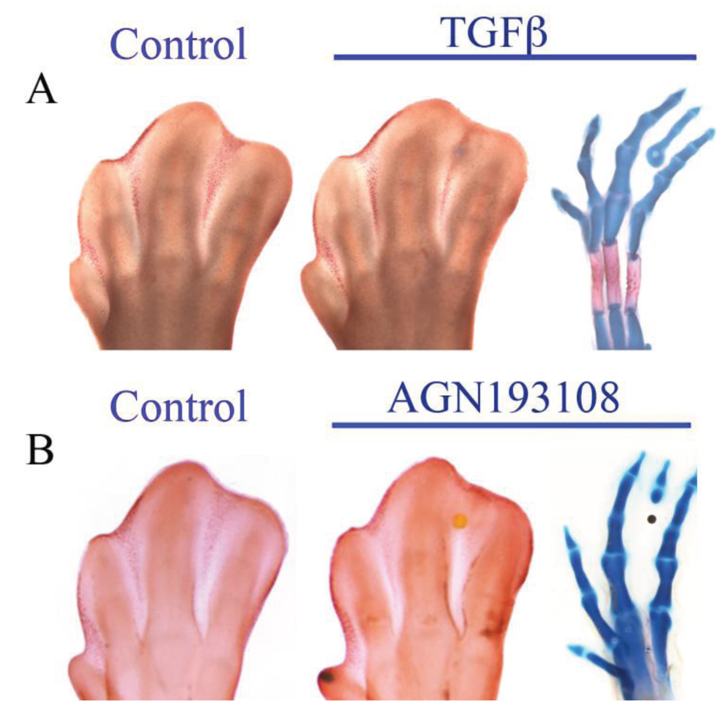
6. Antagonism between RA and TGFβ

On the basis that blocking the function of RA in interdigital tissue of chick embryos results in ectopic digit formation [27,68], it has been suggested that RA may promote cell death and inhibit cartilage differentiation (Figure 5) [21]. Sox9 is the master gene for chondrogenesis and is induced as early as 30 minutes after a stimulus of TGFβ/Activin signaling in interdigital tissue [23]. Subsequently, a molecular cascade begins that culminates in the formation of an ectopic digit and inhibition of cell death (Figure 5) [21,84]. During autopod formation, Sox9 expression is observed in presumptive digital rays and presumptive interdigital tissue [84]. In contrast, when Sox9 is deleted from undifferentiated mesenchymal cells of limb buds in a conditionally mutant mouse, the skeletal elements do not develop in the autopod, but instead massive cell death is observed in the regions in which digits would have been developed [85].
Interestingly, in the synpolydactyly mutant mouse model, Raldh2 is down-regulated, which results in the reduction of RA levels and duplication of one or more digits and the fusion of two or more digits [69]. The intrauterine treatment with RA of these animals rescues the normal phenotype [69]. On this basis it is possible to speculate that RA inhibits the recruitment of mesenchymal cells to the chondrogenic lineage, hence becoming cells responsive to the cell death program, whereas cells committed to chondrogenesis in digital rays that express Cyp26b1 become unresponsive to the cell death program, because CYP26b1 regulates active RA levels, promoting oxidation of RA to polar metabolites [63].
On the other hand, it is interesting to note that Irx1 and Irx2 are also expressed in the boundary of skeletal primordia and non-cartilage tissue. RA inhibits these genes, whereas TGFβ has an inductive effect [29]. RA treatment at the tips of digits induces cell death, but before the commencement of cell death, Irx1 and Irx2 expression is inhibited and Sox9 expression is down-regulated (Figure 4 and Figure 6) [29]. In conclusion, the expression of Irx genes in response to the antagonism between TGFβ and RA might delimit the boundaries of skeletal elements [29]. Recently, it was shown that RA might participate to define the digit–interdigit junction, where tissue remodeling occurs, by induction of Hmgn1 and Fgf18 expression at the digit–interdigit junction [63]. Interestingly, in another work, it was suggested that Irx expression in the boundary of skeletal primordia and non-cartilage tissue (digit-interdigit junction) may reflect the range of diffusion of TGFβ to promote chondrogenesis, and IRX proteins may repress the expression of genes involved in the cell death process, hence possibly allowing the formation of the prospective perichondrium [29].
Figure 5. Activation of transforming growth factor beta (TGFβ) signaling or inhibition of RAR inhibits interdigital cell death and induces the formation of an ectopic digit. (A) TGFβ or (B) pan-RAR antagonist (AGN193108) inhibits cell death and induces the formation of an ectopic digit after 4 days of treatment. The inhibition of RA signaling with a pan-RAR antagonist (AGN193108) gives rise to the same phenotype as treatment with TGFβ. For both treatments neutral red staining indicates interdigital cell death. The image of the ectopic digit after TGFβ treatment was taken from [29], doi:10.1371/journal.pone.0058549.g003.
Jdb 02 00138 g005

7. Concluding Remarks

The antagonism between FGF and RA signaling plays a crucial role in controlling interdigital cell death and outgrowth of the limb. Likewise, the antagonism between RA and TGFβ/Activin signaling in the embryonic chick limb controls cell differentiation during digit formation.
RA promotes cell death by inducing the expression of BMPs, which inhibit Fgf8 expression (Figure 6A). In addition, RA inhibits the expression of Irx1 and Irx2 genes in interdigital tissue. Interestingly, inhibition of Irx genes is not mediated by BMPs (Figure 6A). However, it is known that Irx genes inhibit the expression of Msx and Bmp4 genes during embryonic development of Drosophila and Xenopus [71,77,78,79,80,81,82,83], suggesting that IRX in the interdigital tissue might have a protective function against the role of BMPs on promoting cell death [29]. On the other hand, the mutant mouse fused toes (characterized by deletion of six genes, which include cluster B of the Irx genes) exhibits massive cell death simultaneously with up-regulation of Bmp4 and Dkk1, and down-regulation of Fgf8 and Fgf10 [83]. It is reasonable to speculate that RA is required to inhibit Irx genes to allow BMPs to carry out their effect in the molecular cascade that culminates in the onset of cell death. In other systems, the deletion of Irx genes gives rise to promotion of cell death [80,81]. Also, RA may promote the expression of the pro-apoptotic factor Bax, as well as tissue-transglutaminase and stromelysin 3 genes and inhibit BAG-1, a protein with the ability to bind to the anti-apoptotic protein BCL2 [63,76,86,87]. Overall it is possible to speculate that RA may orchestrate the expression of several genes whose proteic products are necessary to be activated or inhibited once the molecular cascade of cell death is activated (Figure 6A).
Figure 6. Schematic representation of antagonism between (A) RA and FGF8 and antagonism between (B) RA and TGFβ. (C) Represents the integrated model with both antagonistic interactions (see the text for details).
Jdb 02 00138 g006
Inhibition of Fgf8 depends on BMPs and, because RA induces expression of Bmp2, Bmp4, and Bmp7, it has been suggested that the antagonism between RA and FGF signaling may be responsible for distal interdigital cell death (Figure 6A) [76]. In mouse embryonic limbs, distal interdigital cell death depends on down-regulation of Fgf8 in the AER by action of RA [76]. Furthermore, FGF8 promotes cell survival probably by inducing the expression of Cyp26b1 in the undifferentiated mesenchyme underneath the AER and distal ectoderm and by inhibiting Raldh2 expression, thereby protecting interdigital tissue from the actions of RA in the induction of the interdigital cell death cascade (Figure 6A) [76].
Regarding chondrocyte differentiation, RA inhibits Sox9 and, in consequence, chondrogenesis [29,68], probably by preventing the cellular recruitment to the chondrocyte lineage (Figure 6B). Once inhibition of Sox9 expression occurs, Irx genes at the boundary of skeletal elements are inhibited and subsequently cell death occurs [29]. TGFβ promotes expression of Irx genes at the boundary of skeletal elements, and on this basis, it is possible to speculate that Irx gene expression in these regions protects cells committed to chondrocytic lineage against the signals that promote interdigital cell death (Figure 6B) [29]. Also, Cyp26b1 is expressed at developing digits and probably this enzyme protects cartilage tissue from the actions of RA (Figure 6B) [63].
In conclusion, RA is a molecule that interacts with other signaling pathways in an orchestrated manner to regulate the spatial-temporal expression of several genes involved in the activation of cell death or inhibition of cell differentiation (Figure 6C).

Acknowledgments

This study was partially supported by CONACyT grants 53484 and 168642, DGAPA-UNAM grants IN214511, IN220808 and IN213314. Martha Elena Díaz-Hernández and Alberto Jesús Ríos Flores were recipients of a scholarship from CONACYT and students in the Ph.D. program: Programa de Doctorado en Ciencias Biomédicas, UNAM. The authors thank Karen Camargo Sosa for the experiments of Figure 5 and Claudio Iván Galván-Hernández for making the graphic representation in Figure 6.

Author Contributions

MED-H, RFA-B and JC-M have written about digit development and programmed cell death in the limb mesoderm. MED-H, AJR-F, MB and JC-M have written about retinoic acid signaling and antagonisms between RA and FGF and between RA and TGFβ. All authors participated in the design of figures and tables.

Conflicts of Interest

The authors declare no conflict of interest.

References and Notes

  1. Tabin, C.; Wolpert, L. Rethinking the proximodistal axis of the vertebrate limb in the molecular era. Genes Dev. 2007, 21, 1433–1442. [Google Scholar] [CrossRef]
  2. Fernandez-Teran, M.; Ros, M.A. The apical ectodermal ridge: Morphological aspects and signaling pathways. Int. J. Dev. Biol. 2008, 52, 857–871. [Google Scholar] [CrossRef] [Green Version]
  3. ten Berge, D.; Brugmann, S.A.; Helms, J.A.; Nusse, R. Wnt and FGF signals interact to coordinate growth with cell fate specification during limb development. Development 2008, 135, 3247–3257. [Google Scholar] [CrossRef]
  4. Mariani, F.V.; Ahn, C.P.; Martin, G.R. Genetic evidence that FGFs have an instructive role in limb proximal-distal patterning. Nature 2008, 453, 401–405. [Google Scholar] [CrossRef]
  5. Montero, J.A.; Hurle, J.M. Deconstructing digit chondrogenesis. BioEssays 2007, 29, 725–737. [Google Scholar] [CrossRef]
  6. Montero, J.A.; Hurle, J.M. Sculpturing digit shape by cell death. Apoptosis 2010, 15, 365–375. [Google Scholar] [CrossRef]
  7. Montero, J.A.; Lorda-Diez, C.I.; Ganan, Y.; Macias, D.; Hurle, J.M. Activin/tgfbeta and bmp crosstalk determines digit chondrogenesis. Dev. Biol. 2008, 321, 343–356. [Google Scholar] [CrossRef]
  8. Towers, M.; Wolpert, L.; Tickle, C. Gradients of signalling in the developing limb. Curr. Opin. Cell Biol. 2012, 24, 181–187. [Google Scholar] [CrossRef]
  9. Towers, M.; Tickle, C. Growing models of vertebrate limb development. Development 2009, 136, 179–190. [Google Scholar] [CrossRef]
  10. Tickle, C. Making digit patterns in the vertebrate limb. Nat. Rev. Mol. Cell Biol. 2006, 7, 45–53. [Google Scholar] [CrossRef]
  11. Charite, J.; de Graaff, W.; Shen, S.; Deschamps, J. Ectopic expression of hoxb-8 causes duplication of the zpa in the forelimb and homeotic transformation of axial structures. Cell 1994, 78, 589–601. [Google Scholar] [CrossRef]
  12. Charite, J.; McFadden, D.G.; Olson, E.N. The bhlh transcription factor dhand controls sonic hedgehog expression and establishment of the zone of polarizing activity during limb development. Development 2000, 127, 2461–2470. [Google Scholar]
  13. Noji, S.; Nohno, T.; Koyama, E.; Muto, K.; Ohyama, K.; Aoki, Y.; Tamura, K.; Ohsugi, K.; Ide, H.; Taniguchi, S.; et al. Retinoic acid induces polarizing activity but is unlikely to be a morphogen in the chick limb bud. Nature 1991, 350, 83–86. [Google Scholar] [CrossRef]
  14. Wanek, N.; Gardiner, D.M.; Muneoka, K.; Bryant, S.V. Conversion by retinoic acid of anterior cells into zpa cells in the chick wing bud. Nature 1991, 350, 81–83. [Google Scholar] [CrossRef]
  15. Riddle, R.D.; Johnson, R.L.; Laufer, E.; Tabin, C. Sonic hedgehog mediates the polarizing activity of the zpa. Cell 1993, 75, 1401–1416. [Google Scholar] [CrossRef]
  16. Wang, B.; Fallon, J.F.; Beachy, P.A. Hedgehog-regulated processing of gli3 produces an anterior/posterior repressor gradient in the developing vertebrate limb. Cell 2000, 100, 423–434. [Google Scholar] [CrossRef]
  17. Litingtung, Y.; Dahn, R.D.; Li, Y.; Fallon, J.F.; Chiang, C. Shh and gli3 are dispensable for limb skeleton formation but regulate digit number and identity. Nature 2002, 418, 979–983. [Google Scholar] [CrossRef]
  18. te Welscher, P.; Zuniga, A.; Kuijper, S.; Drenth, T.; Goedemans, H.J.; Meijlink, F.; Zeller, R. Progression of vertebrate limb development through shh-mediated counteraction of gli3. Science 2002, 298, 827–830. [Google Scholar] [CrossRef]
  19. Suzuki, T.; Hasso, S.M.; Fallon, J.F. Unique smad1/5/8 activity at the phalanx-forming region determines digit identity. Proc. Natl. Acad. Sci. USA 2008, 105, 4185–4190. [Google Scholar] [CrossRef]
  20. Merino, R.; Ganan, Y.; Macias, D.; Economides, A.N.; Sampath, K.T.; Hurle, J.M. Morphogenesis of digits in the avian limb is controlled by fgfs, tgfbetas, and noggin through bmp signaling. Dev. Biol. 1998, 200, 35–45. [Google Scholar] [CrossRef]
  21. Ganan, Y.; Macias, D.; Duterque-Coquillaud, M.; Ros, M.A.; Hurle, J.M. Role of tgf beta s and bmps as signals controlling the position of the digits and the areas of interdigital cell death in the developing chick limb autopod. Development 1996, 122, 2349–2357. [Google Scholar]
  22. Merino, R.; Macias, D.; Ganan, Y.; Rodriguez-Leon, J.; Economides, A.N.; Rodriguez-Esteban, C.; Izpisua-Belmonte, J.C.; Hurle, J.M. Control of digit formation by activin signalling. Development 1999, 126, 2161–2170. [Google Scholar]
  23. Chimal-Monroy, J.; Rodriguez-Leon, J.; Montero, J.A.; Ganan, Y.; Macias, D.; Merino, R.; Hurle, J.M. Analysis of the molecular cascade responsible for mesodermal limb chondrogenesis: Sox genes and bmp signaling. Dev. Biol. 2003, 257, 292–301. [Google Scholar] [CrossRef]
  24. Hernandez-Martinez, R.; Covarrubias, L. Interdigital cell death function and regulation: New insights on an old programmed cell death model. Dev. Growth Differ. 2011, 53, 245–258. [Google Scholar] [CrossRef]
  25. Zuzarte-Luis, V.; Hurle, J.M. Programmed cell death in the embryonic vertebrate limb. Semin. Cell Dev. Biol. 2005, 16, 261–269. [Google Scholar] [CrossRef]
  26. Dahn, R.D.; Fallon, J.F. Interdigital regulation of digit identity and homeotic transformation by modulated bmp signaling. Science 2000, 289, 438–441. [Google Scholar] [CrossRef]
  27. Ros, M.A.; Piedra, M.E.; Fallon, J.F.; Hurle, J.M. Morphogenetic potential of the chick leg interdigital mesoderm when diverted from the cell death program. Dev. Dynam. 1997, 208, 406–419. [Google Scholar] [CrossRef]
  28. Fernandez-Teran, M.A.; Hinchliffe, J.R.; Ros, M.A. Birth and death of cells in limb development: A mapping study. Dev. Dynam. 2006, 235, 2521–2537. [Google Scholar] [CrossRef]
  29. Diaz-Hernandez, M.E.; Bustamante, M.; Galvan-Hernandez, C.I.; Chimal-Monroy, J. Irx1 and irx2 are coordinately expressed and regulated by retinoic acid, tgfbeta and fgf signaling during chick hindlimb development. PLoS One 2013, 8. [Google Scholar] [CrossRef]
  30. Abarca-Buis, R.F.; Bustamante, M.; Cuervo, R.; Aguilar-Fernandez-de-Lara, D.; Chimal-Monroy, J. Smad8 is expressed in the anterior necrotic zone: Evidence for a role of bone morphogenetic proteins/smad signaling in the activation of a molecular cascade that culminates in cell death. Dev. Growth Differ. 2011, 53, 780–792. [Google Scholar]
  31. Orrenius, S.; Nicotera, P.; Zhivotovsky, B. Cell death mechanisms and their implications in toxicology. Toxicol. Sci. 2011, 119, 3–19. [Google Scholar] [CrossRef]
  32. Ren, D.; Tu, H.C.; Kim, H.; Wang, G.X.; Bean, G.R.; Takeuchi, O.; Jeffers, J.R.; Zambetti, G.P.; Hsieh, J.J.; Cheng, E.H. Bid, bim, and puma are essential for activation of the bax- and bak-dependent cell death program. Science 2010, 330, 1390–1393. [Google Scholar] [CrossRef]
  33. Mace, P.D.; Shirley, S.; Day, C.L. Assembling the building blocks: Structure and function of inhibitor of apoptosis proteins. Cell Death Differ. 2010, 17, 46–53. [Google Scholar] [CrossRef]
  34. Hui, K.K.; Kanungo, A.K.; Elia, A.J.; Henderson, J.T. Caspase-3 deficiency reveals a physiologic role for smac/diablo in regulating programmed cell death. Cell Death Differ. 2011, 18, 1780–1790. [Google Scholar]
  35. Repnik, U.; Stoka, V.; Turk, V.; Turk, B. Lysosomes and lysosomal cathepsins in cell death. Biochim. Biophys. Acta 2012, 1824, 22–33. [Google Scholar]
  36. Lindsten, T.; Ross, A.J.; King, A.; Zong, W.X.; Rathmell, J.C.; Shiels, H.A.; Ulrich, E.; Waymire, K.G.; Mahar, P.; Frauwirth, K.; et al. The combined functions of proapoptotic bcl-2 family members bak and bax are essential for normal development of multiple tissues. Mol. Cell 2000, 6, 1389–1399. [Google Scholar] [CrossRef]
  37. Yoshida, H.; Kong, Y.Y.; Yoshida, R.; Elia, A.J.; Hakem, A.; Hakem, R.; Penninger, J.M.; Mak, T.W. Apaf1 is required for mitochondrial pathways of apoptosis and brain development. Cell 1998, 94, 739–750. [Google Scholar] [CrossRef]
  38. Cecconi, F.; Alvarez-Bolado, G.; Meyer, B.I.; Roth, K.A.; Gruss, P. Apaf1 (ced-4 homolog) regulates programmed cell death in mammalian development. Cell 1998, 94, 727–737. [Google Scholar] [CrossRef]
  39. Chautan, M.; Chazal, G.; Cecconi, F.; Gruss, P.; Golstein, P. Interdigital cell death can occur through a necrotic and caspase-independent pathway. Curr. Biol. 1999, 9, 967–970. [Google Scholar] [CrossRef]
  40. Zuzarte-Luis, V.; Berciano, M.T.; Lafarga, M.; Hurle, J.M. Caspase redundancy and release of mitochondrial apoptotic factors characterize interdigital apoptosis. Apoptosis 2006, 11, 701–715. [Google Scholar] [CrossRef]
  41. Huang, C.; Hales, B.F. Role of caspases in murine limb bud cell death induced by 4-hydroperoxycyclophosphamide, an activated analog of cyclophosphamide. Teratology 2002, 66, 288–299. [Google Scholar] [CrossRef]
  42. Milligan, C.E.; Prevette, D.; Yaginuma, H.; Homma, S.; Cardwell, C.; Fritz, L.C.; Tomaselli, K.J.; Oppenheim, R.W.; Schwartz, L.M. Peptide inhibitors of the ice protease family arrest programmed cell death of motoneurons in vivo and in vitro. Neuron 1995, 15, 385–393. [Google Scholar] [CrossRef]
  43. Zuzarte-Luis, V.; Montero, J.A.; Torre-Perez, N.; Garcia-Porrero, J.A.; Hurle, J.M. Cathepsin d gene expression outlines the areas of physiological cell death during embryonic development. Dev. Dynam. 2007, 236, 880–885. [Google Scholar] [CrossRef]
  44. Zuzarte-Luis, V.; Montero, J.A.; Kawakami, Y.; Izpisua-Belmonte, J.C.; Hurle, J.M. Lysosomal cathepsins in embryonic programmed cell death. Dev. Biol. 2007, 301, 205–217. [Google Scholar] [CrossRef]
  45. Montero, J.A.; Lorda-Diez, C.I.; Certal, A.C.; Moreno, N.; Rodriguez-Leon, J.; Torriglia, A.; Hurle, J.M. Coordinated and sequential activation of neutral and acidic dnases during interdigital cell death in the embryonic limb. Apoptosis 2010, 15, 1197–1210. [Google Scholar] [CrossRef]
  46. McCulloch, D.R.; Nelson, C.M.; Dixon, L.J.; Silver, D.L.; Wylie, J.D.; Lindner, V.; Sasaki, T.; Cooley, M.A.; Argraves, W.S.; Apte, S.S. Adamts metalloproteases generate active versican fragments that regulate interdigital web regression. Dev. Cell 2009, 17, 687–698. [Google Scholar] [CrossRef]
  47. Arteaga-Solis, E.; Gayraud, B.; Lee, S.Y.; Shum, L.; Sakai, L.; Ramirez, F. Regulation of limb patterning by extracellular microfibrils. J. Cell Biol. 2001, 154, 275–281. [Google Scholar] [CrossRef]
  48. Bose, K.; Nischt, R.; Page, A.; Bader, B.L.; Paulsson, M.; Smyth, N. Loss of nidogen-1 and -2 results in syndactyly and changes in limb development. J. Biol. Chem. 2006, 281, 39620–39629. [Google Scholar] [CrossRef]
  49. Diaz-Mendoza, M.J.; Lorda-Diez, C.I.; Montero, J.A.; Garcia-Porrero, J.A.; Hurle, J.M. Interdigital cell death in the embryonic limb is associated with depletion of reelin in the extracellular matrix. Cell Death Dis. 2013, 4. [Google Scholar] [CrossRef]
  50. Debeer, P.; Schoenmakers, E.F.; Twal, W.O.; Argraves, W.S.; De Smet, L.; Fryns, J.P.; Van De Ven, W.J. The fibulin-1 gene (fbln1) is disrupted in a t(12;22) associated with a complex type of synpolydactyly. J. Med. Genet. 2002, 39, 98–104. [Google Scholar] [CrossRef]
  51. Ohkubo, N.; Vitek, M.P.; Morishima, A.; Suzuki, Y.; Miki, T.; Maeda, N.; Mitsuda, N. Reelin signals survival through src-family kinases that inactivate bad activity. J. Neurochem. 2007, 103, 820–830. [Google Scholar] [CrossRef]
  52. Clagett-Dame, M.; Knutson, D. Vitamin A in reproduction and development. Nutrients 2011, 3, 385–428. [Google Scholar] [CrossRef]
  53. Rhinn, M.; Dolle, P. Retinoic acid signalling during development. Development 2012, 139, 843–858. [Google Scholar] [CrossRef]
  54. Farjo, K.M.; Moiseyev, G.; Nikolaeva, O.; Sandell, L.L.; Trainor, P.A.; Ma, J.X. Rdh10 is the primary enzyme responsible for the first step of embryonic vitamin a metabolism and retinoic acid synthesis. Dev. Biol. 2011, 357, 347–355. [Google Scholar] [CrossRef]
  55. Niederreither, K.; Dolle, P. Retinoic acid in development: Towards an integrated view. Nat. Rev. Genet. 2008, 9, 541–553. [Google Scholar] [CrossRef]
  56. Fujii, H.; Sato, T.; Kaneko, S.; Gotoh, O.; Fujii-Kuriyama, Y.; Osawa, K.; Kato, S.; Hamada, H. Metabolic inactivation of retinoic acid by a novel p450 differentially expressed in developing mouse embryos. EMBO J. 1997, 16, 4163–4173. [Google Scholar] [CrossRef]
  57. Haque, M.; Anreola, F. The cloning and characterization of a novel cytochrome p450 family, cyp26, with specificity toward retinoic acid. Nutr. Rev. 1998, 56, 84–85. [Google Scholar] [CrossRef]
  58. Niederreither, K.; Vermot, J.; Schuhbaur, B.; Chambon, P.; Dolle, P. Embryonic retinoic acid synthesis is required for forelimb growth and anteroposterior patterning in the mouse. Development 2002, 129, 3563–3574. [Google Scholar]
  59. Cunningham, T.J.; Chatzi, C.; Sandell, L.L.; Trainor, P.A.; Duester, G. Rdh10 mutants deficient in limb field retinoic acid signaling exhibit normal limb patterning but display interdigital webbing. Dev. Dynam. 2011, 240, 1142–1150. [Google Scholar] [CrossRef]
  60. Sandell, L.L.; Sanderson, B.W.; Moiseyev, G.; Johnson, T.; Mushegian, A.; Young, K.; Rey, J.P.; Ma, J.X.; Staehling-Hampton, K.; Trainor, P.A. Rdh10 is essential for synthesis of embryonic retinoic acid and is required for limb, craniofacial, and organ development. Genes Dev. 2007, 21, 1113–1124. [Google Scholar] [CrossRef]
  61. Ahuja, H.S.; James, W.; Zakeri, Z. Rescue of the limb deformity in hammertoe mutant mice by retinoic acid-induced cell death. Dev. Dynam. 1997, 208, 466–481. [Google Scholar] [CrossRef]
  62. Zakeri, Z.; Quaglino, D.; Ahuja, H.S. Apoptotic cell death in the mouse limb and its suppression in the hammertoe mutant. Dev. Biol. 1994, 165, 294–297. [Google Scholar] [CrossRef]
  63. Zhao, X.; Brade, T.; Cunningham, T.J.; Duester, G. Retinoic acid controls expression of tissue remodeling genes hmgn1 and fgf18 at the digit-interdigit junction. Dev. Dynam. 2010, 239, 665–671. [Google Scholar] [CrossRef]
  64. Duester, G. Retinoic acid synthesis and signaling during early organogenesis. Cell 2008, 134, 921–931. [Google Scholar] [CrossRef]
  65. Campo-Paysaa, F.; Marletaz, F.; Laudet, V.; Schubert, M. Retinoic acid signaling in development: Tissue-specific functions and evolutionary origins. Genesis 2008, 46, 640–656. [Google Scholar] [CrossRef]
  66. Kastner, P.; Mark, M.; Ghyselinck, N.; Krezel, W.; Dupe, V.; Grondona, J.M.; Chambon, P. Genetic evidence that the retinoid signal is transduced by heterodimeric rxr/rar functional units during mouse development. Development 1997, 124, 313–326. [Google Scholar]
  67. Lohnes, D.; Mark, M.; Mendelsohn, C.; Dolle, P.; Dierich, A.; Gorry, P.; Gansmuller, A.; Chambon, P. Function of the retinoic acid receptors (rars) during development (I). Craniofacial and skeletal abnormalities in rar double mutants. Development 1994, 120, 2723–2748. [Google Scholar]
  68. Rodriguez-Leon, J.; Merino, R.; Macias, D.; Ganan, Y.; Santesteban, E.; Hurle, J.M. Retinoic acid regulates programmed cell death through bmp signalling. Nat. Cell Biol. 1999, 1, 125–126. [Google Scholar] [CrossRef]
  69. Kuss, P.; Villavicencio-Lorini, P.; Witte, F.; Klose, J.; Albrecht, A.N.; Seemann, P.; Hecht, J.; Mundlos, S. Mutant hoxd13 induces extra digits in a mouse model of synpolydactyly directly and by decreasing retinoic acid synthesis. J. Clin. Investig. 2009, 119, 146–156. [Google Scholar]
  70. Merino, R.; Rodriguez-Leon, J.; Macias, D.; Ganan, Y.; Economides, A.N.; Hurle, J.M. The bmp antagonist gremlin regulates outgrowth, chondrogenesis and programmed cell death in the developing limb. Development 1999, 126, 5515–5522. [Google Scholar]
  71. Grotewold, L.; Ruther, U. The fused toes (ft) mouse mutation causes anteroposterior and dorsoventral polydactyly. Dev. Biol. 2002, 251, 129–141. [Google Scholar] [CrossRef]
  72. Sun, X.; Mariani, F.V.; Martin, G.R. Functions of fgf signalling from the apical ectodermal ridge in limb development. Nature 2002, 418, 501–508. [Google Scholar] [CrossRef]
  73. Pajni-Underwood, S.; Wilson, C.P.; Elder, C.; Mishina, Y.; Lewandoski, M. Bmp signals control limb bud interdigital programmed cell death by regulating fgf signaling. Development 2007, 134, 2359–2368. [Google Scholar] [CrossRef]
  74. Merino, R.; Ganan, Y.; Macias, D.; Rodriguez-Leon, J.; Hurle, J.M. Bone morphogenetic proteins regulate interdigital cell death in the avian embryo. Ann. N. Y. Acad. Sci. 1999, 887, 120–132. [Google Scholar]
  75. Montero, J.A.; Ganan, Y.; Macias, D.; Rodriguez-Leon, J.; Sanz-Ezquerro, J.J.; Merino, R.; Chimal-Monroy, J.; Nieto, M.A.; Hurle, J.M. Role of fgfs in the control of programmed cell death during limb development. Development 2001, 128, 2075–2084. [Google Scholar]
  76. Hernandez-Martinez, R.; Castro-Obregon, S.; Covarrubias, L. Progressive interdigital cell death: Regulation by the antagonistic interaction between fibroblast growth factor 8 and retinoic acid. Development 2009, 136, 3669–3678. [Google Scholar] [CrossRef]
  77. Gomez-Skarmeta, J.L.; Modolell, J. Iroquois genes: Genomic organization and function in vertebrate neural development. Curr. Opin. Genet. Dev. 2002, 12, 403–408. [Google Scholar] [CrossRef]
  78. Villa-Cuesta, E.; Modolell, J. Mutual repression between msh and iro-c is an essential component of the boundary between body wall and wing in drosophila. Development 2005, 132, 4087–4096. [Google Scholar] [CrossRef]
  79. Gomez-Skarmeta, J.; de La Calle-Mustienes, E.; Modolell, J. The wnt-activated xiro1 gene encodes a repressor that is essential for neural development and downregulates bmp4. Development 2001, 128, 551–560. [Google Scholar]
  80. Glavic, A.; Gomez-Skarmeta, J.L.; Mayor, R. Xiro-1 controls mesoderm patterning by repressing bmp-4 expression in the spemann organizer. Dev. Dynam. 2001, 222, 368–376. [Google Scholar] [CrossRef]
  81. Feijoo, C.G.; Saldias, M.P.; De la Paz, J.F.; Gomez-Skarmeta, J.L.; Allende, M.L. Formation of posterior cranial placode derivatives requires the iroquois transcription factor irx4a. Mol. Cell. Neurosci. 2009, 40, 328–337. [Google Scholar] [CrossRef]
  82. Chuang, H.N.; Cheng, H.Y.; Hsiao, K.M.; Lin, C.W.; Lin, M.L.; Pan, H. The zebrafish homeobox gene irxl1 is required for brain and pharyngeal arch morphogenesis. Dev. Dynam. 2010, 239, 639–650. [Google Scholar] [CrossRef]
  83. van Tuyl, M.; Liu, J.; Groenman, F.; Ridsdale, R.; Han, R.N.; Venkatesh, V.; Tibboel, D.; Post, M. Iroquois genes influence proximo-distal morphogenesis during rat lung development. Am. J. Physiol. Lung Cell. Mol. Physiol. 2006, 290, L777–L789. [Google Scholar] [CrossRef]
  84. Chimal-Monroy, J.; Abarca-Buis, R.F.; Cuervo, R.; Diaz-Hernandez, M.; Bustamante, M.; Rios-Flores, J.A.; Romero-Suarez, S.; Farrera-Hernandez, A. Molecular control of cell differentiation and programmed cell death during digit development. IUBMB Life 2011, 63, 922–929. [Google Scholar] [CrossRef]
  85. Akiyama, H.; Chaboissier, M.C.; Martin, J.F.; Schedl, A.; de Crombrugghe, B. The transcription factor sox9 has essential roles in successive steps of the chondrocyte differentiation pathway and is required for expression of sox5 and sox6. Genes Dev. 2002, 16, 2813–2828. [Google Scholar] [CrossRef]
  86. Dupe, V.; Ghyselinck, N.B.; Thomazy, V.; Nagy, L.; Davies, P.J.; Chambon, P.; Mark, M. Essential roles of retinoic acid signaling in interdigital apoptosis and control of bmp-7 expression in mouse autopods. Dev. Biol. 1999, 208, 30–43. [Google Scholar] [CrossRef]
  87. Crocoll, A.; Herzer, U.; Ghyselinck, N.B.; Chambon, P.; Cato, A.C. Interdigital apoptosis and downregulation of bag-1 expression in mouse autopods. Mech. Dev. 2002, 111, 149–152. [Google Scholar] [CrossRef]

