Article

Ca2+-Transport through Plasma Membrane as a Test of Auxin Sensitivity

by
Anastasia A. Kirpichnikova
1,†,
Elena L. Rudashevskaya
1,†,‡,
Vladislav V. Yemelyanov
1,2 and
Maria F. Shishova
1,*
1
Department of Plant Physiology and Biochemistry, St. Petersburg State University, Universitetskaya emb. 7/9, St. Petersburg 199034, Russia
2
Department of Genetics and Biotechnology, St. Petersburg State University, Universitetskaya emb. 7/9, St. Petersburg 199034, Russia
*
Author to whom correspondence should be addressed.
These authors contributed equally to this work.
Current address: Institute of Medical Chemistry, Medical University of Vienna; Waehringer strasse 10, Vienna 1090, Austria
Plants 2014, 3(2), 209-222; https://doi.org/10.3390/plants3020209
Submission received: 5 November 2013 / Revised: 9 March 2014 / Accepted: 13 March 2014 / Published: 26 March 2014
(This article belongs to the Special Issue Calcium Signaling in Plants)

Abstract

:
Auxin is one of the crucial regulators of plant growth and development. The discovered auxin cytosolic receptor (TIR1) is not involved in the perception of the hormone signal at the plasma membrane. Instead, another receptor, related to the ABP1, auxin binding protein1, is supposed to be responsible for the perception at the plasma membrane. One of the fast and sensitive auxin-induced reactions is an increase of Ca2+ cytosolic concentration, which is suggested to be dependent on the activation of Ca2+ influx through the plasma membrane. This investigation was carried out with a plasmalemma enriched vesicle fraction, obtained from etiolated maize coleoptiles. The magnitude of Ca2+ efflux through the membrane vesicles was estimated according to the shift of potential dependent fluorescent dye diS-C3-(5). The obtained results showed that during coleoptiles ageing (3rd, 4th and 5th days of seedling etiolated growth) the magnitude of Ca2+ efflux from inside-out vesicles was decreased. Addition of ABP1 led to a recovery of Ca2+ efflux to the level of the youngest and most sensitive cells. Moreover, the efflux was more sensitive, responding from 10−8 to 10−6 M 1-NAA, in vesicles containing ABP1, whereas native vesicles showed the highest efflux at 10−6 M 1-NAA. We suggest that auxin increases plasma membrane permeability to Ca2+ and that ABP1 is involved in modulation of this reaction.

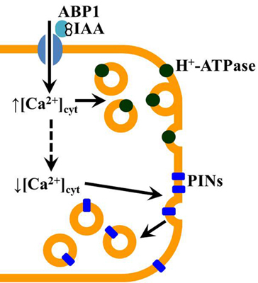
Graphical Abstract

1. Introduction

One of the most frequent questions, which are faced by investigators of auxin effects is: How can this simple molecule trigger such a huge diversity of physiological reactions? During the last decades, an enormous amount of data on auxin signaling has accumulated. A big step in understanding the auxin mechanism of action was made after elucidation of the soluble hormone-receptor TIR1 and the closely related AFB proteins [1,2]. Nevertheless, not all auxin-induced reactions are mediated by activation of TIR1-signaling. A number of primary reactions, e.g., changes in membrane potential, cytosol acidification, elevation in cytosolic Ca2+ concentration, activation of PLA2, protoplast swelling, protein cycling, etc., require another type of receptor, localized in the plasma membrane [3,4,5,6,7]. Nevertheless, the process of auxin sensing at cell surface is still under investigation. The auxin binding protein 1 (ABP1) is considered to be a part of plasma membrane (PM) receptor or closely linked to it [7,8,9,10,11]. The ABP1 appears to be a dimer of 22-kDa subunit, and can easily be solubilized by detergent or acetone from membranes [12,13,14]. A number of ABP1 genes are known to encode the protein in different plants [15,16,17,18,19]. The ABP1 protein has a single N-glycosylation site, which binds a mannose type glycan [14,20,21]. Two conservative domains (Box A, responsible for auxin binding, and Box B) and an ER targeting marker C-terminal KDEL tetrapeptide were determined in the ABP1 structure [21,22,23]. The main ABP1 pool is localized in ER where it is supposed to be inactive. Only about 2% of the protein is secreted to extracellular space to fulfill its physiological function [24].
It was supposed that ABP1 might be coupled to a transmembrane docking protein [25,26]. Unfortunately, up to now little is known about the nature and function of this protein. Based on the knowledge of different animal hormone-receptor complexes at the PM, several models were developed for the auxin receptor. The first one suggested that a kinase could fulfill the role of a transmembrane domain [27]. Another model implicates G-protein-coupled receptor as the auxin one [28]. The third model assumes that ABP1 binds/interacts with a Ca2+-permeable ion channel, which fulfills the role of a docking protein [29]. It is possible that ABP1 has more than one binding partner, differing in plant tissues and stage of development.
The absence of ABP1 or its reduction leads to significant changes: arrest of embryo development and elongation intensity [30,31]. Even heterozygous ABP1/abp1 insertion mutants show a number of developmental disturbances confirmed by reduction of sensitivity to auxin and shift in the intensity of early auxin-regulated Aux/IAAs genes expression [32,33]. Decrease in ABP1 via antisense transformation leads to significant decrease in elongation intensity [31] and cell enlargement/protoplast swelling [34,35,36].
It was shown earlier that addition of exogenous ABP1 to a model system like protoplasts increased the amplitude of auxin-induced PM hyperpolarization [37]. Recently, a fast ABP1-related auxin-induced shift in the membrane potential (MP) was shown in a similar model system, by use of a sensitive fluorescent dye [38]. The advantage of the latter investigation was the ascertainment that the effect was triggered even by the C-terminal peptide of ABP1 and was blocked by antibodies against it. Overexpression of ABP1 enhances the K+-transport by activation of K+-channels and quantity of their expression [39,40]. Thus, it could be concluded that ABP1 is an important modulator of cell sensitivity to the hormone at plasma membrane, but the mechanism of this regulation is still debated.
One of the fast and sensitive reactions triggered by auxin is an elevation of Ca2+ concentration in the cytosol. This reaction was estimated for different plant cells, including maize coleoptile parenchyma cells [9,41] Most probably it reflects the activation of plasma membrane channels, permeable for Ca2+ [9]. The coleoptile is a juvenile organ, the main function of which is to protect the first leaf at the initial stages of grass seedling development. Coleoptiles are very sensitive to auxin [42]. In maize coleoptiles, the native growth slows down tremendously from the 3rd to 5th day of seedling development [43]. The most intensive growth decrement appears at transition from the 3rd to 4th day of seedling development [44]. This phenomenon coincides with a loss of auxin-induced growth of coleoptile segments [43] and a significant decrease of auxin induced [Ca2+]cyt elevation [44]. Thus, a possible reduction in cell sensitivity to the hormone is due to probable changes in auxin signal perception and early transduction. The current investigation focuses on the involvement of a plasma membrane Ca2+-transport system in auxin signal perception under the control of ABP1.

2. Results and Discussion

The intensity of Ca2+ transport through vesicle membranes, obtained from maize coleoptiles of different ages was estimated as ΔMP, determined by a shift in fluorescence of diS-C3-(5) dye, commonly used to test transmembrane potential not only in purified vesicles, but also at whole cell level, like protoplast or bacterial cell [45,46].
Our model system contained two types of vesicles: right-side-out, which copy the native cell orientation, and inside-out ones. Only Ca2+ ions had a gradient across the vesicle membrane (Figure 1a). Addition of IAA into the incubation medium led to a fast shift of dye fluorescence (Figure 1b), similar to our earlier results [47]. The detected shift in MP was due to Ca2+ efflux from the vesicles. We assume that right-side-out vesicles do not participate in ΔMP generation because transport of Ca2+ out of the cell is carried out by active systems like Ca2+-ATPase and by the Ca2+/proton antiporter systems (for review see [48]). Conditions for activation of these transporters were absent; therefore, the estimated ΔMP was due to flux of Ca2+ ions across membranes of inverted vesicles, which correspond to the flow directed into cell in vivo. The revealed IAA-induced shift in MP was similar to the effect obtained after addition of 1-NAA, an active synthetic auxin, but not after 2-NAA addition, a non-active synthetic analogue (Figure 1b).
Figure 1. Auxin-induced generation of the membrane potential in a model system represented by plasma membrane vesicles from maize coleoptile cells isolated at the 3rd day of seedling development. (a) Scheme of vesicles loading (inside-out orientation of vesicle), cyt—cytosolic side of vesicle, arrow—direction of Ca2+ flux; (b) Single traces of diS-C3-(5) fluorescence shift after auxin addition, arrow—addition of auxin.
Plants 03 00209 g001
The fluorescence intensity of diS-C3-(5) is calibrated mainly in a model system with gradients of K+ and Na+ ions. The maximal amplitude of dye fluorescence in our model system decreases to −110 mV of the K+-diffusion potential as calculated by the Nernst equation when we added valinomycin into the Na+ incubation medium containing K+-loaded vesicles [47]. According to earlier results the IAA-induced change in dye fluorescence did not exceed −30 to 33 mV in case of membrane vesicles purified from coleoptiles at 4th day of seedling development. In the absence of Na+, detected changes of the amplitude of the auxin-induced fluorescence signal are presented in Figure 2 in arbitrary units.
Figure 2. Auxin-induced generation of the membrane potential in a model system. Plasma membrane vesicles from maize coleoptile cells were isolated at the 3rd, 4th and 5th day of seedling development. Histograms represent mean values of the shift in diS-C3-(5) intensity of fluorescence (If) in arbitrary units (AU) ± SEM.
Plants 03 00209 g002
The distinct auxin concentration dependence of the fluorescence difference was determined. In our experiments, IAA and 1-NAA caused a maximum effect by addition of hormone at 10−6 M (Figure 2). Similar results were obtained with vesicles isolated from seedling of all tested ages. However, the amplitude of the fluorescence signal decreased in vesicles from older seedlings. Only in the youngest seedlings the effect of IAA was slightly higher than that of 1-NAA. The 2-NAA-induced shift of diS-C3-(5) fluorescence increased slightly but almost linearly with concentration. The reaction did not exceed 4 arbitrary units (Figure 2). Moreover, no difference was obtained for 2-NAA during seedling ageing. Thus we conclude that this reaction is auxin-specific.
The obtained results show that physiologically active native and synthetic auxins trigger transport of Ca2+ through plasma membrane. The magnitude of this transport decreases within seedlings ageing. That coincides well with earlier results on protoplasts from maize-coleoptile cells, which showed a decrement of [Ca2+]cyt elevation after auxin addition correlated with age of the coleoptile [44]. All this points out a possible decrease of auxin sensitivity at the plasma membrane and early transduction steps, like cytosolic Ca2+ elevation, over ageing [44]. If the idea is correct, that ABP1 is an important component of a hormone receptor at the plasma membrane then addition of this protein to the model system might restore the sensitivity for auxin lost within ageing.
Therefore, we modified the model system by loading of ABP1 (10−9 M) into the vesicles (Figure 3a). Addition of IAA 10−6 M to this system did not change the dynamics of fluorescent response and almost did not affect the amplitude in case of the youngest seedlings (Figure 3b).
Figure 3. Effect of auxin binding protein 1 (ABP1) on auxin-induced generation of the membrane potential in a model system represented by plasma membrane vesicles from maize coleoptile cells at the 3rd day of seedling development. (a) Scheme of vesicles loading (inside-out orientation of vesicle), cyt—cytosolic side of vesicle, arrow—direction of Ca2+ flux; (b) Single traces of diS-C3-(5) fluorescence shift after auxin addition.
Plants 03 00209 g003
The presence of ABP1 inside the vesicles led to significant changes in concentration dependence of the response. IAA and 1-NAA-triggered Ca2+ efflux at the 3rd day of development did not increase the maximum amplitude but it reached a maximum sensitivity at 10−8 M (Figure 4). Thus, we found a significant increase in PM sensitivity to auxins.
Figure 4. The role of ABP1 in determination of auxin-induced generation of the membrane potential in a model system, represented by plasma membrane vesicles from maize coleoptile cells at the 3rd, 4th and 5th day of seedling development. Histograms represent mean values of the shift in diS-C3-(5) intensity of fluorescence (If) in arbitrary units (AU) ± SEM.
Plants 03 00209 g004
A change in auxin sensitivity was shown in older seedlings at 4th and especially at 5th day of seedling development (Figure 4). However, in none of the cases did ABP1 addition increase the maximum value of ΔMP generated in vesicles obtained at the 3rd day of seedling development, when the sensitivity to the hormone had its highest value. Thus, it might be concluded that ABP1 is a limiting factor, which determines the sensitivity of the plant cell at the cell surface. A developmental decrease in ABP1 concentration may occur with seedling ageing toward the end of coleoptile physiological function, and may be coincided with a slowdown of the Ca2+ transport through the plasma membrane. Supplementary ABP1 restored the amplitude of Ca2+ transport and, as we assume, increased sensitivity to the hormone.
Special attention was paid to ABP1 because recently more evidences indicate an important role of this protein in the perception of the auxin signal at the plasma membrane. In a number of electrophysiological investigations done on tobacco leaf protoplasts maize ABP1 and antibodies against it strongly affected sensitivity to auxin [26,27,49]. Addition of ABP1 to protoplasts from plants transformed with rol genes of Agrobacterium rhizogenes raised sensitivity to auxin 100- to 1000-fold. It was supposed that ABP1 might increase the number of active perception units at plasma membrane, which shift protoplast sensitivity [27]. Both stimulatory effects of ABP1 and inhibitory effects of antibodies to this protein were also found in another electrophysiological model system—the whole cell patch clamp [50]. ABP1 mediated an auxin-induced shift in cytosolic pH and a flux of K+ [51,52]. Recent investigations showed that overexpression of ABP1 enhanced sensitivity of guard cells to auxin [40] and affected enlargement and division of plant cells [31,53,54].
Besides, the whole ABP1 protein, also its binding domain, as represented by the surface of an antibody D16 having auxin activity and the C-terminal peptide of ABP1 have physiological activity. D16 was able to trigger MP hyperpolarisation [55] and stimulation of the anion channel [37,56] in the absence of auxin. On the other hand, a synthetic peptide containing 12 residues of the C-terminus of ABP1, could mimic the action of high auxin concentrations in regulation of the K+ current, MP value and cytosol alkalinization in guard cells [38,51,52]. Thus, the suggestion was that this peptide played an important role in auxin-ABP1 coupling to intracellular signal cascades [57].
All listed results indicate the importance of ABP1 at the plasma membrane level but does not reveal the mechanism of its action. Recent publications frequently assume that ABP1 participates in regulation of endocytosis [58,59]. Endomembrane trafficking is a process of great importance, which maintains the intracellular re-localization of macromolecules and membranes by secretion and endocytosis. In plant cells traffic of newly synthesized proteins, translocation to the endoplasmic reticulum (ER), and subsequent protein processing and targeting occur via vesicle trafficking through the secretory pathway. Vesicle secretion is a very important process in plant cell growth and signaling [60]. Several publications showed that during auxin-dependent formation of lateral roots, the endomembrane system plays an important role, establishing cellular localization and polarity of the auxin transporters [61,62,63]. Polarity of auxin transporters leads to the formation of auxin gradients determining both initiation of lateral root and the establishment of the primordium cell patterning [64,65]. According to the recently accepted opinion, ABP1 effects are due to ROP GTPase signaling [59]. Nevertheless, the mechanism of transition of auxin signal from extracellular ABP1 to ROP-signaling in the cytosol is still under investigation.
In this manuscript we present data on a fast auxin activation of Ca2+ transport through the plasma membrane. This effect is paralleled by the increase of cytosolic Ca2+ concentration found before [44]. The latter event might trigger another process, namely vesicle secretion. A fast initiation of exocytosis by elevation of [Ca2+]cyt is well known for neurons and now is shown for plant cells [66,67,68,69]. Even if taking into consideration that exocytosis in animal cells is much more sensitive to cytosolic Ca2+ in comparison to cereal coleoptile protoplasts, the phenomenon might indicate common mechanisms of signaling. Analysis of electronic micrographs of oat coleoptile cells reveals a ~12% difference in number of vesicles which are correlated with alterations in growth capacity of the tested cells, preferentially in cells which start elongation [70]. We assume the secretory mechanism is the same in cells from the same tissue, whereas the pool size of vesicles may vary from cell to cell in identical cell types. Elevation of [Ca2+] induced by auxin is estimated to be in the range of approximate 100 nM after auxin treatment [71,72]. This is enough to activate the secretory system within 10–20 s after the increase in Ca2+ concentration [66].
The importance of exocytosis in the mechanism of auxin action nicely correlates with an earlier suggestion about hormone-induced increase in a number of H+-ATPases in plasma membrane via vesicle secretion [73], which will lead along with post-transcriptional phosphorylation [74] to membrane hyperpolarization, shift in ion transport through plasmalemma, increase in acidification of cell wall and cell extension further on. The amplitude of Ca2+ rise might be considered as a threshold. Our data show that it depends on the amount of ABP1 and the external concentration of Ca2+ ([72], data presented here). Exocytosis is usually accompanied by recycling of vesicles. Electron micrograph data shows that about 60% of delivered vesicles to plasma membrane are recycled [75]. The intensity of recycling will depend on ROP GTPase signaling [76] and concentration of the cytosolic Ca2+ concentration [77]. Recently, ROP GTPase signaling was suggested to be linked to ABP1 [78] in support to our model (Figure 5). If cells already pass a step of elongation and have a loss or significant decrease in ABP1 perception facility then this could be another reason for the suggested scheme modulation.
Figure 5. Hypothetical scheme of auxin perception at the plasma membrane and primary signal transduction events in coleoptile cells at early stages of development. In physiologically young coleoptile cell auxin interacts with extracellular ABP1 and triggers Ca2+ transport inside the cell ([72], data presented here). Cytosolic Ca2+ elevation might initiate exocytosis [66]. This will result in the increase in H+-ATPases protein in the plasma membrane [73]. Further Rho-like GTPase from plants (ROPs) signaling cascade, which is suggested to be linked to ABP1 [78], leads to endocytosis and redistribution of plasma membrane (PM) proteins, including auxin transporters PINs.
Plants 03 00209 g005

3. Experimental

3.1. Plant Material

Maize seedlings (Zea mays L., cv. Moldavsky-215) were grown in darkness at 26 ± 1 °C and 95% relative humidity. Seedlings were illuminated for 4 min every 24 h with a weak light in order to get straight coleoptiles and decrease mesocotyl growth. Seedlings were grown 48 h on wet filter paper and were thereafter transferred on glass tube raft on 1/10 Chesnokov nutrient solution. The ratios between different organs of the seedlings (leaf: coleoptile: mesocotyl: root) in cm were approximately 1.4:1.9:1.6:4.4; 2.3:4.2:2.9:9.1; 7.5:4.9:5.6:10.4, for 3-, 4- and 5- day-old seedlings, respectively.

3.2. Measurement of Calcium Transport Rate across Plasma Membrane Vesicles from Maize Coleoptile Cells

The fraction enriched with plasma membranes was isolated from the decapitated coleoptiles of 3, 4 and 5-day-old etiolated maize seedlings. The membrane fraction was prepared by differential centrifugation with subsequent purification in a sucrose density gradient [47]. The purified membranes were collected at the interface between sucrose layers of 1.13 and 1.17 g/cm3 density. Inhibitor analysis showed no appreciable contamination with vacuolar membranes. The membrane fraction contained vesicles of right-side-out and inside-out orientation in the proportion 1:1, according to the enzymatic test with alamethicin.
Membrane vesicles were loaded by osmotic shock with a medium containing 150 mM K2SO4, 1 mM Tris-MES, 150 mM sucrose, pH 6.8 (K+-medium) or with 1 mM CaCl2 in addition (Ca2+ + K+-medium). ABP1 10−9 M, was added to the loading medium depending on the experiment scheme. After loading, the vesicles were concentrated by centrifugation (99,500× g for 1 h). The pellet was re-suspended in the K+-medium.
The change in membrane potential (MP), generated by ion transport through the membrane vesicles due to Ca2+ ion gradients, was recorded by the change in fluorescence intensity of 3,3'-dipropylthiodicarbocyanine iodide (diS-C3-(5)) fluorescent probe (Molecular Probes, Eugene, OR, USA) [79]. This dye is highly sensitive and can be used at a low concentration (0.8 μM). The fluorescence intensity is proportional to MP in a wide range. Fluorescence was measured with a spectrofluorometer constructed similar to MPF44-a Hitachi-Perkin-Elmer model (for details see [47]). Experiments were performed with a standard 1-cm quartz cuvette (volume of 0.3 mL). Excitation and emission wavelengths were 570 and 668 nm, respectively. Fluorescence was measured at an angle of 90 to the direction of excitation light. Aliquots of the dye solution and vesicle preparations were added to the cuvette with K+-incubation medium. Auxins were added after mixing the membrane fraction with the incubation medium and reaching a steady level in MP.
Experiments were performed in 6–7 replicates in 3–4 independent assays. Figures are made with GraphPad Prism 6 and represent mean values and standard error of the mean (SEM) for n = 4–8.

4. Conclusions

In summary, we would like to suggest the following hypothetical scheme for auxin perception at the plasma membrane and further primary transduction (Figure 5). The sensitivity of coleoptile cells to auxin is determined by the amount of ABP1, which is postulated to vary during elongation growth. At high external Ca2+ concentration auxin induces a fast (10–20 s) elevation of the cytosolic concentration of this ion. In young cells, containing a vesicle pool, this may lead to activation of exocytosis. A further intensification of H+-ATPase activity, a shift in membrane potential and magnitude of ion transport, cell wall acidification and initiation of elongation growth is supposed to occur. The cytosolic concentration of Ca2+ decreases rapidly and, in turn, auxin may induce a shift in endocytosis activity, which is followed by redistribution of PIN proteins and further formation of a new local auxin gradient.

Abbreviations

ABP1
auxin binding protein 1
AFB
auxin F-box protein
AU
arbitrary units
[Ca2+]cyt
concentration of Ca2+ in cytoplasm
cyt
cytosol
diS-C3-(5)
3,3'-dipropylthiodicarbocyanine iodide
ER
endoplasmic reticulum
IAA
indole-3-acetic acid
MP
membrane potential
1-NAA
naphthalene-1-acetic acid
PLA2
phospholipase A2
PM
plasma membrane
SEM
standard error of the mean
TIR1
transport inhibitor resistant 1

Acknowledgments

Authors particularly want to thank R. Napier for a kind supply with ABP1 samples. Financial support from Russian Foundation of Basic Research (projects 13-04-00945-a and 12-04-01029-a) and Ministry of Education and Science of Russian Federation (agreement No. 8093 from 23.07.2012, project 2012-1.2.1-12-000-1013-003) is greatly appreciated.

Author Contributions

Anastasia A. Kirpichnikova performed the experiments, summarize a state of the art in the field and prepared a manuscript; Elena L. Rudashevskaya conducted the experiments, analyzed the data and prepared the manuscript; Vladislav V. Yemelyanov analyzed and discussed the data, prepared the manuscript, Maria F. Shishova designed the experiments, analyzed the data, prepared the manuscript and suggested the final scheme.

Conflicts of Interest

The authors declare no conflict of interest.

References

  1. Dharmasiri, N.; Dharmasiri, S.; Estelle, M. The F-box protein TIR1 is an auxin receptor. Nature 2005, 435, 441–445. [Google Scholar] [CrossRef]
  2. Kepinski, S.; Leyser, O. The Arabidopsis F-box protein TIR1 is an auxin receptor. Nature 2005, 435, 446–451. [Google Scholar] [CrossRef]
  3. Badescu, G.O.; Napier, R.M. Receptors for auxin: Will it all end in TIRs? Trends Plant Sci. 2006, 11, 217–223. [Google Scholar] [CrossRef]
  4. Scherer, G.F.; Zahn, M.; Callis, J.; Jones, A.M. A role for phospholipase A in auxin-regulated gene expression. FEBS Lett. 2007, 581, 4205–4211. [Google Scholar] [CrossRef]
  5. Mockaitis, K.; Estelle, M. Auxin receptors and plant development: A new signaling paradigm. Annu. Rev. Cell Dev. Biol. 2008, 24, 55–80. [Google Scholar] [CrossRef]
  6. Vanneste, S.; Friml, J. Auxin: A trigger for change in plant development. Cell 2009, 136, 1005–1016. [Google Scholar] [CrossRef]
  7. Scherer, G.F. Auxin-binding-protein1, the second auxin receptor: What is the significance of a two-receptor concept in plant signal transduction? J. Exp. Bot. 2011, 62, 3339–3357. [Google Scholar] [CrossRef]
  8. Napier, R.M.V.; Venis, M.A. Tansley review No-79—Auxin action and auxin-binding proteins. New Phytol. 1995, 129, 167–201. [Google Scholar] [CrossRef]
  9. Shishova, M.; Lindberg, S. A new perspective on auxin perception. J. Plant Physiol. 2010, 167, 417–422. [Google Scholar] [CrossRef]
  10. Perrot-Rechenmann, C. Cellular responses to auxin: Division versus expansion. Cold Spring Harb. Perspect. Biol. 2010, 2. [Google Scholar] [CrossRef]
  11. Sauer, M.; Kleine-Vehn, J. Auxin binding protein1: The outsider. Plant Cell 2011, 23, 2033–2043. [Google Scholar] [CrossRef]
  12. Venis, M.A. Solubilisation and partial purification of auxin-binding sites of corn membranes. Nature 1977, 266, 268–269. [Google Scholar] [CrossRef]
  13. Shimomura, S.; Sotobayashi, T.; Futai, M.; Fukui, T. Purification and properties of an auxin-binding protein from maize shoot membranes. J. Biochem. 1986, 99, 1513–1524. [Google Scholar]
  14. Napier, R.M.; Venis, M.A.; Bolton, M.A.; Richardson, L.I.; Butcher, D.W. Preparation and characterisation of monoclonal and polyclonal antibodies to maize membrane auxin-binding protein. Planta 1988, 176, 519–526. [Google Scholar] [CrossRef]
  15. Palme, K. Molecular analysis of plant signaling elements: Relevance of eukaryotic signal transduction models. Int. Rev. Cytol. 1992, 132, 223–283. [Google Scholar] [CrossRef]
  16. Shimomura, S.; Liu, W.; Inohara, N.; Watanabe, S.; Futai, M. Structure of the gene for an auxin-binding protein and a gene for 7SL RNA from Arabidopsis thaliana. Plant Cell Physiol. 1993, 34, 633–637. [Google Scholar]
  17. Shimomura, S.; Watanabe, S.; Ichikawa, H. Characterization of auxin-binding protein 1 from tobacco: Content, localization and auxin-binding activity. Planta 1999, 209, 118–125. [Google Scholar] [CrossRef]
  18. Lazarus, C.M.; Macdonald, H. Characterization of a strawberry gene for auxin-binding protein, and its expression in insect cells. Plant Mol. Biol. 1996, 31, 267–277. [Google Scholar] [CrossRef]
  19. Watanabe, S.; Shimomura, S. Cloning and expression of two genes encoding auxin-binding proteins from tobacco. Plant Mol. Biol. 1998, 36, 63–74. [Google Scholar] [CrossRef]
  20. Löbler, M.S.K.; Hesse, T.; Klämbt, D. Auxin receptors in target tissues. In Molecular Biology of Plant Growth Control; Fox, J.E.J.M., Alan, R., Eds.; Liss: New York, NY, USA, 1987; pp. 279–288. [Google Scholar]
  21. Hesse, T.; Feldwisch, J.; Balshusemann, D.; Bauw, G.; Puype, M.; Vandekerckhove, J.; Lobler, M.; Klambt, D.; Schell, J.; Palme, K. Molecular cloning and structural analysis of a gene from Zea mays (L.) coding for a putative receptor for the plant hormone auxin. EMBO J. 1989, 8, 2453–2461. [Google Scholar]
  22. Pelham, H.R. Control of protein exit from the endoplasmic reticulum. Annu. Rev. Cell Biol. 1989, 5, 1–23. [Google Scholar] [CrossRef]
  23. Jones, A.M. Auxin-binding proteins. Annu. Rev. Plant Physiol. Plant Mol. Biol. 1994, 45, 393–420. [Google Scholar] [CrossRef]
  24. Diekmann, W.; Venis, M.A.; Robinson, D.G. Auxins induce clustering of the auxin-binding protein at the surface of maize coleoptile protoplasts. Proc. Natl. Acad. Sci. USA 1995, 92, 3425–3429. [Google Scholar] [CrossRef]
  25. Klambt, D. A view about the function of auxin-binding proteins at plasma membranes. Plant Mol. Biol. 1990, 14, 1045–1050. [Google Scholar] [CrossRef]
  26. Barbier-Brygoo, H.; Ephritikhine, G.; Klambt, D.; Maurel, C.; Palme, K.; Schell, J.; Guern, J. Perception of the auxin signal at the plasma membrane of tobacco mesophyll protoplasts. Plant J. 1991, 1, 83–93. [Google Scholar] [CrossRef]
  27. Barbier-Brygoo, H. Tracking auxin receptors using functional approaches. Crit. Rev. Plant Sci. 1995, 14, 1–25. [Google Scholar] [CrossRef]
  28. MacDonald, H. Auxin perception and signal transduction. Physiol. Plant 1997, 100, 423–430. [Google Scholar] [CrossRef]
  29. Shishova, M. Membrane Mechanism of Auxin Action on Plant Cell. Ph.D. Thesis, St. Petersburg University, Moscow, Russia, 1999. [Google Scholar]
  30. Chen, J.G.; Shimomura, S.; Sitbon, F.; Sandberg, G.; Jones, A.M. The role of auxin-binding protein 1 in the expansion of tobacco leaf cells. Plant J. 2001, 28, 607–617. [Google Scholar]
  31. Chen, J.-G.; Ullah, H.; Young, J.C.; Sussman, M.R.; Jones, A.M. ABP1 is required for organized cell elongation and division in Arabidopsis embryogenesis. Genes Dev. 2001, 15, 902–911. [Google Scholar] [CrossRef]
  32. Effendi, Y.; Rietz, S.; Fischer, U.; Scherer, G.F. The heterozygous abp1/ABP1 insertional mutant has defects in functions requiring polar auxin transport and in regulation of early auxin-regulated genes. Plant J. 2011, 65, 282–294. [Google Scholar] [CrossRef]
  33. Effendi, Y.; Scherer, G.F. Auxin binding-protein1 (ABP1), a receptor to regulate auxin transport and early auxin genes in an interlocking system with PIN proteins and the receptor TIR1. Plant Signal. Behav. 2011, 6, 1101–1103. [Google Scholar] [CrossRef]
  34. Steffens, B.; Feckler, C.; Palme, K.; Christian, M.; Bottger, M.; Luthen, H. The auxin signal for protoplast swelling is perceived by extracellular ABP1. Plant J. 2001, 27, 591–599. [Google Scholar] [CrossRef]
  35. Christian, M.S.B.; Schenck, D.; Burmester, S.; Böttger, M.; Lüthen, H. How does auxin enhance cell elongation? Roles of auxin-binding proteins and potassium channels in growth control. Plant Biol. 2006, 8, 346–352. [Google Scholar] [CrossRef]
  36. Yamagami, M.; Haga, K.; Napier, R.M.; Iino, M. Two distinct signaling pathways participate in auxin-induced swelling of pea epidermal protoplasts. Plant Physiol. 2004, 134, 735–747. [Google Scholar] [CrossRef]
  37. Barbier-Brygoo, H.; Zimmermann, S.; Thomine, S.; White, I.R.; Millner, P.; Guern, J. Elementary response chains at the plasma membrane involve external ABP1 and multiple electrogenic ion transport proteins. Plant Growth Regul. 1996, 18, 23–28. [Google Scholar] [CrossRef]
  38. Dahlke, R.I.; Luethen, H.; Steffens, B. ABP1: An auxin receptor for fast responses at the plasma membrane. Plant Signal. Behav. 2010, 5, 1–3. [Google Scholar] [CrossRef]
  39. Philippar, K.; Fuchs, I.; Luthen, H.; Hoth, S.; Bauer, C.S.; Haga, K.; Thiel, G.; Ljung, K.; Sandberg, G.; Bottger, M.; et al. Auxin-induced K+ channel expression represents an essential step in coleoptile growth and gravitropism. Proc. Natl. Acad. Sci. USA 1999, 96, 12186–12191. [Google Scholar] [CrossRef]
  40. Bauly, J.M.; Sealy, I.M.; Macdonald, H.; Brearley, J.; Droge, S.; Hillmer, S.; Robinson, D.G.; Venis, M.A.; Blatt, M.R.; Lazarus, C.M.; et al. Overexpression of auxin-binding protein enhances the sensitivity of guard cells to auxin. Plant Physiol. 2000, 124, 1229–1238. [Google Scholar] [CrossRef]
  41. Monshausen, G.D.; Miller, N.D.; Murphy, A.S.; Gilroy, S. Dynamics of auxin-dependent Ca2+ and pH signaling in root growth revealed by integrating high-resolution imaging with automated computer vision-based analysis. Plant J. 2011, 65, 309–318. [Google Scholar] [CrossRef]
  42. Cleland, R.E. The outer epidermis of Avena and maize coleoptiles is not a unique target for auxin in elongation growth. Planta 1991, 186, 75–80. [Google Scholar]
  43. Rudashevskaya, E.L.; Yemelyanov, V.V.; Kirpichnikova, A.A.; Burova, E.A.; Bobinova, O.A.; Shishova, M.F. The dependence of age changes in maize coleoptile and mesocotyl growth activity on indole-3-acetic acid content. Bull. St. Petersburg Univ. 2002, 3, 99–106. [Google Scholar]
  44. Shishova, M.; Yemelyanov, V.; Rudashevskaya, E.; Lindberg, S. A shift in sensitivity to auxin within development of maize seedlings. J. Plant Physiol. 2007, 164, 1323–1330. [Google Scholar] [CrossRef]
  45. Oka, K.; Naitou, S.; Yoshida, M.; Ishikawa, H.; Ohta, E.; Sakata, M. Membrane potential measurement of protoplasdts isolated from Vigna mungo hypocotil using a fluorescent probe, diS-C3-(5). Plant Cell Physiol. 1987, 28, 843–849. [Google Scholar]
  46. Suzuki, H.; Wang, Z.Y.; Yamakoshi, M.; Kobayashi, M.; Nozawa, T. Probing the transmembrane potential of bacterial cells by voltage-sensitive dyes. Anal. Sci. 2003, 19, 1239–1242. [Google Scholar] [CrossRef]
  47. Shishova, M.F.; Inge-Vechtomova, N.I.; Vykhvalov, K.A.; Rudashevskaya, E.L.; Polevoi, V.V. Auxin-dependent transport of K+ and Ca2+ across the membrane of plasmalemma vesicles from coleoptile cells. Russ. J. Plant Physiol. 1998, 45, 67–73. [Google Scholar]
  48. Kudla, J.; Batistic, O.; Hashimoto, K. Calcium signals: The lead currency of plant information processing. Plant Cell 2010, 22, 541–563. [Google Scholar] [CrossRef]
  49. Barbier-Brygoo, H.; Ephritikhine, G.; Klambt, D.; Ghislain, M.; Guern, J. Functional evidence for an auxin receptor at the plasmalemma of tobacco mesophyll protoplasts. Proc. Natl. Acad. Sci. USA 1989, 86, 891–895. [Google Scholar] [CrossRef]
  50. Rück, A.P.K.; Venis, M.A.; Napier, R.M.; Felle, H. Patch-clamp analysis establishes a role for an auxin-binding protein in the auxin stimulation of plasma membrane current in Zea mays protoplasts. Plant J. 1993, 4, 41–46. [Google Scholar]
  51. Thiel, G.; Blatt, M.R.; Fricker, M.D.; White, I.R.; Millner, P. Modulation of K+ channels in Vicia stomatal guard cells by peptide homologs to the auxin-binding protein C-terminus. Proc. Natl. Acad. Sci. USA 1993, 90, 11493–11497. [Google Scholar] [CrossRef]
  52. Gehring, C.A.; McConchie, R.M.; Venis, M.A.; Parish, R.W. Auxin-binding-protein antibodies and peptides influence stomatal opening and alter cytoplasmic pH. Planta 1998, 205, 581–586. [Google Scholar] [CrossRef]
  53. Fellner, M.E.G.; Barbier-Brygoo, H.; Guern, J. An antibody raised to a maize auxin-binding protein has inhibitory effects on cell division of tobacco mesophyll protoplasts. Plant Physiol. Biochem. 1996, 34, 133–138. [Google Scholar]
  54. Jones, A.M.; Im, K.H.; Savka, M.A.; Wu, M.J.; DeWitt, N.G.; Shillito, R.; Binns, A.N. Auxin-dependent cell expansion mediated by overexpressed auxin-binding protein 1. Science 1998, 282, 1114–1117. [Google Scholar] [CrossRef]
  55. Venis, M.A.; Napier, R.M.; Barbier-Brygoo, H.; Maurel, C.; Perrot-Rechenmann, C.; Guern, J. Antibodies to a peptide from the maize auxin-binding protein have auxin agonist activity. Proc. Natl. Acad. Sci. USA 1992, 89, 7208–7212. [Google Scholar]
  56. Zimmermann, S.; Thomine, S.; Guern, J.; Barbier-Brygoo, H. An anion current at the plasma membrane of tobacco protoplasts shows ATP-dependent voltage regulation and is modulated by auxin. Plant J. 1994, 6, 707–716. [Google Scholar]
  57. Leblanc, N.; Perrot-Rechenmann, C.; Barbier-Brygoo, H. The auxin-binding protein Nt-ERabp1 alone activates an auxin-like transduction pathway. FEBS Lett. 1999, 449, 57–60. [Google Scholar] [CrossRef]
  58. Robert, S.; Kleine-Vehn, J.; Barbez, E.; Sauer, M.; Paciorek, T.; Baster, P.; Vanneste, S.; Zhang, J.; Simon, S.; Covanova, M.; et al. ABP1 mediates auxin inhibition of clathrin-dependent endocytosis in Arabidopsis. Cell 2010, 143, 111–121. [Google Scholar] [CrossRef]
  59. Murphy, A.S.; Peer, W.A. Vesicle trafficking: ROP-RIC roundabout. Curr. Boil. 2012, 22, R576–R578. [Google Scholar] [CrossRef]
  60. Bassham, D.C.; Blatt, M.R. SNAREs: Cogs and coordinators in signaling and development. Plant Physiol. 2008, 147, 1504–1515. [Google Scholar] [CrossRef]
  61. Geldner, N.; Friml, J.; Stierhof, Y.D.; Jurgens, G.; Palme, K. Auxin transport inhibitors block PIN1 cycling and vesicle trafficking. Nature 2001, 413, 425–428. [Google Scholar] [CrossRef]
  62. Dhonukshe, P.; Aniento, F.; Hwang, I.; Robinson, D.G.; Mravec, J.; Stierhof, Y.D.; Friml, J. Clathrin-mediated constitutive endocytosis of PIN auxin efflux carriers in Arabidopsis. Curr. Boil. 2007, 17, 520–527. [Google Scholar] [CrossRef]
  63. Friml, J. Subcellular trafficking of PIN auxin efflux carriers in auxin transport. Eur. J. Cell Boil. 2010, 89, 231–235. [Google Scholar] [CrossRef]
  64. Benkova, E.; Michniewicz, M.; Sauer, M.; Teichmann, T.; Seifertova, D.; Jurgens, G.; Friml, J. Local, efflux-dependent auxin gradients as a common module for plant organ formation. Cell 2003, 115, 591–602. [Google Scholar] [CrossRef]
  65. Perez-Henriquez, P.; Raikhel, N.V.; Norambuena, L. Endocytic trafficking towards the vacuole plays a key role in the auxin receptor SCF(TIR)-independent mechanism of lateral root formation in A. thaliana. Mol. Plant 2012, 5, 1195–1209. [Google Scholar]
  66. Sutter, J.U.; Homann, U.; Thiel, G. Ca2+-stimulated exocytosis in maize coleoptile cells. Plant Cell 2000, 12, 1127–1136. [Google Scholar]
  67. Sutter, J.U.; Denecke, J.; Thiel, G. Synthesis of vesicle cargo determines amplitude of Ca2+-sensitive exocytosis. Cell Calcium 2012, 52, 283–288. [Google Scholar] [CrossRef]
  68. Thiel, G.; Sutter, J.U.; Homann, U. Ca2+-sensitive and Ca2+-insensitive exocytosis in maize coleoptile protoplasts. Pflug. Archiv. Eur. J. Physiol. 2000, 439, R152–R153. [Google Scholar] [CrossRef]
  69. Campanoni, P.; Blatt, M.R. Membrane trafficking and polar growth in root hairs and pollen tubes. J. Exp. Bot. 2007, 58, 65–74. [Google Scholar] [CrossRef]
  70. Quaite, E.; Parker, R.E.; Steer, M.W. Plant cell extension: Structural implications for the origin of the plasma membrane. Plant Cell Environ. 1983, 6, 429–432. [Google Scholar] [CrossRef]
  71. Felle, H. Auxin causes oscillations of cytosolic free calcium and pH in Zea mays coleoptiles. Planta 1988, 174, 495–499. [Google Scholar] [CrossRef]
  72. Shishova, M.; Lindberg, S. Auxin induces an increase of Ca2+ concentration in the cytosol of wheat leaf protoplasts. J. Plant Physiol. 2004, 161, 937–945. [Google Scholar] [CrossRef]
  73. Hager, A.; Debus, G.; Edel, H.G.; Stransky, H.; Serrano, R. Auxin induces exocytosis and the rapid synthesis of a high-turnover pool of plasma-membrane H+-ATPase. Planta 1991, 185, 527–537. [Google Scholar]
  74. Takahashi, K.; Hayashi, K.; Kinoshita, T. Auxin activates the plasma membrane H+-ATPase by phosphorylation during hypocotyl elongation in Arabidopsis. Plant Physiol. 2012, 159, 632–641. [Google Scholar] [CrossRef]
  75. Phillips, G.D.; Preshaw, C.; Steer, M.W. Dictyosome vesicle production and plasma membrane turnover in auxin-stimulated outer epidermal cells of coleoptile segments from Avena sativa (L.). Protoplasma 1988, 145, 59–65. [Google Scholar] [CrossRef]
  76. Yalovsky, S.; Bloch, D.; Sorek, N.; Kost, B. Regulation of membrane trafficking, cytoskeleton dynamics, and cell polarity by ROP/RAC GTPases. Plant Physiol. 2008, 147, 1527–1543. [Google Scholar] [CrossRef]
  77. Hwang, J.U.; Gu, Y.; Lee, Y.J.; Yang, Z. Oscillatory ROP GTPase activation leads the oscillatory polarized growth of pollen tubes. Mol. Biol. Cell 2005, 16, 5385–5399. [Google Scholar] [CrossRef]
  78. Xu, T.; When, M.; Nagawa, S.; Fu, Y.; Chen, J.-G.; Wu, M.-J.; Perrot-Rechenmann, K.; Friml, J.; Jones, A.; Yang, Z. Cell surface- and Rho GTPase-based auxin signaling controls cellular interdigitation in Arabidopsis. Cell 2011, 143, 99–110. [Google Scholar]
  79. Ivkova, M.N.; Pechatnikov, V.A.; Ivkov, V.G.; Pletnev, V.V. Mechanism of the fluorescent response of carbocyanine probe diS-C3-(5) to membrane potential change. Biofizika 1983, 28, 160–170. [Google Scholar]