Communication

Biological Activities of Extracts from Aerial Parts of Salvia pachyphylla Epling Ex Munz

by
Gabriela Almada-Taylor
1,
Laura Díaz-Rubio
1,
Ricardo Salazar-Aranda
2,
Noemí Waksman de Torres
2,
Carla Uranga-Solis
3,
José Delgadillo-Rodríguez
4,
Marco A. Ramos
1,
José M. Padrón
5,
Rufina Hernández-Martínez
3,* and
Iván Córdova-Guerrero
1,*
1
Facultad de Ciencias Químicas e Ingeniería, Universidad Autónoma de Baja California, Calzada Universidad 14418, 22390 Tijuana, Mexico
2
Departamento de Química Analítica, Facultad de Medicina, Universidad Autónoma de Nuevo León, Madero y Dr. Aguirre Pequeño, 64460 Monterrey, Mexico
3
Centro de Investigación Científica y Educación Superior de Ensenada (CICESE), Carretera Tijuana-Ensenada 3918, 22860 Ensenada, Mexico
4
Facultad de Ciencias, Universidad Autónoma de Baja California, Carretera Tijuana-Ensenada km. 103, 22860 Ensenada, Mexico
5
Instituto Universitario de Bio-Orgánica “Antonio González”, Universidad de La Laguna, Avenida Astrofísico Francisco Sánchez 2, 38206 La Laguna, Spain
*
Authors to whom correspondence should be addressed.
Plants 2018, 7(4), 105; https://doi.org/10.3390/plants7040105
Submission received: 18 October 2018 / Revised: 13 November 2018 / Accepted: 18 November 2018 / Published: 23 November 2018
(This article belongs to the Special Issue Lamiaceae Species: Biology, Ecology and Practical Uses)

Abstract

:
The antioxidant, antimicrobial, antiproliferative, and enzyme inhibitory properties of five extracts from aerial parts of Salvia pachyphylla Epling ex Munz were examined to assess the prospective of this plant as a source of natural products with therapeutic potential. These properties were analyzed by performing a set of standard assays. The extract obtained with dichloromethane showed the most variety of components, as they yielded promising results in all completed assays. Furthermore, the extract obtained with ethyl acetate exhibited the greatest antioxidant activity, as well as the best xanthine oxidase inhibitory activity. Remarkably, both extracts obtained with n-hexane or dichloromethane revealed significant antimicrobial activity against the Gram-positive bacteria; additionally, they showed greater antiproliferative activity against three representative cell lines of the most common types of cancers in women worldwide, and against a cell line that exemplifies cancers that typically develop drug resistance. Despite that, other extracts were less active, such as the methanolic or aqueous; their results are promising for the isolation and identification of novel bioactive molecules.

1. Introduction

The genus Salvia belongs to a large family of flowering plants, Lamiaceae, which comprises about 252 genera and 7200 species [1,2]. Several species of Salvia are cultivated for their aromatic features and serve as flavorings, food condiments, cosmetics and perfume additives, and folk medicines [3]. Considering the latter, scrutiny of their chemical constituents have revealed the presence of a vast assortment of active compounds, some of them with antibacterial [4,5,6,7], antiviral [8,9], antitumor [10,11,12,13], antioxidant [14,15,16,17], antidiabetic [18,19], and antiparasitic [20] properties. Additionally, some species have been used etnopharmacologically for the treatment of mental and nervous illness [21], as well as for gastrointestinal conditions [22,23]. Furthermore, phytochemical studies have led to the isolation of many types of diterpenoids, such as abietane, ictexane, labdane, neoclerodane, and phenalenone [24,25,26], triterpenes and sterols [27], along with anthocyanins, coumarins, polysaccharides, flavonoids, and phenolic acids [22].
Salvia pachyphylla Epling ex Munz (blue sage) is a perennial herbaceous plant distributed from the state of California (USA) to the peninsula of Baja California (Mexico) [28]. The traditional medicine of Native American communities has taken advantage of the curative goods of blue sage and, currently, serves to treat flu symptoms, menstrual depression, and hysteria [29]. Several abietane diterpenoids with pharmacological properties have been isolated from the aerial parts of S. pachyphylla [30]. Considering the therapeutic potential of this plant, our study was directed towards identifying specific biological activities existing in different extracts from the aerial parts of S. pachyphylla. This approach represents the initial stage of a major survey aimed to isolate and identify phytochemicals with pharmacological potential.
Despite the recent dominance of synthetic chemistry as the foremost method to generate new or improved therapeutic agents, there is still a potential for plants to serve as a natural source of novel drugs [31]. Interestingly, the chemical diversity of natural products is complementary to the diversity found in synthetic libraries. However, natural products are sterically more complex and have a greater diversity because of their long evolutionary selection process [32].
Examples of successful medicines derived from natural products include antibiotics, enzyme inhibitors, immunosuppressive drugs, and antiparasitic agents [33]. The antitumor area is likely the greatest impact of drugs derived from plants, where vinblastine, vincristine, taxol, and camptothecin have improved the effectiveness of chemotherapy of some of the deadliest cancers.
Here, we reported the antioxidant, antimicrobial, antiproliferative, and enzyme inhibition properties of five extracts (obtained with n-hexane, dichloromethane, ethyl acetate, methanol, and water) from the aerial parts of S. pachyphylla. These properties were examined performing a set of standard in vitro assays.

2. Results

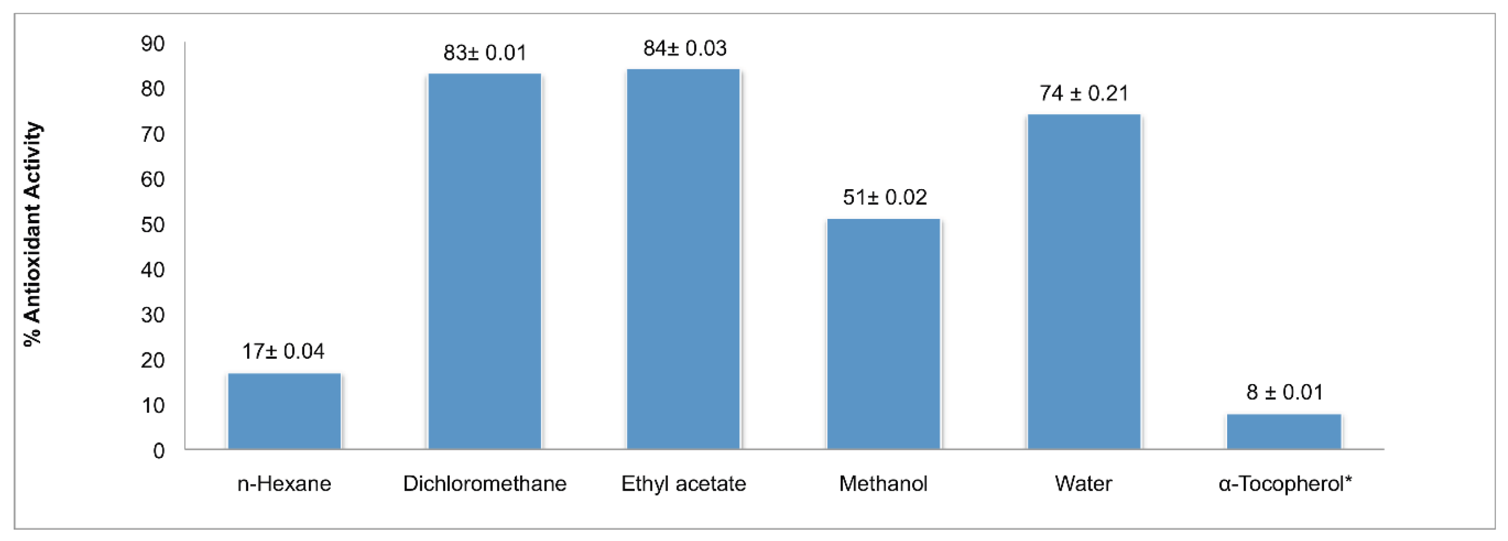
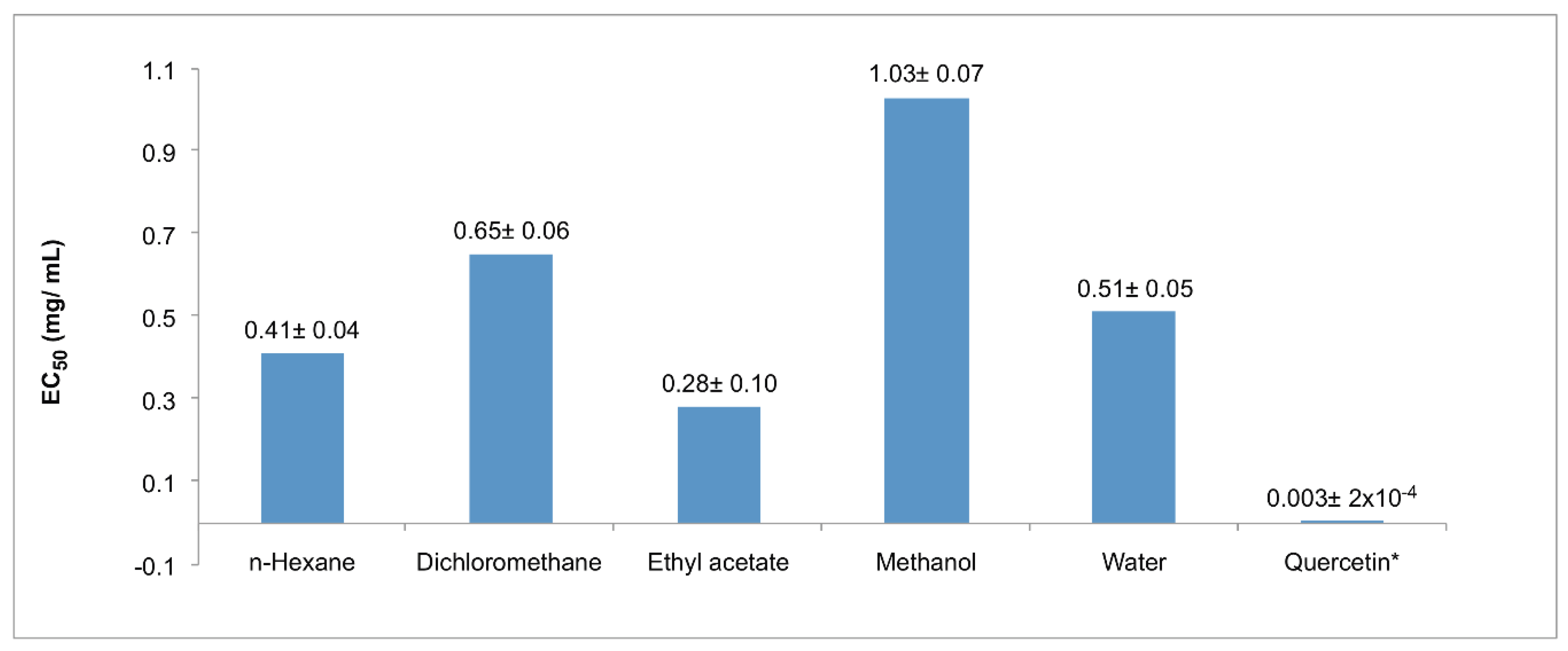
2.1. Antioxidant Screening

The antioxidant activity was evaluated using the β-carotene-linoleic acid assay and the 1,1-diphenyl-2-picrylhydrazyl DPPH radical-scavenging capacity assay (Figure 1 and Figure 2). In the β-carotene-linoleic test, the best activity was detected in the ethyl acetate extract (84%), immediately followed by the dichloromethane extract (83%); all five extracts showed higher activity, as compared with the reference compound α-tocopherol (8%). On the other hand, in the DPPH system, the ethyl acetate extract remained at the top in this activity, revealing an EC50 of 0.28 mg/mL. In addition, the extracts obtained with n-hexane or water showed similar values (0.41 and 0.51 mg/mL, respectively). Remarkably, neither of the extracts exhibited a comparative value with quercetin, the reference compound (0.003 mg/mL).

2.2. Antimicrobial Activity

The antimicrobial activity was examined by determining the minimum inhibitory concentrations (MIC) using five bacterial strains and three antibiotics as the reference (Table 1). Interestingly, the extracts obtained with n-hexane or dichloromethane showed significant activity against the Gram-positive Staphylococcus aureus and Enterococcus faecalis, as well as for the Gram-negative Escherichia coli. Furthermore, E. coli also exhibited considerable sensitivity to the ethyl acetate extract. Remarkably, the methanolic and the aqueous extracts were inactive against the all bacteria tested. Moreover, Klebsiella pneumoniae and Acinetobacter baumannii were insensitive to all S. pachyphylla extracts examined.

2.3. Xanthine and Acetylcholinesterase Inhibitory Assay

The enzymatic evaluation results are shown on Table 2. In the acetylcholinesterase inhibition assay, the extracts did not show a remarkable activity, only the dichloromethane extract presented a slight activity with an IC50 of 191.7 µg/mL; however, such a result is far away from the positive control galantamine (0.278 µg/mL). In the xanthine oxidase inhibition assay, better results were obtained, with IC50 values for the ethyl acetate and methanol extracts standing out with 11.7 and 19.5 µg/mL, respectively, although they did not surpass the drug allopurinol, used as control (0.842 µg/mL). The rest of the extracts did not show significant activity.

2.4. Antiproliferative Activity
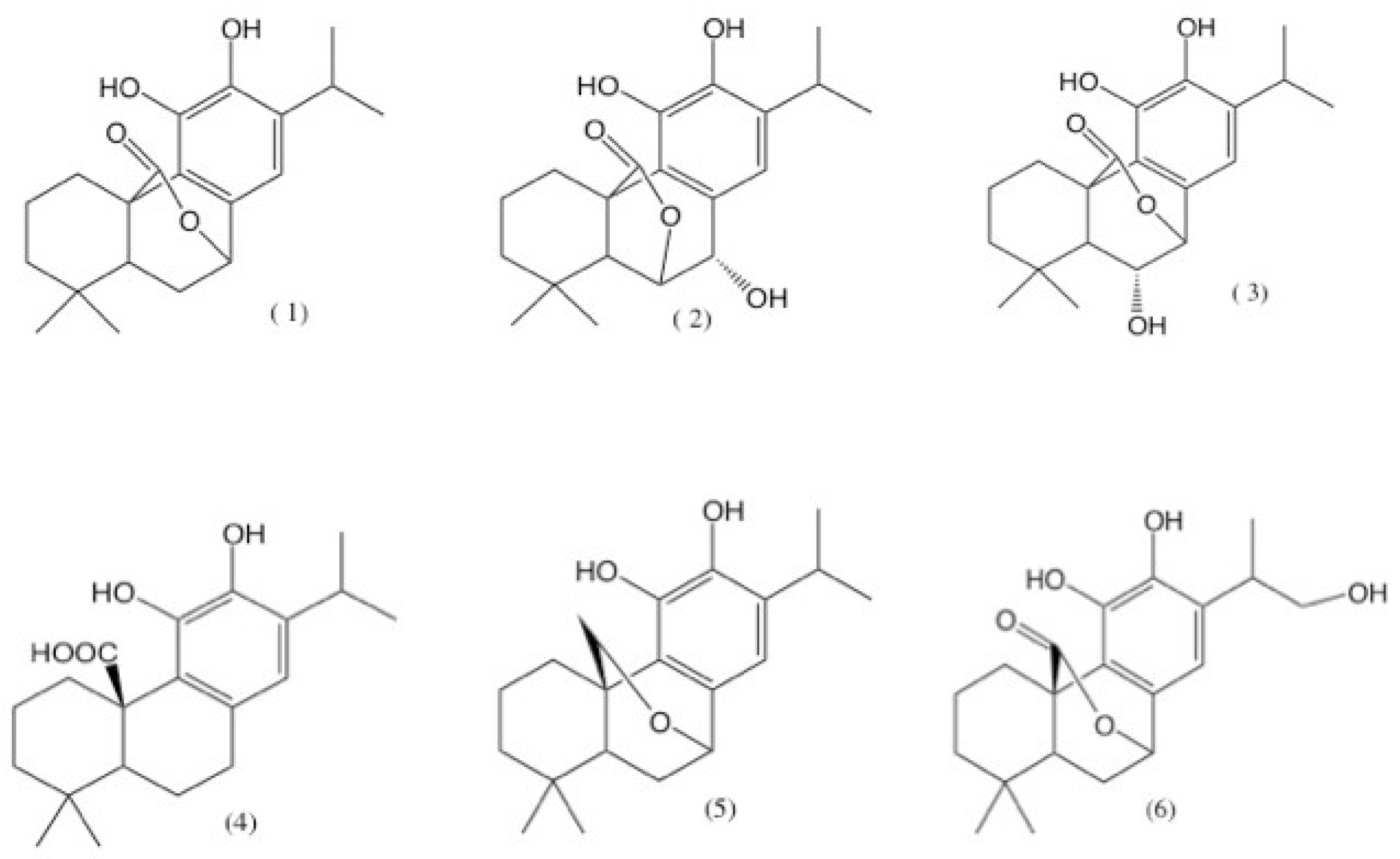
The antiproliferative activity was obtained by measuring the concentration needed to decrease cell propagation by 50% (GI50) using six human cancer cell lines and three well-known anti-cancer drugs (Table 3). All extracts exhibited a degree of effectiveness against all cell lines tested. Specifically, extracts obtained with dichloromethane or n-hexane were the most active against all the evaluated cell lines, showing GI50 values between 5.4 and 11 µg/mL. Both extracts showed higher cytotoxicity against cell lines SW1573, T-47D, and WiDr, with concentrations of 6.6, 11, and 8.6 µg/mL and 7.7, 9.9, and 9.9 µg/mL for n-hexane and dichloromethane extracts, respectively; in both cases, the extracts surpassed the positive control etoposide (GI50 of 15, 22, and 23 µg/mL against SW1573, T-47D, and WiDr, respectively) and cisplatin (GI50 of 15 and 26 µg/mL against T-47D and WiDr, respectively).

3. Discussion

Oxidative stress plays a key role in the development of several pathophysiological conditions, such as neurodegenerative and cardiovascular diseases, cancer, and diabetes; natural antioxidants ingested in the daily diet protect the cells against the damage produced by an excess of reactive oxygen species (ROS) [34,35,36,37]. There are several techniques for determining the antioxidant capacity; the difference between these methods lies in the assay principle and the experimental conditions [38]. We worked with two of the most used techniques for the antiradical evaluation: DPPH and ABTS•+ [39], Their high popularity is due to the speed, reproducibility, and simplicity of their procedures. On the other hand, β-carotene gives information regarding the capacity of lipidic peroxidation inhibition [40]. Several studies suggested a good antioxidant potential from Salvia species around the world, mainly because of the presence of diterpenes, such as carnosol (1), rosmanol (2), and isorosmanol (3) (Figure 3) [41]. These three compounds are also described in a phytochemical study of S. pachyphylla by Guerrero et al. [30].
Cuvelier et al. [41] also made an antioxidant evaluation of isolated diterpenes from Salvia officinalis such as carnosol and rosmanol by the Method of Antioxidative Power (AOP test) [42], and established that the activity of all these components was related to their phenolic structure. Phenolic diterpenes are widely known to be excellent antioxidants [43,44].
In another study with abietane diterpenes isolated from a dichloromethane extract of Salvia officinalis L, made by Miura et al. [45] with the Oil Stability Index (OSI) method [46] and the radical scavenging activity on the DPPH radical, these diterpenes exhibited a remarkably strong antioxidant activity, which was comparable to the one of the standard α-tocopherol. The authors mention that the compounds showing strong antioxidant activity were commonly included an ortho-dihydroxy group in the molecule structure, suggesting that the antioxidant activity is due to the presence of this ortho-dihydroxy group on the C-ring. This characteristic is also found in the metabolites mentioned above that can be found in S. pachyphylla extracts.
In the present work, the ethyl acetate extract showed a better antioxidant activity for the two evaluated techniques, along with the dichloromethane extract in the β-carotene assay. These results are in accordance to those obtained by Şenol et al. (2010) and Loizzo et al. (2010) [47,48], where the intermediate polarity extracts (ethyl acetate) from different Salvia species presented better results for the same antioxidant techniques of β-carotene and the DPPH radical. The phytochemical background, described above with plants of the Salvia genus, suggests that the abietane-type diterpenes can be found in the intermediate polarity extracts of S. pachyphylla and could be the responsible for its antioxidant activity.
For the antimicrobial susceptibility analysis, one of the more employed techniques is the calculation of the minimal inhibitory concentration (MIC), which allows researchers to determine easily the minimum quantity of an analyte capable of inhibiting the visible growth of a microorganism [49]. In the antimicrobial evaluation, n-hexane and dichloromethane extracts only matched the MIC of oxacillin in the ORSA (125 µg/mL) and got near to cephalotin with the same bacteria (62.5 µg/mL). Although the reference antibiotics surpassed the extracts activities, it can be noticed that n-hexane and dichloromethane extracts presented a better activity against Gram-positive bacteria S. aureus and E. faecalis, and only with E. coli on the Gram-negative evaluated bacteria. From the higher-polarity extracts (ethyl acetate, methanol, and water), only ethyl acetate presented some degree of activity with ORSA and E. coli; however, this was still far away from the reference antibiotics. Methanol and water extracts had even higher MIC than the n-hexane and dichloromethane ones. While our extracts were less active than the references, it can be pointed out that in general, the better results were obtained from the low to medium polarity extracts, against the Gram-positive bacteria. Our results are consistent with those described by Vlietinck et al. [50], who suggested that Gram-positive bacteria are significantly more susceptible to plant-derived extracts. This may be attributed to the fact that the cell wall in Gram-positive bacteria consist of a single layer, while the Gram-negative cell wall is a multilayered and quite complex structure [51]. Previous studies suggested that the antibacterial activity from Salvia extracts over Gram-negative bacteria such as E. coli depends on the nature of the studied extract [52]. As has been mentioned before, the biological activity of the Salvia extracts is related to the presence of abietane diterpenes [53]; in a bioassay-guided study of Rosmarinus officinalis L, several extracts were evaluated against different microorganisms responsible for initiating dental caries, Enterococcus faecalis among them, and their MICs showed that the higher antibacterial activity was from an extract where carnosic acid (4) and carnosol (1) (Figure 3) were identified as the major compounds by HPLC analysis. As stated before, these metabolites are also found in S. pachyphylla.
On the other hand, Oluwatuyi et al. [54], as part of a project to characterize plant-derived natural products that modulate bacterial multidrug resistance (MDR), conducted a bioassay-guided fractionation of a chloroform extract of the aerial parts of Rosmarinus officinalis, leading to the characterization of the diterpenes carnosic acid (1) and carnosol (2). The antibacterial activities of these natural products make them interesting potential targets for synthesis.
The screening of natural products in the search of medically relevant enzyme inhibitors remains a viable approach for isolation of novel compounds with specific pharmacological properties, which still has a great potential for further studies. Here, two activity assays were used to identify enzyme inhibitors, within each extract, with therapeutic potential (Table 2): xanthine oxidase (XO) and acetylcholinesterase (AChE) inhibition assays. Nowadays, the potential of natural products has not yet been explored in the search of new treatments for the control of Xanthine Oxidase related diseases [55]. Xanthine oxidase catalyzes the oxidation of hypoxanthine to xanthine and uric acid; however, under certain conditions, this can generate superoxide. It has been proved that XO inhibitors can be helpful for the treatment of liver disease and gout [56]. Acetylcholinesterase catalyzes the hydrolysis of the neurotransmitter acetylcholine into choline and acetic acid [57]. Modulation of acetylcholine levels using acetylcholinesterase inhibitors is among the major strategies to address diverse neurodegenerative diseases [58]. Remarkably, extracts with a prospective inhibitory effect showed a concentration-dependent trend and IC50 values were estimated, with the ethyl acetate extract that exhibited a significant effect over XO, while the dichloromethane extract showed a considerable effect on AChE. Unfortunately, other extracts showed a little or poor after-effect on either of the tested enzyme activities, hence, they were considered as inactive.
Regarding the antiproliferative activity, the effects of the n-hexane and dichloromethane extracts over A2780 (ovarian carcinoma), HBL-100 (breast carcinoma), and HeLa (cervix adenocarcinoma), which represent three of the most common cancers in women worldwide, were noticeable. Remarkable results were also obtained against colorectal adenocarcinoma (WiDr), which exemplifies cell lines that typically show drug resistance [59]. Interestingly, these results are different from those previously reported by Guerrero et al. [30]. They tested pure diterpenes isolated from the aerial parts of two species, S. clevelandi and S. pachyphylla, which were less effective than our extracts. According to the results obtained, from all tested diterpenes, the most active were carnosol (1), 20-deoxocarnosol (5), and 16-hydroxycarnosol (6) (Figure 3) for five different cell lines. We thought that the main difference resides in the nature of the sample, as our results were generated using total extracts, suggesting a possible synergistic effect. Despite other extracts being less active, their results remain promising for pursuing novel molecules with cytotoxic effect.

4. Materials and Methods
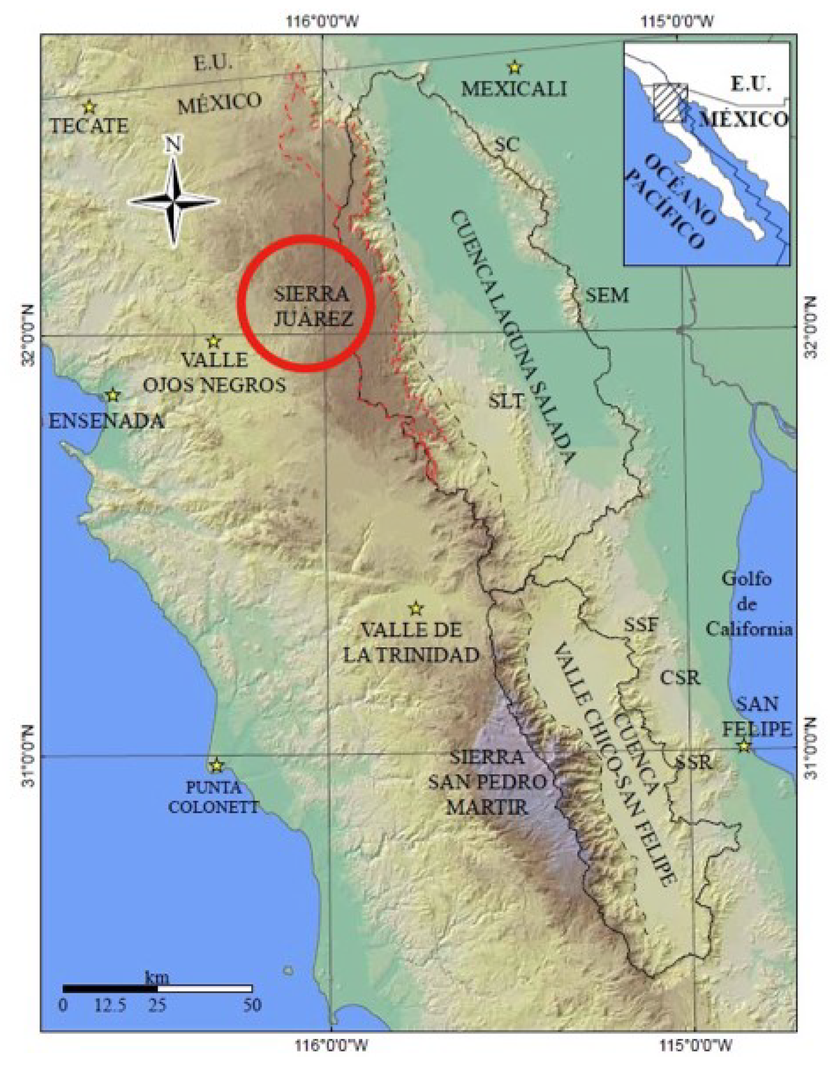
4.1. Plant Material

The aerial parts (leaf, flower, and stem) of S. pachyphylla were collected in lands of the Sierra Juarez-Constitution National Park, Ensenada, B.C., México, at an elevation of 1630 m and coordinates of N: 32°01′41″; W: 115°56′11″ (Figure 4). Identification and authentication were carried out by Dr. José Delgadillo, and the voucher specimen (BCMEX9783) was deposited in the herbarium of the Autonomous University of Baja California, at Ensenada. Aerial parts (1.3 kg) were air-dried for a week, under shade (to reduce moisture content). The dried material was ground to fine powder and stored at 4 °C until use.

4.2. Preparation Extracts

Crude extracts were obtained through the classical Soxhlet method. Five different thimbles were uniformly packed, each one with 75 g of fine powder. The extraction was carried out using different solvents, one for each one of the thimbles (250 mL): n-hexane (HX), dichloromethane (DC), ethyl acetate (EA), methanol (MT), and distilled water (AQ). The extraction process was stopped until the solvent in siphon tube became colorless. Each extract was filtered and dried at 40 °C, using a rotary evaporator (Buchi Rotavapor® R-215), until a solid or semi-solid residue was yielded. Each residue was separately further lyophilized to get a dry solid matter: HX = 60.3 g, DC = 7.9 g, EA = 24.2 g, MT = 9.9 g, and AQ = 14.5 g. All solids were kept in air tight bottles and stored at 4 °C until use.

4.3. β-Carotene-Linoleic Acid Assay

The antioxidant activity was assayed by the coupled oxidation of β-carotene and linoleic acid as described by Burda and Oleszek [60] with minor modifications. 1 mL of a β-carotene solution (0.2 mg/mL in chloroform) was added to an emulsion containing 0.018 mL of linoleic acid and 0.2 mL of Tween-20. Chloroform was removed (under a nitrogen environment), 50 mL of aerated deionized water (DO of 9.7 mg/L) was slowly added, and the mixture was vigorously agitated to form a stable emulsion. 5 mL of this emulsion was transferred to test tubes containing the corresponding sample (2 mg) of each extract. Immediately, the absorbance was measured at 470 nm (A470, zero time). All tubes were then incubated at 50 °C and A470 values were registered every 15 min for 2 h. A control without the antioxidant was prepared aside and α-tocopherol was used as a reference compound. The antioxidant activity (AA) was expressed as the percentage of inhibition of β-carotene bleaching, as compared to the control, and calculated using the following formula:
AA (%) = [1 − (AS0AS120/AC0AC120)] × 100
where AS and AC represent the A470 value of the sample and control, respectively, and the superscript numbers denote the time of the initial and final measurement (0 and 120 min). All determinations were performed in duplicate and replicated at least three times.

4.4. DPPH Radical-Scavenging Capacity Assay

The radical-scavenging activity was performed as described by Burda and Oleszek [60] with slight modifications. For the evaluation of each extract, a stock solution (4 mg/mL) was prepared and serially two-fold diluted (down to 0.003 mg/mL) with methanol. An aliquot of each dilution (1 mL) was mixed with 1 mL of a methanolic solution of 1,1-diphenyl-2-picrylhydrazyl (DPPH at 0.03 mg/mL). At the same time, a control containing 1 mL of methanol and 1 mL of the DPPH solution was prepared. The mixtures were incubated at room temperature in the dark for 5 min. Using methanol as a blank, the absorbance was quantified at 517 nm (A517). The radical-scavenging activity was calculated as the percentage of DPPH decoloration using the following formula:
DPPH (%) = [1 − (A/B) × 100]
where A and B represent the A517 value of the control and sample, respectively. All determinations were performed in duplicate and replicated at least three times. For each extract, the percentage of DPPH decoloration was plotted against the concentration of each dilution. The concentration required to decrease the absorbance of DPPH by 50% was obtained by interpolation, from a linear regression analysis, and expresses the EC50 value. Quercetin was used as a reference compound.

4.5. Antimicrobial Assay

Antibacterial activity was tested using a microdilution assay following the National Committee for Clinical Laboratory Standards (NCCLS), due its precision and reproducibility [61,62], and MIC was defined as the lowest concentration that prevents visible growth of bacteria. Six strains of microorganisms were used for antimicrobial testing including Gram-positive bacteria (Enterococcus faecalis, Staphylococcus aureus, and Oxacillin-resistant Staphylococcus aureus) and Gram-negative bacteria (Acinetobacter baumannii, Escherichia coli, and Klebsiella pneumoniae); all strains were provided by the Regional Center for Infection Diseases, School of Medicine, Autonomous University of Nuevo Leon (Monterrey, Mexico). All strains were plated on a Müeller–Hinton agar (Becton Dickinson) and incubated at 37 °C for 24 h. Four or five colony forming units were suspended in saline solution and the optical density was adjusted to the turbidity of the 0.5 McFarland Standard. Working suspensions were prepared by a 1:50 dilution in Müeller–Hinton broth. For the evaluation of each extract, a stock solution (6 mg/mL in 5% DMSO) was prepared and serially two-fold diluted with Müeller–Hinton broth in a 96-well microtiter plate (down to 0.5 μg/mL, final highest DMSO concentration 0.83%). One volume (0.1 mL) of working suspension was added to each well. The antibiotics cephalothin, oxacillin, and vancomycin were used as reference compounds. Controls without bacterial cells (medium control) and without extract or antibiotic (growth control) were prepared aside. Plates were incubated at 37 °C for 48 h and growth was visually examined.

4.6. Xanthine Oxidase Inhibition Assay

There are several assays employed for the Xanthine oxidase activity quantification, the more widely used is by the spectrophotometric determination of uric acid [63], the final reaction product. In this study, the inhibition of the XO activity was evaluated using the easy and sensitive protocol described by Havlik et al. [64]. A reaction solution containing 0.4 mL of 120 mM phosphate buffer (pH 7.8) and 0.33 mL of 150 mM of xanthine was supplemented with 0.25 mL of inhibitor solution (extract or reference) and mixed thoroughly. The reaction was started by adding 0.02 mL of XO enzyme solution (0.5 U/mL). After 3 min of incubation at 24 °C, the uric acid formation was determined by measuring the absorbance at 295 nm (A295). A reaction without inhibitor was used as control and allopurinol served as a reference compound. The inhibition percentage of XO activity was calculated using the following formula:
XO inhibition (%) = [1 − (As/Ac)] ×100
where AS and AC represent the initial velocity of reactions with sample and control, respectively. All determinations were performed in duplicate and replicated at least three times. The concentration required to decrease the activity of XO by 50% was obtained by interpolation, from a linear regression analysis, and expresses the IC50 value.

4.7. Acetylcholinesterase (AChE) Inhibition Assay

The acetylcholinesterase inhibition activity was determined with the technique described by Adewusi et al. [65], which is a modification of the widely known and employed Ellman method [66]. For each determination, wells of a microtiter plate were filled with 25 μL of 15 mM acetylthiocholine iodide (in water), 125 μL of 3 mM DTNB in buffer C (50 mM Tris-HCl, pH 8.0, containing 0.1 M NaCl and 0.02 M MgCl2·6 H2O), 72.5 μL of buffer B (50 mM Tris-HCl, pH 8.0, containing 0.1% BSA), and 2.5 μL of inhibitor solution (extract or reference, in DMSO) and were mixed thoroughly. Absorbance was measured at 412 nm (A412) every 45 s, three times consecutively. Thereafter, 25 μL of AChE enzyme solution (0.2 U/mL) was added to each well and A412 was measured five times consecutively every 45 s. A reaction without inhibitor was used as control and galantamine served as the reference compound. Any increase in absorbance due to the spontaneous hydrolysis of the substrate was corrected by subtracting the A412 before adding the enzyme. The inhibition percentage of AChE activity was calculated using the following formula:
AChE inhibition (%) = [1 − (As/Ac)] ×100
where AS and AC represent the initial velocity of reactions with sample and control, respectively. All determinations were performed in duplicate and replicated at least three times. The concentration required to decrease the activity of AChE by 50% was obtained by interpolation, from a sigmoidal regression analysis, and expresses the IC50 value.

4.8. Cell lines and Culture Conditions

Five human cancer cell lines were used in this study: A2780 (ovarian carcinoma), HBL-100 (breast carcinoma), HeLa (cervix adenocarcinoma), SW1573 (lung carcinoma), T-47D (breast ductal carcinoma), and WiDr (colorectal adenocarcinoma). All line cells were maintained in RPMI (Roswell Park Memorial Institute) 1640 media supplemented with 5% heat-inactivated FCS (Fetal Calf Serum) and 2 mM l-glutamine at 37 °C, 5% CO2, and 95% humidity. Exponentially growing cells were trypsinized and resuspended in medium containing 2% FCS and antibiotics (100 U/mL of penicillin G and 0.1 mg/mL of streptomycin). Single cell suspensions showing >97% viability, by trypan blue dye exclusion assay, were subsequently counted. After counting, dilutions were made to give the appropriate cell densities required for antiproliferative testing.

4.9. Antiproliferative Assay

Antiproliferative testing was performed using the Sulforhodamine B assay (SRB), the preferred high-throughput assay from National Cancer Institute (NCI, NIH, USA), as reported by Miranda et al. [59], with slight modifications. The SRB assay is based on the dye union to the basic amino acids of the cellular proteins, and the colorimetric evaluation provides an estimation of the total protein mass, which is related to the number of cells. This assay presents an excellent linearity, sensibility, and low costs compared to others [67]. Each extract was initially dissolved in DMSO at 400 times the desired maximum concentration to test. Six thousand cells were inoculated to each well of a microtiter plate (100 μL of a suspension of 6 × 10⁴ cells per mL). One day after plating, all testing samples and reference compounds were added to corresponding wells (triplicated). Samples were tested in six decimal serial dilutions starting at 250 µg/mL. Control cultures were tested against equivalent concentrations of DMSO (0.25% as a negative control). After 48 h of incubation at permissive conditions, cell cultures were treated with 25 μL ice-cold 50% TCA and fixed at 4 °C for 60 min. Thereafter, the SRB assay was performed. The absorbance of each well was measured at 492 nm (A492). All absorbance values were corrected for background A492 (control wells containing just culture media). As recommended by the NCI, the concentration that causes 50% growth inhibition, GI50 value, was corrected by count at time zero; thus, GI50 is the concentration where [(T − T0)/(C − T0)] = 0.5. The absorbance of the test well after 48 h is T, the absorbance at time zero is T0, and the absorbance of the control is C. For all these calculations, Excel spreadsheets were used. GI50 values were computed from the dose-response curves.

5. Conclusions

In conclusion, aerial parts of S. pachyphylla have prospective potential as a source of natural products that could act as antioxidants, antimicrobial and antiproliferative compounds, or enzyme inhibitors.
Although our findings could be the outcome of a synergistic effect, they support the notion of aiming our next approach towards the isolation and identification of novel molecules with therapeutic potential.

Author Contributions

G.A.-T., performed the investigation and wrote the original draft of the paper; L.D.-R., R.S.-A., J.D.-R., and J.M.P. adjusted the methodology of the different experiments realized.; N.W.d.T., J.M.P., and I.C.-G. did the formal analysis of the results generated.; M.A.R., R.H.-M., and C.U.-S. did the review and editing of the paper.; R.H.-M. managed the funding acquisition.; I.C.-G. conducted the supervision and project administration.

Funding

This research received no external funding.

Acknowledgments

The authors would like to thank the Facultad de Ciencias Químicas e Ingeniería, Universidad Autónoma de Baja California, for the support given and for allowing the use of their facilities during this investigation. Additionally, to CONACYT, who supported G.A.-T. with a scholarship during her PhD studies during the realization of this investigation.

Conflicts of Interest

The authors declare no conflict of interest.

References

  1. Mabberley, D.J. The Plant-Book, a Portable Dictionary of Vascular Plants, 2nd ed.; University of Cambridge: Cambridge, UK, 1997; pp. 385–635. ISBN 0521414210. [Google Scholar]
  2. Harley, R.M.; Atkins, S.; Budantsev, A.L.; Cantino, P.D.; Conn, B.J.; Grayer, R.; Harley, M.M.; De Kok, R.P.J.; Krestovskaja, T.; Morales, R. Labiatae. In The Families and Genera of Vascular Plants, 1st ed.; Springer: Berlin, Germany, 2004; pp. 167–282. ISBN 3540405933. [Google Scholar]
  3. Firdous, S.; Dadass, A.K.; Khan, K.M.; Usmani, S.B.; Ahmad, V.U. A new triterpenoid from the leaves of Salvia triloba. Fitoterapia 1999, 70, 326–327. [Google Scholar] [CrossRef]
  4. Ikram, M.; Haq, I. Screening of medicinal plants for antimicrobial activity. Fitoterapia 1980, 51, 231–235. [Google Scholar]
  5. González, A.G.; Abad, T.; Jimenez, I.A.; Ravelo, A.G.; Zahira Aguiar, J.G.L.; San Andres, L.; Plasencia, M.; Herrera, J.R.; Moujir, L. A first study of antibacterial activity of diterpenes isolated from some Salvia species (Lamiaceae). Biochem. Syst. Ecol. 1989, 7, 293–296. [Google Scholar] [CrossRef]
  6. Moujir, L.; Gutierrez-Navarro, A.M.; San Andres, L.; Luis, J.G. Structure-antimicrobial activity relationships of abietane diterpenes from Salvia species. Phytochemistry 1993, 34, 1493–1495. [Google Scholar] [CrossRef]
  7. Moujir, L.; Gutierrez-Navarro, A.M.; San Andres, L.; Luis, J.G. Bioactive diterpenoids isolated from Salvia mellifera. Phytother. Res. 1996, 10, 172–174. [Google Scholar] [CrossRef]
  8. Cui, X.Y.; Wang, Y.L.; Kokudo, N.; Fang, D.Z.; Tang, W. Traditional Chinese medicine and related active compounds against hepatitis B virus infection. Biosci. Trends 2010, 4, 39–47. [Google Scholar] [PubMed]
  9. Alim, A.; Goze, I.; Goze, H.M.; Tepe, B. In vitro antimicrobial and antiviral activities of the essential oil and various extracts of Salvia cedronella Boiss. J. Med. Plants Res. 2009, 3, 413–419. [Google Scholar]
  10. Hu, S.; Chen, S.M.; Li, X.K.; Qin, R.; Mei, Z.N. Antitumor effects of Chi-Shen extract from Salvia miltiorrhiza and Paeoniae radix on human hepatocellular carcinoma cells. Acta Pharmacol. Sin. 2007, 28, 1215–1223. [Google Scholar] [CrossRef] [PubMed]
  11. Dat, N.T.; Jin, X.; Lee, J.H.; Lee, D.; Hong, Y.S.; Lee, K.; Kim, Y.H.; Lee, J.J. Abietane diterpenes from Salvia miltiorrhiza inhibit the activation of hypoxia-inducible factor-1. J. Nat. Prod. 2007, 70, 1093–1097. [Google Scholar] [CrossRef] [PubMed]
  12. Wang, X.H.; Bastow, K.F.; Sun, C.M.; Lin, Y.L.; Yu, H.J.; Don, M.J.; Wu, T.S.; Nakamura, S.; Lee, K.H. Antitumor agents. 239. Isolation, structure elucidation, total synthesis, and anti-breast cancer activity of neo-tanshinlactone from Salvia miltiorrhiza. J. Med. Chem. 2004, 47, 5816–5819. [Google Scholar] [CrossRef] [PubMed]
  13. Ulubelen, A.; Topcu, G.; Tan, N.; Lin, L.J.; Cordell, G.A. Microstegiol, a rearranged diterpene from Salvia microstegia. Phytochemistry 1992, 31, 2419–2421. [Google Scholar] [CrossRef]
  14. Kabouche, A.; Kabouche, Z.; Öztürk, M.; Kolak, U.; Topçu, G. Antioxidant abieetane diterpenoids from Salvia barrelieri. Food Chem. 2007, 102, 1281–1287. [Google Scholar] [CrossRef]
  15. Weng, X.C.; Wang, W. Antioxidant activity of compounds isolated from Salvia plebeia. Food Chem. 2000, 71, 489–493. [Google Scholar] [CrossRef]
  16. Tepe, B.; Sokmen, M.; Akpulat, H.A.; Sokmen, A. Screening of the antioxidant potentials of six Salvia species from Turkey. Food Chem. 2006, 95, 200–204. [Google Scholar] [CrossRef]
  17. Kamatou, G.P.P.; Viljoen, A.M.; Steenkamp, P. Antioxidant, antiinflammatory activities and HPLC analysis of South African Salvia species. Food Chem. 2010, 119, 684–688. [Google Scholar] [CrossRef]
  18. Hitokoto, H.; Morozumi, S.; Wauke, T.; Saiki, S.; Kurata, H. Inhibitory effects of spices on growth and toxin production of toxigenic fungi. Appl. Environ. Microbiol. 1980, 39, 818–822. [Google Scholar] [PubMed]
  19. Han, Y.M.; Oh, H.; Na, M.; Kim, B.S.; Oh, W.K.; Kim, B.Y.; Jeong, D.G.; Ryu, S.E.; Sok, D.E.; Ahn, J.S. PTP1B inhibitory effect of abietane diterpenes isolated from Salvia miltiorrhiza. Biol. Pharm. Bull. 2005, 28, 1795–1797. [Google Scholar] [CrossRef] [PubMed]
  20. Sairafianpour, M.; Christensen, J.; Staerk, D.; Budnik, B.A.; Kharazmi, A.; Bagherzadeh, K.; Jaroszewski, J.W. Leishmanicidal, antiplasmodial, and cytotoxic activity of novel diterpenoid 1,2-quinones from Perovskia abrotanoides: New source of tanshinones. J. Nat. Prod. 2001, 64, 1398–1403. [Google Scholar] [CrossRef] [PubMed]
  21. Maklad, Y.A.; Aboutabl, E.A.; El-Sherei, M.M.; Meselhy, K.M. Bioactivity studies of Salvia transsylvanica (Schur ex Griseb) grown in Egypt. Phytother. Res. 1999, 13, 147–150. [Google Scholar] [CrossRef]
  22. Lu, Y.; Foo, L.Y. Polyphenolics of Salvia. A review. Phytochemistry 2002, 59, 117–140. [Google Scholar] [CrossRef]
  23. Tepe, B.; Dönmez, E.; Unlu, M.; Candan, F.; Dimitra, D.; Vardar-Unlu, G. Antimicrobial and antioxidative activities of the essential oils and methanol extracts of Salvia cryptantha (Montbret et Aucher ex Benth.) and Salvia multicaulis (Vahl). Food Chem. 2004, 84, 519–525. [Google Scholar] [CrossRef]
  24. Kusumi, T.; Ooi, T.; Hayashi, T.; Kakisawa, H. A diterpenoid phenalenone from Salvia miltiorrhiza. Phytochemistry 1985, 24, 2118–2120. [Google Scholar] [CrossRef]
  25. Habibi, Z.; Eftekhar, F.; Samiee, K.; Rustaiyan, A. Structure and antibacterial activity of a new labdane diterpenoid from Salvia leriaefolia. J. Nat. Prod. 2000, 63, 270–271. [Google Scholar] [CrossRef] [PubMed]
  26. Nieto, M.; García, E.E.; Giordano, O.S.; Tonn, C.E. Icetexane and abietane diterpenoids from Salvia gilliessi. Phytochemistry 2000, 53, 911–915. [Google Scholar] [CrossRef]
  27. Rauter, A.P.; Branco, I.; Lopes, R.G.; Justino, J.; Silva, F.V.M.; Noronha, J.P.; Cabrita, E.J.; Brouard, I.; Bermejo, J. A new lupine triterpenetriol and anticholinesterase activity of Salvia sclareoides. Fitoterapia 2007, 78, 474–481. [Google Scholar] [CrossRef] [PubMed]
  28. Taylor, R. The Origin and Adaptive Radiation of Salvia pachyphylla (Lamiaceae). Master’s Thesis, Northern Arizona University, Flagstaff, Arizona, 2002. [Google Scholar]
  29. Alonso-Castro, A.J.; Villarreal, M.L.; Salazar-Olivo, L.A.; Gomez-Sanchez, M.; Dominguez, F.; Garcia-Carranca, A. Mexican medicinal plants used for cancer treatment: Pharmacological, phytochemical and ethnobotanical studies. J. Ethnopharmacol. 2011, 133, 945–972. [Google Scholar] [CrossRef] [PubMed]
  30. Guerrero, I.C.; Andrés, L.S.; León, L.G.; Machín, R.P.; Padrón, J.M.; Luis, J.G.; Delgadillo, J. Abietane diterpenoids from Salvia pachyphylla and S. clevelandii with cytotoxic activity against human cancer cell lines. J. Nat. Prod. 2006, 69, 1803–1805. [Google Scholar] [CrossRef] [PubMed]
  31. Kviecinski, M.R.; Felipe, K.B.; Schoenfelder, T.; de Lemos Wiese, L.P.; Rossi, M.H.; Gonçalez, E.; Felicio, J.D.; Filho, D.W.; Pedrosa, R.C. Study of the antitumor potential of Bidens pilosa (Asteraceae) used in Brazilian folk medicine. J. Ethnopharmacol. 2008, 117, 69–75. [Google Scholar] [CrossRef] [PubMed]
  32. Rates, S.K.M. Plants as source of drugs. Toxicon 2001, 39, 603–613. [Google Scholar] [CrossRef]
  33. Harvey, A.L.; Waterman, P. The continuing contribution of biodiversity to drug discovery. Curr. Opin. Drug Discov. Dev. 1998, 1, 71–76. [Google Scholar]
  34. Ighodaro, O.M. Molecular pathways associated with oxidative stress in diabetes mellitus. Biomed. Pharmacother. 2018, 108, 656–662. [Google Scholar] [CrossRef] [PubMed]
  35. Klaunig, J.E.; Wang, Z. Oxidative stress in carcinogenesis. Curr. Opin. Toxicol. 2018, 7, 116–121. [Google Scholar] [CrossRef]
  36. Taleb, A.; Ahmad, K.A.; Ihsan, A.U.; Qu, J.; Lin, N.; Hezam, K.; Koju, N.; Hui, L.; Qilong, D. Antioxidant effects and mechanism of silymarin in oxidative stress induced cardiovascular diseases. Biomed. Pharmacother. 2018, 102, 689–698. [Google Scholar] [CrossRef] [PubMed]
  37. Tramutola, A.; Lanzillotta, C.; Perluigi, M.; Butterfield, D.A. Oxidative stress, protein modification and Alzheimer disease. Brain Res. Bull. 2017, 133, 88–96. [Google Scholar] [CrossRef] [PubMed]
  38. Joon, K.M.; Takayuki, S. Antioxidant Assays for Plant and Food Components. J. Agric. Food Chem. 2009, 57, 1655–1666. [Google Scholar] [CrossRef]
  39. Reşat, A.; Mustafa, Ö.; Kubilay, G.; Esra, Ç. Antioxidant Activity/Capacity Measurement. 1. Classification, Physicochemical Principles, Mechanisms, and Electron Transfer (ET)-Based Assays. J. Agric. Food Chem. 2016, 64, 997–1027. [Google Scholar] [CrossRef]
  40. Floegel, A.; Dae-Ok, K.; San-Jin, C.; Sung, I.K.; Ock, K.C. Comparison of ABTS/DPPH assays to measure antioxidant capacity in popular antioxidant-rich US foods. J. Food Compos. Anal. 2011, 24, 1043–1048. [Google Scholar] [CrossRef]
  41. Cuvelier, M.E.; Berset, C.; Richard, H. Antioxidant Constituents in Sage (Salvia officinalis). J. Agric. Food. Chem. 1994, 42, 665–669. [Google Scholar] [CrossRef]
  42. Cuvelier, M.E.; Berset, C.; Richard, H. Use of a new test for determining comparative antioxidative activity of BHA, BHT, alpha- and gamma-tocopherols and extracts from rosemary and sage. Sci. Aliments 1990, 10, 797–806. [Google Scholar]
  43. Nakatani, N.; Inatani, R. Structure of rosmanol, a new antioxidant from rosemary. Agric. Biol. Chem. 1981, 45, 2385–2386. [Google Scholar] [CrossRef]
  44. Nakatani, N.; Inatani, R. A new diterpene lactone, rosmadial, from rosemary. Agric. Biol. Chem. 1983, 47, 353–358. [Google Scholar] [CrossRef]
  45. Miura, K.; Kikuzaki, H.; Nakatani, N. Antioxidant Activity of Chemical Components from Sage (Salvia officinalis L.) and Thyme (Thymus vulgaris L.) Measured by the Oil Stability Index Method. J. Agric. Food Chem. 2002, 50, 1845–1851. [Google Scholar] [CrossRef] [PubMed]
  46. Nakatani, N.; Tachibana, Y.; Kikuzaki, H. Establishment of a model substrate oil for antioxidant activity Assessment by Oil Stability Index Method. J. Am. Oil Chem. Soc. 2001, 78, 19–23. [Google Scholar] [CrossRef]
  47. Şenol, F.S.; Orhan, I.; Celep, F.; Kahraman, A.; Doğan, M.; Yilmaz, G.; Şener, B. Survey of 55 Turkish Salvia taxa for their acetylcholinesterase inhibitory and antioxidant activities. Food Chem. 2010, 120, 34–43. [Google Scholar] [CrossRef]
  48. Loizzo, M.R.; Tundis, R.; Conforti, F.; Menichini, F.; Bonesi, M.; Nadjafi, F.; Frega, N.G.; Menichini, F. Salvia leriifolia Benth (Lamiaceae) extract demonstrates in vitro antioxidant properties and cholinesterase inhibitory activity. Nutr. Res. 2010, 30, 823–830. [Google Scholar] [CrossRef] [PubMed]
  49. Sánchez-García, E.; Castillo-Hernández, S.L.; García-Palencia, P. Actividad Antimicrobiana. In Investigación en Plantas de Importancia Médica, 1st ed.; Rivas-Morales, C., Oranday-Cardenas, M.A., Verde-Star, M.J., Eds.; Publisher: Barcelona, España, 2016; Volume 1, pp. 77–100. ISBN 978-84-944673-7-0. [Google Scholar]
  50. Vlietinck, A.J.; Van, H.L.; Totte, J.; Lasure, A.; Vanden, B.D.; Rwangabo, P.C.; Mvukiyumwami, J. Screening of hundred Rwandese medicinal plants for antimicrobial and antiviral properties. J. Ethnopharmacol. 1995, 46, 31–47. [Google Scholar] [CrossRef]
  51. Abu-Darwish, M.S.; Al-Ramamneh, E.A.D.M.; Kyslychenko, V.S.; Karpiuk, U.V. The antimicrobial activity of essential oils and extracts of some medicinal plants grown in Ash-shoubak region—South of Jordan. Pak. J. Pharm. Sci. 2012, 25, 239–246. [Google Scholar] [PubMed]
  52. Ghorbani, A.; Esmaeilizadeh, M. Pharmacological properties of Salvia officinalis and its components. J. Tradit. Complement. Med. 2017, 7, 433–440. [Google Scholar] [CrossRef] [PubMed]
  53. González, C.M.A. Aromatic abietane diterpenoids: Their biological activity and synthesis. Nat. Prod. Rep. 2015, 32, 684–704. [Google Scholar] [CrossRef] [PubMed]
  54. Oluwatuyi, M.; Kaatz, G.W.; Gibbons, S. Antibacterial and resistance modifying activity of Rosmarinus officinalis. Phytochemistry 2004, 65, 3249–5324. [Google Scholar] [CrossRef] [PubMed]
  55. Hudaib, M.M.; Tawaha, K.A.; Mohammad, M.K.; Assaf, A.M.; Issa, A.Y.; Alali, F.Q.; Aburjai, T.A.; Bustanji, Y.K. Xanthine oxidase inhibitory activity of the methanolic extracts of selected Jordanian medicinal plants. Pharmacogn. Mag. 2011, 7, 320–324. [Google Scholar] [CrossRef] [PubMed]
  56. Sahgal, G.; Ramanathan, S.; Sasidharan, S.; Mordi, M.N.; Ismail, S.; Mansor, S.M. In vitro antioxidant and xanthine oxidase inhibitory activities of methanolic Swietenia mahagoni seed extracts. Molecules 2009, 14, 4476–4485. [Google Scholar] [CrossRef] [PubMed]
  57. Čolović, M.B.; Krstić, D.Z.; Lazarević-Pašti, T.D.; Bondžić, A.M.; Vasić, V.M. Acetylcholinesterase Inhibitors: Pharmacology and Toxicology. Curr. Neuropharmacol. 2013, 11, 315–335. [Google Scholar] [CrossRef] [PubMed]
  58. Hasnat, M.A.; Pervin, M.; Lim, B.O. Acetylcholinesterase inhibition and in vitro and in vivo antioxidant activities of Ganoderma lucidum grown on germinated brown rice. Molecules 2013, 18, 6663–6678. [Google Scholar] [CrossRef] [PubMed]
  59. Miranda, P.O.; Padrón, J.M.; Padrón, J.I.; Villar, J.; Martín, V.S. Prins-type synthesis and SAR study of cytotoxic alkyl chloro dihydropyrans. Chem. Med. Chem. 2006, 1, 323–329. [Google Scholar] [CrossRef] [PubMed]
  60. Burda, S.; Oleszek, W. Antioxidant and antiradical activities of flavonoids. J. Agric. Food Chem. 2001, 49, 2774–2779. [Google Scholar] [CrossRef] [PubMed]
  61. Alanís-Garza, B.A.; González-González, G.M.; Salazar-Aranda, R.; Waksman de Torres, N.; Rivas-Galindo, V.M. Screening of antifungal activity of plants from the northeast of Mexico. J. Ethnopharmacol. 2007, 114, 468–471. [Google Scholar] [CrossRef] [PubMed]
  62. Salazar-Aranda, R.; Pozos, M.E.; Cordero, P.; Perez, J.; Salinas, M.C.; Waksman, N. Determination of the antioxidant activity of plants from northeast Mexico. Pharm. Biol. 2008, 46, 166–170. [Google Scholar] [CrossRef]
  63. Shintani, H. Determination of Xanthine Oxidase. Pharm. Anal. Acta 2013, S7, 4. [Google Scholar] [CrossRef]
  64. Havlik, J.; de la Huebra, R.G.; Hejtmankova, K.; Fernandez, J.; Simonova, J.; Melich, M.; Rada, V. Xanthine oxidase inhibitory properties of Czech medicinal plants. J. Ethnopharmacol. 2010, 132, 461–465. [Google Scholar] [CrossRef] [PubMed]
  65. Adewusi, E.A.; Moodley, N.; Steenkamp, V. Antioxidant and acetylcholinesterase inhibitory activity of selected southern African medicinal plants. S. Afr. J. Bot. 2011, 77, 638–644. [Google Scholar] [CrossRef]
  66. Mathew, M.; Subramanian, S. In Vitro Screening for Anti-Cholinesterase and Antioxidant Activity of Methanolic Extracts of Ayurvedic Medicinal Plants Used for Cognitive Disorders. PLoS ONE 2014, 9, e86804. [Google Scholar] [CrossRef] [PubMed]
  67. Kuete, V.; Karaosmanoglu, O.; Sivas, H. Anticancer Activities of African Medicinal Spices and Vegetables In Medicinal Spices and Vegetables from Africa, 1st ed.; Kuete, V., Ed.; Academic Press: Cambridge, MA, USA, 2017; Volume 1, pp. 271–297. ISBN 9780128092866. [Google Scholar]
Figure 1. β-carotene-linoleic acid assay of extracts from aerial parts of Salvia pachyphylla. * Used as a reference compound. Values are mean ± SD, n = 3.
Plants 07 00105 g001
Figure 2. DPPH radical-scavenging capacity assay of extracts from aerial parts of S. pachyphylla. * Used as a reference compound. Values are mean ± SD, n = 3.
Plants 07 00105 g002
Figure 3. Abietanic diterpenes: carnosol (1), rosmanol (2), isorosmanol (3), carnosic acid (4), 20-deoxocarnosol (5), and 16-hydroxycarnosol (6).
Plants 07 00105 g003
Figure 4. Sierra Juarez-Constitution National Park (marked by red circle), Ensenada, BC, México.
Plants 07 00105 g004
Table 1. In vitro antimicrobial activity of extracts from aerial parts of S. pachyphylla.
Antimicrobial Activity (Minimum Inhibitory Concentrations (MIC), μg/mL)
Extracts or ControlsS. aureusORSAaE. faecalisE. coliK. pneumoniaeA. baumannii
n-Hexane62.5125250250>1000>1000
Dichloromethane62.5125250250>1000>1000
Ethyl acetate1000250>1000250>1000>1000
Methanol1000>1000>1000>1000>1000>1000
Water1000>1000>1000>1000>1000>1000
Oxacillin *0.4812531.20.487>1000>1000
Cephalothin *0.4862.531.21>100062.5
Vancomycin *0.481.951.95>250>1000250
* Used as a reference compound. a Oxacillin-resistant Staphylococcus aureus.
Table 2. Acetylcholinesterase (AChE) and Xanthine Oxidase (XO) inhibitory activity of the extracts from aerial parts of S. pachyphylla.
IC50 (μg/mL)
Extracts or ControlsAChEXO
n-hexane>400254.5 ± 31.7
Dichloromethane191.7 ± 13.186 ± 6.0
Ethylacetate314.3 ± 43.211.7 ± 2.4
Methanol>40019.5 ± 0.47
Water>40061.8 ± 0.9
Galantamine *0.278 ± 0.01ND
Allopurinol *ND0.842 ± 0.078
* Used as a reference compound. Values are mean ± SD, n = 3. ND, not determined.
Table 3. Antiproliferative activity of extracts from the aerial parts of S. pachyphylla.
GI50 (µg/mL)
Extracts or ControlsA2780HBL-100HeLaSW1573T-47DWiDr
n-hexane6.05.96.16.6118.6
Dichloromethane5.46.78.37.79.99.9
Ethylacetate6.51840153853
Methanol34647170>100>100
Water52557774>100>100
Cisplatin *ND1.92.0 3.0 1526
Etoposide *ND2.33.0 15 2223
Camptothecin *NDND0.6 0.252.01.8
* Used as a reference compound. ND, not determined. Human cancer cell lines: A2780, ovarian carcinoma; HBL-100, breast carcinoma; HeLa, cervix adenocarcinoma; SW1573, lung carcinoma; T-47D, breast ductal carcinoma; and WiDr, colorectal adenocarcinoma.

