Perforating Granuloma Annulare — An Unusual Subtype of a Common Disease

Abstract
:1. Introduction
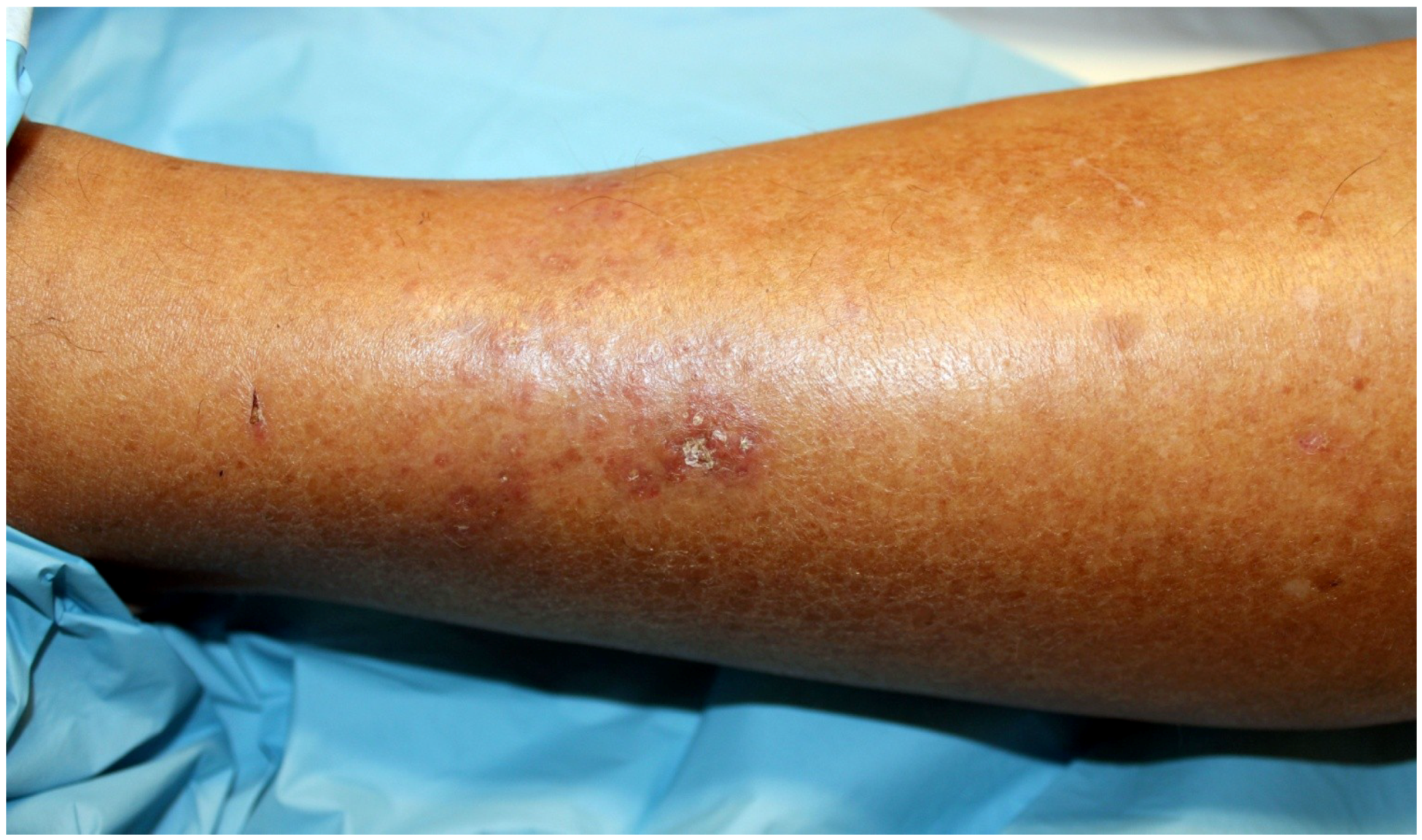
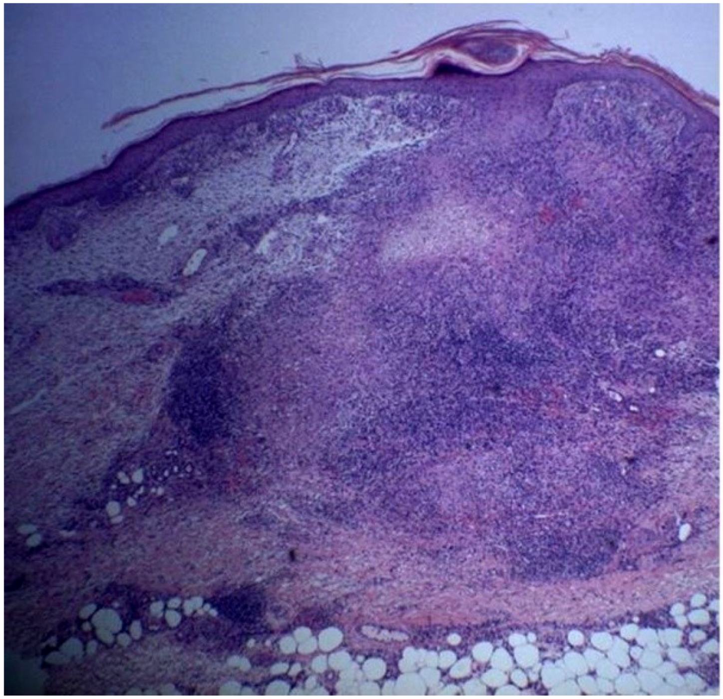
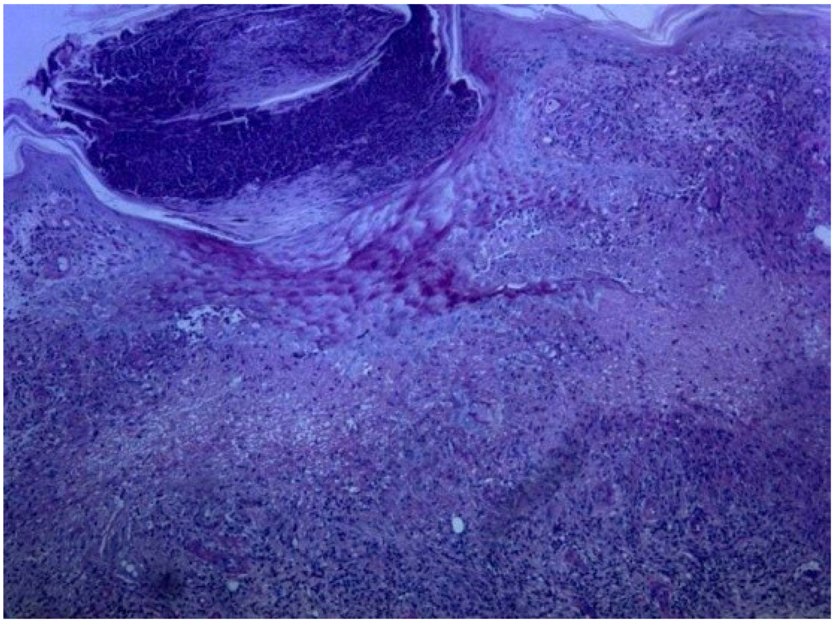
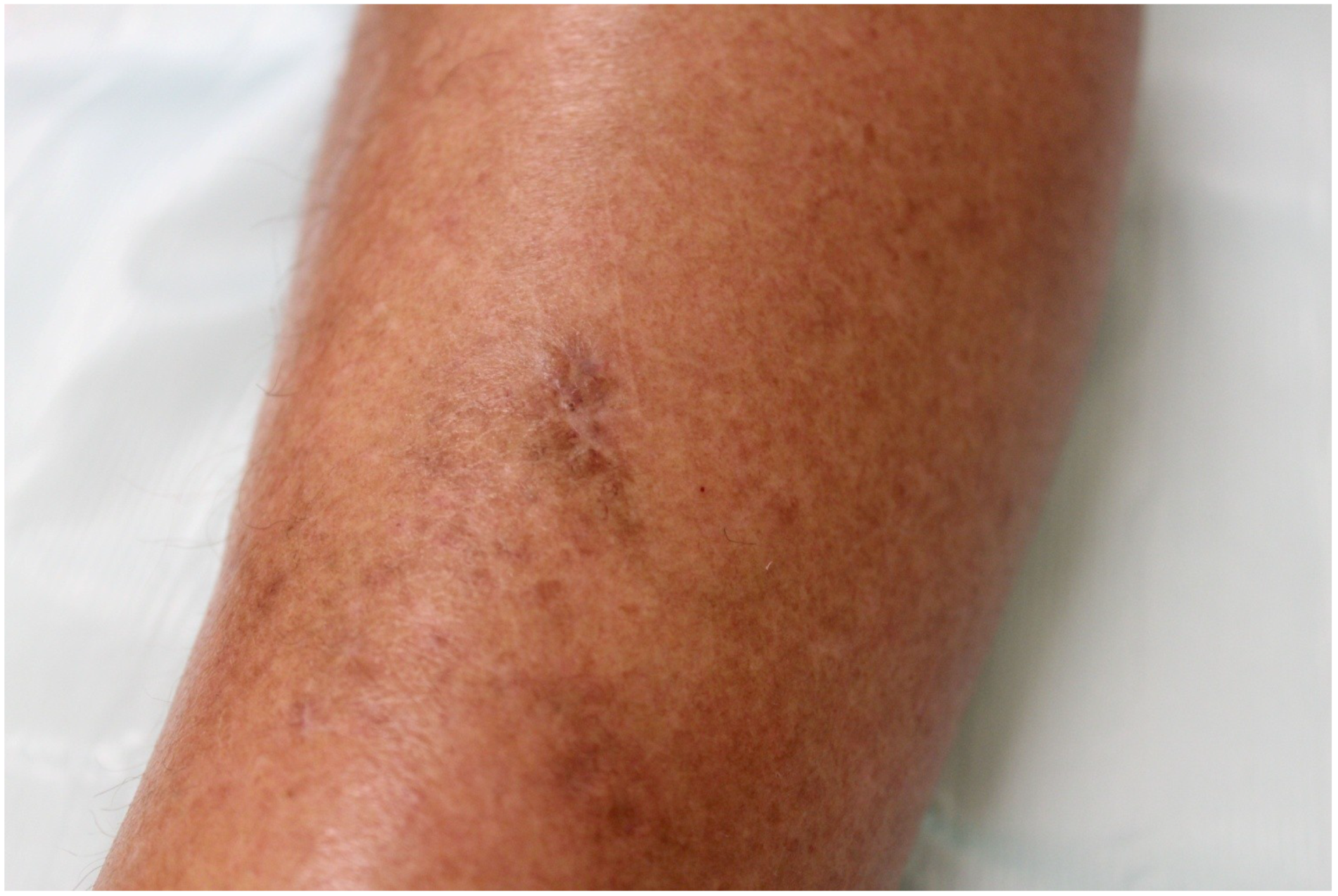
2. Case Report


3. Discussion

4. Conclusions
Author Contributions
Conflicts of Interest
References
- Igraffea, A.; Vinovrski, T.; Zhou, L.H. What is your diagnosis? Perforating granuloma annulare. Cutis 2011, 87, 123–124. [Google Scholar]
- Samlaska, S.P.; Sandberg, G.D.; Maggio, K.L.; Sakas, E.L. Generalized perforating granuloma annulare. J. Am. Acad. Dermatol. 1992, 27, 319–322. [Google Scholar]
- Dornelles, S.I.; Poziomczyk, C.S.; Boff, A.; Köche, B.; Dornelles Mde, A.; Richter, G.K. Generalized perforating granuloma annulare. An. Bras. Dermatol. 2011, 86, 327–331. [Google Scholar]
- Töröcsik, D.; Bárdos, H.; Hatalyák, Z.; Dezső, B.; Losonczy, G.; Paragh, L.; Péter, Z.; Balázs, M.; Remenyik, E.; Adány, R. Detection of factor XIII-A is a valuable tool for distinguishing dendritic cells and tissue macrophages in granuloma annulare and necrobiosis lipoidica. J. Eur. Acad. Dermatol. Venereol. 2014, 28, 1087–1096. [Google Scholar]
- Zaba, L.C.; Fuentes-Duculan, J.; Steinman, R.M.; Krueger, J.G.; Lowes, M.A. Normal human dermis contains distinct populations of CD11c+BDCA-1+ dendritic cells and CD163+FXIII-A+ macrophages. J. Clin. Invest. 2007, 117, 2517–2525. [Google Scholar]
- Penas, P.F.; Jones-Caballero, M.; Fraga, J.; Sánchez-Pérez, J.; García-Díez, A. Perforating granuloma annulare. Int J. Dermatol. 1997, 36, 340–348. [Google Scholar]
- Ismail, A.; Beckum, K.; McKay, K. Transepithelial elimination in sarcoidosis: A frequent finding. J. Cutan. Pathol. 2014, 41, 22–27. [Google Scholar] [PubMed]
- Freiman, A.; Ting, P.; Miller, M.; Greenaway, C. Papulonecrotic tuberculid: A rare form of cutaneous tuberculosis. Cutis 2005, 75, 341–346. [Google Scholar] [PubMed]
- Hoque, S.R.; Ameen, M.; Holden, C.A. Acquired reactive perforating collagenosis: Four patients with a giant variant treated with allopurinol. Br. J. Dermatol. 2006, 154, 759–762. [Google Scholar] [PubMed]
- Golitz, L. Follicular and perforating disorders. J. Cutan. Pathol. 1985, 12, 282–288. [Google Scholar] [PubMed]
- Rios-Buceta, L.; Amigo-Echenaqusia, A.; Sols-Candelas, M.; Fraga-Fernandez, J.; Fernandez-Herrera, J. Elastosis perforans serpiginosa with simultaneous onset in two sisters. Int. J. Dermatol. 1993, 32, 879–881. [Google Scholar]
- Thornsberry, L.A.; English, J.C., 3rd. Etiology, diagnosis, and therapeutic management of granuloma annulare: an update. Am. J. Clin. Dermatol. 2013, 14, 279–290. [Google Scholar] [PubMed]
- Farrar, C.W.; Bell, H.K.; Dobson, C.M.; Sharpe, G.R. Perforating granuloma annulare presenting on the ears. Br. J. Dermatol. 2002, 147, 1026–1028. [Google Scholar] [PubMed]
- Wollina, U. Granuloma annulare disseminatum responding to fumaric acid esters. Dermatol Online J. 2008, 14, 12. [Google Scholar] [PubMed]
- Kerker, B.J.; Huang, C.P.; Morison, W.L. Photochemotherapy of generalized granuloma annulare. Arch. Dermatol. 1990, 126, 359–361. [Google Scholar] [CrossRef] [PubMed]
- Looney, M.; Smith, K.M. Isotretinoin in the treatment of granuloma annulare. Ann. Pharmocother. 2004, 38, 494–497. [Google Scholar] [CrossRef]
- Plotner, A.N.; Mutasim, D.F. Successful treatment of disseminated granuloma annulare with methotrexate. Br. J. Dermatol. 2010, 163, 1123–1124. [Google Scholar] [PubMed]
- Poppe, H.; Poppe, L.M.; Goebeler, M.; Trautmann, A. Treatment of disseminated granuloma annulare with oral vitamin E: “primum nil nocere”. Dermatology 2013, 227, 83–88. [Google Scholar] [CrossRef] [PubMed]
- Murdaca, G.; Colombo, B.M.; Barabino, G.; Caiti, M.; Cagnati, P.; Puppo, F. Anti-tumor necrosis factor-α treatment with infliximab for disseminated granuloma annulare. Am. J. Clin. Dermatol. 2010, 11, 437–439. [Google Scholar] [CrossRef] [PubMed]
- Hertl, M.S.; Haendle, I.; Schuler, G.; Hertl, M. Rapid improvement of recalcitrant disseminated granuloma annulare upon treatment with the tumour necrosis factor-alpha inhibitor, infliximab. Br. J. Dermatol. 2005, 152, 552–555. [Google Scholar] [PubMed]
© 2014 by the authors; licensee MDPI, Basel, Switzerland. This article is an open access article distributed under the terms and conditions of the Creative Commons Attribution license (http://creativecommons.org/licenses/by/3.0/).
Share and Cite
Alves, J.; Barreiros, H.; Bártolo, E. Perforating Granuloma Annulare — An Unusual Subtype of a Common Disease. Healthcare 2014, 2, 338-345. https://doi.org/10.3390/healthcare2030338
Alves J, Barreiros H, Bártolo E. Perforating Granuloma Annulare — An Unusual Subtype of a Common Disease. Healthcare. 2014; 2(3):338-345. https://doi.org/10.3390/healthcare2030338
Chicago/Turabian StyleAlves, João, Hugo Barreiros, and Elvira Bártolo. 2014. "Perforating Granuloma Annulare — An Unusual Subtype of a Common Disease" Healthcare 2, no. 3: 338-345. https://doi.org/10.3390/healthcare2030338