Review

Recent Advances in Electrochemical-Based Sensing Platforms for Aflatoxins Detection

1
Biosensor Lab, Department of Chemistry, BITS, Pilani K. K. Birla Goa Campus, Zuarinagar, 403726 Goa, India
2
BAE Laboratoire, Université de Perpignan Via Domitia, 52 Avenue Paul Alduy, 66860 Perpignan, France
3
Department of Chemistry, National Institute of Technology, Warangal, 506004 Telangana, India
4
Interdisciplinary Research Centre in Biomedical Materials (IRCBM), COMSATS Institute of Information Technology (CIIT), Lahore 54000, Pakistan
*
Author to whom correspondence should be addressed.
Chemosensors 2017, 5(1), 1; https://doi.org/10.3390/chemosensors5010001
Submission received: 25 August 2016 / Revised: 15 December 2016 / Accepted: 20 December 2016 / Published: 26 December 2016
(This article belongs to the Special Issue Electrochemical Immunosensors and Aptasensors)

Abstract

:
Mycotoxin are small (MW ~700 Da), toxic secondary metabolites produced by fungal species that readily colonize crops and contaminate them at both pre- and post-harvesting. Among all, aflatoxins (AFs) are mycotoxins of major significance due to their presence in common food commodities and the potential threat to human health worldwide. Based on the severity of illness and increased incidences of AFs poisoning, a broad range of conventional and analytical detection techniques that could be useful and practical have already been reported. However, due to the variety of structural analogous of these toxins, it is impossible to use one common technique for their analysis. Numerous recent research efforts have been directed to explore alternative detection technologies. Recently, immunosensors and aptasensors have gained promising potential in the area of sample preparation and detection systems. These sensors offer the advantages of disposability, portability, miniaturization, and on-site analysis. In a typical design of an aptasensor, an aptamer (ssDNA or RNA) is used as a bio-recognition element either integrated within or in intimate association with the transducer surface. This review paper is focused on the recent advances in electrochemical immuno- and aptasensing platforms for detection of AFs in real samples.

Graphical Abstract

1. Introduction

With the increasing incidence and stubbornly high mycotoxin mortality around the world, the earlier diagnosis of mycotoxin contamination has drawn significant attention. The presence of mycotoxin in food and feed due to their associated toxic effects on human health and the environment has now became a primary concern [1]. Mycotoxins are the toxic fungal metabolites produced by fungi (micromycetes and macromycetes) under specific conditions of temperature and moisture [2]. The optimal condition of temperature for mycotoxin—producing molds ranging between 24 °C and 35 °C and a relative humidity of ≥70%. Toxicity of these metabolites in human and warm-blooded animals is commonly known as mycotoxicosis. More than 300 mycotoxins (aflatoxins, ochratoxins, trichothecane) commonly exist, but only some of them are practically important. Among all, the most commonly studied groups of mycotoxins are aflatoxins (AFs). Initially, AFs were isolated and identified after the death of young turkeys on poultry farms in England, which were found to be related due to the consumption of a Brazilian peanut meal. AFs are the difuranocoumarin derivatives mainly produced by Aspergillus parasiticus, Aspergillus flavus, and rarely by Aspergillus nomius [3]. AFs are often present in corn, peanuts, nuts, almonds, milk, cheese, and wide varieties of agriculture foodstuffs and beverages [4,5]. They have been classified based on their fluorescent properties under ultraviolet light (365 nm) and chromatographic mobility into different structural analogs, such as B-group (cyclopentane ring, blue fluorescence), G-group (lactone ring, yellow-green fluorescence), and a metabolite of B-group known as AFM1 and AFM2 (Figure 1). Among AFs, AFB1 is the most common and highly toxic contaminant responsible for more than 75% of all AF contamination in food and animal feed [6]. Subsequent exposure of AFB-contaminated feed to lactating animals leads to secretion of AFM1 and M2 in milk through the hydroxylation reaction mechanism. AFM1 and AFM2 are quite stable during milk pasteurization, as well as dairy product processing, which may persist to the final stage during human consumption [7,8,9].

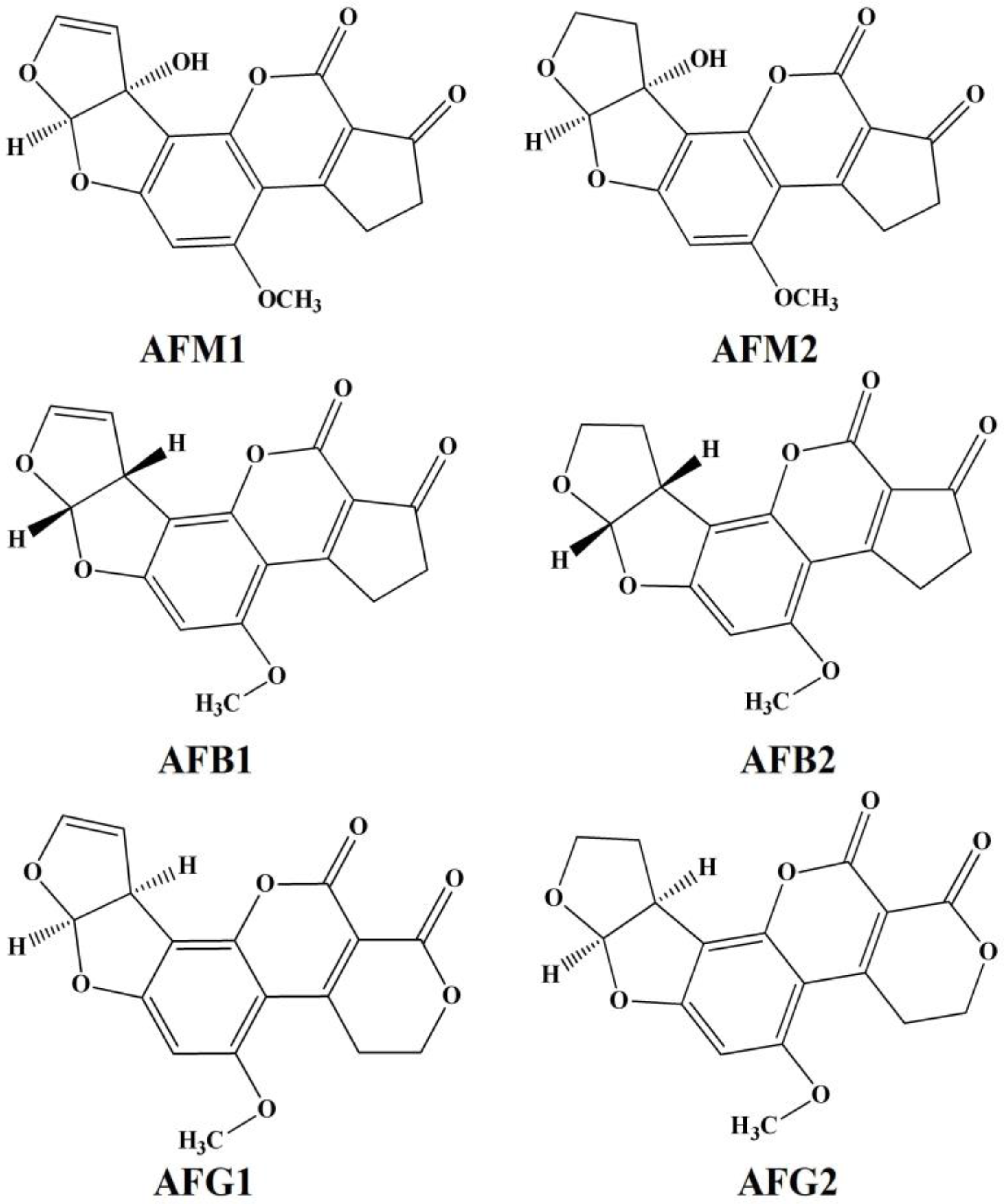
1.1. Toxicity of AFs

The biotransformation of AFB1 comprises the derivatives of AFM1, aflatoxin-exo-8,9-epoxide, AFQ1. Among them, the AFM1 and AFQ1 are less reactive and easily eliminated from the body through urination [10]. However, aflatoxin B1-8,9-exo-epoxide is a known mutagen, which is electrophilic in nature and capable of forming covalent bonds with nucleophilic sites of macromolecules, such as nucleic acids and proteins [11]. These covalent bond formations determines the formation of aflatoxin B1-DNA adducts, which results in mutagenic and carcinogenic effects of AFB1, such as G→T transversion mutation and attacks on the guanine base of DNA. This introduces the mutation in the normal cells and formation of various types of carcinomas in humans, especially in liver [12]. Typically, the AFB1 mutation can cause hepatocellular carcinoma, point mutation, inversion of base sequences (DNA and RNA), and destruction of protein structures [13]. The epoxide attacks at the position of seventh (7th) guanine nitrogen (both DNA and RNA), altering the hybridization of nucleic acids and transcription process. AFB1 has a negative impact on carbohydrate metabolism, which results in the reduction in hepatic glycogen and increased blood glucose levels. Although the toxic effects of AFM1 are less than that of AFB1, nevertheless, it causes the oxidative damage due to intracellular radical generation, DNA intercalation, base impairment, teratogenicity, birth defects, and genetic mutation [14].
According to the report of the Food and Agricultural Organization (FAO) on mycotoxin published in 2004, globally 99 countries had fixed the maximum stringent limits for mycotoxins in food and food products. In 2012, the Rapid Alert System for Food and Feed (RASFF) declared the AFs as one of the principle hazards in European Union [15,16,17]. To minimize the production losses and ensure the safety of human health, the European Commission (EC) has established the maximum stringent limits for most of the mycotoxins in food and food products as mentioned in the Commission Regulation (EC number 1881/2006), as well as through methods of sampling and analysis for their control (EC number 401/2006) [18,19]. Table 1 summarizes the permissible limits of aflatoxins in food by different agencies such as; European Union (EU), US Food and Drugs Administration (USFDA), the Codex Alimentous Commission (CAC), and the Food Safety and Standards Authority of India (FSSAI) [18,20].

1.2. Monitoring of Aflatoxins (AFs)

AF contamination seriously influences the quality of agricultural production, animal feeds, food quality, and other dietary products with potential threats to the human health and the environment, due to economic losses. Considering the above facts, the rapid, sensitive, and accurate detection of AF contamination in food and feed products, agriculture, and exposure levels in the human body require regular screening and risk monitoring. The reported classical methods for AFs detection are based on the chromatographic techniques, such as thin-layer chromatography [21,22,23], high-performance liquid chromatography [24,25], liquid chromatography coupled with mass spectroscopy [26,27,28], high-performance liquid chromatography coupled with fluorescence detection [29,30,31], and liquid chromatography/atmospheric pressure chemical ionization mass spectrometry (LC/APCI-MS) [32]. However, the inherent properties involved in the chromatographic techniques, such as long and complicated sample pre-treatment procedures, expensive instruments, and the requirement of trained technicians, limits their wider utility in high-throughput and on-site analysis of samples. The traditional immunoassays, mainly enzyme-linked immunosorbent assay (ELISA) are commonly used to detect AFs. However, the disadvantages involved in the immunoassay, such as long reaction time, difficulty in the automation of the process, in vivo production of antibodies and low sensitivity in different assays, decrease their involvement in real samples analysis. Some innovation and enhancement in the development of immunoassay such integration of nanomaterial, miniaturization have been reported. Meanwhile, the emergence of biosensing techniques has been witnessed as an alternative to the above problems. In the present review, we will discuss novel electrochemical biosensors and assay platforms for the detection of aflatoxins in different food matrices.

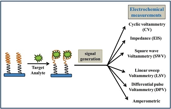
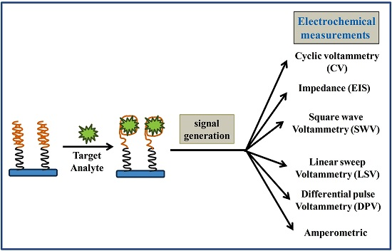
2. Biosensors (as an Alternative Tool)

A biosensor is a compact analytical device used for the detection of a target analyte based on optical, thermal, piezoelectric, and electrochemical signal generation, which are generated by the interaction between the recognition element and analyte of interest [33]. The molecular recognition elements are, consequently, the key for biosensors since their binding affinity and specificity greatly influences the sensor performance. Initially, the recognition elements (antibodies, enzymes, isolated receptors, etc.) were isolated from living organism i.e., goats, mice, horses. The antibodies were generated by animal immunization when it responds to the different antigens such as toxins, drugs, chemicals, virus particles, spores, and other foreign substrates [34]. Currently, synthetic or bio-engineered recognition elements are available in the laboratory, including antibodies, enzymes, molecularly-imprinted polymers, and lectins with the improved features of selectivity and specificity in biosensing. The specific and selective interaction between a particular antibody and an antigen is the basic principle involved in immunoassays. The results obtained from these immunoassays must be reproducible and repeatable in order to enable proper detection of analytes. Depending upon the technique used, immunosensors can be optical [35], mass-sensitive [36], and electrochemical [37]. Among these, electrochemical immunosensors are widely used, since they involve comparatively inexpensive, simple, and easy to use instruments. Immunosensors based on screen-printed electrodes (SPEs) are very convenient to use as they are easy to fabricate, portable, suitable for mass production, and provide inexpensive kits for the rapid and accurate detection and quantification of antigens and antibodies in a sample matrix. However, this is possible only when such a system is thoroughly characterized, well optimized, and immobilized on the surface of the electrodes.
Unfortunately, these recognition elements exhibit certain limitations. For instance, the antibodies and enzymes are sensitive to working pH and temperature, which is reflected in the short shelf life and irreversible denaturation [38]. The need of animal immunization for antibody production, which often involves the animal suffering, batch to batch variation, and difficulty in labeling of the specific recognition site, decreases the wide utility of antibodies. Finally, due to the requirement of immobilization and extensive washing in antibody-based affinity assays, it is difficult to carry out the homogeneous assays [39]. Therefore, it is highly desirable to seek the alternative ligands or recognition elements as a new platform for biosensing and analytical applications.

Aptamer

In the last decades, aptamers have attracted tremendous interest in therapeutic and bioanalytical applications, either used as an active separation material in chromatography or as recognition material in biosensing applications to replace commonly-used bio-receptors [40,41,42,43,44,45]. Aptamers are synthetic oligonucleotides sequences (30–100 nucleotides) with high affinity and specificity to recognize their cognate target molecules, ranging from small ions to large peptides. Upon target recognition, the aptamer folds into a specific 3D structure known as the antiparallel G-quadruplex aptamer complex form. Most of the aptamers are obtained through a combinatorial process called a systemic evolution of ligands by exponential enrichment (SELEX) from vast populations of random sequences. In SELEX, a random oligonucleotides library is exposed to the specific analyte of interest under a set of pH, ionic strength, and temperature conditions. However, it is difficult to optimize the SELEX parameters and select the aptamer sequence with high binding efficiency but, once optimized, it will reflect the sensing environment for the detection of target molecules [46]. The effect of structural analogs or interference against target molecules might hinder the aptamer synthesis. The selection of complex real matrices such as extracts, food, or bacterial samples for testing, ensure that the synthesized aptamer has the potential to work in real samples and detect mycotoxins. Considering these factors, there remain a number of mycotoxins for which aptamers could be selected. Another potential advantage of aptamer technology over the antibody is that the selection of oligonucleotides sequences can be rationally determined and altered to optimize molecular recognition performance. The high binding sequences can be partitioned from the sequences lacking affinity against the target. For small molecules, such as mycotoxins, the SELEX is often achieved by the immobilizing of target molecules to a solid phase or beads, allowing the easy removal of unwanted sequences through multiple washing steps. As recognition elements, aptamers offer many advantages over conventional antibodies. Due to their small size, high affinity, specificity, ease of denaturation, high stability (especially DNA aptamers), ease of modification, and labeling, aptamers have gained significant potential for developing practical, inexpensive and robust biosensing platforms [47,48]. Even though the promising potential of aptamer-based sensing strategies exist in the food industry, therapeutics, and clinical diagnostics, only a few aptamer-based products are commercializedThis review surveys the recent literature dealing with immuno- and aptasensors for AF detection. These studies can open the way to novel analytical devices of commercial interest with several advantages, such as miniaturization, portability, disposability, low sample requirement, and suitability for practical and on-site applications.

3. Electrochemical Immunosensors for AFs Detections

In the existing literature, several electrochemical immunosensing platforms have been reported for AFB1 and AFM1 detections. Firstly, an indirect competitive ELISA was performed on SPE electrodes using DPV analysis for AFB1 detection. The presented method was successfully applied for detection of AFB1 in barley samples with high sensitivity and good recoveries. The SPE-based ELISA showed better analytical performance than spectrophotometric ELISA with a LOD of 30 pg/mL [49]. After one year, a direct HRP-linked chronoamperometric immunosensor was developed for detection of AFM1 in milk samples [50]. Obtained results showed that, using SPEs, AFM1 can be measured up to 25 pg/mL with a dynamic working range between 30 and 160 pg/mL. Meanwhile, in a study by Parker et al., it was concluded that the presence of divalent ion (calcium) is highly recommended to stabilize the milk samples on metal electrodes [51]. For multi-analyte determinations, a competitive ELISA combined with 96-well screen-printed microplate-based multichannel electrochemical detection was developed for AFB1 detection in corn samples using intermittent pulse amperometry (IPA) [52]. The author reported a LOD of 30 pg/mL with the high throughput ELISA procedure. In the last decades, nanomaterial-based signal amplification strategies for conventional ELISA and electrochemical detection have been applied for AFM1 detection [53,54]. For AFB1 detection, an impedimetric immunosensor based on colloidal gold and silver electrodeposition for AFM1 detection was developed by Vig et al. [55]. The signal amplification was carried out by silver electrodeposition using chronoamperometry. The results of calculating charge transfer resistance (EIS signal) correspond to the amount of AFM1 present. Obtained results were further compared with linear sweep voltammetry (LSV) measurements. In the same context, an indirect enzymatic immunosensor for AFB1 detection was fabricated on gold electrodes using signal amplification strategies based on silver electrodeposition. LSV measurements were carried out to quantify the metal silver, which corresponds to the amount of AFB1 in rice samples [37]. Bacher et al., reported a label-free impedimetric immunosensor based on antibody-coupled silver wire for detection of AFM1 in milk samples [56]. The anti-AFM1 mAb and AFM1 interaction were quantified on the basis of impedance change at 10 mV potential. The developed sensor has the highest sensitivity with a LOD of 1 pg/mL with an analysis time of 20 min. In order to miniaturize and improve the sensitivity and selectivity of a conventional electrode system, a gold microelectrode array was used as a platform for AF analysis. Parker et al., reported the development of direct competitive ELISA based on the gold microelectrode array for direct analysis of AFM1 in milk samples [57].
Integration of nanotechnology or nanostructures in biosensing applications improves the analytical performance of the electrochemical biosensing methods owing to their high surface area impact ratio, excellent surface catalytic activity, ease of preparation, and bioconjugation [58]. Glassy carbon electrodes (GCEs) or modified GCE surfaces have been widely used for preparing electrochemical immunosensors for AF detection. After capturing the analyte on the sensor surface, the electrochemical signal change is measured by DPV, EIS, and amperometry measurements [59,60,61,62]. Similarly, single-walled carbon nanotubes (SWNTs) have also been widely used in the development of electrochemical immunosensing platforms. For AFB1 detection in corn powder, an AFB1-BSA immobilized conjugate on SWNTs/chitosan-modified GCE surfaces was used for development of an electrochemical-based indirect competitive immunoassay [63]. Graphene oxide (GO), has been used for fabricating electrochemical immunosensors for mycotoxin detection. Recently, GO based electrochemiluminescent and EIS immunosensors have been developed for AFM1 and AFB1 detection [64,65]. Immunosensing platforms reported for AF detection based on electrochemical signal generation have been summarized in Table 2.

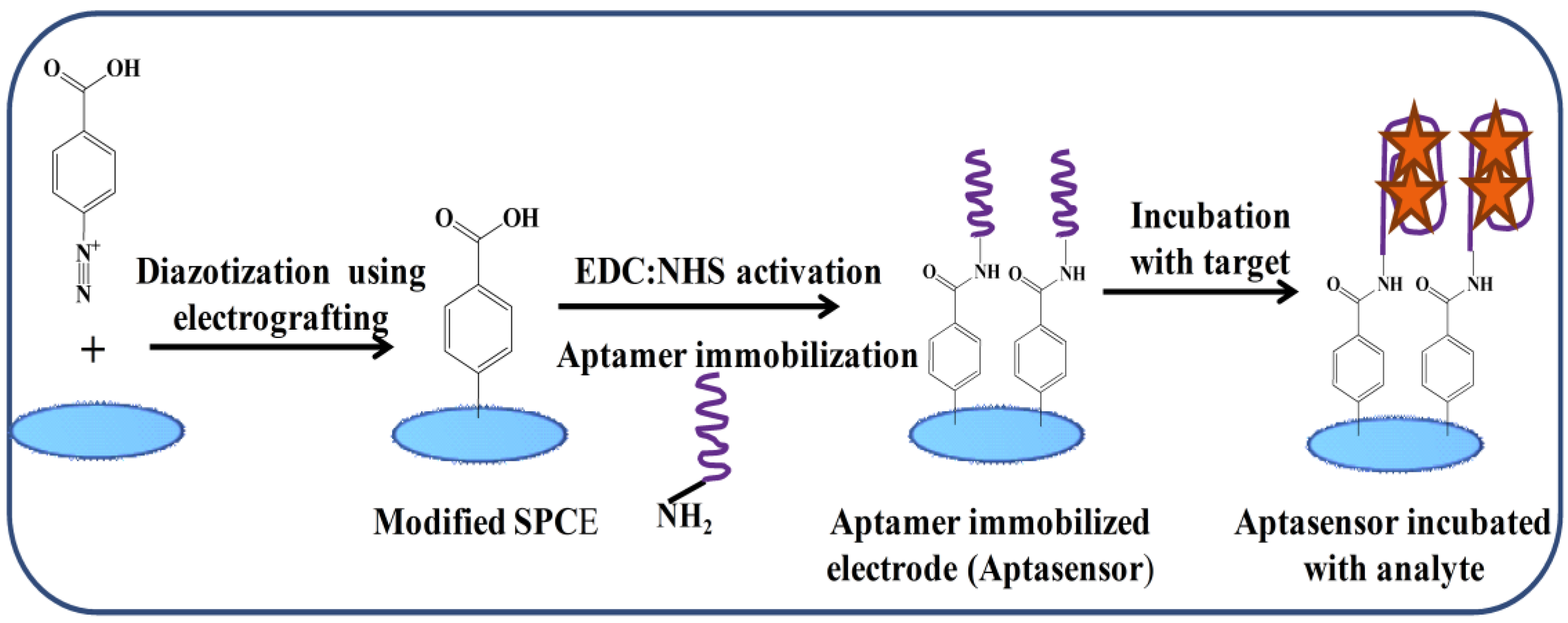
4. Aptamer-Based Sensing Strategies for Aflatoxins (AFs) Detection

Recently, aptamer sequences possessing a high affinity for AFs (different structural analogs) have been reported in either publications or patents (Table 3) [66,67,68,69,70]. In the present scenario, the problems associated with the analysis of complex samples, such as blood, serum, and cellular extracts, have been solved using electrochemical biosensors [71,72].
In the last decades, the development of electrochemical aptasensing platforms has gained considerable attention in the analysis of target analytes. Immobilization of aptamers on the electrode surface is highly important. Several immobilization techniques, such as thiolation, diazonium coupling, and click chemistry, are reported [73,74,75]. Among all, diazotization coupling provides better immobilization impact due to the lack of leakage of bio-recognition elements on storage. The generation of the electrochemical signal corresponds to the amount of analyte present. The electrochemical aptasensors can be easily modified and offer the advantages of high sensitivity, selectivity, stability, compatibility with microfabrication, disposability, portability, high detection speed, and the requirement of low sample volume. Based on these advantages, electrochemical sensors appear to be well suited for practical applications. In the last decade, a large number of papers and reviews have been published in this field.

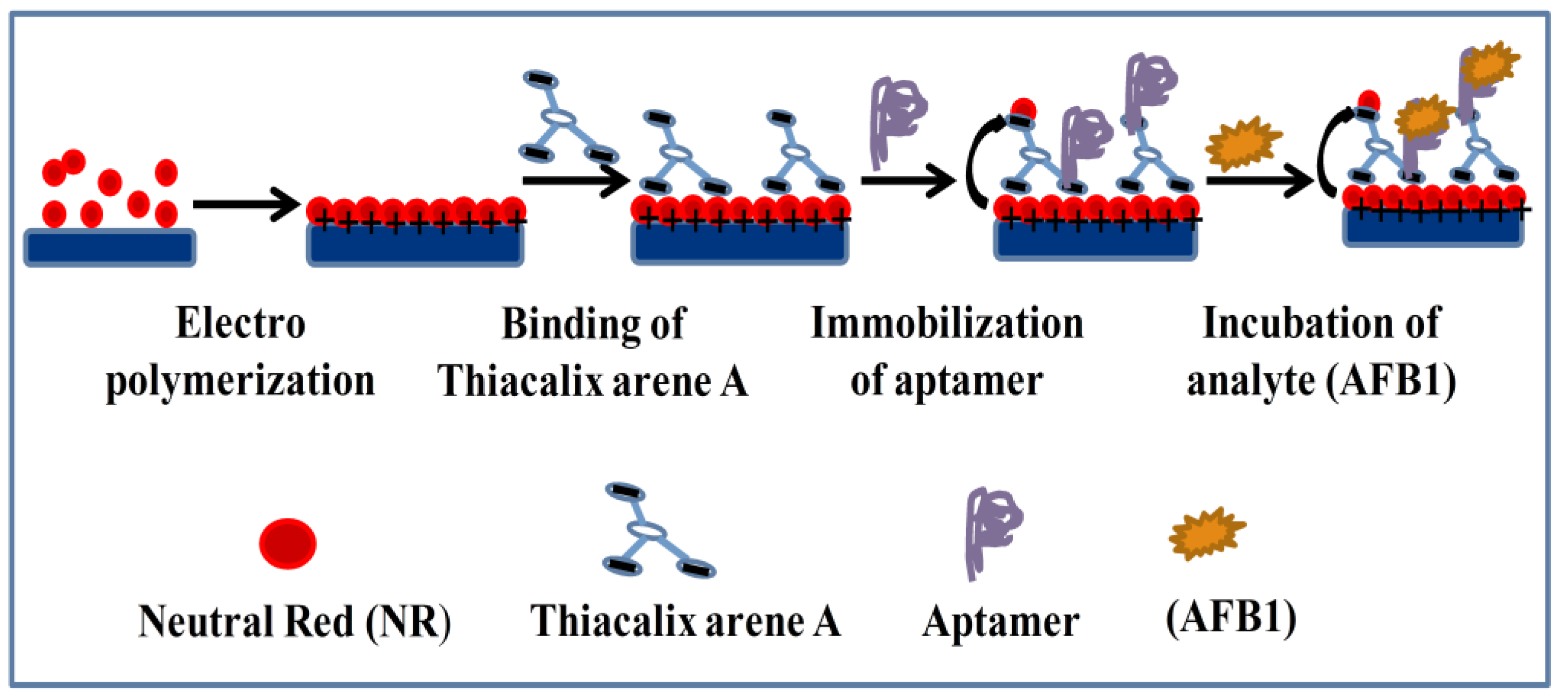
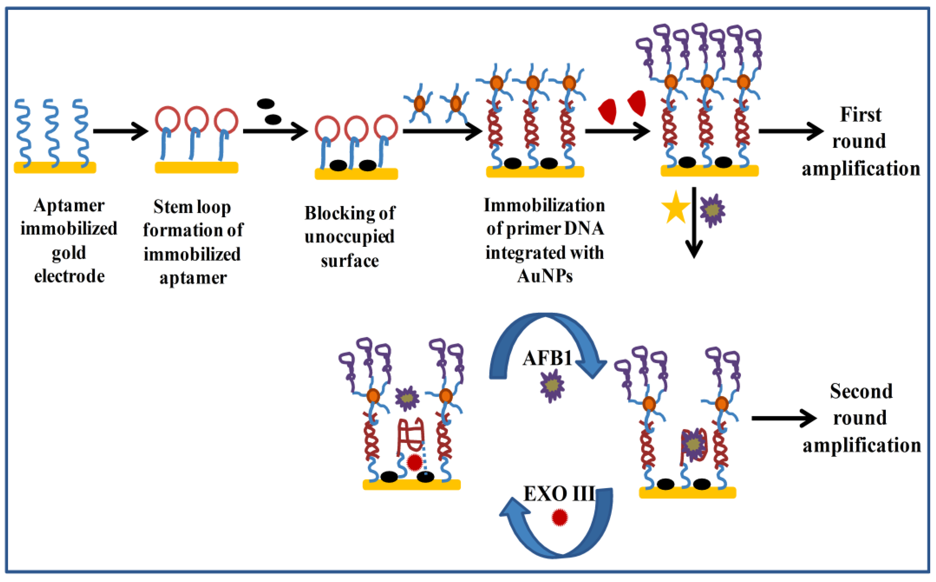
Electrochemical Aptasensors for AFs Detection

In recent years, the development of aptasensors for detection of toxins and environmental pollutants has gained significant attention. The merging of aptamer capabilities and the versatility of nanomaterials has opened new strategies for the amplified detection of mycotoxins. The various developed electrochemical aptasensors for AFs detection has been summarized in Table 4. Nguyen et al. have reported the label-free electrochemical aptasensor for detection of AFM1 [69]. For the construction of electrochemical aptasensor, a Fe3O4 polyaniline (Fe3O4/PANi) film was polymerized on the interdigitated electrode (IDE) for AFM1 detection. Immobilized aptamers as affinity capture reagents, and magnetic nanoparticles for signal amplification were employed in construction of sensing platform. For AFM1 quantification, label-free and direct measurements of the AFM1 aptamer on the Fe3O4/PANi interface were carried out using cyclic (CV) and square wave voltammetry (SWV). The developed aptasensor showed a LOD of 1.98 ng/L with a good sensitivity in the range 6–60 ng/L. Later, the aptasensor performance was successfully demonstrated in milk samples. A DNA biosensor based on the interaction of AFM1 and a self-assembled metal supported lipid bilayer membrane (s-BLMs) and its effect on DNA hybridization was reported [76]. The interactions of AFM1 with s-BLMs was composed of egg phosphatidylcholine, responsible for an increase in ion current, which corresponds to the concentration of toxin. The presence of ssDNA causes an increase in ion current across s-BLMs, whereas the current decrease is due to the formation of double-stranded DNA (dsDNA). The captured signal decreases in the presence of the toxin and increases the time to reach equilibrium. This aptasensor provided the rapid (<1 min) detection and the low detection limit (0.5 nM) of AFM1 based on the measurements of the initial rate of hybridization. Dinckaya et al. reported the development of an impedimetric DNA biosensor for detection of AFM1 in milk and dairy products [77]. The DNA biosensor was constructed by covalent immobilization of the thio-modified single-stranded DNA (ss-HSDNA) on the gold surface using self-assembled monolayer. Using impedance spectroscopy, a detection limit of 1–14 ng/mL was obtained.
Very recently, an impedimetric aptasensor for detection of AFM1 was reported by Istamboulie et al. [78]. In this work, the hexaethyleneglycol-modified oligonucleotides (seven base pair sequences) of anti-AFM1 aptamer were immobilized on the diazotized screen-printed carbon electrode (SPCEs) via a carbodiimide coupling reaction. The fabricated aptasensor was characterized at each step using CV and EIS using ferri/ferrocyanide as a redox probe. A dynamic range of 2–150 ng/L AFM1 was obtained with a LOD of 1.15 ng/L. For real sample analysis, a simple filtration through a 0.2 mm polytetrafluoroethylene (PTFE) membrane was carried out to allow the determination of AFM1 in milk samples.
Castillo et al. reported the development of an electrochemical aptasensor using polyamidoamine PAMAM dendrimers for AFB1 detection [79]. For sensor fabrication, a single-stranded (ss) amino-modified DNA aptamer highly specific to AFB1 was immobilized on the assembly of a multilayer framework of immobilized PAMAM dendrimers on the gold electrode. The CV and EIS measurements were performed to capture the signal response by means of redox indicators: K[Fe(CN)6]3−/4−. The aptasensor allowed AFB1 determination in the range of 0.1–10 nM AFB1. The sensor possesses the LOD of 0.40 ± 0.03 nM, with a stability of 60 h at 4 °C. In previous years, the use of mediators, such as methylene blue, ferrocene, ferri-ferrocyanide, and methylene green in the electrochemical sensor has been successfully reported for a decrease in potential and amplification of signals [83,84,85,86]. Previously, an electrochemical aptasensor based on the electropolymerization of neutral red on the electrode surface for detection of AFB1 has been reported by Evtugyn et al. [80]. The aptasensor was prepared using covalent immobilization of anti-AFB1 DNA aptamer to the polycarboxylated macrocyclic ligand immobilized (Thiacalix arene A) on an electropolymerised layer of neutral red, which acts as a redox probe (Figure 2). For quantitative measurements of AFB1, CV and EIS measurements were carried out. The developed aptasensor showed a LOD of 0.05 nM with EIS in 4-(2-hydroxyethyl)-1-piperazineethanesulfonic acid (HEPES) binding buffer. It was reported that the developed protocol provides the enhancement in stability of the surface layer and improved reproducibility of the voltammetric signal in multiple food matrices, such as peanuts, cashew nuts, white wine, and soy sauce.
Very recently, a label-free EIS aptasensor for the detection of AFB1 in alcoholic beverages [81]. An EIS aptasensor was fabricated over SPCEs via immobilization of anti-AFB1 aptamer using a diazonium coupling mechanism (Figure 3). In this work, the two different sequences of an anti-AFB1 aptamer were used and compared for their analytical performance. On incubation of AFB1, a dynamic detection range from 0.125 to 16 ng/mL was obtained with a LOD of 0.12 and 0.25 ng/mL for seqA and seqB. The performance of the EIS aptasensor was successfully demonstrated in alcoholic beverages (beer and wine samples) with recoveries between 92% and 102%. The developed aptasensor offers the advantages of disposability and portability for on-site analysis. Among the reported electrochemical techniques, one important strategy is the designing of a switchable on-off electrochemical aptasensor, which results in a signal upon target recognition depending upon conformational changes. However, for the “signal-off” electrochemical sensor, it is generally recognized that the suppression of the signal alters the sensitivity and specificity of the developed platforms [87]. Presently, to effectively avoid the inherent drawbacks of signal-off biosensors, the integration of different amplification approaches became attractive, such as in vitro DNA amplification. Based on the above facts, an enzyme-based signal amplification electrochemical aptasensing platform for ultrasensitive detection of AFB1 has been reported by Zheng et al. [82]. In this work, a heteroenzyme-based two-round signal amplification electrochemical aptasensor approach was designed for AFB1 detection. In the first round, the telomerase-based amplification led to the generation of a high current signal, which increases the detection range (Figure 4), whereas in the second round the EXO III-based amplification led to the generation of an observable signal response corresponding to the trace concentrations of AFB1. Based on the advantage of the two-round signal amplification strategy, the sensitivity and detection range of proposed electrochemical aptasensors were greatly improved.

5. Safety Notes

AFs are highly carcinogenic and should be handled with extreme care. After use, the AF-contaminated labwares must be decontaminated with an aqueous solution of sodium hypochlorite (5%). The AFs are subject to light degradation; therefore, the samples must be protected from daylight and standards must be stored in amber-colored vials. For aqueous solutions of AFs, the use of non-acid-washed glassware may result in the loss of AF, thus, special attention and precautions should be paid in cleaning new glassware, which should be soaked in dilute acid (10% sulfuric acid) for several hours and then thoroughly rinsed with distilled water to remove all of the traces of acid [88,89].

6. Conclusions and Future Perspectives

Various analytical methods employed in the analysis of aflatoxins in agricultural, food, crops, and feeds have been reported. Over the well-established antigen-antibody-based (immunosensor) detection systems, the aptamer-based (aptasensors) strategies have been explored due to their inherent practical benefits over the antibodies as recognition elements. Preferably, a detection method should be able to detect the target analyte at very low levels with high specificity. In this context, immunosensors with a very high level of analytical performance; lower LODs, high stability with high precision and accuracy, has been reported. Despite of their numerous advantages, the immunosensors still require some improvements for better analysis of food and environmental samples, whereas the in vitro design and selection of the aptamer sequences allow the unparalleled control over binding conditions and possible cross-reactivity. The SELEX experiments can be carefully designed, including the counter selections against toxins or other possible interferences. Additionally, the selection of aptamer directly in complex matrices, such as extracts from the crops or food, could help to ensure their reliable performances in real-world samples. Considering these factors, there is scope to explore the SELEX process for selection of aptamer against a series of mycotoxins for which aptamers are not known.
It is worth noting that although many sensitive methods have been described for the analysis of AFs, based on electrochemical signal generation. The EIS aptasensors offers the advantages of disposability, portability, miniaturization, and on-site analysis. Therefore, the development of simple, label-free, rapid, and sensitive tools that are based on electrochemical responses can provide versatile, portable, sensitive, and accurate devises for AFs on-site detection. The discussed signal amplification strategies possess the significant potential to overcome bottleneck in the traditional signal-off biosensor. One of the major breakthrough studies could be the integration of signal amplification strategies with sensing platforms based on screen printed electrodes.

Acknowledgements

Atul Sharma (A.S.) and Kotagiri Yugender Goud (K.Y.G.) would like to thanks EUPHRATES (ERASMUS Mundus) Doctoral Fellowship program. A.S. would also acknowledge NFBSFARA (Project No. PHT/4007/2013-14), ICAR, New Delhi, India for Senior Research Fellowship. Authors would like to thank the reviewers and academic editors for their valuable inputs to improve the quality of manuscript.

Conflicts of Interest

The authors declare no conflict of interest.

References

  1. Guo, X.; Wen, F.; Zheng, N.; Luo, Q.; Wang, H.; Wang, H.; Li, S.; Wang, J. Development of an ultrasensitive aptasensor for the detection of aflatoxin B1. Biosens. Bioelectron. 2014, 56, 340–344. [Google Scholar] [CrossRef] [PubMed]
  2. Boonen, J.; Malysheva, S.V.; Taevernier, L.; Diana Di Mavungu, J.; De Saeger, S.; De Spiegeleer, B. Human skin penetration of selected model mycotoxins. Toxicology 2012, 301, 21–32. [Google Scholar] [CrossRef] [PubMed]
  3. Yao, H.; Hruska, Z.; Mavungu, J.D.D. Developments in detection and determination of aflatoxins. World Mycotoxin J. 2015, 8, 181–191. [Google Scholar] [CrossRef]
  4. Bakirci, I. A study on the occurrence of aflatoxin M1 in milk and milk products produced in van province of turkey. Food Control 2001, 12, 47–51. [Google Scholar] [CrossRef]
  5. Sharma, A.; Catanante, G.; Hayat, A.; Istamboulie, G.; Ben Rejeb, I.; Bhand, S.; Marty, J.L. Development of structure switching aptamer assay for detection of aflatoxin M1 in milk sample. Talanta 2016, 158, 35–41. [Google Scholar] [CrossRef] [PubMed]
  6. Ayub, M.; Sachan, D. Dietary factors affecting aflatoxin B1 carcinogenicity. Malays. J. Nutr. 1997, 3, 161–197. [Google Scholar]
  7. Codex Committee on Food Additives and Contaminants. Comments Submitted on the Draft Maximum Level for Aflatoxin M1 in Milk, CL CX/FAC 01/20; Codex Alimentarious Commission Netherlands: Hague, The Netherlands, 2001; pp. 1–9. [Google Scholar]
  8. Stroka, J.; Anklam, E. New strategies for the screening and determination of aflatoxins and the detection of aflatoxin-producing moulds in food and feed. TrAC Trends Anal. Chem. 2002, 21, 90–95. [Google Scholar] [CrossRef]
  9. Badea, M.; Micheli, L.; Messia, M.C.; Candigliota, T.; Marconi, E.; Mottram, T.; Velasco-Garcia, M.; Moscone, D.; Palleschi, G. Aflatoxin M1 determination in raw milk using a flow-injection immunoassay system. Anal. Chim. Acta 2004, 520, 141–148. [Google Scholar] [CrossRef] [Green Version]
  10. Wild, C.P.; Turner, P.C. The toxicology of aflatoxins as a basis for public health decisions. Mutagenesis 2002, 17, 471–481. [Google Scholar] [CrossRef] [PubMed]
  11. Wacoo, A.P.; Wendiro, D.; Vuzi, P.C.; Hawumba, J.F. Methods for detection of aflatoxins in agricultural food crops. J. Appl. Chem. 2014, 2014, 706291. [Google Scholar] [CrossRef]
  12. De Oliveira, C.A.; Germano, P.M. Aflatoxins: Current concepts on mechanisms of toxicity and their involvement in the etiology of hepatocellular carcinoma. Rev. Saude Publ. 1997, 31, 417–424. [Google Scholar]
  13. Levy, D.D.; Groopman, J.D.; Lim, S.E.; Seidman, M.M.; Kraemer, K.H. Sequence specificity of aflatoxin B1-induced mutations in a plasmid replicated in xeroderma pigmentosum and DNA repair proficient human cells. Cancer Res. 1992, 52, 5668–5673. [Google Scholar] [PubMed]
  14. Hamid, A.S.; Tesfamariam, I.G.; Zhang, Y.; Zhang, Z.G. Aflatoxin B1-induced hepatocellular carcinoma in developing countries: Geographical distribution, mechanism of action and prevention. Oncol. Lett. 2013, 5, 1087–1092. [Google Scholar] [PubMed]
  15. Marin, S.; Ramos, A.J.; Cano-Sancho, G.; Sanchis, V. Mycotoxins: Occurrence, toxicology, and exposure assessment. Food Chem. Toxicol. 2013, 60, 218–237. [Google Scholar] [CrossRef] [PubMed]
  16. Official Journal of the European Union. Commission Regulation (EU) No 165/2010 of 26 February 2010 Amending Regulation (EC) No 1881/2006 Setting Maximum Levels for Certain Contaminants in Foodstuffs as Regards Aflatoxins; OJ L 50; Official Journal of the European Union: Brusseles, Belgium, 2010; pp. 8–126. [Google Scholar]
  17. Official Journal of the European Union. Commission Regulation (EU) No 1058/2012 of 12 November 2012 Amending Regulation (EC) No 1881/2006 as Regards Maximum Levels for Aflatoxins in Dried Figs Text with EEA Relevance; OJ L 313; Official Journal of the European Union: Brusseles, Belgium, 2012; pp. 14–15. [Google Scholar]
  18. Official Journal of the European Union. Commission Regulation (EC) No. 1881/2006 of 19 December 2006 Setting Maximum Levels for Certain Contaminants in Foodstuffs; Official Journal of the European Union: Brusseles, Belgium, 2006; pp. L364/5–L364/24. [Google Scholar]
  19. Official Journal of the European Union. Commission Regulation (EC) No. 401/2006 Laying down the Methods of Sampling and Analysis for the Official Control of the Levels of Mycotoxins in Foodstuffs; Official Journal of the European Union: Brusseles, Belgium, 2006; Volume L70, p. 1234. [Google Scholar]
  20. Dorsm, G.C.; Caldas, S.C.; Feddern, V.; Bemvenuti, R.; Hackbart, H.C.D.S.; De Souza, M.M.; Oliveira, M.D.S.; Buffon, J.G.; Primel, E.G.; Furlong, E.B. Aflatoxins: Contamination, Analysis and Control, Aflatoxins-Biochemistry and Molecular Biology; Guevara-Gonzalez, R.G., Ed.; InTech: Rijeka, Croatia, 2011. [Google Scholar]
  21. Soares, L.M.; Rodriguez-Amaya, D.B. Survey of aflatoxins, ochratoxin a, zearalenone, and sterigmatocystin in some brazilian foods by using multi-toxin thin-layer chromatographic method. J Assoc. Off. Anal. Chem. 1989, 72, 22–26. [Google Scholar] [PubMed]
  22. Stroka, J.; Otterdijk, R.V.; Anklam, E. Immunoaffinity column clean-up prior to thin-layer chromatography for the determination of aflatoxins in various food matrices. J. Chromatogr. A 2000, 904, 251–256. [Google Scholar] [CrossRef]
  23. Stroka, J.; Anklam, E. Development of a simplified densitometer for the determination of aflatoxins by thin-layer chromatography. J. Chromatogr. A 2000, 904, 263–268. [Google Scholar] [CrossRef]
  24. Rodríguez Velasco, M.L.; Calonge Delso, M.M.; Ordónez Escudero, D. ELISA and HPLC determination of the occurrence of aflatoxin M1 in raw cow’s milk. Food Addit. Contam. 2003, 20, 276–280. [Google Scholar] [CrossRef] [PubMed]
  25. Turner, N.W.; Subrahmanyam, S.; Piletsky, S.A. Analytical methods for determination of mycotoxins: A review. Anal. Chim. Acta 2009, 632, 168–180. [Google Scholar] [CrossRef] [PubMed]
  26. Spanjer, M.C.; Rensen, P.M.; Scholten, J.M. LC–MS/MS multi-method for mycotoxins after single extraction, with validation data for peanut, pistachio, wheat, maize, cornflakes, raisins and figs. Food Addit. Contam. Part A 2008, 25, 472–489. [Google Scholar] [CrossRef] [PubMed]
  27. Vahl, M.; Jørgensen, K. Determination of aflatoxins in food using LC/MS/MS. Zeitschrift für Lebensmitteluntersuchung und Forschung A 1998, 206, 243–245. (In German) [Google Scholar] [CrossRef]
  28. Monbaliu, S.; Van Poucke, C.; Detavernier, C.L.; Dumoulin, F.; Van De Velde, M.; Schoeters, E.; Van Dyck, S.; Averkieva, O.; Van Peteghem, C.; De Saeger, S. Occurrence of mycotoxins in feed as analyzed by a multi-mycotoxin lc-ms/ms method. J. Agric. Food Chem. 2010, 58, 66–71. [Google Scholar] [CrossRef] [PubMed]
  29. Braga, S.M.; de Medeiros, F.D.; de Oliveira, E.J.; Macedo, R.O. Development and validation of a method for the quantitative determination of aflatoxin contaminants in maytenus ilicifolia by HPLC with fluorescence detection. Phytochem. Anal. 2005, 16, 267–271. [Google Scholar] [CrossRef] [PubMed]
  30. Navas, S.A.; Sabino, M.; Rodriguez-Amaya, D.B. Aflatoxin M1 and ochratoxin a in a human milk bank in the city of São Paulo, Brazil. Food Addit. Contam. 2005, 22, 457–462. [Google Scholar] [CrossRef] [PubMed]
  31. Iha, M.H.; Barbosa, C.B.; Favaro, R.M.; Trucksess, M.W. Chromatographic method for the determination of aflatoxin M1 in cheese, yogurt, and dairy beverages. J. AOAC Int. 2011, 94, 1513–1518. [Google Scholar] [CrossRef] [PubMed]
  32. Tanaka, H.; Takino, M.; Sugita-Konishi, Y.; Tanaka, T. Development of a liquid chromatography/time-of-flight mass spectrometric method for the simultaneous determination of trichothecenes, zearalenone and aflatoxins in foodstuffs. Rapid Commun. Mass Spectrom. 2006, 20, 1422–1428. [Google Scholar] [CrossRef] [PubMed]
  33. Thevenot, D.R.; Tóth, K.; Durst, R.A.; Wilson, G.S. Electrochemical biosensors: Recommended definitions and classification. Pure Appl. Chem. 1999, 71, 2333–2338. [Google Scholar] [CrossRef]
  34. Radi, A.-E. Electrochemical aptamer-based biosensors: Recent advances and perspectives. Int. J. Electrochem. 2011, 2011, 863196. [Google Scholar] [CrossRef]
  35. Sassolas, A.; Catanante, G.; Hayat, A.; Marty, J.-L. Development of an efficient protein phosphatase-based colorimetric test for okadaic acid detection. Anal. Chim. Acta 2011, 702, 262–268. [Google Scholar] [CrossRef] [PubMed]
  36. Mishra, G.K.; Sharma, A.; Bhand, S. Ultrasensitive detection of streptomycin using flow injection analysis-electrochemical quartz crystal nanobalance (FIA-EQCN) biosensor. Biosens. Bioelectron. 2015, 67, 532–539. [Google Scholar] [CrossRef] [PubMed]
  37. Tan, Y.; Chu, X.; Shen, G.-L.; Yu, R.-Q. A signal-amplified electrochemical immunosensor for aflatoxin B1 determination in rice. Anal. Biochem. 2009, 387, 82–86. [Google Scholar] [CrossRef] [PubMed]
  38. Jayasena, S.D. Aptamers: An emerging class of molecules that rival antibodies in diagnostics. Clin. Chem. 1999, 45, 1628–1650. [Google Scholar] [PubMed]
  39. Zhou, W.; Huang, P.-J.J.; Ding, J.; Liu, J. Aptamer-based biosensors for biomedical diagnostics. Analyst 2014, 139, 2627–2640. [Google Scholar] [CrossRef] [PubMed]
  40. Ellington, A.D.; Szostak, J.W. In vitro selection of RNA molecules that bind specific ligands. Nature 1990, 346, 818–822. [Google Scholar] [CrossRef] [PubMed]
  41. Stojanovic, M.N.; de Prada, P.; Landry, D.W. Fluorescent sensors based on aptamer self-assembly. J. Am. Chem. Soc. 2000, 122, 11547–11548. [Google Scholar] [CrossRef]
  42. Bunka, D.H.J.; Stockley, P.G. Aptamers come of age—At last. Nat. Rev. Microbiol. 2006, 4, 588–596. [Google Scholar] [CrossRef] [PubMed]
  43. Keefe, A.D.; Pai, S.; Ellington, A. Aptamers as therapeutics. Nat. Rev. Drug Discov. 2010, 9, 537–550. [Google Scholar] [CrossRef] [PubMed]
  44. Hayat, A.; Marty, J.L. Aptamer based electrochemical sensors for emerging environmental pollutants. Front. Chem. 2014, 2, 41. [Google Scholar] [CrossRef] [PubMed]
  45. Sharma, A.; Hayat, A.; Mishra, R.K.; Catanante, G.; Shahid, S.A.; Bhand, S.; Marty, J.L. Design of a fluorescence aptaswitch based on the aptamer modulated nano-surface impact on the fluorescence particles. RSC Adv. 2016, 6, 65579–65587. [Google Scholar] [CrossRef]
  46. McKeague, M.; McConnell, E.M.; Cruz-Toledo, J.; Bernard, E.D.; Pach, A.; Mastronardi, E.; Zhang, X.; Beking, M.; Francis, T.; Giamberardino, A.; et al. Analysis of in vitro aptamer selection parameters. J. Mol. Evol. 2015, 81, 150–161. [Google Scholar] [CrossRef] [PubMed]
  47. Mairal, T.; Ozalp, V.C.; Lozano Sanchez, P.; Mir, M.; Katakis, I.; O’Sullivan, C.K. Aptamers: Molecular tools for analytical applications. Anal. Bioanal. Chem. 2008, 390, 989–1007. [Google Scholar] [CrossRef] [PubMed]
  48. Wang, J.; Lv, R.; Xu, J.; Xu, D.; Chen, H. Characterizing the interaction between aptamers and human IgE by use of surface plasmon resonance. Anal. Bioanal. Chem. 2008, 390, 1059–1065. [Google Scholar] [CrossRef] [PubMed]
  49. Ammida, N.H.S.; Micheli, L.; Palleschi, G. Electrochemical immunosensor for determination of aflatoxin B1 in barley. Anal. Chim. Acta 2004, 520, 159–164. [Google Scholar] [CrossRef]
  50. Micheli, L.; Grecco, R.; Badea, M.; Moscone, D.; Palleschi, G. An electrochemical immunosensor for aflatoxin m1 determination in milk using screen-printed electrodes. Biosens. Bioelectron. 2005, 21, 588–596. [Google Scholar] [CrossRef] [PubMed] [Green Version]
  51. Parker, C.O.; Tothill, I.E. Development of an electrochemical immunosensor for aflatoxin M1 in milk with focus on matrix interference. Biosens. Bioelectron. 2009, 24, 2452–2457. [Google Scholar] [CrossRef] [PubMed] [Green Version]
  52. Piermarini, S.; Micheli, L.; Ammida, N.H.S.; Palleschi, G.; Moscone, D. Electrochemical immunosensor array using a 96-well screen-printed microplate for aflatoxin B1 detection. Biosens. Bioelectron. 2007, 22, 1434–1440. [Google Scholar] [CrossRef] [PubMed] [Green Version]
  53. Kanungo, L.; Pal, S.; Bhand, S. Miniaturised hybrid immunoassay for high sensitivity analysis of aflatoxin M1 in milk. Biosens. Bioelectron. 2011, 26, 2601–2606. [Google Scholar] [CrossRef] [PubMed]
  54. Paniel, N.; Radoi, A.; Marty, J.-L. Development of an electrochemical biosensor for the detection of aflatoxin M1 in milk. Sensors 2010, 10, 9439–9448. [Google Scholar] [CrossRef] [PubMed]
  55. Vig, A.; Radoi, A.; Muñoz-Berbel, X.; Gyemant, G.; Marty, J.-L. Impedimetric aflatoxin M1 immunosensor based on colloidal gold and silver electrodeposition. Sens. Actuators B Chem. 2009, 138, 214–220. [Google Scholar] [CrossRef]
  56. Bacher, G.; Pal, S.; Kanungo, L.; Bhand, S. A label-free silver wire based impedimetric immunosensor for detection of aflatoxin M1 in milk. Sens. Actuators B Chem. 2012, 168, 223–230. [Google Scholar] [CrossRef]
  57. Parker, C.O.; Lanyon, Y.H.; Manning, M.; Arrigan, D.W.; Tothill, I.E. Electrochemical immunochip sensor for aflatoxin M1 detection. Anal. Chem. 2009, 81, 5291–5298. [Google Scholar] [CrossRef] [PubMed]
  58. Wang, X.; Niessner, R.; Tang, D.; Knopp, D. Nanoparticle-based immunosensors and immunoassays for aflatoxins. Anal. Chim. Acta 2016, 912, 10–23. [Google Scholar] [CrossRef] [PubMed]
  59. Masoomi, L.; Sadeghi, O.; Banitaba, M.H.; Shahrjerdi, A.; Davarani, S.S.H. A non-enzymatic nanomagnetic electro-immunosensor for determination of aflatoxin b1 as a model antigen. Sens. Actuators B Chem. 2013, 177, 1122–1127. [Google Scholar] [CrossRef]
  60. Owino, J.; Arotiba, O.; Hendricks, N.; Songa, E.; Jahed, N.; Waryo, T.; Ngece, R.; Baker, P.; Iwuoha, E. Electrochemical immunosensor based on polythionine/gold nanoparticles for the determination of aflatoxin B1. Sensors 2008, 8, 8262–8274. [Google Scholar] [CrossRef] [PubMed]
  61. Zaijun, L.; Zhongyun, W.; Xiulan, S.; Yinjun, F.; Peipei, C. A sensitive and highly stable electrochemical impedance immunosensor based on the formation of silica gel-ionic liquid biocompatible film on the glassy carbon electrode for the determination of aflatoxin B1 in bee pollen. Talanta 2010, 80, 1632–1637. [Google Scholar] [CrossRef] [PubMed]
  62. Sun, A.-L.; Qi, Q.-A.; Dong, Z.-L.; Liang, K.Z. An electrochemical enzyme immunoassay for aflatoxin B1 based on bio-electrocatalytic reaction with room-temperature ionic liquid and nanoparticle-modified electrodes. Sens. Instrum. Food Qual. Saf. 2008, 2, 43–50. [Google Scholar] [CrossRef]
  63. Zhang, X.; Li, C.-R.; Wang, W.-C.; Xue, J.; Huang, Y.-L.; Yang, X.-X.; Tan, B.; Zhou, X.-P.; Shao, C.; Ding, S.-J.; et al. A novel electrochemical immunosensor for highly sensitive detection of aflatoxin B1 in corn using single-walled carbon nanotubes/chitosan. Food Chem. 2016, 192, 197–202. [Google Scholar] [CrossRef] [PubMed]
  64. Gan, N.; Zhou, J.; Xiong, P.; Hu, F.; Cao, Y.; Li, T.; Jiang, Q. An ultrasensitive electrochemiluminescent immunoassay for aflatoxin M1 in milk, based on extraction by magnetic graphene and detection by antibody-labeled cdte quantumn dots-carbon nanotubes nanocomposite. Toxins 2013, 5, 865–883. [Google Scholar] [CrossRef] [PubMed]
  65. Wang, D.; Hu, W.; Xiong, Y.; Xu, Y.; Li, M.C. Multifunctionalized reduced graphene oxide-doped polypyrrole/pyrrolepropylic acid nanocomposite impedimetric immunosensor to ultra-sensitively detect small molecular aflatoxin B1. Biosens. Bioelectron. 2015, 63, 185–189. [Google Scholar] [CrossRef] [PubMed]
  66. Le, C.L.; Cruz-Aguado, J.; Penner, G.A. DNA Ligands for Aflatoxin and Zearalenone. US Patent No. US20120225494 A1, 6 September 2012. [Google Scholar]
  67. Ma, X.; Wang, W.; Chen, X.; Xia, Y.; Wu, S.; Duan, N.; Wang, Z. Selection, identification, and application of aflatoxin B1 aptamer. Eur. Food Res. Technol. 2014, 238, 919–925. [Google Scholar] [CrossRef]
  68. Ma, X.; Wang, W.; Chen, X.; Xia, Y.; Duan, N.; Wu, S.; Wang, Z. Selection, characterization and application of aptamers targeted to aflatoxin B2. Food Control 2015, 47, 545–551. [Google Scholar] [CrossRef]
  69. Nguyen, B.H.; Tran, L.D.; Do, Q.P.; Nguyen, H.L.; Tran, N.H.; Nguyen, P.X. Label-free detection of aflatoxin M1 with electrochemical Fe3O4/polyaniline-based aptasensor. Mater. Sci. Eng. C 2013, 33, 2229–2234. [Google Scholar] [CrossRef] [PubMed]
  70. Malhotra, S.; Pandey, A.K.; Rajput, Y.S.; Sharma, R. Selection of aptamers for aflatoxin M1 and their characterization. J. Mol. Recognit. 2014, 27, 493–500. [Google Scholar] [CrossRef] [PubMed]
  71. Zuo, X.; Song, S.; Zhang, J.; Pan, D.; Wang, L.; Fan, C. A target-responsive electrochemical aptamer switch (TREAS) for reagentless detection of nanomolar ATP. J. Am. Chem. Soc. 2007, 129, 1042–1043. [Google Scholar] [CrossRef] [PubMed]
  72. Zuo, X.; Xiao, Y.; Plaxco, K.W. High specificity, electrochemical sandwich assays based on single aptamer sequences and suitable for the direct detection of small-molecule targets in blood and other complex matrices. J. Am. Chem. Soc. 2009, 131, 6944–6945. [Google Scholar] [CrossRef] [PubMed]
  73. Adams, N.M.; Jackson, S.R.; Haselton, F.R.; Wright, D.W. Design, synthesis, and characterization of nucleic-acid-functionalized gold surfaces for biomarker detection. Langmuir 2012, 28, 1068–1082. [Google Scholar] [CrossRef] [PubMed]
  74. Goud, K.Y.; Sharma, A.; Hayat, A.; Catanante, G.; Gobi, K.V.; Gurban, A.M.; Marty, J.L. Tetramethyl-6-carboxyrhodamine quenching-based aptasensing platform for aflatoxin B1: Analytical performance comparison of two aptamers. Anal. Biochem. 2016, 508, 19–24. [Google Scholar] [CrossRef] [PubMed]
  75. Hayat, A.; Sassolas, A.; Marty, J.-L.; Radi, A.-E. Highly sensitive ochratoxin A impedimetric aptasensor based on the immobilization of azido-aptamer onto electrografted binary film via click chemistry. Talanta 2013, 103, 14–19. [Google Scholar] [CrossRef] [PubMed]
  76. Siontorou, C.G.; Nikolelis, D.P.; Miernik, A.; Krull, U.J. Rapid methods for detection of aflatoxin M1 based on electrochemical transduction by self-assembled metal-supported bilayer lipid membranes (s-BLMs) and on interferences with transduction of DNA hybridization. Electrochim. Acta 1998, 43, 3611–3617. [Google Scholar] [CrossRef]
  77. Dinçkaya, E.; Kınık, Ö.; Sezgintürk, M.K.; Altuğ, Ç.; Akkoca, A. Development of an impedimetric aflatoxin M1 biosensor based on a DNA probe and gold nanoparticles. Biosens. Bioelectron. 2011, 26, 3806–3811. [Google Scholar] [CrossRef] [PubMed]
  78. Istamboulié, G.; Paniel, N.; Zara, L.; Granados, L.R.; Barthelmebs, L.; Noguer, T. Development of an impedimetric aptasensor for the determination of aflatoxin M1 in milk. Talanta 2016, 146, 464–469. [Google Scholar] [CrossRef] [PubMed]
  79. Castillo, G.; Spinella, K.; Poturnayová, A.; Šnejdárková, M.; Mosiello, L.; Hianik, T. Detection of aflatoxin B1 by aptamer-based biosensor using pamam dendrimers as immobilization platform. Food Control 2015, 52, 9–18. [Google Scholar] [CrossRef]
  80. Evtugyn, G.; Porfireva, A.; Stepanova, V.; Sitdikov, R.; Stoikov, I.; Nikolelis, D.; Hianik, T. Electrochemical aptasensor based on polycarboxylic macrocycle modified with neutral red for aflatoxin B1 detection. Electroanalysis 2014, 26, 2100–2109. [Google Scholar] [CrossRef]
  81. Yugender Goud, K.; Catanante, G.; Hayat, A.; Satyanarayana, M.; Vengatajalabathy Gobi, K.; Marty, J.L. Disposable and portable electrochemical aptasensor for label free detection of aflatoxin B1 in alcoholic beverages. Sens. Actuators B Chem. 2016, 235, 466–473. [Google Scholar] [CrossRef]
  82. Zheng, W.; Teng, J.; Cheng, L.; Ye, Y.; Pan, D.; Wu, J.; Xue, F.; Liu, G.; Chen, W. Hetero-enzyme-based two-round signal amplification strategy for trace detection of aflatoxin B1 using an electrochemical aptasensor. Biosens. Bioelectron. 2016, 80, 574–581. [Google Scholar] [CrossRef] [PubMed]
  83. Wu, J.; Chu, H.; Mei, Z.; Deng, Y.; Xue, F.; Zheng, L.; Chen, W. Ultrasensitive one-step rapid detection of ochratoxin a by the folding-based electrochemical aptasensor. Anal. Chim. Acta 2012, 753, 27–31. [Google Scholar] [CrossRef] [PubMed]
  84. Evtugyn, G.; Porfireva, A.; Ivanov, A.; Konovalova, O.; Hianik, T. Molecularly imprinted polymerized methylene green as a platform for electrochemical sensing of aptamer–thrombin interactions. Electroanalysis 2009, 21, 1272–1277. [Google Scholar] [CrossRef]
  85. Wang, Y.; He, X.; Wang, K.; Ni, X.; Su, J.; Chen, Z. Electrochemical detection of thrombin based on aptamer and ferrocenylhexanethiol loaded silica nanocapsules. Biosens. Bioelectron. 2011, 26, 3536–3541. [Google Scholar] [CrossRef] [PubMed]
  86. Kwon, D.; Jeong, H.; Chung, B.H. Label-free electrochemical detection of human α-thrombin in blood serum using ferrocene-coated gold nanoparticles. Biosens. Bioelectron. 2011, 28, 454–458. [Google Scholar] [CrossRef] [PubMed]
  87. Yin, H.; Zhou, Y.; Chen, C.; Zhu, L.; Ai, S. An electrochemical signal ‘off-on’ sensing platform for microrna detection. Analyst 2012, 137, 1389–1395. [Google Scholar] [CrossRef] [PubMed]
  88. Dragacci, S.; Grosso, F. Immunoaffinity column cleanup with liquid chromatography for determination of aflatoxinm1 in liquid milk: Collaborative study. J. AOAC Int. 2001, 84, 437–443. [Google Scholar] [PubMed]
  89. Kanungo, L.; Bhand, S. Fluorimetric immunoassay for multianalysis of aflatoxins. J. Anal. Methods Chem. 2013, 2013, 584964. [Google Scholar] [CrossRef] [PubMed]
Figure 1. Structure of commonly found aflatoxins (AFs).
Chemosensors 05 00001 g001
Figure 2. Schematic representation of an electrochemical aptasensor used for determination of AFB1 using electropolymerized modified electrodes (scheme illustration from [80]).
Chemosensors 05 00001 g002
Figure 3. Schematic representation of an electrochemical aptasensor used for determination of AFB1 using diazotized SPCEs (scheme illustration from [81]).
Chemosensors 05 00001 g003
Figure 4. Schematic representation of two signals amplified signal on an electrochemical aptasensor for AFB1 detection (scheme illustration from [82]).
Chemosensors 05 00001 g004
Table 1. The regulatory standard for aflatoxins (AFs).
MatrixMaximum Permissible Level
EUUSFDACACFSSAI
Milk and Milk based products25 pg/mL (AFM1)500 pg/mL (AFM1)500 pg/mL (AFM1)500 pg/mL (AFM1)
50 pg/mL (AFM1)
Nuts and dried food5 µg/kg (AFB1)20 µg/kg-TotalNot specified30 µg/kg
10 µg/kg-Total
Groundnuts & dried fruits and their processed products2 µg/kg (AFB1)20 µg/kg-Total15 µg/kg-Total30 µg/kg
4 µg/kg-Total
Cereals2 µg/kg (AFB1)20 µg/kg-TotalNot specified30 µg/kg
4 µg/kg-Total
Table 2. Summary of literature reports describing electrochemical immunosensors for aflatoxins detection.
AnalyteMethodLODMatrixReference
AFB1ELISA with DPV30 pg/mLBarley[49]
AFM1Amperomertic25 pg/mLMilk[50]
AFB1intermittent pulse amperometry (IPA)30 pg/mLCorn[52]
AFM1EIS15 ng/LMilk[55]
AFB1LSV0.06 ng/mLRice[37]
AFM1EIS1 pg/mLMilk[56]
AFM1EIS8 ng/LMilk[57]
AFB1DPV0.07 ng/mL-[58]
AFM1DPV0.2 ng/mLPBS[59]
0.7 ng/mL-[60]
AFB1EIS0.01 ng/mLBee pollen[61]
AFB1Amperometric0.05 ng/mLHuman serum and grape samples[62]
AFB1DPV3.5 pg/mLCorn powder[63]
AFM1Electrochemiluminescent (ECL)0.3 pg/mLMilk[64]
AFB1EIS0.5 pg/mLCorn samples[65]
Table 3. Aptamer sequences for aflatoxins (AFs).
TargetAptamer LengthBase Pair Sequences (No.)SequencesKd (nM)Ref.
AFB15016GTTGGGCACGTGTTGTCTCTCTGTGTCTCGTGCCCTTCGCTAGGCCCACAN.R.[66]
8026AGCAGCACAGAGGTCAGATGGTGCTATCATGCGCTCAATGGGAGACTTTAGCTGCCCCCACCTATGCGTGCTACCGTGAA11.29 ± 1.27[67]
AFB28026AGCAGCACAGAGGTCAGATGCTGACACCCTGGACCTTGGGATTCCGGAAGTTTTCCGGTACCTATGCGTGCTACCGTGAA9.83 ± 0.99[68]
AFM1217ACTGCTAGAGATTTTCCACATN.R.[69]
7224ATCCGTCACACCTGCTCTGACGCTGGGGTCGACCCGGAGAAATGCATTCCCCTGTGGTGTTGGCTCCCGTAT35.6 ± 2.6[70]
Table 4. The reported literature based on electrochemical aptasensors for detection of Aflatoxins.
TargetMethod UsedLimit of Detection (LOD)MatrixRef.
AFM1Cyclic (CV) and square wave voltammetry (SWV)1.98 ng/LMilk[69]
AFM1Amperomertic0.5 nMMilk[76]
AFM1Electrochemical impedance spectroscopy (EIS)N.R.Milk[77]
AFM1CV and SWV1.98 ng/LMilk[78]
AFB1CV and EIS0.40 ± 0.03 nMpeanuts-corn snacks[79]
AFB1CV0.10 nMpeanuts, cashew nuts, white wine and soy sauce[80]
EIS0.05 nM
AFB1EIS0.12 ng/mL (seqA)Alcoholic beverages[81]
0.25 ng/mL (seqB)
AFB1SWV0.6 × 10−4 ng/LCorn[82]

