Communication

Bryonolic Acid Blocks Cancer Cell Clonogenicity and Invasiveness through the Inhibition of Fatty Acid: Cholesteryl Ester Formation

by
Farid Khallouki
1,2,3,
Robert Wyn Owen
1,
Sandrine Silvente-Poirot
3,* and
Marc Poirot
3,*
1
Division of Preventive Oncology, National Center for Tumor Diseases, Im Neuenheimer Feld 460 and German Cancer Research Center (DKFZ), Im Neuenheimer Feld 581, Heidelberg 69120, Germany
2
Team of Endocrinology and Nutrition Physiology, Faculté des Sciences et Techniques d’Errachidia (FSTE), Université Moulay Ismail, 509, Boutalamine, Errachidia 52000, Morocco
3
Cancer Research Center of Toulouse, Unité Mixte de Recherche (UMR) 1037 Institut National de la Santé et de la Recherche Médicale (INSERM)-University of Toulouse III, Toulouse F-31037, France
*
Authors to whom correspondence should be addressed.
Biomedicines 2018, 6(1), 21; https://doi.org/10.3390/biomedicines6010021
Submission received: 15 January 2018 / Revised: 28 January 2018 / Accepted: 9 February 2018 / Published: 12 February 2018
(This article belongs to the Special Issue Plant Derived Biomedicines)

Abstract

:
Bryonolic acid (BrA) is a pentacyclic triterpene present in several plants used in African traditional medicine such as Anisophyllea dichostyla R. Br. Here we investigated the in vitro anticancer properties of BrA. We report that BrA inhibits acyl-coA: cholesterol acyl transferase (ACAT) activity in rat liver microsomes in a concentration-dependent manner, blocking the biosynthesis of the cholesterol fatty acid ester tumour promoter. We next demonstrated that BrA inhibits ACAT in intact cancer cells with an IC50 of 12.6 ± 2.4 µM. BrA inhibited both clonogenicity and invasiveness of several cancer cell lines, establishing that BrA displays specific anticancer properties. BrA appears to be more potent than the other pentacyclic triterpenes, betulinic acid and ursolic acid studied under similar conditions. The inhibitory effect of BrA was reversed by exogenous addition of cholesteryl oleate, showing that ACAT inhibition is responsible for the anticancer effect of BrA. This report reveals new anticancer properties for BrA.

Graphical Abstract

1. Introduction

Natural compounds are an important source of pharmacologically active molecules that have been continuously characterized for years in terms of structure and pharmacological potencies and use for drug development [1]. Among the natural molecules of plant and microorganism origin, triterpenoids occupy a central role. Triterpenoids constitute a large group of polycyclic compounds derived from squalene or 30-carbon acyclic equivalents [2]. Triterpenoids include biologically active sterols, steroids and saponins. The long extended acyclic precursors are the substrate of various enzymes, which can transform them into stereo-selective polycyclic compounds [2]. Bryonolic acid (BrA) (Figure 1A) is a triterpenoid compound detected and identified in the root of various Cucurbitaceae [3] as well as in the Coriaceae [4] and Anisophylleaceae families [5].
Pentacyclic triterpenoids have been reported to display pharmacological properties suggesting they may have utilities in the protection against several degenerative diseases including cancers [1,6,7,8,9,10]. Deregulation of cholesterol metabolism are involved in various pathologies, including cardiovascular diseases, neurodegenerative diseases [11,12], the control of the immune response [13,14], cancer [15,16,17,18] and ageing [19,20]. We recently reported the chemopreventive properties of several natural substances such as the mammalian steroidal alkaloid dendrogenin A [17,21,22,23,24], auraptene, a prenyloxycoumarin from Citrus species [25] and vitamin E including tocopherol and tocotrienol compounds [26]. We found that these compounds had capacities to inhibit different mitogenic pathways in cancer cells. These pathways include the oestrogen receptor alpha (ERα), cholesterol-5,6-epoxide hydrolase (ChEH) and acyl-co-A cholesterol acyl transferase (ACAT). The ACAT catalysed the esterification of cholesteryl esters with fatty acids and these fatty acid:cholesteryl esters were recently shown to act as tumour promoters making ACAT a new target in cancer research [13,18,25,27,28,29,30]. We report that BrA displayed anticancer properties through the inhibition of cholesterol esterification.

2. Materials and Methods

2.1. Chemicals and Reagents

Bryonolic acid (BrA) was purified and characterized exactly as described earlier [5]. Betulinic and ursolic acids were purchased from Extrasynthese (Lyon Nord, Genay, France). Acetic acid, acetonitrile, dichloromethane (DCM) and anhydrous sodium sulphate were obtained from Merck (Darmstadt, Germany). 3-[decyldimethylsilyl]-N-[2-(4-methylphenyl)-1-phenylethyl]-propanamide (Sah 58-035) was kindly provided by A. Suter at Novartis (Basel, Switzerland). [3H]17βEstradiol, [14C]oleyl-CoA and [14C] cholesterol were from Perkin Elmer (Waltham, MA, USA). The radiochemical purity of the compounds was verified by thin-layer chromatography (TLC) and was greater than 98%. Solvents were from Sigma, Fischer, Scharlau or VWR. TLC plates were from Macherey Nagel. Normal silica Sep-Pack cartridges were from Waters. All other chemicals and reagents were purchased from Sigma-Aldrich (Saint-Louis, MO, USA).

2.2. Assays for Cholesterol Esterification (ACAT) Inhibition

Rat liver microsomes were prepared as described previously [31]. The 105,000 g microsomal pellet was resuspended in 0.1 M phosphate buffer, pH 7.4, 1 mM EDTA and 2 mM dithiothreitol at a protein concentration of 5 mg/mL. The ACAT activity was assayed by measuring the formation of cholesteryl [14C]oleate from endogenous cholesterol in the microsomal fraction and exogenous [14C]oleyl-coA as the substrate, following the procedure described previously [31]. ACAT activity is expressed as the percentage of the activity measured in the absence of inhibitors (control assay with solvent vehicle). ACAT activity was assayed using 40 μM [14C]oleyl-CoA in the presence or absence of 1, 5, 10, 25 and 50 μM concentrations of the tested compounds. The concentration of compound required to inhibit ACAT by 50% (IC50) was calculated using Prism software, version 4.0 (GraphPad Software Inc., San Diego, CA, USA). The IC50 values were calculated with data from triplicate assays at each drug concentration.

2.3. Cell Culture

MCF-7, MDA-MB231 (MB-231) and U-87 cells were from the American Type Culture Collection (Manassas, VA, USA) and NIH-3T3-CCK2R-E151A cells (3T3-EA) were obtained as described previously [32]. MCF-7 were routinely grown in RPMI 1640 growth medium containing 5% foetal bovine serum (FBS) (Invitrogen, Carlsbad, CA, USA), 2 mM glutamine and 50 U/mL penicillin and 50 U/mL streptomycin. E151A were grown in DMEM containing 10% FBS, 2 mM glutamine and 50 U/mL of both penicillin and streptomycin. Cells were incubated at 37 °C in a humidified 5% CO2/air atmosphere.

2.4. Assay for ACAT Activity in Intact Cells

MCF-7, MB-231, U-87 and 3T3-EA cells were plated on six-well plates (40,000 cells/well). ACAT activity in intact cells was measured as described previously [25]. Cells were preincubated for 15 min. with solvent vehicle or increasing concentrations of BrA ranging from 1 to 200 μM in complete medium. [14C]Cholesterol (0.2 μCi/well) was added and the cells were incubated for 24 h. At the end of the incubation, intracellular and secreted lipids in the supernatant were extracted and then separated by TLC as described previously [30]. Free and esterified cholesterol were identified using purified 14C commercial standards and the radioactivity of each individual lipid was quantified using a phosphor screen (Storm; GE Healthcare, Chicago, IL, USA). ACAT activity is expressed as the percentage of the ACAT activity measured in the absence of inhibitors (cells treated with solvent vehicle).

2.5. Oestrogen Receptor Binding Assay

Competition binding to ERα and ERβ was measured exactly as described previously [26].

2.6. Cholesterol Epoxide Hydrolase (ChEH) Assays

Inhibition of ChEH activity was measured as described previously on a whole-cell assay using MCF-7 cells [33]. [14C]α-CE (10 Ci/mol) was synthesized as described previously [34]. Cells were treated for 24 h with 0.6 μM [14C]α-CE. The final assay volume was 150 μL containing 130 μL of buffer (50 mM Tris, pH 7.4 and 150 mM KCl), 10 μL of microsomal proteins (15 mg/mL) and 10 μL of acetonitrile (6.7%) containing the test compound/drug and the labelled α-CE.
The mixtures were incubated over a period of 0 to 30 min. The tubes were placed in ice cold distilled water to stop the reaction and the addition of 1.5 mL chloroform/methanol (2:1) and reaction buffer (500 mL) followed. After vortexing, the lower phase was aspirated and the residual aqueous phase was extracted with chloroform (1.5 mL). The organic extracts were combined and dried under a gentle stream of nitrogen, followed by suspension in ethanol (60 µL). Over 90% of the radioactivity was present in the organic extracts.
Samples were applied to TLC plates that had been heated previously for 1 h at 100 °C and were developed using ethyl acetate. The regions corresponding to authentic CE and cholestane-3β,5α,6β-triol standards were visualized by iodine vapour. Radioactive metabolites were visualized using a Storm apparatus (GE Healthcare) and quantified by densitometry with the software ImageQuant version 5.2 (GE Healthcare). Cell culture: 3T3-EA was obtained as previously described [32] and was grown in DMEM medium containing 10% foetal bovine serum (Invitrogen, Carlsbad, CA, USA), 2 mM glutamine, 50 units/mL of both penicillin and streptomycin. Cells were incubated at 37 °C in a humidified 5% CO2/air atmosphere at 37 °C.

2.7. Measurement of the Effect of Compounds on Cholesterol Esterification

Cells were plated on 6-well plates. The cells were preincubated for 15 min with solvent vehicle, Sah 058-035 (10 µM), BrA (50 μM), ursolic acid (50 µM) and betulinic acid (50 μM) in complete medium. [14C]cholesterol (0.2 μCi/well) was added and cells were incubated for 24 h. At the end of the incubation, intracellular lipids were extracted as described in [30] and then separated by TLC as described previously [11]. Free and esterified cholesterol were identified using purified [14C] standards and the radioactivity associated with each individual lipid was quantified using a Phosphoscreen (Storm; Molecular Dynamics). ACAT activity is expressed as the percentage of the ACAT activity measured in the absence of inhibitors (cells treated with solvent vehicle).

2.8. Clonogenic Assay

Following trypsinization of the cells, they were plated in tissue culture plates (60 mm) at a density of 500–1000 per plate. After adherence for 24 h, the drugs were added at the chosen final concentrations from freshly prepared stock solutions. The plates were washed twice with serum-free medium after 24 to 72 h incubation. Fresh medium was added and the plates were incubated at 37 °C until the colonies became visible. After incubation, the plates were washed with PBS and stained using Coomassie brilliant blue. The stained colonies were counted and are reported as the percentage of control cells (ethanol treated (0.01% (v/v)).

2.9. Cell Invasion Assays

Cells were added to six-well plates (40,000 cells/well) containing Dulbecco's Modified Eagle Medium (DMEM) and 10% FCS. Following incubation for 24 h, the cells were treated for a further 24 h in the presence of the test compounds, or else vehicle in DMEM and 2% Fetal Calf Serum (FCS). The cells were harvested and counted. Cells (20,000) were placed in serum-free DMEM on the surface of Nunc filters (8 mm diameter, 8 mm pore size; Nalgen Nunc International, Rochester, NY, USA). The filters were coated with growth factor-reduced Matrigel (250 mg/mL Matrigel; BD Biosciences, San Jose, CA, USA) in the presence of the test compounds or else vehicle. The base of the filter was filled with 10% FCS/DMEM. Following incubation for 48 h at 37 °C, cells which had invaded the Matrigel and attached themselves to the base of the filter were fixed, stained with Giemsa stain and counted microscopically.

2.10. Statistical Analysis

Values are the mean ± S.E.M. of three independent experiments, each carried out in duplicate. Statistical analysis was carried out using the Student’s t-test for unpaired variables. *, **, *** in the Figure refer to p-values of <0.05, <0.001 and <0.0001 respectively, compared with control cells that were treated with solvent vehicle alone.

3. Results and Discussion

Bryonolic acid (D-C friedoolean-8-en-3β-ol-29 oic acid) belongs to the family of the D:C fridooleane and this structural class of triterpene has interesting stereochemical features and biological activities [7]. We tested here if BrA could affect cancer cell proliferation and invasiveness. Cholesteryl esters have been recently identified as tumour promoters by showing they stimulated cancer cells clonogenicity and invasiveness and the inhibition of the cholesteryl esterification of fatty acids (ACAT) has been shown to represent a promising target for cancer management [18,29,30,35,36,37]. Several pentacyclic triterpenoids including betulinic acid and ursolic acid were reported to inhibit ACAT [38,39,40] suggesting that other pentacyclic triterpenoids could be ACAT inhibitors. We first investigated the effect of BrA on ACAT activity from rat liver microsomes and established that it inhibited cholesterol esterification in a concentration-dependent manner (Table 1).
The measured IC50 was 12.6 ± 2.4 µM. Betulinic acid and ursolic acid inhibited ACAT with an IC50 of 18.5 ± 2.1 µM and 71.4 ± 5.1 µM respectively (Table 1).
To determine whether this inhibition of ACAT measured in vitro was equivalent in cancer cells, we measured ACAT inhibition in whole cell assays as previously reported [25,30]. We found that BrA potently inhibits ACAT on different cancer cell lines and in a genetically engineered cell line in which cholesteryl esters were shown to be responsible of their tumorigenicity (Table 2). BrA displayed an efficacy on whole cells, in the same range as in the in vitro assay while a decrease of efficacy and a loss of ACAT inhibition was observed with betulinic acid and ursolic acid respectively (Table 2).
We next investigated the putative impact of BrA on other targets involved in the chemoprevention of breast cancers such as ERα [41] and cholesterol-5,6-epoxide hydrolase (ChEH) [15,21,42] and found that BrA does not modulate ER and does not inhibit ChEH (Table 3).
Because cholesteryl esters are tumour promoters, we next investigated the effect of BrA on cell clonogenicity and invasiveness. We found that 25 µM BrA inhibited cancer cell colony formation by more than 50% in the four different tested cell lines (Figure 2A).
Tests on invasiveness were also positive and BrA showed a better efficacy than betulinic acid in this effect. The inhibition of invasiveness measured for BrA paralleled the ACAT inhibition measured on the whole cell assays. The inhibition of invasiveness induced by BrA was observable in the four cell lines (Figure 2B). The less potent effect being on the breast cancer cell line MCF-7 in which cholesterol esterification is weaker than in other tested cell lines [25,30,43]. Next, we found that the addition of cholesteryl oleate to cells treated with BrA blocked the inhibition of cell invasiveness triggered by BrA (Figure 2C). Taken together these data shows that BrA is more potent that betulinic acid and ursolic acid in the inhibition of cell invasiveness showing that BrA in vitro blocks the most poisonous parameters in cancer development, which are tumour invasiveness and colony formation.
BrA is a pentacyclic triterpene present in several plants. Although some pharmacological properties were proposed for BrA, little was known on its putative impact on cancer cells, except on cytotoxicity [8]. We thus investigated its impact on cholesterol esterification on liver extracts and on whole cell assays and established that BrA was a potent ACAT inhibitor. BrA was less potent than the prototypical ACAT inhibitor Sah 58-035 but more potent than two other pentacyclic terpenoids betulinic acid and ursolic acid that are well studied for their pharmacological properties [7,44,45]. Although ACAT inhibition was earlier used to screen compounds with steroidal backbones for putative antiatheromatous properties [31], recent data from the literature showed that cholesteryl esters of fatty acids displayed tumour promoter properties and that ACAT inhibition in cancer cells blocked cancer cell invasiveness and clonogenicity and activate the lymphocyte T CD8+ antitumor activity [13,18,27,29,30,35,36]. We report here for the first time that BrA displays ACAT inhibition and inhibits cancer cell clonogenicity and invasiveness. This effect was observable on the ERα positive breast cancer cell line MCF-7, on the triple negative breast cancer cell line MDA-MB-231, on the glioblastoma U-87 and on the transgenic tumorigenous cell line 3T3-EA. This suggests that BrA could display anticancer properties on cancer cells of different tissue origin, opening up a broad range of putative anticancer applications for BrA. Its impact on in vivo tumour cancer in curative and chemopreventive settings deserves further investigation.

Acknowledgments

This study was supported by the Institut National de la Santé et de la Recherché Médicale, the Conseil Régional Midi-Pyrénées and the Institut National du Cancer through the ResisTH network. F.K. was supported by a post-doctoral fellowship from the Institut National du Cancer.

Author Contributions

Conception and design: Sandrine Silvente-Poirot and Marc Poirot. Acquisition of data: Farid Khallouki, Robert Wyn Owen, Marc Poirot and Sandrine Silvente-Poirot. Analysis and interpretation of data: Farid Khallouki, Robert Wyn Owen, Sandrine Silvente-Poirot and Marc Poirot; Writing, review, and/or revision of the manuscript: Farid Khallouki, Robert Wyn Owen, Sandrine Silvente-Poirot and Marc Poirot. Study supervision: Sandrine Silvente-Poirot and Marc Poirot.

Conflicts of Interest

The authors declare no conflict of interest.

Abbreviations

BrABryonolic acid
Sah 058-0353-[decyldimethylsilyl]-N-[2-(4-methylphenyl)-1-phenylethyl]-propanamide
ACATAcyl-CoA:cholesterol Acyl Transferase

References

  1. Balunas, M.J.; Kinghorn, A.D. Drug discovery from medicinal plants. Life Sci. 2005, 78, 431–441. [Google Scholar] [CrossRef] [PubMed]
  2. Xu, R.; Fazio, G.C.; Matsuda, S.P. On the origins of triterpenoid skeletal diversity. Phytochemistry 2004, 65, 261–291. [Google Scholar] [CrossRef] [PubMed]
  3. Takeda, T.; Kondo, T.; Mizukami, H.; Ogihara, Y. Bryonolic acid production in hairy roots of Trichosanthes kirilowii Max. var Japonica Kitam. Transformed with Agrobacterium rhizogenes and its cytotoxic activity. Chem. Pharm. Bull. 1994, 42, 730–732. [Google Scholar] [CrossRef] [PubMed]
  4. Chang, Y.S.; Lin, M.S.; Jiang, R.L.; Huang, S.C.; Ho, L.K. 20-Epibryonolic acid, phytosterols and ellagic acid from Coriaria intermedia. Phytochemistry 1996, 42, 559–560. [Google Scholar] [CrossRef]
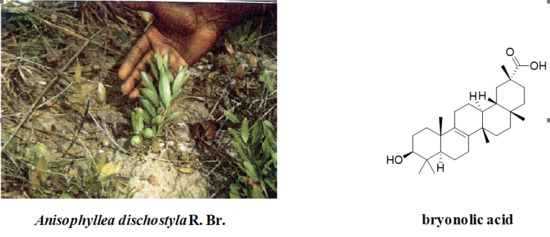
  5. Khallouki, F.; Hull, W.E.; Owen, R.W. Characterization of a rare triterpenoid and minor phenolic compounds in the root bark of Anisophyllea dichostyla R. Br. Food Chem. Toxicol. 2009, 47, 2007–2012. [Google Scholar] [CrossRef] [PubMed]
  6. Dietz, B.M.; Hajirahimkhan, A.; Dunlap, T.L.; Bolton, J.L. Botanicals and Their Bioactive Phytochemicals for Women’s Health. Pharmacol. Rev. 2016, 68, 1026–1073. [Google Scholar] [CrossRef] [PubMed]
  7. Bishayee, A.; Ahmed, S.; Brankov, N.; Perloff, M. Triterpenoids as potential agents for the chemoprevention and therapy of breast cancer. Front. Biosci. 2011, 16, 980–996. [Google Scholar] [CrossRef]
  8. Kongtun, S.; Jiratchariyakul, W.; Kummalue, T.; Tan-Ariya, P.; Kunnachak, S.; Frahm, A.W. Cytotoxic properties of root extract and fruit juice of Trichosanthes cucumerina. Planta Med. 2009, 75, 839–842. [Google Scholar] [CrossRef] [PubMed]
  9. Gatbonton-Schwager, T.N.; Letterio, J.J.; Tochtrop, G.P. Bryonolic acid transcriptional control of anti-inflammatory and antioxidant genes in macrophages in vitro and in vivo. J. Nat. Prod. 2012, 75, 591–598. [Google Scholar] [CrossRef] [PubMed]
  10. Que, J.; Ye, M.; Zhang, Y.; Xu, W.; Li, H.; Xu, W.; Chu, K. Bryonolic acid, a triterpenoid, protect against N-methyl-d-aspartate-induced neurotoxicity in PC12 Cells. Molecules 2016, 21, 418. [Google Scholar] [CrossRef] [PubMed]
  11. Doria, M.; Maugest, L.; Moreau, T.; Lizard, G.; Vejux, A. Contribution of cholesterol and oxysterols to the pathophysiology of Parkinson’s disease. Free Radic. Biol. Med. 2016, 101, 393–400. [Google Scholar] [CrossRef] [PubMed]
  12. Zhang, J.; Liu, Q. Cholesterol metabolism and homeostasis in the brain. Protein Cell 2015, 6, 254–264. [Google Scholar] [CrossRef] [PubMed]
  13. Yang, W.; Bai, Y.; Xiong, Y.; Zhang, J.; Chen, S.; Zheng, X.; Meng, X.; Li, L.; Wang, J.; Xu, C.; et al. Potentiating the antitumour response of CD8+ T cells by modulating cholesterol metabolism. Nature 2016, 531, 651–655. [Google Scholar] [CrossRef] [PubMed]
  14. Fessler, M.B. The intracellular cholesterol landscape: Dynamic integrator of the immune response. Trends Immunol. 2016, 37, 819–830. [Google Scholar] [CrossRef] [PubMed]
  15. Leignadier, J.; Dalenc, F.; Poirot, M.; Silvente-Poirot, S. Improving the efficacy of hormone therapy in breast cancer: The role of cholesterol metabolism in SERM-mediated autophagy, cell differentiation and death. Biochem. Pharmacol. 2017, 144, 18–28. [Google Scholar] [CrossRef] [PubMed]
  16. Kloudova, A.; Guengerich, F.P.; Soucek, P. The role of oxysterols in human cancer. Trends Endocrinol. Metab. 2017, 28, 485–496. [Google Scholar] [CrossRef] [PubMed]
  17. Silvente-Poirot, S.; Poirot, M. Cancer. Cholesterol and cancer, in the balance. Science 2014, 343, 1445–1446. [Google Scholar] [CrossRef] [PubMed]
  18. Silvente-Poirot, S.; Poirot, M. Cholesterol metabolism and cancer: The good, the bad and the ugly. Curr. Opin. Pharmacol. 2012, 12, 673–676. [Google Scholar] [CrossRef] [PubMed]
  19. Morgan, A.E.; Mooney, K.M.; Wilkinson, S.J.; Pickles, N.A.; Mc Auley, M.T. Cholesterol metabolism: A review of how ageing disrupts the biological mechanisms responsible for its regulation. Ageing Res. Rev. 2016, 27, 108–124. [Google Scholar] [CrossRef] [PubMed]
  20. Zarrouk, A.; Vejux, A.; Mackrill, J.; O’Callaghan, Y.; Hammami, M.; O’Brien, N.; Lizard, G. Involvement of oxysterols in age-related diseases and ageing processes. Ageing Res. Rev. 2014, 18, 148–162. [Google Scholar] [CrossRef] [PubMed]
  21. Segala, G.; David, M.; de Medina, P.; Poirot, M.; Serhan, N.; Vergez, F.; Mougel, A.; Saland, E.; Carayon, K.; Leignadier, J.; et al. Dendrogenin A drives LXR to trigger lethal autophagy in cancers. Nat. Commun. 2017, 8, 1903. [Google Scholar] [CrossRef] [PubMed]
  22. Dalenc, F.; Poirot, M.; Silvente-Poirot, S. Dendrogenin A: A mammalian metabolite of cholesterol with tumor suppressor and neurostimulating properties. Curr. Med. Chem. 2015, 22, 3533–3549. [Google Scholar] [CrossRef] [PubMed]
  23. De Medina, P.; Paillasse, M.R.; Segala, G.; Voisin, M.; Mhamdi, L.; Dalenc, F.; Lacroix-Triki, M.; Filleron, T.; Pont, F.; Saati, T.A.; et al. Dendrogenin A arises from cholesterol and histamine metabolism and shows cell differentiation and anti-tumour properties. Nat. Commun. 2013, 4, 1840. [Google Scholar] [CrossRef] [PubMed]
  24. De Medina, P.; Paillasse, M.R.; Payre, B.; Silvente-Poirot, S.; Poirot, M. Synthesis of new alkylaminooxysterols with potent cell differentiating activities: Identification of leads for the treatment of cancer and neurodegenerative diseases. J. Med. Chem. 2009, 52, 7765–7777. [Google Scholar] [CrossRef] [PubMed]
  25. De Medina, P.; Genovese, S.; Paillasse, M.R.; Mazaheri, M.; Caze-Subra, S.; Bystricky, K.; Curini, M.; Silvente-Poirot, S.; Epifano, F.; Poirot, M. Auraptene is an inhibitor of cholesterol esterification and a modulator of estrogen receptors. Mol. Pharmacol. 2010, 78, 827–836. [Google Scholar] [CrossRef] [PubMed]
  26. Khallouki, F.; de Medina, P.; Caze-Subra, S.; Bystricky, K.; Balaguer, P.; Poirot, M.; Silvente-Poirot, S. Molecular and biochemical analysis of the estrogenic and proliferative properties of Vitamin E compounds. Front. Oncol. 2015, 5, 287. [Google Scholar] [CrossRef] [PubMed]
  27. Bandyopadhyay, S.; Li, J.; Traer, E.; Tyner, J.W.; Zhou, A.; Oh, S.T.; Cheng, J.X. Cholesterol esterification inhibition and imatinib treatment synergistically inhibit growth of BCR-ABL mutation-independent resistant chronic myelogenous leukemia. PLoS ONE 2017, 12, e0179558. [Google Scholar] [CrossRef] [PubMed]
  28. De Gonzalo-Calvo, D.; Lopez-Vilaro, L.; Nasarre, L.; Perez-Olabarria, M.; Vazquez, T.; Escuin, D.; Badimon, L.; Barnadas, A.; Lerma, E.; Llorente-Cortes, V. Intratumor cholesteryl ester accumulation is associated with human breast cancer proliferation and aggressive potential: A molecular and clinicopathological study. BMC Cancer 2015, 15, 460. [Google Scholar] [CrossRef] [PubMed]
  29. Yue, S.; Li, J.; Lee, S.Y.; Lee, H.J.; Shao, T.; Song, B.; Cheng, L.; Masterson, T.A.; Liu, X.; Ratliff, T.L.; et al. Cholesteryl ester accumulation induced by PTEN loss and PI3K/AKT activation underlies human prostate cancer aggressiveness. Cell Metab. 2014, 19, 393–406. [Google Scholar] [CrossRef] [PubMed] [Green Version]
  30. Paillasse, M.R.; de Medina, P.; Amouroux, G.; Mhamdi, L.; Poirot, M.; Silvente-Poirot, S. Signaling through cholesterol esterification: A new pathway for the cholecystokinin 2 receptor involved in cell growth and invasion. J. Lipid Res. 2009, 50, 2203–2211. [Google Scholar] [CrossRef] [PubMed]
  31. De Medina, P.; Payre, B.L.; Bernad, J.; Bosser, I.; Pipy, B.; Silvente-Poirot, S.; Favre, G.; Faye, J.C.; Poirot, M. Tamoxifen is a potent inhibitor of cholesterol esterification and prevents the formation of foam cells. J. Pharmacol. Exp. Ther. 2004, 308, 1165–1173. [Google Scholar] [CrossRef] [PubMed]
  32. Gales, C.; Sanchez, D.; Poirot, M.; Pyronnet, S.; Buscail, L.; Cussac, D.; Pradayrol, L.; Fourmy, D.; Silvente-Poirot, S. High tumorigenic potential of a constitutively active mutant of the cholecystokinin 2 receptor. Oncogene 2003, 22, 6081–6089. [Google Scholar] [CrossRef] [PubMed]
  33. De Medina, P.; Paillasse, M.R.; Segala, G.; Poirot, M.; Silvente-Poirot, S. Identification and pharmacological characterization of cholesterol-5,6-epoxide hydrolase as a target for tamoxifen and AEBS ligands. Proc. Natl. Acad. Sci. USA 2010, 107, 13520–13525. [Google Scholar] [CrossRef] [PubMed]
  34. Segala, G.; de Medina, P.; Iuliano, L.; Zerbinati, C.; Paillasse, M.R.; Noguer, E.; Dalenc, F.; Payre, B.; Jordan, V.C.; Record, M.; et al. 5,6-Epoxy-cholesterols contribute to the anticancer pharmacology of tamoxifen in breast cancer cells. Biochem. Pharmacol. 2013, 86, 175–189. [Google Scholar] [CrossRef] [PubMed]
  35. Li, J.; Gu, D.; Lee, S.S.; Song, B.; Bandyopadhyay, S.; Chen, S.; Konieczny, S.F.; Ratliff, T.L.; Liu, X.; Xie, J.; et al. Abrogating cholesterol esterification suppresses growth and metastasis of pancreatic cancer. Oncogene 2016, 35, 6378–6388. [Google Scholar] [CrossRef] [PubMed]
  36. Geng, F.; Cheng, X.; Wu, X.; Yoo, J.Y.; Cheng, C.; Guo, J.Y.; Mo, X.; Ru, P.; Hurwitz, B.; Kim, S.H.; et al. Inhibition of SOAT1 suppresses glioblastoma growth via blocking SREBP-1-mediatedlipogenesis. Clin. Cancer Res. 2016, 22, 5337–5348. [Google Scholar] [CrossRef] [PubMed]
  37. Uda, S.; Accossu, S.; Spolitu, S.; Collu, M.; Angius, F.; Sanna, F.; Banni, S.; Vacca, C.; Murru, E.; Mulas, C.; et al. A lipoprotein source of cholesteryl esters is essential for proliferation of CEM-CCRF lymphoblastic cell line. Tumor Biol. 2012, 33, 443–453. [Google Scholar] [CrossRef] [PubMed]
  38. Lin, Y.; Vermeer, M.A.; Trautwein, E.A. Triterpenic acids present in Hawthorn lower plasma cholesterol by inhibiting intestinal ACAT activity in hamsters. Evid. Based Complement. Altern. Med. 2011, 2011, 801272. [Google Scholar] [CrossRef] [PubMed]
  39. Lee, W.S.; Im, K.R.; Park, Y.D.; Sung, N.D.; Jeong, T.S. Human ACAT-1 and ACAT-2 inhibitory activities of pentacyclic triterpenes from the leaves of Lycopus lucidus TURCZ. Biol. Pharm. Bull. 2006, 29, 382–384. [Google Scholar] [CrossRef] [PubMed]
  40. Tabas, I.; Chen, L.L.; Clader, J.W.; McPhail, A.T.; Burnett, D.A.; Bartner, P.; Das, P.R.; Pramanik, B.N.; Puar, M.S.; Feinmark, S.J.; et al. Rabbit and human liver contain a novel pentacyclic triterpene ester with acyl-CoA: Cholesterol acyltransferase inhibitory activity. J. Biol. Chem. 1990, 265, 8042–8051. [Google Scholar] [PubMed]
  41. Jordan, V.C. Chemoprevention of breast cancer with selective oestrogen-receptor modulators. Nat. Rev. Cancer 2007, 7, 46–53. [Google Scholar] [CrossRef] [PubMed]
  42. Silvente-Poirot, S.; Poirot, M. Cholesterol epoxide hydrolase and cancer. Curr. Opin. Pharmacol. 2012, 12, 696–703. [Google Scholar] [CrossRef] [PubMed]
  43. Kedjouar, B.; de Medina, P.; Oulad-Abdelghani, M.; Payre, B.; Silvente-Poirot, S.; Favre, G.; Faye, J.C.; Poirot, M. Molecular characterization of the microsomal tamoxifen binding site. J. Biol. Chem. 2004, 279, 34048–34061. [Google Scholar] [CrossRef] [PubMed]
  44. Alqahtani, A.; Hamid, K.; Kam, A.; Wong, K.H.; Abdelhak, Z.; Razmovski-Naumovski, V.; Chan, K.; Li, K.M.; Groundwater, P.W.; Li, G.Q. The pentacyclic triterpenoids in herbal medicines and their pharmacological activities in diabetes and diabetic complications. Curr. Med. Chem. 2013, 20, 908–931. [Google Scholar] [PubMed]
  45. Huang, M.; Lu, J.J.; Huang, M.Q.; Bao, J.L.; Chen, X.P.; Wang, Y.T. Terpenoids: Natural products for cancer therapy. Expert Opin. Investig. Drugs 2012, 21, 1801–1818. [Google Scholar] [CrossRef] [PubMed]
Figure 1. Chemical structures of the tested triterpenoid compounds.
Biomedicines 06 00021 g001
Figure 2. (A) Effect of Bryonolic acid on colony formation. Cells were treated with 25 µM BrA and the number of colonies was measured compared with solvent vehicle-treated cells (taken to be 100%); (B) after 24 h of pre-treatment with either the solvent vehicle or 25 μM betulinic acid or BrA, cell invasion was assayed using Matrigel-coated filters as described under Materials and Methods. After 48 h, cells on the lower surface of the filters were stained and counted under a phase-contrast microscope; (C) Effect of cholesteryl oleate on the inhibition of MB-231 and U87 cell invasiveness by BrA. Cells were pre-treated for 24 h with cholesteryl oleate (4 µg/mL) and layered on the top of Matrigel-coated filters in serum-free medium for 48 h (+CO) or cells were treated similarly with the solvent vehicle (−CO). After 48 h, cells on the lower surface of the filters were stained and counted. Values are expressed relative to that of cells treated with the solvent vehicle (−CO) and are the mean ± SEM of three separate experiments. Values are expressed relative to those of cells treated with the solvent vehicle (control) and are the mean ± S.E.M. of three to six separate experiments performed in triplicate. (* p < 0.05, ** p < 0.01, *** p < 0.001).
Biomedicines 06 00021 g002
Table 1. Effect bryonolic acid, betulinic acid and ursolic acid on cholesterol esterification (ACAT activity). ACAT activity was measured on in rat liver microsomes. Rat liver microsomes (60 μg of protein) were incubated with 10 different concentrations of each drug and 40 μM [14C]oleyl-CoA. Cholesteryl esters were quantified as described under the “Materials and Methods” section. ACAT activity is expressed as the percentage of the ACAT activity measured in the absence of inhibitors (control with solvent vehicle).
CompoundsIC50 in µM
sah 58-0350.65 ± 0.22
bryonolic acid12.6 ± 2.4
betulinic acid18.5 ± 2.1
ursolic acid71.4 ± 5.1
Table 2. Inhibition of ACAT activity in intact MCF-7, MB231, U-87 and 3T3-EA cells. Cells were treated as described in the legend of Table 1 for 24 h. Free and esterified cholesterol were quantified as described in the “Materials and Methods” section. ACAT activity is expressed as the percentage of the ACAT activity measured in the absence of inhibitors (control with solvent vehicle). IC50 values were determined using the iterative curve fitting program GraphPad Prism (version 6.0). Values are the average of three experiments ± S.E.M., each carried out in duplicate. n.m.: not measurable.
MCF-7MB-231U-873T3-EA
CompoundsIC50 in µM
sah 58-0355.1 ± 0.59.1 ± 1.49.2 ± 2.27.4 ± 1.8
bryonolic acid22.5 ± 3.729.5 ± 5.517.5 ± 4.819.4 ± 7.6
betulinic acid53.7± 4.258.3 ± 8.161.4 ± 9.460.2 ± 8.0
ursolic acidn.m.n.m.n.m.n.m
Table 3. Competition assays on ERs and measure of ChEH inhibition. Binding of radiolabelled 17β-estradiol (E2) to the ERα and ERβ were measured at different concentrations of compounds as described under “Materials and Methods.” For the ChEH inhibition tests, 150 μg of rat liver microsomal proteins and 10 and 20 μM concentrations of [14C]α-CE with increasing concentrations of compounds ranging from 0.01 to 1000 μM were used under the conditions described under Materials and Methods. Values are the mean ± S.E.M. from three independent experiments. n.m., no measurable inhibition of binding.
CompoundsERαERβChEH
IC50 in nM
E21.2 ± 0.51.7 ± 0.7n.m.
tamoxifen45.7 ± 5.158.4 ± 7.1122.6
bryonolic acidn.m.n.m.n.m.
betulinic acidn.m.n.m.n.m.
ursolic acidn.m.n.m.n.m.

