Next Issue
Volume 6, December
Previous Issue
Volume 6, June


Biomedicines, Volume 6, Issue 3 (September 2018) – 21 articles

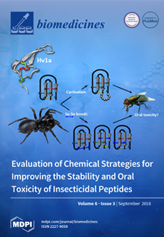
Cover Story (view full-size image): Spider venoms are rich in insecticidal peptides, but as venoms are usually injected, there has been no evolutionary pressure to produce venom peptides with oral activity. Nevertheless, some orally active venom peptides were recently reported, although their oral activity was significantly lower than by injection. Therefore, we systematically evaluated several chemical strategies to increase the oral potency of the insecticidal spider-venom peptide ω-HXTX-Hv1a (Hv1a). N- to C-terminal cyclisation and replacement of disulfide by diselenide bonds failed to improve oral activity. Cyclization of Hv1a dramatically reduced the translocation of Hv1a across the insect midgut, highlighting the importance of bioavailability for oral activity. Hadronyche versuta photo courtesy of Mike Gray (Australian Museum) View the paper here.
  • Issues are regarded as officially published after their release is announced to the table of contents alert mailing list.
  • You may sign up for e-mail alerts to receive table of contents of newly released issues.
  • PDF is the official format for papers published in both, html and pdf forms. To view the papers in pdf format, click on the "PDF Full-text" link, and use the free Adobe Reader to open them.
Order results
Result details
Section
Select all
Export citation of selected articles as:
14 pages, 2287 KiB  
Article
Intravenous Infusion of Nucleated Peripheral Blood Cells Restores Fertility in Mice with Chemotherapy-Induced Premature Ovarian Failure
by Abdeljabar El Andaloussi, Prosper Igboeli, Amero Amer and Ayman Al-Hendy
Biomedicines 2018, 6(3), 93; https://doi.org/10.3390/biomedicines6030093 - 15 Sep 2018
Cited by 8 | Viewed by 6447
Abstract
Cancer treatment with specific chemotherapeutic agents has been well documented to have an adverse impact on female fertility leading to premature ovarian failure (POF). The objective of this study is to investigate if chemotherapeutic induced POF can be reversed by the infusion of [...] Read more.
Cancer treatment with specific chemotherapeutic agents has been well documented to have an adverse impact on female fertility leading to premature ovarian failure (POF). The objective of this study is to investigate if chemotherapeutic induced POF can be reversed by the infusion of autologous nucleated peripheral blood cells (PBMC). To reach our goal, mice were treated with a single intraperitoneal injections of busulfan and cyclophosphamide to induce POF. This was followed by transfusion of PBMC. The ovarian morphology and functional parameters were monitored by radioimmunoassay, real-time PCR, immunofluorescence and immunohistochemistry analysis. Our study showed that chemotherapy (CTX) protracted estrous cycle period and repressed E2 production. In addition, CTX decreased the expressions of steroidogenesis markers, CYP-17 synthesis, StAR (steroidogenic acute regulatory protein), and Connexin-43 protein expression in the ovarian follicles. We also observed reduced numbers and sizes of the primordial and primary follicles in CTX-treated mice compared to untreated controls (p < 0.05). When both CTX and untreated control groups were stimulated with gonadotrophin, the control group produced ten times more ova than the CTX group. Finally, the treatment of premature ovarian failure induced by CTX with autologous PBMC transfusion resulted in over-expression and a statistically significant increase in several stem cell markers and restoration of fertility. Infusion with PBMC in CTX further decreased the estrous cycle length by 2.5 times (p < 0.01). We found that transfusion of autologous PBMC to mice with chemotherapy induced POF was very effective at restoring fertility. These results are similar to other studies using bone marrow derived mesenchymal stem cells. Full article
Show Figures

Graphical abstract

9 pages, 1015 KiB  
Article
Aflibercept Nanoformulation Inhibits VEGF Expression in Ocular In Vitro Model: A Preliminary Report
by Shannon J. Kelly, Anjali Hirani, Vishal Shahidadpury, Aum Solanki, Kathleen Halasz, Sheeba Varghese Gupta, Brian Madow and Vijaykumar Sutariya
Biomedicines 2018, 6(3), 92; https://doi.org/10.3390/biomedicines6030092 - 11 Sep 2018
Cited by 20 | Viewed by 5113
Abstract
Age-related macular degeneration (AMD) is one of the leading causes of blindness in the United States, affecting approximately 11 million patients. AMD is caused primarily by an upregulation of vascular endothelial growth factor (VEGF). In recent years, aflibercept injections have been used to [...] Read more.
Age-related macular degeneration (AMD) is one of the leading causes of blindness in the United States, affecting approximately 11 million patients. AMD is caused primarily by an upregulation of vascular endothelial growth factor (VEGF). In recent years, aflibercept injections have been used to combat VEGF. However, this treatment requires frequent intravitreal injections, leading to low patient compliance and several adverse side effects including scarring, increased intraocular pressure, and retinal detachment. Polymeric nanoparticles have demonstrated the ability to deliver a sustained release of drug, thereby reducing the necessary injection frequency. Aflibercept (AFL) was encapsulated in poly lactic-co-glycolic acid (PLGA) nanoparticles (NPs) via double emulsion diffusion. Scanning electron microscopy showed the NPs were spherical and dynamic light scattering demonstrated that they were uniformly distributed (PDI < 1). The encapsulation efficiency and drug loading were 75.76% and 7.76% respectively. In vitro release studies showed a sustained release of drug; 75% of drug was released by the NPs in seven days compared to the full payload released in 24 h by the AFL solution. Future ocular in vivo studies are needed to confirm the biological effects of the NPs. Preliminary studies of the proposed aflibercept NPs demonstrated high encapsulation efficiency, a sustained drug release profile, and ideal physical characteristics for AMD treatment. This drug delivery system is an excellent candidate for further characterization using an ocular neovascularization in vivo model. Full article
Show Figures

Figure 1

20 pages, 870 KiB  
Review
Resveratrol: A Double-Edged Sword in Health Benefits
by Bahare Salehi, Abhay Prakash Mishra, Manisha Nigam, Bilge Sener, Mehtap Kilic, Mehdi Sharifi-Rad, Patrick Valere Tsouh Fokou, Natália Martins and Javad Sharifi-Rad
Biomedicines 2018, 6(3), 91; https://doi.org/10.3390/biomedicines6030091 - 09 Sep 2018
Cited by 624 | Viewed by 31740
Abstract
Resveratrol (3,5,4′-trihydroxy-trans-stilbene) belongs to polyphenols’ stilbenoids group, possessing two phenol rings linked to each other by an ethylene bridge. This natural polyphenol has been detected in more than 70 plant species, especially in grapes’ skin and seeds, and was found in discrete amounts [...] Read more.
Resveratrol (3,5,4′-trihydroxy-trans-stilbene) belongs to polyphenols’ stilbenoids group, possessing two phenol rings linked to each other by an ethylene bridge. This natural polyphenol has been detected in more than 70 plant species, especially in grapes’ skin and seeds, and was found in discrete amounts in red wines and various human foods. It is a phytoalexin that acts against pathogens, including bacteria and fungi. As a natural food ingredient, numerous studies have demonstrated that resveratrol possesses a very high antioxidant potential. Resveratrol also exhibit antitumor activity, and is considered a potential candidate for prevention and treatment of several types of cancer. Indeed, resveratrol anticancer properties have been confirmed by many in vitro and in vivo studies, which shows that resveratrol is able to inhibit all carcinogenesis stages (e.g., initiation, promotion and progression). Even more, other bioactive effects, namely as anti-inflammatory, anticarcinogenic, cardioprotective, vasorelaxant, phytoestrogenic and neuroprotective have also been reported. Nonetheless, resveratrol application is still being a major challenge for pharmaceutical industry, due to its poor solubility and bioavailability, as well as adverse effects. In this sense, this review summarized current data on resveratrol pharmacological effects. Full article
Show Figures

Graphical abstract

16 pages, 1860 KiB  
Article
Evaluation of Chemical Strategies for Improving the Stability and Oral Toxicity of Insecticidal Peptides
by Volker Herzig, Aline Dantas De Araujo, Kathryn P. Greenwood, Yanni K.-Y. Chin, Monique J. Windley, Youmie Chong, Markus Muttenthaler, Mehdi Mobli, Neil Audsley, Graham M. Nicholson, Paul F. Alewood and Glenn F. King
Biomedicines 2018, 6(3), 90; https://doi.org/10.3390/biomedicines6030090 - 28 Aug 2018
Cited by 11 | Viewed by 4915
Abstract
Spider venoms are a rich source of insecticidal peptide toxins. Their development as bioinsecticides has, however, been hampered due to concerns about potential lack of stability and oral bioactivity. We therefore systematically evaluated several synthetic strategies to increase the stability and oral potency [...] Read more.
Spider venoms are a rich source of insecticidal peptide toxins. Their development as bioinsecticides has, however, been hampered due to concerns about potential lack of stability and oral bioactivity. We therefore systematically evaluated several synthetic strategies to increase the stability and oral potency of the potent insecticidal spider-venom peptide ω-HXTX-Hv1a (Hv1a). Selective chemical replacement of disulfide bridges with diselenide bonds and N- to C-terminal cyclization were anticipated to improve Hv1a resistance to proteolytic digestion, and thereby its activity when delivered orally. We found that native Hv1a is orally active in blowflies, but 91-fold less potent than when administered by injection. Introduction of a single diselenide bond had no effect on the susceptibility to scrambling or the oral activity of Hv1a. N- to C-terminal cyclization of the peptide backbone did not significantly improve the potency of Hv1a when injected into blowflies and it led to a significant decrease in oral activity. We show that this is likely due to a dramatically reduced rate of translocation of cyclic Hv1a across the insect midgut, highlighting the importance of testing bioavailability in addition to toxin stability. Full article
(This article belongs to the Special Issue Discovery and Development of Constrained Peptide Ligands)
Show Figures

Graphical abstract

11 pages, 628 KiB  
Review
Contributions of Thyroid Hormone to Cancer Metastasis
by Shaker A. Mousa, Gennadi V. Glinsky, Hung-Yun Lin, Osnat Ashur-Fabian, Aleck Hercbergs, Kelly A. Keating and Paul J. Davis
Biomedicines 2018, 6(3), 89; https://doi.org/10.3390/biomedicines6030089 - 22 Aug 2018
Cited by 39 | Viewed by 5600
Abstract
Acting at a cell surface receptor on the extracellular domain of integrin αvβ3, thyroid hormone analogues regulate downstream the expression of a large panel of genes relevant to cancer cell proliferation, to cancer cell survival pathways, and to tumor-linked angiogenesis. Because αvβ3 is [...] Read more.
Acting at a cell surface receptor on the extracellular domain of integrin αvβ3, thyroid hormone analogues regulate downstream the expression of a large panel of genes relevant to cancer cell proliferation, to cancer cell survival pathways, and to tumor-linked angiogenesis. Because αvβ3 is involved in the cancer cell metastatic process, we examine here the possibility that thyroid hormone as l-thyroxine (T4) and the thyroid hormone antagonist, tetraiodothyroacetic acid (tetrac), may respectively promote and inhibit metastasis. Actions of T4 and tetrac that are relevant to cancer metastasis include the multitude of synergistic effects on molecular levels such as expression of matrix metalloproteinase genes, angiogenesis support genes, receptor tyrosine kinase (EGFR/ERBB2) genes, specific microRNAs, the epithelial–mesenchymal transition (EMT) process; and on the cellular level are exemplified by effects on macrophages. We conclude that the thyroid hormone-αvβ3 interaction is mechanistically linked to cancer metastasis and that modified tetrac molecules have antimetastatic activity with feasible therapeutic potential. Full article
Show Figures

Graphical abstract

14 pages, 3980 KiB  
Article
Improved Methods for the Rapid Formation and Prevention of Advanced Glycation End Products (AGEs) In Vitro by Coupling to the Hypoxanthine/Xanthine Oxidase Assay System
by Samuel Marques, Teresa Trevisan, Carlos Maia, Andrea Breuer and Robert W. Owen
Biomedicines 2018, 6(3), 88; https://doi.org/10.3390/biomedicines6030088 - 15 Aug 2018
Cited by 8 | Viewed by 4671
Abstract
Advanced glycation end products (AGEs) represent a set of molecules that contribute directly to the initiation and aggravation of diseases associated with ageing. AGEs are produced by the reaction between reducing sugars (or α-dicarbonyl compounds), proteins, and amino acid residues. Previous in vitro [...] Read more.
Advanced glycation end products (AGEs) represent a set of molecules that contribute directly to the initiation and aggravation of diseases associated with ageing. AGEs are produced by the reaction between reducing sugars (or α-dicarbonyl compounds), proteins, and amino acid residues. Previous in vitro methods using non-enzymatic procedures described in the literature require an incubation period of 1–3 weeks to generate AGEs. In this study, the reaction time for the formation of AGEs (48 and 3 h) was significantly reduced by adaptation of methods previously described in the literature and coupling them to the free radical generation system termed hypoxanthine/xanthine oxidase assay. The incorporation of this assay into the experimental system accelerated the production of AGEs as a result of the formation of reactive oxygen species (ROS), as shown by increased fluorescence. The capacity of different classes of chemical compounds (aminoguanidine, chlorogenic acid, rutin, and methanol extracts of Hancornia speciosa Gomes) to inhibit protein glycation by acting as scavenging agents of α-dicarbonyl species was evaluated. Aminoguanidine and, especially, rutin identified in the leaf extracts of H. speciosa Gomes showed a high capacity to act as scavengers of reactive carbonyl species RCS-trapping, resulting in the inhibition of AGEs formation. Full article
Show Figures

Graphical abstract

18 pages, 604 KiB  
Review
New Development of Biomarkers for Gastrointestinal Cancers: From Neoplastic Cells to Tumor Microenvironment
by Jiajia Zhang, Shafat Quadri, Christopher L. Wolfgang and Lei Zheng
Biomedicines 2018, 6(3), 87; https://doi.org/10.3390/biomedicines6030087 - 13 Aug 2018
Cited by 8 | Viewed by 4997
Abstract
Biomarkers refer to a plethora of biological characteristics that can be quantified to facilitate cancer diagnosis, forecast the prognosis of disease, and predict a response to treatment. The identification of objective biomarkers is among the most crucial steps in the realization of individualized [...] Read more.
Biomarkers refer to a plethora of biological characteristics that can be quantified to facilitate cancer diagnosis, forecast the prognosis of disease, and predict a response to treatment. The identification of objective biomarkers is among the most crucial steps in the realization of individualized cancer care. Several tumor biomarkers for gastrointestinal malignancies have been applied in the clinical setting to help differentiate between cancer and other conditions, facilitate patient selection for targeted therapies, and to monitor treatment response and recurrence. With the coming of the immunotherapy age, the need for a new development of biomarkers that are indicative of the immune response to tumors are unprecedentedly urgent. Biomarkers from the tumor microenvironment, tumor genome, and signatures from liquid biopsies have been explored, but the majority have shown a limited prognostic or predictive value as single biomarkers. Nevertheless, use of multiplex biomarkers has the potential to provide a significantly increased diagnostic accuracy compared to traditional single biomarker. A comprehensive analysis of immune-biomarkers is needed to reveal the dynamic and multifaceted anti-tumor immunity and thus imply for the rational design of assays and combinational strategies. Full article
(This article belongs to the Special Issue Cancer Biomarkers and Targets in Digestive Organs)
Show Figures

Figure 1

21 pages, 294 KiB  
Review
Emerging Medical Treatments for Meningioma in the Molecular Era
by Fares Nigim, Hiroaki Wakimoto, Ekkehard M. Kasper, Linda Ackermans and Yasin Temel
Biomedicines 2018, 6(3), 86; https://doi.org/10.3390/biomedicines6030086 - 06 Aug 2018
Cited by 28 | Viewed by 6018
Abstract
Meningiomas are the most common type of primary central nervous system tumors. Approximately, 80% of meningiomas are classified by the World Health Organization (WHO) as grade I, and 20% of these tumors are grade II and III, considered high-grade meningiomas (HGMs). Clinical control [...] Read more.
Meningiomas are the most common type of primary central nervous system tumors. Approximately, 80% of meningiomas are classified by the World Health Organization (WHO) as grade I, and 20% of these tumors are grade II and III, considered high-grade meningiomas (HGMs). Clinical control of HGMs, as well as meningiomas that relapse after surgery, and radiation therapy is difficult, and novel therapeutic approaches are necessary. However, traditional chemotherapies, interferons, hormonal therapies, and other targeted therapies have so far failed to provide clinical benefit. During the last several years, next generation sequencing has dissected the genetic heterogeneity of meningioma and enriched our knowledge about distinct oncogenic pathways driving different subtypes of meningiomas, opening up a door to new personalized targeted therapies. Molecular classification of meningioma allows a new design of clinical trials that assign patients to corresponding targeted agents based on the tumor genetic subtypes. In this review, we will shed light on emerging medical treatments of meningiomas with a particular focus on the new targets identified with genomic sequencing that have led to clinical trials testing novel compounds. Moreover, we present recent development of patient-derived preclinical models that provide platforms for assessing targeted therapies as well as strategies with novel mechanism of action such as oncolytic viruses. Full article
Show Figures

Graphical abstract

14 pages, 259 KiB  
Review
Stem-Like Signature Predicting Disease Progression in Early Stage Bladder Cancer. The Role of E2F3 and SOX4
by Joaquim Bellmunt
Biomedicines 2018, 6(3), 85; https://doi.org/10.3390/biomedicines6030085 - 02 Aug 2018
Cited by 25 | Viewed by 4625
Abstract
The rapid development of the cancer stem cells (CSC) field, together with powerful genome-wide screening techniques, have provided the basis for the development of future alternative and reliable therapies aimed at targeting tumor-initiating cell populations. Urothelial bladder cancer stem cells (BCSCs) that were [...] Read more.
The rapid development of the cancer stem cells (CSC) field, together with powerful genome-wide screening techniques, have provided the basis for the development of future alternative and reliable therapies aimed at targeting tumor-initiating cell populations. Urothelial bladder cancer stem cells (BCSCs) that were identified for the first time in 2009 are heterogenous and originate from multiple cell types; including urothelial stem cells and differentiated cell types—basal, intermediate stratum and umbrella cells Some studies hypothesize that BCSCs do not necessarily arise from normal stem cells but might derive from differentiated progenies following mutational insults and acquisition of tumorigenic properties. Conversely, there is data that normal bladder tissues can generate CSCs through mutations. Prognostic risk stratification by identification of predictive markers is of major importance in the management of urothelial cell carcinoma (UCC) patients. Several stem cell markers have been linked to recurrence or progression. The CD44v8-10 to standard CD44-ratio (total ratio of all CD44 alternative splicing isoforms) in urothelial cancer has been shown to be closely associated with tumor progression and aggressiveness. ALDH1, has also been reported to be associated with BCSCs and a worse prognosis in a large number of studies. UCC include low-grade and high-grade non-muscle invasive bladder cancer (NMIBC) and high-grade muscle invasive bladder cancer (MIBC). Important genetic defects characterize the distinct pathways in each one of the stages and probably grades. As an example, amplification of chromosome 6p22 is one of the most frequent changes seen in MIBC and might act as an early event in tumor progression. Interestingly, among NMIBC there is a much higher rate of amplification in high-grade NMIBC compared to low grade NMIBC. CDKAL1, E2F3 and SOX4 are highly expressed in patients with the chromosomal 6p22 amplification aside from other six well known genes (ID4, MBOAT1, LINC00340, PRL, and HDGFL1). Based on that, SOX4, E2F3 or 6q22.3 amplifications might represent potential targets in this tumor type. Focusing more in SOX4, it seems to exert its critical regulatory functions upstream of the Snail, Zeb, and Twist family of transcriptional inducers of EMT (epithelial–mesenchymal transition), but without directly affecting their expression as seen in several cell lines of the Cancer Cell Line Encyclopedia (CCLE) project. SOX4 gene expression correlates with advanced cancer stages and poor survival rate in bladder cancer, supporting a potential role as a regulator of the bladder CSC properties. SOX4 might serve as a biomarker of the aggressive phenotype, also underlying progression from NMIBC to MIBC. The amplicon in chromosome 6 contains SOX4 and E2F3 and is frequently found amplified in bladder cancer. These genes/amplicons might be a potential target for therapy. As an existing hypothesis is that chromatin deregulation through enhancers or super-enhancers might be the underlying mechanism responsible of this deregulation, a potential way to target these transcription factors could be through epigenetic modifiers. Full article
(This article belongs to the Special Issue Stem Cells and Cancer Therapeutics)
14 pages, 254 KiB  
Review
Relapsing Polychondritis: An Updated Review
by Francesco Borgia, Roberta Giuffrida, Fabrizio Guarneri and Serafinella P. Cannavò
Biomedicines 2018, 6(3), 84; https://doi.org/10.3390/biomedicines6030084 - 02 Aug 2018
Cited by 67 | Viewed by 13142
Abstract
Relapsing polychondritis is an immune-mediated systemic disease characterized by recurrent episodes of inflammation of cartilaginous and proteoglycan-rich tissues, resulting in progressive anatomical deformation and functional impairment of the involved structures. Auricular and nasal chondritis and/or polyarthritis represent the most common clinical features, but [...] Read more.
Relapsing polychondritis is an immune-mediated systemic disease characterized by recurrent episodes of inflammation of cartilaginous and proteoglycan-rich tissues, resulting in progressive anatomical deformation and functional impairment of the involved structures. Auricular and nasal chondritis and/or polyarthritis represent the most common clinical features, but potentially all types of cartilage may be involved. Because of the pleomorphic nature of the disease, with non-specific symptoms at the onset, the diagnosis of relapsing polychondritis is often delayed. In this review article we provide a comprehensive look into clinical presentation, laboratory and instrumental investigations, diagnostic criteria, and therapeutic options. Full article
12 pages, 755 KiB  
Article
Straw Wine Melanoidins as Potential Multifunctional Agents: Insight into Antioxidant, Antibacterial, and Angiotensin-I-Converting Enzyme Inhibition Effects
by Vlasios Goulas, Demetra Nicolaou, George Botsaris and Alexandra Barbouti
Biomedicines 2018, 6(3), 83; https://doi.org/10.3390/biomedicines6030083 - 02 Aug 2018
Cited by 11 | Viewed by 4424
Abstract
Numerous studies provide robust evidence for a protective effect of red wine against many diseases. This bioactivity has been mainly associated with phenolic fractions of wines. However, the health effects of melanoidins in red sweet wines has been ignored. The goal of the [...] Read more.
Numerous studies provide robust evidence for a protective effect of red wine against many diseases. This bioactivity has been mainly associated with phenolic fractions of wines. However, the health effects of melanoidins in red sweet wines has been ignored. The goal of the present work was to unravel the antioxidant, antimicrobial, and angiotensin-I-converting enzyme (ACE) inhibitory properties of straw sweet wine melanoidins. Results demonstrated that melanoidins have a potential antioxidant activity, determined by 2,2-Diphenyl-1-picrylhydrazyl (DPPH) and Ferric reducing antioxidant power (FRAP) assays. The antimicrobial activity of melanoidins was also tested against Listeria monocytogenes, Salmonella Enteritidis, and Escherichia coli. Minimum inhibitory concentration (MIC) of isolated melanoidins against three bacterial strains ranged from 5 mg mL−1 to 10 mg mL−1. Finally, the ACE inhibitory effect of isolated melanoidins was evaluated, as it is linked with antihypertensive activity. Results showed that they have ACE-inhibitory activity ranging from 58.2 ± 5.4% to 75.3 ± 6.4% at a concentration level of 2 mg mL−1. Furthermore, the chemical properties of isolated melanoidins were determined. Results demonstrated that the skeleton of straw wine melanoidins is mainly composed of carbohydrates, and bear significant numbers of phenolic compounds that may play critical roles in their functional properties. Overall, this study describing the chemical composition and functional properties of melanoidin fractions isolated from a straw wine highlights that they can be exploited as functional agents for multiple purposes. Finally, melanoidins are an unexplored source of bioactive molecules in straw wines except from polyphenols that contribute to the health effects. Full article
(This article belongs to the Special Issue Antioxidants and Antimicrobial Compounds from Natural Sources)
Show Figures

Graphical abstract

21 pages, 667 KiB  
Review
Evidence for the Involvement of the Master Transcription Factor NF-κB in Cancer Initiation and Progression
by Yu Rou Puar, Muthu K Shanmugam, Lu Fan, Frank Arfuso, Gautam Sethi and Vinay Tergaonkar
Biomedicines 2018, 6(3), 82; https://doi.org/10.3390/biomedicines6030082 - 27 Jul 2018
Cited by 166 | Viewed by 7434
Abstract
Nuclear factor kappa-light-chain-enhancer of activated B cells (NF-κB) is responsible for the regulation of a large number of genes that are involved in important physiological processes, including survival, inflammation, and immune responses. At the same time, this transcription factor can control the expression [...] Read more.
Nuclear factor kappa-light-chain-enhancer of activated B cells (NF-κB) is responsible for the regulation of a large number of genes that are involved in important physiological processes, including survival, inflammation, and immune responses. At the same time, this transcription factor can control the expression of a plethora of genes that promote tumor cell proliferation, survival, metastasis, inflammation, invasion, and angiogenesis. The aberrant activation of this transcription factor has been observed in several types of cancer and is known to contribute to aggressive tumor growth and resistance to therapeutic treatment. Although NF-κB has been identified to be a major contributor to cancer initiation and development, there is evidence revealing its role in tumor suppression. This review briefly highlights the major mechanisms of NF-κB activation, the role of NF-κB in tumor promotion and suppression, as well as a few important pharmacological strategies that have been developed to modulate NF-κB function. Full article
(This article belongs to the Special Issue Roles of NF-κB in Cancer and Their Therapeutic Approaches 2019)
Show Figures

Graphical abstract

16 pages, 1019 KiB  
Review
Progress in Adenoviral Capsid-Display Vaccines
by Marija Vujadinovic and Jort Vellinga
Biomedicines 2018, 6(3), 81; https://doi.org/10.3390/biomedicines6030081 - 26 Jul 2018
Cited by 17 | Viewed by 5664
Abstract
Adenoviral vectored vaccines against infectious diseases are currently in clinical trials due to their capacity to induce potent antigen-specific B- and T-cell immune responses. Heterologous prime-boost vaccination with adenoviral vector and, for example, adjuvanted protein-based vaccines can further enhance antigen-specific immune responses. Although [...] Read more.
Adenoviral vectored vaccines against infectious diseases are currently in clinical trials due to their capacity to induce potent antigen-specific B- and T-cell immune responses. Heterologous prime-boost vaccination with adenoviral vector and, for example, adjuvanted protein-based vaccines can further enhance antigen-specific immune responses. Although leading to potent immune responses, these heterologous prime-boost regimens may be complex and impact manufacturing costs limiting efficient implementation. Typically, adenoviral vectors are engineered to genetically encode a transgene in the E1 region and utilize the host cell machinery to express the encoded antigen and thereby induce immune responses. Similarly, adenoviral vectors can be engineered to display foreign immunogenic peptides on the capsid-surface by insertion of antigens in capsid proteins hexon, fiber and protein IX. The ability to use adenoviral vectors as antigen-display particles, with or without using the genetic vaccine function, greatly increases the versatility of the adenoviral vector for vaccine development. This review describes the application of adenoviral capsid antigen-display vaccine vectors by focusing on their distinct advantages and possible limitations in vaccine development. Full article
(This article belongs to the Special Issue Adenoviruses: From Virus to Medicine)
Show Figures

Graphical abstract

9 pages, 673 KiB  
Communication
GABA Levels in Left and Right Sensorimotor Cortex Correlate across Individuals
by Nicolaas A. J. Puts, Stefanie Heba, Ashley D. Harris, Christopher John Evans, David J. McGonigle, Martin Tegenthoff, Tobias Schmidt-Wilcke and Richard A. E. Edden
Biomedicines 2018, 6(3), 80; https://doi.org/10.3390/biomedicines6030080 - 24 Jul 2018
Cited by 10 | Viewed by 3915
Abstract
Differences in γ-aminobutyric acid (GABA) levels measured with Magnetic Resonance Spectroscopy have been shown to correlate with behavioral performance over a number of tasks and cortical regions. These correlations appear to be regionally and functionally specific. In this study, we test the hypothesis [...] Read more.
Differences in γ-aminobutyric acid (GABA) levels measured with Magnetic Resonance Spectroscopy have been shown to correlate with behavioral performance over a number of tasks and cortical regions. These correlations appear to be regionally and functionally specific. In this study, we test the hypothesis that GABA levels will be correlated within individuals for functionally related regions—the left and right sensorimotor cortex. In addition, we investigate whether this is driven by bulk tissue composition. GABA measurements using edited MRS data were acquired from the left and right sensorimotor cortex in 24 participants. T1-weighted MR images were also acquired and segmented to determine the tissue composition of the voxel. GABA level is shown to correlate significantly between the left and right regions (r = 0.64, p < 0.03). Tissue composition is highly correlated between sides, but does not explain significant variance in the bilateral correlation. In conclusion, individual differences in GABA level, which have previously been described as functionally and regionally specific, are correlated between homologous sensorimotor regions. This correlation is not driven by bulk differences in voxel tissue composition. Full article
(This article belongs to the Special Issue Neural Mechanisms of Learning)
Show Figures

Graphical abstract

12 pages, 933 KiB  
Review
Pathogens and Their Effect on Exosome Biogenesis and Composition
by Leandra B. Jones, Courtnee’ R. Bell, Kartz E. Bibb, Linlin Gu, Mamie T. Coats and Qiana L. Matthews
Biomedicines 2018, 6(3), 79; https://doi.org/10.3390/biomedicines6030079 - 23 Jul 2018
Cited by 55 | Viewed by 7351
Abstract
Exosomes are nanosized membrane microvesicles (30–100 nm) that have the capability to communicate intercellularly and transport cell components (i.e., miRNA, mRNA, proteins and DNA). Exosomes are found in nearly every cell type (i.e., mast cells, dendritic, tumor, and macrophages). There have been many [...] Read more.
Exosomes are nanosized membrane microvesicles (30–100 nm) that have the capability to communicate intercellularly and transport cell components (i.e., miRNA, mRNA, proteins and DNA). Exosomes are found in nearly every cell type (i.e., mast cells, dendritic, tumor, and macrophages). There have been many studies that have shown the importance of exosome function as well as their unique packaging and targeting abilities. These characteristics make exosomes ideal candidates to act as biomarkers and therapeutics for disease. We will discuss the biogenesis, composition, and relationship of exosomes with non-viral microbial infections including gram-negative bacteria, gram-positive bacteria, Leishmania and Trypanosoma cruzi. Full article
Show Figures

Graphical abstract

15 pages, 3017 KiB  
Review
The Local Atomic Structure of Colloidal Superparamagnetic Iron Oxide Nanoparticles for Theranostics in Oncology
by Elena Kuchma, Stanislav Kubrin and Alexander Soldatov
Biomedicines 2018, 6(3), 78; https://doi.org/10.3390/biomedicines6030078 - 18 Jul 2018
Cited by 11 | Viewed by 5073
Abstract
The paper contains an overview of modern spectroscopic methods for studying the local atomic structure of superparamagnetic nanoparticles based on iron oxide (SPIONs), which are an important class of materials promising for theranostics in oncology. Practically important properties of small and ultra small [...] Read more.
The paper contains an overview of modern spectroscopic methods for studying the local atomic structure of superparamagnetic nanoparticles based on iron oxide (SPIONs), which are an important class of materials promising for theranostics in oncology. Practically important properties of small and ultra small nanoparticles are determined primarily by their shape, size, and features of the local atomic, electronic, and magnetic structures, for the study of which the standard characterization methods developed for macroscopic materials are not optimal. The paper analyzes results of the studies of SPIONs local atomic structure carried out by X-ray absorption spectroscopy at synchrotron radiation sources and Mössbauer spectroscopy during the last decade. Full article
Show Figures

Graphical abstract

14 pages, 263 KiB  
Review
Breast Cancer Stem Cells
by Judy S. Crabtree and Lucio Miele
Biomedicines 2018, 6(3), 77; https://doi.org/10.3390/biomedicines6030077 - 17 Jul 2018
Cited by 51 | Viewed by 7762
Abstract
Breast cancer stem cells (BCSC) have been implicated in tumor initiation, progression, metastasis, recurrence, and resistance to therapy. The origins of BCSCs remain controversial due to tumor heterogeneity and the presence of such small side populations for study, but nonetheless, cell surface markers [...] Read more.
Breast cancer stem cells (BCSC) have been implicated in tumor initiation, progression, metastasis, recurrence, and resistance to therapy. The origins of BCSCs remain controversial due to tumor heterogeneity and the presence of such small side populations for study, but nonetheless, cell surface markers and their correlation with BCSC functionality continue to be identified. BCSCs are driven by persistent activation of developmental pathways, such as Notch, Wnt, Hippo, and Hedgehog and new treatment strategies that are aimed at these pathways are in preclinical and clinical development. Full article
(This article belongs to the Special Issue Stem Cells and Cancer Therapeutics)
Show Figures

Graphical abstract

13 pages, 537 KiB  
Review
Immune Profiling of Cancer Patients Treated with Immunotherapy: Advances and Challenges
by Lorenzo Pilla and Cristina Maccalli
Biomedicines 2018, 6(3), 76; https://doi.org/10.3390/biomedicines6030076 - 02 Jul 2018
Cited by 10 | Viewed by 5121
Abstract
The recent advances in immunotherapy and the availability of novel drugs to target the tumor microenvironment have dramatically changed the paradigm of cancer treatment. Nevertheless, a significant proportion of cancer patients are unresponsive or develop resistance to these treatments. With the aim to [...] Read more.
The recent advances in immunotherapy and the availability of novel drugs to target the tumor microenvironment have dramatically changed the paradigm of cancer treatment. Nevertheless, a significant proportion of cancer patients are unresponsive or develop resistance to these treatments. With the aim to increase the clinical efficacy of immunotherapy, combinations of agents and standard therapies with complementary actions have been developed mostly on an empirical base, since their mechanisms of actions are not yet fully dissected. The characterization of immune responsiveness and its monitoring along with the treatment of cancer patients with immunotherapy can provide insights into the mechanisms of action of these therapeutic regimens and contribute to the optimization of patients’ stratification and of combination strategies and to the prediction of treatment-related toxicities. Thus far, none of the immunomonitoring strategies has been validated for routine clinical practice. Moreover, it is becoming clear that the genomic and molecular make-up of tumors and of the infiltrating immune system represent important determinants of the clinical responses to immunotherapy. This review provides an overview of different approaches for the immune profiling of cancer patients and discusses their advantages and limitations. Recent advances in genomic-based assays and in the identification of host genomic relationships with immune responses represent promising approaches to identify molecular determinants and biomarkers to improve the clinical efficacy of cancer immunotherapy. Full article
(This article belongs to the Special Issue Dissecting the Immunological Landscape of Human Malignancies)
Show Figures

Graphical abstract

16 pages, 784 KiB  
Article
The Effect of Training-Induced Visual Imageability on Electrophysiological Correlates of Novel Word Processing
by Laura Bechtold, Marta Ghio and Christian Bellebaum
Biomedicines 2018, 6(3), 75; https://doi.org/10.3390/biomedicines6030075 - 01 Jul 2018
Cited by 7 | Viewed by 4607
Abstract
The concreteness effect (CE) describes a processing advantage for concrete over abstract words. Electrophysiologically, the CE manifests in higher N400 and N700 amplitudes for concrete words. The contribution of the stimulus-inherent imageability to the electrophysiological correlates of the CE is not yet fully [...] Read more.
The concreteness effect (CE) describes a processing advantage for concrete over abstract words. Electrophysiologically, the CE manifests in higher N400 and N700 amplitudes for concrete words. The contribution of the stimulus-inherent imageability to the electrophysiological correlates of the CE is not yet fully unraveled. This EEG study focused on the role of imageability irrespective of concreteness by examining the effects of training-induced visual imageability on the processing of novel words. In two training sessions, 21 healthy participants learned to associate novel words with pictures of novel objects as well as electron-microscopical structures and were additionally familiarized with novel words without any picture association. During a post-training EEG session, participants categorized trained novel words with or without picture association, together with real concrete and abstract words. Novel words associated with novel object pictures during the training elicited a higher N700 than familiarized novel words without picture-association. Crucially, this training-induced N700 effect resembled the CE found for real words. However, a CE on the N400 was found for real words, but no effect of imageability in novel words. The results suggest that the N400 CE for real words depends on the integration of multiple semantic features, while mere visual imageability might contribute to the CE in the N700 time window. Full article
(This article belongs to the Special Issue Neural Mechanisms of Learning)
Show Figures

Graphical abstract

21 pages, 1269 KiB  
Review
Targeting TGFβ Signaling to Address Fibrosis Using Antisense Oligonucleotides
by James T. March, Golnoush Golshirazi, Viktorija Cernisova, Heidi Carr, Yee Leong, Ngoc Lu-Nguyen and Linda J. Popplewell
Biomedicines 2018, 6(3), 74; https://doi.org/10.3390/biomedicines6030074 - 25 Jun 2018
Cited by 19 | Viewed by 7119
Abstract
Fibrosis results from the excessive accumulation of extracellular matrix in chronically injured tissue. The fibrotic process is governed by crosstalk between many signaling pathways. The search for an effective treatment is further complicated by the fact that there is a degree of tissue-specificity [...] Read more.
Fibrosis results from the excessive accumulation of extracellular matrix in chronically injured tissue. The fibrotic process is governed by crosstalk between many signaling pathways. The search for an effective treatment is further complicated by the fact that there is a degree of tissue-specificity in the pathways involved, although the process is not completely understood for all tissues. A plethora of drugs have shown promise in pre-clinical models, which is not always borne out translationally in clinical trial. With the recent approvals of two antisense oligonucleotides for the treatment of the genetic diseases Duchenne muscular dystrophy and spinal muscular atrophy, we explore here the potential of antisense oligonucleotides to knockdown the expression of pro-fibrotic proteins. We give an overview of the generalized fibrotic process, concentrating on key players and highlight where antisense oligonucleotides have been used effectively in cellular and animal models of different fibrotic conditions. Consideration is given to the advantages antisense oligonucleotides would have as an anti-fibrotic therapy alongside factors that would need to be addressed to improve efficacy. A prospective outlook for the development of antisense oligonucleotides to target fibrosis is outlined. Full article
(This article belongs to the Special Issue Antisense Therapy)
Show Figures

Graphical abstract

17 pages, 7188 KiB  
Article
siRNAs Targeting Growth Factor Receptor and Anti-Apoptotic Genes Synergistically Kill Breast Cancer Cells through Inhibition of MAPK and PI-3 Kinase Pathways
by Nur Izyani Kamaruzman, Snigdha Tiash, Maeirah Ashaie and Ezharul Hoque Chowdhury
Biomedicines 2018, 6(3), 73; https://doi.org/10.3390/biomedicines6030073 - 22 Jun 2018
Cited by 18 | Viewed by 4567
Abstract
Breast cancer, the second leading cause of female deaths worldwide, is usually treated with cytotoxic drugs, accompanied by adverse side-effects, development of chemoresistance and relapse of disease condition. Survival and proliferation of the cancer cells are greatly empowered by over-expression or over-activation of [...] Read more.
Breast cancer, the second leading cause of female deaths worldwide, is usually treated with cytotoxic drugs, accompanied by adverse side-effects, development of chemoresistance and relapse of disease condition. Survival and proliferation of the cancer cells are greatly empowered by over-expression or over-activation of growth factor receptors and anti-apoptotic factors. Identification of these key players that cross-talk to each other, and subsequently, knockdown with their respective siRNAs in a synchronous manner could be a promising approach to precisely treat the cancer. Since siRNAs demonstrate limited cell permeability and unfavorable pharmacokinetic behaviors, pH-sensitive nanoparticles of carbonate apatite were employed to efficiently carry the siRNAs in vitro and in vivo. By delivering selective siRNAs against the mRNA transcripts of the growth factor receptors, such as ER, ERBB2 (HER2), EGFR and IGFR, and anti-apoptotic protein, such as BCL2 in human (MCF-7 and MDA-MB-231) and murine (4T1) breast cancer cell lines, we found that ESR1 along with BCL-2, or with ERBB2 and EGFR critically contributes to the growth/survival of the cancer cells by activating the MAPK and PI-3 kinase pathways. Furthermore, intravenous delivery of the selected siRNAs aiming to suppress the expression of ER/BCL2 and ER/ERBB2/EGFR groups of proteins led to a significant retardation in tumor growth in a 4T1-induced syngeneic mouse model. Full article
Show Figures

Figure 1

Previous Issue
Next Issue
Back to TopTop